PROSPECTIVE STUDY OF HIGH DOSE RATE
BRACHYTHERAPY IN CERVICAL CANCER TREATMENT
USING COBALT-60 RADIONUCLIDE SOURCE
INSTITUTION
DEPARTMENT OF RADIOTHERAPY MADRAS MEDICAL COLLEGE
RAJIV GANDHI GOVERNMENT GENERAL HOSPITAL CHENNAI - 600003.
DISSERTATION SUBMITTED IN PARTIAL FULFILLMENT OF MD BRANCH IX RADIOTHERAPY EXAMINATION MAY 2019
CERTIFICATE
This is to certify that the dissertation entitled
“PROSPECTIVE STUDY OF HIGH DOSE RATE BRACHYTHERAPY IN CERVICAL CANCER TREATMENT USING COBALT-60 RADIONUCLIDE SOURCE” submitted by Dr.MEENAKSHI.S.B, in partial fulfillment for the award of the
degree of Doctor of Medicine in Radiotherapy by the Tamil Nadu
Dr.MG.R. Medical University, Chennai is a bonafide record of the
work done by her in the Department of Radiotherapy, Madras
Medical College during the academic year 2016-2019.
DEAN,
Madras Medical College,
Rajiv Gandhi Government General Hospital Chennai - 600 003.
PROFESSOR & HOD, Department of Radiotherapy, Madras Medical College,
CERTIFICATE OF THE GUIDE
This is to certify that the dissertation entitled
“PROSPECTIVE STUDY OF HIGH DOSE RATE BRACHYTHERAPY IN CERVICAL CANCER TREATMENT USING COBALT-60 RADIONUCLIDE SOURCE” submitted by Dr.MEENAKSHI.S.B, in partial fulfillment for the award of the
degree of Doctor of Medicine in Radiotherapy by the Tamil Nadu
Dr.MG.R. Medical University, Chennai is a bonafide record of
original work done by her under my guidance and supervision in
the Department of Radiotherapy, Madras Medical College during
the academic year 2016-2019.
DECLARATION
I solemnly declare that the dissertation titled “ PROSPECTIVE STUDY OF
HIGH DOSE RATE BRACHYTHERAPY IN CERVICAL CANCER
TREATMENT USING COBALT-60 RADIONUCLIDE SOURCE.’’ was done in
department of radiotherapy, Madras Medical college and Rajiv Gandhi Government
General Hospital, Chennai during June 2017 to September 2018 under guidance and
supervision of Prof. Dr .N. V .KALAIYARASI.
The dissertation is submitted to The Tamil Nadu Dr. M.G.R Medical
UNIVERSITY towards the fulfilment for the award of M.D. Degree (Branch IX) in
Radiotherapy.
Dr.S.B.MEENAKSHI MD Radiotherapy
Post Graduate, Madras Medical College Chennai
ACKNOWLEDGEMENT
I thank THE LORD ALMIGHTY, for his eternal grace and guidance in
helping me finish this study.
I express my sincere gratitude to Prof.Dr.R.JAYANTHI, M.D.FRCP, Dean,
Madras Medical College, Chennai - 03, who has been a continuous source of
encouragement. I am grateful to her for permitting me to conduct this study.
I express my sincere gratitude to Prof. SUDHA SESHAIYAN M.D., Vice
Principal, Madras Medical College, Chennai – 03 for her kind words of encouragement.
I express my gratitude to Chairman Dr.C. RAJENDRAN M.D.and the members
of the Ethical Committee Council of Madras Medical College and Rajiv Gandhi Govt.
General Hospital, Chennai - 03, affiliated to The Tamil Nadu Dr. M. G. R Medical
University, Chennai - 32 for having approved my study and for his valuable suggestions.
I am extremely grateful to Prof. Dr.N.V.KALAIYARASI, MDRT, DCH,
Professor & Head, Department of Radiotherapy, Madras Medical College and Rajiv
Gandhi Government General Hospital, Chennai - 03 for bringing out an attitude of
questioning everything and not accepting anything at face value. For her probing
questions to get to the basics of all subjects and making us think. I have benefitted
I am gratified to Prof. Dr.GIRIDHARAN., M.D,DMRD, Additional
Professor , for his practical understanding of the subjects, the assignments he gave
which helped me to improve my knowledge about the subject and he plays a major
role in instituting the habit of daily reading. The feedback what he gives for the
assignment and project paved way to explore many areas of the subject.
I am grateful to Prof. MUTHUVEL., Professor & HOD, Department of
Radiological Physics, for the support, encouragement and motivation rendered
throughout the study period.
I wish to express my sincere gratitude to all the Assistant professors of our
department (past and present) for guiding me during my study period. They guided me
in acquiring the cases, planning the treatment, executing the chemotherapy and
radiotherapy, manage the side effects during the treatment and much more.
Dr. P. K. BASKAR, M.D.R.T
Dr. S. MADHUMATHI, M.D.R.T
Dr.SUNDARESAN, M.D.R.T
Dr.SANJAL KUMAR, M.D.R.T
Dr.VIJEY KARTHIK, M.D.R.T
Dr.POONKODI, M.D.R.T
Dr.JEYASHANKAR, M.D.R.T
Dr.SENTHIL KUMARAN, M.D.R.T
I am indebted to the Medical Physicists of The Department of Radiological
My words of appreciation and gratitude also extends to our department
radiographers Mr. VELU, Mr. VIVEK, Mr. UDAY KUMAR and their team of
radiographers and students for their sincere execution of the treatment, kind support
and service rendered for the patients in this study.
My sincere gratitude goes out to my fellow post graduates and friends of our
department for the magnanimous assistance offered to me throughout the study period.
Finally, I wish to acknowledge my family and the co-operation of my patients
during the study period without whom this study would have been impossible.
CONTENTS
Sl. No. TITLE PAGE No
1. INTRODUCTION 1
2. LITERATURE REVIEW 3
3. AIM OF THE STUDY 43
4. MATERIALS AND METHODS 44
5. RESULTS AND ANALYSIS 54
6. DISCUSSION 64
7. CONCLUSION 75
INTRODUCTION
Cancer cervix is the second commonest malignancy among women,
globally and accounts for nearly 500,000 cases and 250,000 deaths annually.
According to ICMR 2012 Chennai metropolitan ranks first compared to other
metropolitan with 236 per 100,000.Average annual number of cases have been
on a rise since 2012. Over 80% of patients present in locally advanced stage.
Around 80,000 deaths were reported due to cervical cancer in India.
Radiotherapy is an effective treatment modality for carcinoma of uterine cervix.
Radiotherapy in carcinoma cervix comprises usually of a combination of
external beam radiation and intracavitary brachytherapy. The curative potential
of radiotherapy is greatly enhanced by intracavitary brachytherapy. There are
three methods for intracavitary brachytherapy dose delivery system - Low dose
rate (LDR), medium dose rate and High dose rate (HDR) with pros and cons for
each system. The success of brachytherapy depends on the delivery of high
radiation dose to the uterine cervical tumor volume and considerable sparing of
surrounding normal structures. HDR brachytherapy was developed to overcome
the potential disadvantages of LDR brachytherapy radiation exposure to
medical personnel, prolonged treatment time, mandatory hospitalization. HDR
brachytherapy although used successfully for over 30 years, the primary
disadvantage of HDR brachytherapy is the potential late toxicity of large dose
per fraction. But still late tissue complications can be minimized more
displacement (bladder -anteriorly and rectum-posteriorly) is possible because of
shorter treatment time and available retraction devices.
Several studies (including randomized and non-randomized prospective
clinical trials survey of published studies and meta-analysis) have compared
LDR-BT with HDR-BT in the management of cervical cancer2. In summary
these have demonstrable comparable local control, survival and morbidity.
RTOG, GOG have incorporated HDR as a component in the treatment of cancer
cervix. With 5 year survival rate after radiotherapy in the range of 30% to 50%
even for advanced cases of carcinoma cervix, brachytherapy with EBRT has
become the standard of care .For HDR various dose fraction schedules have
been used worldwide. Though iridium 192 has been widely used as a
radionuclide source, in our institute it was our first time using cobalt 60
radionuclide source for treatment. Though there has been an apprehension of
toxicity due to its higher energy [average-1.25MeV], studies have proved that
the two radionuclide sources have comparable physical and dosimetric
properties. Due to their similarity in properties the clinical outcomes on toxicity
are comparable with an additional advantage of less number of change of source
LITERATURE REVIEW
EPIDEMIOLOGYThere is a wide geographical variation in incidence of cancer cervix. The
highest incidence rates are reported from Asia, South America and Africa. Most
of the women belong to lower socioeconomic stratum.
RISK FACTORS3
The predisposing factors include
• Early Sexual intercourse
• Multiple sexual partners
• HPV, HIV (Human immunodeficiency virus), HSV (Herpes simplex
virus but controversial).
• Smoking
Natural History:
Squamous cell carcinoma of the uterine cervix originates at the
squamocolumnar junction (transformation zone) of the endocervical canal. The
lesion is frequently associated with severe cervical dysplasia and carcinoma in
situ usually progressing over 10 to 20 years. The malignant process breaks
through the basement membrane of the epithelium and invades the cervical
stroma. The lesion may eventually manifest as superficial ulceration, exophytic
tumor may spread to the vaginal fornices or to the paracervical and parametrial
tissues with eventual direct invasion of the bladder, rectum or both. Lymphatic
metastasis depends on the stage of the tumor. Distant metastasis commonly
involves lung, spine, and supraclavicular node.
Clinical Presentation
• CIS and early invasive carcinoma can be detected before it becomes
symptomatic by cytological smears.
• Frequent and first manifestation of cancer cervix is post coital bleeding
which may increase to metrorrhagia or menorrhagia.
• Sero sanguineous or yellowish foul-smelling discharge is also noted.
• Fatigue, weakness related to anemia if chronic bleeding occurs.
• Pain in pelvis or hypogastrium due to tumor necrosis or associated ,pelvic
inflammatory disease
• Lumbosacral pain due to para aortic node involvement
• Hematuria, rectal bleeding may appear due to bladder or rectal invasion
by the tumor.
PATHOLOGY
Over 90% of tumors are squamous cell carcinomas. There are 3 types:
• large cell keratinizing
• Small cell type.
They are sub divided according to the degree of differentiation into well,
moderately or poorly differentiated. Verrucous carcinoma is a variant of a very
well differentiated SCC which has a tendency to occur locally but not to
metastasize.
Adenocarcinoma arises from the cylindrical mucosa of the endocervix or
the mucous secreting endocervical glands. Approximately they form 7-10% of
cervical tumors. Mucinous is the most common sub type. Other sub types are
clear cell adenocarcinoma, adenosquamous carcinoma, adenoid cystic
carcinoma, and adenoid basal cell carcinoma. Small cell carcinoma,
neuroendocrine tumors, undifferentiated carcinomas, lymphomas and sarcomas
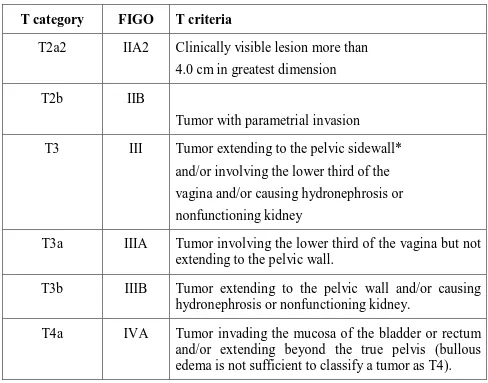
STAGING [AJCC- 8th edition]5
T category FIGO T criteria
Tx Primary tumor cannot be assessed
T0 No evidence of tumor
T1 I Cervical carcinoma confined to cervix
T1a I A Invasive carcinoma diagnosed only by microscopy. Stromal invasion with a maximum depth of 5 mm measured from the base of the epithelium and a horizontal spread of 7mm or less .vascular space invasion ,venous or lymphatic does not affect classification.
T1a1
IA1
Measured stromal invasion of 3mm or less in depth and 7mm or less in horizontal spread.
T1a2
IA2
Measured stromal invasion of more than 3mm and not more than 5mm with a horizontal spread of 7mm or less.
T1b IB Clinically visible lesion confined to the cervix or microscopic lesion greater than T1a/IA2.Includes all macroscopically visible lesions, even those with superficial invasion.
T1b1 IB1 Clinically visible lesion 4cm or less in greatest dimension.
T1b2
IB2
Clinically visible lesion more than 4cm in greatest dimension.
T2
II
Cervical carcinoma invading beyond the
uterus but not to the pelvic wall or to lower third of the vagina
T2a IIA Tumor without parametrial invasion
T2a1 IIA1 Clinically visible lesion 4.0 cm or less
T category FIGO T criteria
T2a2 IIA2 Clinically visible lesion more than
4.0 cm in greatest dimension
T2b IIB
Tumor with parametrial invasion
T3 III Tumor extending to the pelvic sidewall*
and/or involving the lower third of the
vagina and/or causing hydronephrosis or
nonfunctioning kidney
T3a IIIA Tumor involving the lower third of the vagina but not extending to the pelvic wall.
T3b IIIB Tumor extending to the pelvic wall and/or causing hydronephrosis or nonfunctioning kidney.
T4a IVA Tumor invading the mucosa of the bladder or rectum and/or extending beyond the true pelvis (bullous edema is not sufficient to classify a tumor as T4).
The pelvic sidewall is defined as the muscle, fascia, neurovascular
structures and skeletal portions of the bony pelvis. On rectal examination, there
[image:15.595.64.552.68.451.2]Definition of Regional Lymph Node (N) N category FIGO N criteria
Nx Regional lymph nodes cannot be assessed
N0 No regional lymph node metastasis
N0(i+) Isolated tumor cells in regional lymph node(s) no greater than 0.2 mm
N1 Regional lymph node metastasis
Definition of Distant Metastasis (M)
M Category FIGO Stage M Criteria M0 No distant metastasis
STAGE GROUPING
WHEN T is AND N is AND M is Then STAGE GROUPING IS
T1 Any N M0 I
T1a Any N M0 IA
T1a1 Any N M0 IA1
T1a2 Any N M0 IA2
T1b Any N M0 IB
T1b1 Any N M0 IB1
TIb2 Any N M0 IB2
T2 Any N M0 II
T2a Any N M0 IIA
T2a1 Any N M0 IIA1
T2a2 Any N M0 IIA2
T2b Any N M0 IIB
T3 Any N M0 IIII
T3a Any N M0 IIIA
T3b Any N M0 IIIB
T4 Any N M0 IV A
PROGNOSTIC FACTORS
PATIENT FACTORSAGE
The study conducted by Delaloye et al6 says that, age is not a prognostic
factor in carcinoma of the cervix. But Dattoli et al7 reported decreased survival
in women younger than 35 or 40 years, who have a greater frequency of poorly
differentiated tumors.
TUMOUR VOLUME
Piver and Chung8 showed a greater incidence of lymphatic and distant
metastasis and lower survival rates in patients with bulky and barrel-shaped
stage IB and IIA tumors treated by radical hysterectomy. A higher incidence of
pelvic recurrences and distant metastases and a decreased survival rate were
reported by Fletcher9 , Eifel et al10. , and Perez et al11 in patients with larger
tumors treated with irradiation. In stages IB and IIA, higher radiation doses or
combination with an extrafascial hysterectomy improved local tumor
control12,13,14..
ANEMIA
The strongest evidence that anemia plays a causative role in pelvic
recurrence comes from a small randomized study conducted by Princess
Margaret Hospital15 .In all patients, the hemoglobin level was maintained at
least 10 gm%, but in patients in the treatment arm, the hemoglobin level was
regional recurrence rate was significantly higher for the 25 anemic patients in
the control arm than it was for the patients who received transfusions.
Treatment related factors
There is higher chance of recurrence if the duration of treatment is more than 7
Weeks16
OVERVIEW OF TREATMENT POLICY IN CANCER CERVIX All the three standard modalities of oncology namely radiation, surgery
and chemotherapy have stamped their role in the treatment of different stages of
the disease.
ROLE OF SURGERY IN CANCER CERVIX THERAPY The role of radical surgery is limited to pre-invasive and early stages of
invasive growth. In some selected cases it is combined with RT and used as a
salvage procedure to treat local failure after RT. Radical curative surgery can be
done up to FIGO stage IIA which has 5 year survival rates similar to radiation
alone. In early disease surgery is preferred over RT in:
• Patients with Carcinoma in situ, severe dysplasia
• Small volume disease (<4cm),
• Young patients for better preservation of sexual life.
• Endocervical cancers and adenocarcinomas,
Types of surgeries in carcinoma cervix
17:
Radical hysterectomy - Excision of the uterus en bloc with the parametrium (i.e., round, broad, cardinal, and uterosacral ligaments) and the upper one-third
to one-half of the vagina. There are 5 types of radical hysterectomies
• Total (extrafascial) abdominal hysterectomy (class I) consists of removal
of the cervix and adjacent tissues as well as a small cuff of the upper
vagina in a plane outside the pubocervical fascia. There is minimal
disturbance of the ureters and the trigone of the bladder.
• In modified radical extended hysterectomy (class II), the cervix and upper
vagina are removed, including paracervical tissues, and the ureters are
dissected in the paracervical tunnel to their point of entry into the bladder.
This operation may be performed with or without lymphadenectomy.
• Radical abdominal hysterectomy (class III) with bilateral pelvic
lymphadenectomy consists of a wider resection of the parametrial tissues
to the pelvic wall, with dissection of the ureters and mobilization of the
bladder as well as the rectum to allow for more extensive removal of
tissues. Also, a vaginal cuff of at least 2 to 3 cm is always included in the
procedure. A bilateral pelvic lymphadenectomy is usually carried out.
More extensive radical hysterectomies (class IV and V) have been
described, but they are rarely performed.
MICROINVASIVE CARCINOMA (STAGE IA)
The standard treatment for patients with stage IA1 disease is cervical
conization or total (type I) or vaginal hysterectomy. 18The risk of lymph node
metastasis in minimally invasive carcinoma cervix is less than 1%. Because of
this prophylactic lymphadenectomy is not recommended19,20. For patients whose
tumors invade 3 to 5 mm into the stroma (FIGO stage IA2), the risk of nodal
metastases is approximately 5%. Therefore, in such patients, a bilateral pelvic
lymphadenectomy should be performed in conjunction with a modified radical
(type II) hysterectomy. Modified radical hysterectomy is a less extensive
procedure than a classic radical (type III) hysterectomy.
Stage IB and IIA Disease
Early stage IB cervical carcinomas can be treated effectively with
combined external-beam irradiation and brachytherapy or with radical
hysterectomy and bilateral pelvic lymphadenectomy. The goal of both
treatments is to destroy malignant cells in the cervix, para cervical tissues, and
regional lymph nodes21.
In 1997, Landoni et al22 conducted a prospective trial comparing radical surgery
were randomly assigned to receive treatment with type III radical hysterectomy
or a combination of external-beam and Low-Dose Rate intracavitary
radiotherapy. In the surgical arm, findings of parametrial involvement, positive
margins, deep stromal invasion, or positive nodes led to the use of postoperative
pelvic irradiation in 62(54%) of 114 patients with tumors 4 cm or smaller in
diameter and in 46 (84%) of 55 patients with tumors measuring more than 4 cm.
Patients in the radiotherapy arm received a relatively low total dose of radiation
to the cervix, with a median dose to point A of 76 Gy. With a median follow-up
of 87 months, the 5-year actuarial disease-free survival rates for patients treated
in the surgery and radiotherapy groups were 80% and 82%, respectively, for
patients with tumors that were 4 cm or smaller and 63% and 57%, respectively,
for patients with larger tumors. The authors reported a significantly higher rate
of complications in the patients treated with initial surgery, and they attributed
EXTERNAL BEAM RADIATION TREATMENT
Radiotherapy has been used successfully to treat cervical cancer for nearly a
century. Most of the patients present with locally advanced stage of the disease,
and nearly 70% belong to FIGO Stage IIB or III at presentation23.
The combination of external beam radiotherapy (EBRT) and intracavitary
brachytherapy (ICBT) has become a standard treatment modality for locally
advanced cervical cancer. As with radical surgery, the goal of radical
radiotherapy is to sterilize disease in the cervix, paracervical tissues, and
regional lymph nodes in the pelvis. Patients are usually treated with a
combination of external beam irradiation to the pelvis and brachytherapy.
Clinicians balance between external and intracavitary treatment in different
ways, weighting one or the other component more heavily. A homogenous
dose distribution over a large volume can be achieved only by teletherapy. The
external beam radiation is followed by ICA to achieve the highest local control
rate possible and this sequence is also ideal in that the tumor shrinkage caused
by the initial EBRT will bring the anatomy to near normal resulting in an
optimal and uniform dose distribution from a subsequent ICA. EBRT is
delivered before brachytherapy in patients with bulky primary tumors,
exophytic bleeding tumors, necrotic or infected tumors and tumors with
parametrial involvement. However brachytherapy is a critical element in the
small tumors that involve multiple quadrants of the cervix are usually treated
with total doses of 80 to 85 Gy to point A. Although patients with small tumors
may be treated with somewhat smaller fields than patients with more advanced
loco regional disease, care must still be taken to adequately cover the obturator,
external iliac, low common iliac, and presacral nodes24 .
External-beam irradiation is used to deliver a homogeneous dose to the primary
cervical tumor and to potential sites of regional spread. An initial course of
external irradiation may also improve the efficacy of subsequent intracavitary
treatment by shrinking bulky tumor and bringing it within the range of the
high-dose portion of the brachytherapy high-dose distribution. For this reason, patients
with locally advanced disease usually begin with a course of external-beam
treatment. Subsequent brachytherapy exploits the inverse square law to deliver a
high dose to the cervix and parametrial tissues while minimizing the dose to
adjacent normal tissues25. Whitney et al26,27 in the Gynaecologic Oncology
Group (GOG 85) randomly assigned patients with stage IIB to IVA disease to
receive either hydroxyurea or cisplatin-containing chemotherapy during
external-beam irradiation. All three of the cisplatin-containing regimens in these
trials produced local control and survival rates superior to those for the control
arms (hydroxyurea and radiation). Keys et al28 conducted a study in patients
with stage IB tumors measuring at least 4 cm in diameter. They were randomly
assigned to receive radiation alone or radiation plus weekly cisplatin before
have a complete histologic response and were more likely to be disease-free at
the time of preliminary analysis.
In a study conducted by the Southwest Oncology Group and the
Gynecology and Oncology Group (GOG)29, included patients who were treated
with radical hysterectomy and were found to have pelvic lymph node
metastases. Positive margins, or parametrial involvement. Patients were
randomly assigned to receive postoperative pelvic radiation alone or combined
with cisplatin and 5-FU. In the preliminary analysis, patients who received
chemotherapy had a better disease-free survival rate.
The Radiation Therapy Oncology Group30 also conducted a trial in which
radiotherapy alone (including prophylactic para-aortic irradiation) was
compared with pelvic irradiation plus concurrent cisplatin and 5-FU [(8-year OS
(67 vs. 41%)]. This is the only study in which chemotherapy was administered
during both the brachytherapy and external-beam components of treatment. The
results of this trial showed significant difference in the rates of local control,
distant metastasis, overall survival, and disease-free survival favoring in the
treatment arm with chemotherapy.
Eifel et al (RTOG90-01)31,32 conducted a study showed that chemo
radiation followed by brachytherapy improved overall survival in locally
advanced carcinoma cervix compared to external beam radiation and
Concurrent chemo radiation is the treatment of choice for locally
advanced carcinoma cervix. EBRT with concurrent chemotherapy showed
consistent improvement in local disease control, distant metastasis and
survival33.
In Feb 1999 National Cancer Institute (NCI), in clinical announcement
advised that Cisplatin based chemotherapy administered concurrently exhibited
a marked superiority over standard radiotherapy alone regimens in locally
advanced cancer cervix and that furthermore Cisplatin based chemo radiation
has been the standard of treatment for this disease.
BRACHYTHERAPY
Brachytherapy is also known as internal radiotherapy, endocurietherapy
or sealed source radiotherapy. It is a form of radiotherapy where a radiation
source is placed inside or next to the area requiring treatment34. Brachytherapy
procedures were initially performed by inserting the radioactive material
directly into the tumor ("hot" loading), thereby exposing the physicians and
caregivers to high radiation doses. Manually after loaded techniques, whereby
hollow needles, catheters or applicators are first inserted into the tumor, then
loaded with radioactive materials, increased placement accuracy while reducing
the radiation hazards. The introduction of remote controlled insertion of sources
patient was housed in a shielded room while the caregivers were in an adjacent
room monitoring the patient treatment remotely35.
PRINCIPLE OF ICA
ICA delivers a very high dose to the central tumor volume including the
cervix and adjacent tissues with maximum tumor control without crossing the
tolerance of surrounding normal tissue. This is possible because the normal
uterus and vagina are relatively radio-resistant and tolerate relatively high doses
of radiation and there is a rapid fall of dose at a distance from the cervix
protecting the rectum, bladder and small bowel. Intracavitary brachytherapy can
be delivered by low dose rate (LDR), moderate dose rate (MDR) and high dose
rate (HDR) dose delivery systems. The dose rate of LDR is 0.4- 2Gy/ hr., MDR
is 2-12 Gy/hr and HDR is > 12 Gy/hr. As many studies have demonstrated
comparable local control, survival and morbidity HDR-ICA has been widely
incorporated as a component in the treatment of cancer cervix.
ADVANTAGES OF HDR VS. LDR IN CANCER OF CERVIX
• Eliminates radiation exposure hazard for caregivers, visitors.
• Allows shorter treatment times: Less patient discomfort, elimination of
prolonged bed rest reduces hospitalization (due to outpatient therapy),
possibly allows greater displacement of nearby normal tissues (by
reduce rectal and bladder morbidity, possible to treat large number of
patients in institutions having high volume of cervical cancer patients.
• Allows use of smaller diameter sources than are used in LDR-reduces the
need for dilatation of the cervix and need for heavy sedation or general
anesthesia is reduced, physically easier to insert applicator into the
cervix.
• Makes treatment dose distribution (dwell time, dwell position)
optimization possible.
• Allows integration of EBRT and HDR which can lead to a shorter overall
treatment duration and potentially to better tumor control.
THE AMERICAN BRACHYTHERAPY SOCIETY DOSE RECOMMENDATIONS36-
1) Recommended Prescription [earlier]: Low-dose-rate prescription may be
in milligram-hours or in cGy to Point A or LDR primary treatment of
45-50 Gy external-beam plus 40-60 cGy/hr to a cumulative dose of 40-45
Gy. Goal TD should be >85 Gy. High-dose-rate typically prescribed in
one of the following fractionation regimes: 5.5 Gy x 5, 6 Gy x 5, 7 Gy x 4
2) Timing- All treatment, including external-beam and brachytherapy, must
be completed within 56 days from initiation of treatment. High-dose-rate
being given per week during the conclusion of external beam and during
the parametrial boost portion of treatment. Brachytherapy can be initiated
earlier, but not earlier than approximately 20 Gy, if the applicator placed
at this time point would provide adequate tumor coverage and sparing of
normal tissues. Alternatively, if 45 Gy is delivered to the whole pelvis
prior to brachytherapy, two brachytherapy insertions per week should be
given to avoid treatment prolongation of treatment.
3) The updated ABS 2011 Guideline recommends that 3D imaging with
ultrasound, CT or MRI be performed when feasible to estimate the
cervical tumor dimensions and ensure adequate coverage of the tumor.
Normal-tissue dosimetry using 3D parameters results in a more accurate
reflection of doses administered and may provide more reliable indicators
of the risk of toxicity. The dose to point A should be recorded, the goal
should be good coverage (i.e., a D90) of the involved region with EQD2
≥ 80 Gy for patients with either a complete response or a partial response
with residual disease less than 4 cm. For non-responders or those with
tumors larger than 4 cm at the time of brachytherapy, tumor dose
escalation to an EQD2 of 85–90 Gy is recommended for maximizing
local control. Other fractionation regimens with EQD2 in the range of
80–85 Gy are also acceptable, although the larger fraction size, the higher
the risk for normal-tissue toxicity. For the normal tissues, it is
are calculated and the final dose to the bladder, rectum and sigmoid
calculated. Dose limits for the normal tissues are the EQD2 limit for the
rectum and sigmoid is 70–75 Gy and for the bladder is 90 Gy.
CONCEPT OF EQD2
EQD2 is equivalent dose in 2gy fraction, it’s the total dose in 2 gy fraction that
would give the same log kill as the given schedule.
It is a more practical alternative to convert the BED to equivalent total doses
delivered in 2gy fractions.
BED =nd[1+d÷α/β]
EQD2 =BED/(1+2/α/β)
SOURCES FOR HDR INTRACAVITARY BRACHYTHERAPY
A radionuclide with high specific activity (activity per unit mass; Ci/g) is
needed so that treatment dose rates of at least 12 Gy/hr can be achieved without
interstitial brachytherapy. A source no larger than 1 mm in diameter by 4mm
long with an exposure rate of at least IR/sec at l cm is required. The exposure
rate achieved by a small source depends on the chemical form (i.e. relative mass
of non-radioactive atoms) of the source, its density, exposure rate constant of
the radionuclide and photon self-absorption.
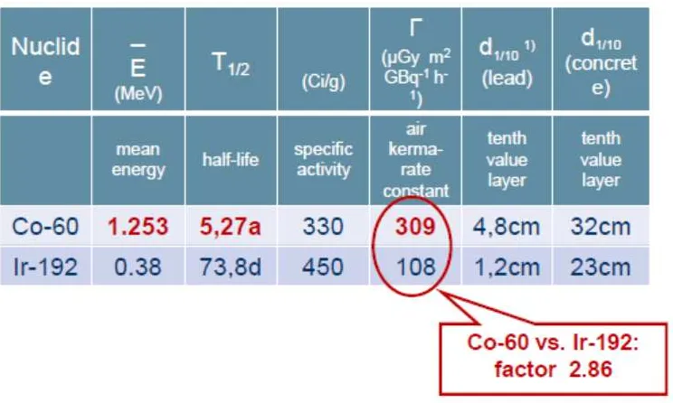
SOURCES
The need for high specific activity sources limits the number of
radioisotopes suitable for HDR remote after loaders. Most HDR units use
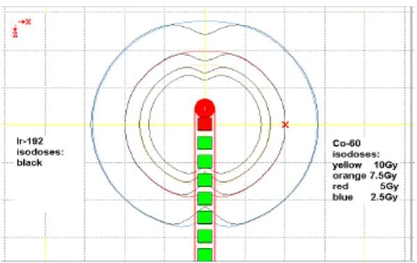
Iridium-192 (192Ir) or cobalt 60 (60Co). 192Ir offer smaller source sizes but
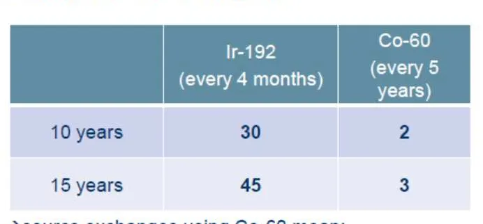
sources must be changed frequently. Most centers exchange their 192Ir sources
every 3 or 4 months (half life 73.8 days) whereas a similar decay fraction for
60
Co takes 5-8years (half life 5.26 years). 60Co is used as an intracavitary HDR
source in the form of small spherical pellets and it emits two high energetic
gamma rays(1.17 &1.34 MeV). The smaller sources permit access to more body
sites via interstitial or intraluminal applications. Based solely on specific
activity considerations 192Ir has been the optimal choice for HDR brachytherapy
and is being widely used radionuclide for the application. With the introduction
of cobalt as a brachytherapy source paradigm has shifted to its use for cost
effectiveness in high burden setups.
Fig-2.1
Fig- 2.2
[image:32.595.98.470.415.638.2]Fig- 2.3
Fig- 2.4
Anisotropy- No difference between the two radio isotopes except dip in the
[image:33.595.148.448.484.673.2]Fig -2.5
[image:34.595.106.472.461.677.2]
Fig -2.7 source change data for both radionuclides
DOSE SPECIFICATION
Since the first application of radium in the treatment of cancer of the
uterus in 1908, several techniques have evolved, most of which are
modifications of the Stockholm technique and the Paris technique. Stockholm37
based techniques was introduced by Forsell and Heyman' at the Radium
hemmet in Stockholm. It is a fractionated course of radiation delivered over a
period of one month. In Stockholm there are three insertions each of 22 hours
separated by 1 -3 weeks. Total prescribed dose - 6500-7100 mg Ra and 4500
per fraction. Paris technique38 is a single application of radium for 120 hrs
(45R/hr). It delivers a dose of 7000- 8000mg-hrs of radium
The Manchester system, which evolved from the Paris technique, uses a
Rubber uterine tandem to hold one to three radium tubes and rubber ovoids,
separated by a rubber spacer, to each hold a radium tube. The radiation is
delivered in at least two applications. In the Fletcher-Suit applicator the tandem
and the ovoids (or the colpostats) are made of stainless steel and then secured to
hollow handles to permit after loading of the sources39.
Brachytherapy plays a very important role in obtaining high cure rates with
minimum complications. Ideal placement of the uterine tandem and vaginal
ovoids produces a pear-shaped distribution, delivering a high dose to the cervix
and paracervical tissues and a reduced dose to the rectum and bladder40. If the
intracavitary placement has been optimized, this can usually be accomplished
without exceeding a dose of 75 Gy to the bladder reference point or 70 Gy to
the rectal reference point, doses that are usually associated with an acceptably
low risk of major complications. The dose to the surface of the lateral wall of
the apical vagina should not usually exceed 120 to 140 Gy. Suboptimal
placements occasionally force compromises in the dose to tumor or normal
tissues. To choose a treatment that optimizes the therapeutic ratio in these
circumstances requires experience and a detailed understanding of factors that
HIGH DOSE RATE APPLICATORS Fletcher-Suit or Fletcher-Suit-DeIco's
These applicators are used for the treatment of gynecological malignancies of
the uterus, cervix and pelvic side walls. The applicator set typically consists of
three rigid intrauterine tandems, with curvature of 15-, 30-, and 45-degree
angles, and a pair of ovoids or colpostats. The tandem and the ovoids (or the
colpostats) are made of stainless steel and then secured to hollow handles to
permit after loading of the sources. CT and MRI compatible applicators are now
available.
The Manchester System
The Manchester system is one of the oldest and the most extensively used
systems in the world. It is characterized by doses to four points: point A, point
B, a bladder point, and a rectum point. The duration of the implant is based on
the dose rate calculated at point A, although the dose at the other points is taken
into consideration in evaluating a treatment plan. With the availability of the
treatment planning computers, most users of the Manchester system examine
the isodose distributions in the frontal and sagittal planes in addition to
obtaining dose at the four designated points. Point A still remains the point of
dose prescription. Point A was originally defined as 2 cm superior to the lateral
vaginal fornix and 2 cm lateral to the cervical canal. Later, it was redefined to
be 2 cm superior to the external cervical os (or cervical end of the tandem), and
Ideally, a point A represents the location where the uterine vessels cross the
ureter. It is believed that the tolerance of these structures is the main limiting
factor in the irradiation of the uterine cervix. Point B represents obturator node.
The International Commission on Radiation Units and Measurements System [ICRU]
The ICRU has recommended a system of dose specification that relates
the dose distribution to the target volume, instead of the dose to a specific point.
The dose is prescribed as the value of an isodose surface that just surrounds the
target volume.
Data required for reporting intracavitary therapy [ICRU-38]41
• Description of the Technique: Minimum information should
include the applicator type, source type and loading and orthogonal
radiographs of the application.
• Total Reference Air Kerma: By this parameter, it is meant the total
air kerma strength of sources times the implant duration. This is
similar to the total milligram-hours of radium or total mg-Ra eq-h
except that the sources are calibrated in units of air kerma strength,
that is, ^Gy m2 h-1.
• Reference Volume. The reference volume is the volume of the
isodose surface that just surrounds the target volume. The
from the external beam. The reference volume for the intracavitary
part of the treatment should be identified and its dimensions
recorded.
Absorbed Dose at Reference Points
Bladder Point: The bladder point is localized by using a Foley catheter,
with the balloon filled with 7 ml of a contrast material. On the frontal
radiograph, the bladder point is marked at the center of the balloon; on the
lateral radiograph, the bladder point is obtained on a line drawn antero
posteriorly through the center of the balloon, at the posterior surface.
Rectal Point: The rectal point is identified on the frontal radiograph at the
midpoint of the ovoid sources (or at the lower end of the intrauterine source).
On the lateral radiograph, the rectal point is located on a line drawn from the
middle of the ovoid sources, 5 mm behind the posterior vaginal wall. The
posterior vaginal wall may be visualized by using radiopaque gauze for the
vaginal packing.
Pelvic Wall Points: On the anteroposterior radiograph, the pelvic wall
points are located at the intersection of a horizontal tangent to superior aspect of
the acetabulum and a vertical line touching the medial aspect of the acetabulum.
On the lateral view, these points are marked as the highest mid distance points
CHARACTERISTICS OF AN IDEAL APPLICATION IN BRACHYTHERAPY42
• The ovoids should fill the vaginal fornices - largest ovoid size to be
used.
• The ovoids should be separated by 0.5 -1.0 cm, admitting the flange on
the tandem.
• The axis of the tandem should be central between the ovoids
• Tandem - 1/3 of the way between Sl -S2 and the symphysis pubis.
• The tandem - midway between the bladder and Sl -S2.
• Tandem should bisect the ovoids. The bladder and rectum should be
packed away from the implant.
DOSIMETRIC CHARACTERISTICS OF BRACHYTHERAPY SOURCES
In general, four factors influence the single-source dose distribution for
photon-emitting sources:
• distance (inverse-square law);
• absorption and scattering in the source core and encapsulation;
• photon attenuation and
• Scattering in the surrounding medium.
RADIOBIOLOGY OF BRACHYTHERAPY AND THE DOSE-RATE EFFECT
The biological effects of radiotherapy depend on dose distribution,
treated volume, dose rate, fractionation and treatment duration. These factors,
have an important role in determining the outcome of external beam
radiotherapy or of brachytherapy.
The biological damage inflicted by irradiation of human cells with
ionizing radiation can be divided into three consecutive steps:
A very short initial physical phase (about 10-18 S), during which photons
atoms (excitation), or ejecting some of them from the atoms (ionization). This is
the energy deposition phase.
A chemical phase, again very short (about 10-3 s), during which ionized
and excited atoms interact, leading either directly or indirectly through the
formation of free radicals to the breakage of chemical bonds. Free radicals are
highly reactive and can induce chemical changes in biologically important
molecules like DNA. Single-strand or double-strand breaks in DNA appears to
be the basic damage leading to biological effects.
A biological phase, much longer (seconds to years), during which the
cells react to the inflicted chemical damage43.Specific repair enzymes can
successfully repair the vast majority of lesions in DNA. A few lesions however
may not be repaired, and may therefore lead to cell death. Cell death is not
immediate and usually occurs during the next cell division (apoptosis is a minor
process in most human cells). The early reactions are seen during the first days
or weeks after irradiation (for example diarrhea or acute mucositis). They are
temporary because the cell deficit is compensated by the repopulation of stem
cells, and subsequently of differentiated cells. Late reactions due to damage to
the late-reacting tissues, for instance blood vessel damage, fibrosis,
telangiectasia, etc., may be seen after months or years. Damage to these late
reacting normal tissues is poorly repaired and is responsible for most severe
complications of radiotherapy. Tolerance of these tissues is the limiting factor
THE FIVE R'S OF RADIOBIOLOGY
A number of biological processes take place during irradiation and
modify the radiation response. These processes are often described as the 5 R’s
of radiobiology44
Each follows a specific time pattern:
• Repair of DNA damage. Both experimental and clinical studies have shown that human tumors strongly differ in radio sensitivity and radio curability
thought to stem from differences in capacity for repair of sub lethal
damage45,46.
• Reassortment or redistribution in the cell cycle. The cell cycle is divided into four consecutive stages: Gl, S, G2 and M. Gl is a gap of apparent
inactivity after a mitosis (M), before DNA synthesis (S-phase) resumes in
view of the following cell division. G2 is a second gap of apparent inactivity
between S phase and M.
• Radiosensitivity - varies along the cell cycle, S being the most resistant phase, and G2 and M the most sensitive. Therefore, cells surviving an
exposure are preferentially in a stage of low sensitivity (Gl), i.e.
synchronized in a resistant cell cycle phase. They progress thereafter
together into S and then to the more sensitive G2 and M phases. A new
irradiation exposure at this time will have a larger biological effect (more
experimental results, redistribution has never been shown to play a
measurable role in the clinic of radiotherapy.
• Repopulation - Cells surviving an irradiation keep proliferating. This increases the number of clonogenic cells, i.e. the number that must
eventually be sterilized to eradicate cancer. Repopulation therefore has a
detrimental effect as far as cancer control is concerned. Stem cells do also
proliferate in normal tissues, which has in this case a protective effect (it
helps the tissue to recover from radiation damage and it adds to DNA repair
in cells).
• Reoxygenation - Because of an inappropriate development of intra tumoral vasculature, every tumor of clinically detectable size contains a large
proportion of poorly oxygenated cells. Also, the proportion of hypoxic cells
increases with the tumor size. Acutely hypoxic cells are far more radio
resistant than well oxygenated cells. This is expressed by the Oxygen
Enhancement Ratio (OER), i.e. the ratio between radiation doses required in
hypoxia and air to produce the same biological effect. Its value is 3, and it
varies very little with dose or with the biological system. Hypoxic cells
usually survive irradiation, but they progressively (re)oxygenate, due to the
better supply of oxygen available after well oxygenated cells have died .This
restores radio sensitivity in the tumor. Several mechanisms are involved, but
reoxygenation occurring at long intervals is probably due to tumor shrinkage
DOSE RATE EFFECTS IN BRACHYTHERAPY
Biological effects of radiation are strongly dependent upon the rate of
dose delivery. The radiobiological processes involved in high dose rate
brachytherapy are in all respects similar to those involved in fractionated
external beam radiation therapy, except for the volume effect, as mentioned
earlier. Repair, repopulation, and reoxygenation, are the main factors
determining outcome. They do not occur during the very short duration of
irradiation (up to 10-15 minutes), but take place between consecutive fractions,
provided the interval is adequate.
Repair. - For brief exposures, the survival fraction S of a cell population
decreases with increasing dose D. It has been mathematically modelled as the
sum of two type of lesions:
Lethal (non-repairable) lesions, with a survival fraction S = exp
(-αD), represented by the tangent to the survival curve at its origin.
Sublethal lesions, non-lethal and potentially repairable, but the
accumulation of which can cause cell death, with a survival fraction S = (-βD2)
The sum of these two components leads to the classical linear-quadratic
Equation proposed by Chadwick & Leenhouts and Kellerer & Rossi.
S=exp-(αD+(βd2).
The survival curve displayed on a semi-logarithmic graph exhibits an
initial shoulder. According to the model, it is proportional to DNA repair
ratio α/β corresponds to the dose at which the contribution of the two factors to
the survival fraction is equal, αD = βD2, and D = α/β. A large α/β corresponds
to a small shoulder (small repair capacity) and a small α/β to a broad shoulder
(large repair capacity).
In summary, a low α/β is characteristic of late-responding normal tissues
and some tumors (0.5 to 6Gy, average 3 Gy), while a higher α/β ratio
characterizes the early-responding normal tissues and carcinomas. (7 to 20 Gy,
average 10 Gy
Radiobiological studies have shown that each successive fraction in a
series equally effective, so the effect (E) of n fractions of size d can be
expressed as:
E= αD+ β D2
Where the total radiation dose D = nd.
Repopulation does not occur in late responding normal tissue during the
course of a 6-7 weeks irradiation, but it plays a role in early reactions and tumor
than 3-4 weeks47 . After this time, accelerated repopulation of fast-growing
tumors may be observed48. For early effects on skin and mucosa (desquamation
and mucositis), the spontaneous tissue kinetics are unchanged until about 10
days after the initiation of Irradiation, when the rate of cell replacement is
accelerated49. It remains very active during the two weeks following irradiation,
and then tends to drop quickly, back to its physiological level.
Reoxygenation. Following a large single dose irradiation, most well
oxygenated cells are killed, and hypoxic cells survive predominantly. Because
aerobic cells have disappeared, the distance between capillary vessels and
hypoxic cells decreases. This allows oxygen to reach hypoxic cell, which
reoxygenate and become more radiosensitive. The process takes between hours
COMPLICATIONS
RTOG-ACUTE TOXICITY [Table-2.1]
Toxicity Grade 1 Grade 2 Grade 3 Grade 4 Grade 5 GENITO URI NARY Frequency of urination or nocturia twice pretreatment habit/dysuria, urgency not requiring medication. Frequency of urination or
nocturia that is
less frequent than
every hour. dysuria urgency, bladder spasm requiring local anaesthetic. Frequency with urgency and nocturia hourly or more frequently/ dysuria, pelvis
pain or bladder
spasm
requiring
regular, frequent
narcotic/ gross
haematuria with
/ without clot
Toxicity Grade 1 Grade 2 Grade 3 Grade 4 Grade 5 Small bowel toxicity Anorexia with
≤5% weight loss
from pretreatment baseline/nausea not requiring antiemetic’s/ abdominal discomfort not requiring parasym patholytic drugs or analgesics. Anorexia with
≤15% weight loss
from pretreatment
baseline/ nausea
and or vomiting
requiring antiemetic’s/ abdominal pain requiring analgesics. Anorexia
with≥15%weight
loss from
pretreatment
baseline or
requiring NG
tube or parentral
support. Nausea
and/or vomiting
requiring tube or
parentral support/ abdominal pain ,severe despite medication /hematemesis or melena/ abdominal
distention[ x ray
Toxicity Grade 1 Grade 2 Grade 3 Grade 4 Grade 5 Rectal toxicity Increased frequency or
change in quality
of bowel habits
not requiring medication/Rectal discomfort not requiring analgesics. Diarrhea requiring parasympatholytic drugs/Mucous discharge not necessitating sanitary pads/rectal or abdominal pain requiring analgesics. Diarrhea requiring parentral support/ severe mucous or bloody discharge necessitating sanitary pads/ abdominal distention [X-ray demonstrates distended loops]
Acute or sub
AIM OF THE STUDY
To evaluate the acute gastrointestinal and genitourinary toxicity profile in
carcinoma cervix patients using cobalt 60 radionuclide source.
SECONDARY END POINTS
• Feasibility of twice weekly brachytherapy
• Response assessment after completion of treatment.
STUDY CENTRE-
• Dept. of Radiotherapy, Barnard Institute of Radiotherapy, Madras
Medical College, Chennai-3
• The study was reviewed and approved by the institutional ethical
committee.
STUDY PERIOD
From June 2017 to September 2018
MATERIALS AND METHODS
CASE SELECTION
Carcinoma cervix patients who had completed their EBRT and slated for
brachytherapy with minimal or no parametrial disease.
NO. OF PATIENTS - 38
INCLUSION CRITERIA
• Biopsy proven newly diagnosed carcinoma cervix
• Age - 30-65 years
• Stage – IB2 –IIIB
• Histology – Squamous cell carcinoma and its variants.
• ECOG 0-2
• Previously not exposed to any chemotherapy or radiotherapy.
• No major life threatening complications
• HIV negative
• Patient should be fit for anesthesia [GA,SA,IV sedation]
• Cystoscopy – for ruling out bladder invasion
• Urine routine, culture and sensitivity-to rule out other causes of cystitis
before brachytherapy.
EXCLUSION CRITERIA
• Age - < 30 and > 65 years.
• ECOG -3 or more
• Stage IVA –involvement of bladder and rectum.
• Inadequate hepatic and renal functions
• Patient not consenting to chemotherapy.
• Previously treated for any other malignancy.
• Metastatic or recurrent disease
• HIV positive patients
• Patient unfit for anesthesia.
INVESTIGATIONS
• Biopsy from the tumor
• Complete blood count, liver function test, renal function test, viral
markers.
• CT scan abdomen and pelvis or MRI- plain and contrast [pretreatment
and post treatment at 6-8 weeks]
• Chest X ray –PA view ,ECG, blood grouping
• Cardiology evaluation for fitness.
DATA COLLECTION AND METHODS
Eligible patients treated with radiotherapy in the form of external beam
radiotherapy to a total dose of 45-50Gy to the whole pelvis. [45-50Gy/180-200
cGy/#/25#/5 days a week [Monday to Friday] using cobalt teletherapy machine.
Followed by brachytherapy to a dose of 21Gy/7 Gy/#/3# with a minimum
gap of 72 hours between each fraction using cobalt 60 source with BEBIG
BRACHYTHERAPY MACHINE.
CONCURRENT CHEMOTHERAPY
Cisplatin 40mg/m2 weekly along with proper premedication.
Patients to be clinically assessed during treatment for toxicity of acute
gastrointestinal and genitourinary symptoms and response graded accordingly.
CT abdomen and pelvis or MRI to be taken 6-8 weeks post treatment for
response assessment. Toxicity to be graded according to RTOG acute toxicity
criteria. Patients were also assessed for their compliance during brachytherapy
sessions.
TREATMENT PLANNING EBRT
The whole pelvis including cervix, vagina, and parametrium with the iliac
RT PORTALS AND BORDERS
SUPERIOR - L4-L5 interspace [to include iliac and hypogastric nodes]
INFERIOR – If vagina uninvolved –lower border of obturator foramen.
If vagina involved- entire vagina upto introitus was included.
LATERAL- 2cm lateral to bony pelvis.
Treatment field was verified using X-ray simulation
PORTALS
AP and PA portals treated if field separation was less than 20cm
Field separation of more than 20cm were treated with 4 field box technique
where the anterior border was kept in front of pubic symphysis and posterior
border at S2-S3 junction.
CHEMOTHERAPY DRUG - cisplatin
DOSE - 40mg/m2 –weekly
SCHEDULE - Weekly once
PREMEDICATIONS – Inj.Dexamethasone
Inj.Ranitidine
Inj.Ondansetron
INTENT OF ADDING CHEMOTHERAPY - Radio sensitizer.
TOTAL NUMBER OF CYCLES - 4 -5 cycles
INTRACAVITARY PROTOCOL
After Teletherapy all patients were assessed for intracavitary application.
Those who were found fit for brachytherapy were subjected to the procedure.
HDR BRACHYTHERAPY
Technique - Remote after loading with cobalt 60 source.
Machine - BEBIG
Activity - 1.82 curie [67.484 GBq]
Intracavitary applicator – Modified fletcher suit with 15 and 20 degree
angulation.
No. of fractions -3 # with a minimum gap of 72 hrs between each fraction.
Dose prescribed to Point A - 7Gy.
PROCEDURE
Under anesthesia [ IV sedation/SA/GA] with patient in lithotomy position
the perineum and upper half of thighs were cleaned with beta dine and draped.
Per vaginal examination done .Urinary bladder was catheterized and 7ml of
diluted contrast(3ml of contrast +4 ml of distilled water) was injected into the
Foleys balloon. Uterine sound was introduced and uterine length measured. The
cervical stopper was adjusted according to the uterine length and fixed. Then
packing was done. Rectal tube inserted. CT simulation was done. The films
were used in our treatment planning system for applicator reconstruction,
defining Point A, Point B, dose prescription to point A and calculating bladder
and rectum points as per ICRU38 .Dose to bladder and rectum was kept less
than 80% of point A. Optimization done using change in dwell position and
time .After obtaining the desired prescription isodose, patient was connected to
the BEBIG machine with cobalt 60 source through catheters (transfer tubes) and
treated.
The Assessment of GI and GU toxicities was done as per schedule
First assessment -after the application of first HDR ICRT brachytherapy
Second assessment – at the time of last HDR ICRT application
Third assessment- one month later to last HDR ICRT application
Fourth assessment-two month later to last HDR ICRT application
ANATOMICAL TOPOGRAPHY AND BRACHYTHERAPY APPLICATOR
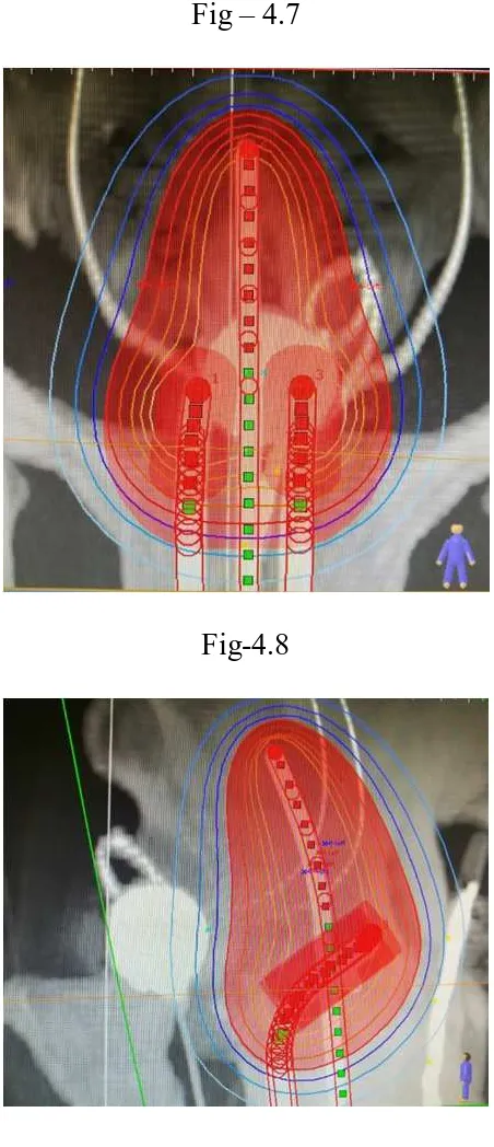
FLETCHER SUIT DELCLOS APPLICATOR [Fig - 4.3]
SIMULATION FILMS [AP VIEW] -fig -4.5
[image:60.595.144.452.450.758.2]ISODOSE DISTRIBUTION IN AP AND LATERAL VIEW
Fig – 4.7
CASE ANALYSIS AND RESULTS
STUDY POPULATION AND COMPLIANCE TO TREATMENT
From June 2017-September 2018 a total of 38 patients of carcinoma
cervix were studied for acute toxicity analysis with brachytherapy using cobalt
60 source. All insertions were done when patient had completed EBRT and fit
for intracavitory insertions. All patients were available for final analysis
CHARACTERISTICS AGE (TABLE-5.1)
AGE [Years] Number of patients [percentage]
30- 40 yrs 01 [3%]
41 -50 yrs 10 [27%]
51-60 yrs 18 [49%]
61 -65 yrs 08 [21%]
FIGURE [5.1]
30-40 yrs
3% 41-50 yrs 27%
[image:62.595.68.527.320.734.2]51-60 yrs [PERCENTAGE] 61-65 yrs
21%
No. of patients
30-40 yrs
41-50 yrs
51-60 yrs
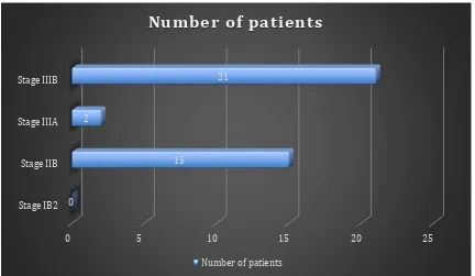
FIGO STAGING (TABLE - 5.2)
STAGE NUMBER OF PATIENTS [%]
IB2 NIL
IIB 15 [39.4%]
IIIA 02 [5.2%]
IIIB 21 [55.2%]
FIGURE-5.2
0 5 10 15 20 25
Stage IB2 Stage IIB Stage IIIA Stage IIIB
0
15 2
21
Number of patients
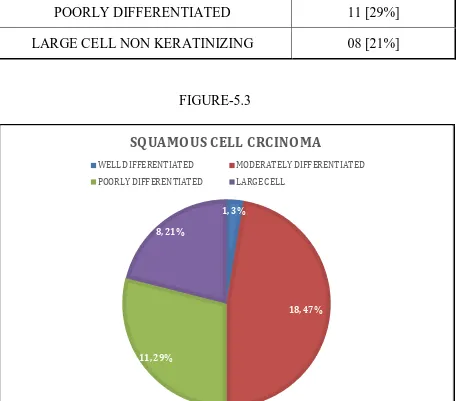
[image:63.595.83.515.339.590.2]Histology (Table- 5.3)
[image:64.595.73.529.220.621.2]
FIGURE-5.3
1, 3%
18, 47%
11, 29% 8, 21%
SQUAMOUS CELL CRCINOMA
WELL DIFFERENTIATED MODERATELY DIFFERENTIATED POORLY DIFFERENTIATED LARGE CELL
SQUAMOUS CELL CARCINOMA NO.OF PATIENTS
WELL DIFFERENTIATED 01 [2.6%]
MODERATELY DIFFERENTIATED 18 [47.3%]
POORLY DIFFERENTIATED 11 [29%]
EBRT+CHEMO (Table-5.4)
EBRT+CHEMO NUMBER OF PATIENTS
RT+CHEMO 38
RT ONLY NIL
TIME INTERVAL BETWEEN EBRT AND BRACHYTHERAPY (Table - 5.5)
TIME INTERVAL NUMBER OF PATIENTS [%]
< 1 WEEK 23 [60.5%]
CHEMOTHERAPY CYCLES (Table - 5.6)
CISPLATIN [CYCLE] NUMBER OF
PATIENTS PERCENTAGE
2 CYCLES 02 5.2%
3 CYCLES 06 15.7%
4 CYCLES 12 31.5%
[image:66.595.68.519.87.561.2]5 CYCLES 18 47.36%
FIGURE-5.4
Majority of the patients in the study were able to tolerate chemotherapy
cycle and were able to complete all 5 weekly schedules.
2 cycles, 2
3 cycles, 6
4 cycles, 12 5 cycles, 18
Cisplatin
PERFORMANCE STATUS (Table-5.7)
ECOG NO.OF PATIENTS
0 03 [7.8%]
1 21 [55.2%]
[image:67.595.70.517.168.546.2]2 14 [36.8%]
FIGURE-5.5
There was a delay of more than a week to 12 days for nearly half of the
patients due to grade 3 - 4 skin reactions due to EBRT and referral from other
centers but this was compensated with twice weekly brachytherapy with a
minimum gap of 72 hrs between each fractionation.
3
21 14
0 5 10 15 20 25
ECOG 0 ECOG 1 ECOG 2
PERFORMANCE STATUS
SECONDARY ASSESMENT
There was no patient related factors for delay in between the fractions
except for 1 patient who had grade III GI toxicity and was managed with
symptomatic and supportive care.
TUMOUR ASSESMENT AFTER EBRT (Table- 5.8)
TUMOUR RESPONSE [CLINICAL] NUMBER OF PATIENTS COMPLETE RESPONSE 27 [71%]
PARTIAL RESPONSE 11 [29%]
[image:68.595.66.528.384.674.2]
FIGURE-5.6
27, 71% 11, 29%
TUMOUR RESPONSE
COMPLETE RESPONSE
BRACHY THERAPY DOSE CHARACTERISTICS. (Table-5.9)
CA CERVIX[1-III] MEDIAN RANGE
EBRT[WHOLE PELVIS] 50GY 45-50 GY
HDR 7/3# -
PT A 7gy 7-7.5GY
ICRU BLD PT 5.6GY 4.2-6GY
ICRU REC PT 5.4GY 3.2-6GY
BED RECT[EBRT+HDR] 124.4 120-133
EQD2 TUMOUR[EBRT+HDR] 83.6GY 78.8-86GY
REACTIONS ASSESED
• NAUSEA
• VOMITING
• DIARRHEA
• URINARY FREQUENCY
(Table- 5.10)
GENITOURINARY TOXICITY [RTOG acute toxicity scale]
GENITO URINARY NO.OF PATIENTS PERCENTAGE
GRADE 0 22 55.2 %
GRADE 1 12 31.5 %
GRADE 2 04 10.52%
GASTROINTESTINAL TOXICITY (Table- 5.11)
GI TOXICITY NO.OF PATIENTS PERCENTAGE
GRADE 0 16 42.1%
GRADE 1 11 28.94%
GRADE 2 09 23.68%
FIGURE 5.7
0 5 10 15 20 25
Grade 0 Grade 1 Grade 2 Garde 3
22
12
4
0 16
11
9
1
![Fig – 4.6 [ LATERAL VIEW]](https://thumb-us.123doks.com/thumbv2/123dok_us/136330.13474/60.595.144.452.450.758/fig-lateral-view.webp)
![FIGURE [5.1]](https://thumb-us.123doks.com/thumbv2/123dok_us/136330.13474/62.595.68.527.320.734/figure.webp)