Rochester Institute of Technology
RIT Scholar Works
Theses
Thesis/Dissertation Collections
5-27-1997
Hanta virus
Rhonna Brown
Follow this and additional works at:
http://scholarworks.rit.edu/theses
This Thesis is brought to you for free and open access by the Thesis/Dissertation Collections at RIT Scholar Works. It has been accepted for inclusion
in Theses by an authorized administrator of RIT Scholar Works. For more information, please contact
.
Recommended Citation
Rochester Institute
ofTechnology
A Thesis
submittedto the
Faculty
ofthe
College
ofImaging
Arts
andSciences
in
candidacy
for
the
degree
ofMaster
ofFine
Arts.
Hanta Virus
by
Rhonna Kaye
Brown
Approvals
Chief Advisor: Robert Wabnitz
Date
Associate Advisor Glen Hintz
Date
Associate
Advisor:
Dr.
Jeffery
Lodge
Date
Chairperson:
Dr.
Thomas Lightfoot
Date
I,
Rhonna Kaye
Brown,
hereby
grant permissionto the
Wallace
Memorial
Library
ofBIT to
reproducemy
thesis
in
wholeor
in
part.Any
reproduction will notbe
for
commercialuse orprofit.
Ill
Table
of
Contents
List
ofDlustrations
iv
Chapter
1.
Introduction
5
Part
I.
The Disease
2.
What Hanta Virus Is
5
3.
What Hanta Virus Does
6
4.
How
One
Contracts Hanta Virus
7
Part
n.
The Thesis
5.
The Fine Art Work
7
6.
The Computer Work
8
ArtWork
xiIV
List
of
Illustrations
Figure
Page
Fig.
1
Deermouse
7
Fig.
2
Lungs,
healthy
anddiseased
8
Fig. 3 Computer
enhancedlungs
with alveoli8
Fig.
4 Graphic
of spray bottle
9
Fig.
5 Intestines
9
Fig.
6 Intestines
transposed
onto mouse9
Fig. 7
Brochure
XI
[image:5.612.58.458.154.405.2]Introduction
Medical
Illustration,
to
me,
means communication.A
medicalillustrator
is
aninterpreter between
the
medical/scientific community
andthe
lay
community.
The
ability
to
be
ableto
understand and communicate withboth
sides
is
essentialto
successin
this
profession.Presenting highly
scientificinformation
in
away
that
the
majority
of people willbe
ableto
understandrequires a special
ability
to
translate, especially
sothat
little
ofthe
information
is
lost
and no onefeels
overwhelmed.This
thesis
representstwo
years ofstudy
in
Fine Arts
as appliedto
Medical Illustration.
I
chosethis
subject notonly
for its
pertinence
but
alsobecause it
allowed meto
illustrate both
humans
and animals.
The
mediaused, the
poster andbrochure,
allowthe
information
to
reachmore people.
The
poster canbe displayed in
a clinicorotherlikely
places andthe
brochure
canbe
taken
home
and read atleisure
by
anyone.They
canbe
printed
for
non-englishspeaking
people aswell.This
way
the
most people canbe
reachedthrough
ahighly
informative
andvery
cost effective means.The
black
and yellowcolors usedthroughout
aremeantto
represent a cautionsign.This is
awarning
to
beware
andto
be
careful around mice andespecially
wildanimals.
It
is
not meant causefear
or wide spread panic.What Hanta Virus Is
The Hanta
virusis
an enveloped retroviruswhichcauses adisease
in
humans known
asHanta Virus
Pulmonary
Syndrome (HPS).
(Hjelle, 1995)
This
virus was
first
isolated
due
to
an outbreak ofHPS in
the
area whereArizona,
New
Mexico,
Colorado,
andUtah
meet otherwiseknown
asthe
Four
Corners
area.
This
particular strain ofHanta
virusis
calledthe
Four
Corners
virus(FC).
The FC
viruslives in
the
digestive
tract
ofdeermice. The
viruslives
as a parasiteinfected,
but it is
believed
that the
virusis
spreadamong
miceby biting
eachother.
The
range ofdeennice
extendsfrom
Mexico
to
Canada
andthe
family
range covers
both
ofthe
Americas. (Eisenberg.
1987)
There
are other virusesthat
are
closely
relatedto
FC
and causeHPS
that
arefound in
rodentsin
the
samefamily
(Muridae)
asthe
deermouse.(CDC,
293)
Such
rodents asthe
cottonrat,
harvest
mouse,
andthe
white-footed mouse allcarry
virusesthat
can causeHPS.
(Hjelle, 1995)
It is
possiblethat
more speciesin
this
family
carry
someform
ofthe
virus and researchis
continuing
in
orderto
learn
more.What Hanta Virus Does
Hanta
virusis
found
in the
rodent'ssaliva,
feces,
andurine.(Ing,1994)
FC
is
an aerosol and canbe
inhaled,
if
a person comesinto
contactwithinfected
dust from
mouse urine andfeces.
It
can alsobe
caughtby being
bitten.
Once
inside
the
body,
the
virusquickly
takes
effect andis believed
to
attackthe
alveolar cells ofthe
lungs. Symptoms
appear as aflu-like
prodrome(Hjelle,
1995)
and caninclude
persistentfever,
chills,
headache,
myalgia, nausea,
vomiting,
and non-productivecough.(CDC,
291)
The
prodromeis
followed
by
dyspnea,
cough,
thrombocytopenia,
severehemodynamic
instability
andneutrophiliaas well as an
interstitial
infiltrate
ofthe
lungs
that
resembles a widevariety
of otherdiseases.
(Hjelle,
1995)
As
the
alveolar cells are attacked anddie,
they
leave
holes
in
the
lining
ofthe
lung
tissue.
The
blood
vessels are exposed andtear
easily.There is
also aninflammatory
response andthe
lungs
fill
up
with
fluid
andblood
from
the
leaking
capillaries.After
the
virushas
runit's
course andthe
infection
subsides, the
lungs begin
to
heal.
Scarring
then
occursand can
be
extensive.If
the
damage
to
alveolar celllining
ofthe
lung
is
too
much, thick
scartissue
willdevelop limiting
the
amount of gas exchangepossible.
Some
alveoli collapse while others expand astheir
wallsthicken
withHow One Contracts
Hanta
Virus
The
virusis
mostcommonly
contracted viadust
breathed
in.
Sweeping
and
vacuuming
arethe two
methodswhichtypically
causedust
to
stir up.To
avoid
breathing
in
dust,
use afiltering
mask.When
handling
mice ortheir
remains,
be
sureto
wear gloves.(Ing, 1994)
Use
ahousehold
cleanser(Ing,
1994)
to
thoroughly
wetthe
areato
be
cleanedbefore
removing
any
remains ordroppings.
This
willhelp
preventdust
from wafting into
the
air.Hanta
viruscannot
be
treated
or cured.There
is
notreatment protocol,
however if detected
soon
enough,
it
canbe
managed.(Hjelle,
1995)
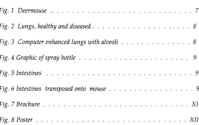
The Illustrations
The
poster gives generalinformation
aboutthe
virus and showsthe
disease
through
illustrations. The
brochure
gives more specificinformation
about
the
disease
andhow
to
preventit
as wellas contains allthe
illustrations
from
the
poster.People
can seethe
poster andbe informed
and can alsotake
home
abrochure
to
remind <*>them
ofthe
poster and getmore
information.
The illustrations
werea combinationof
traditional
illustrations
andcomputer generated
illustra
tions.
The
traditional
artwas painted
in
watercoloron
hotpress
watercolorboard
andis
shownin
figures
1
and2.
To
paintthe
mouse(Fig.
1),
flat
washeswere usedto
build up
volume and shape.
Then,
using
a size00000 brush
anddry-brush technique,
alayer
of ''fur" was paintedevenly
overthe
mouse.With
aflat
washbrush
and [image:8.612.85.551.345.697.2]plainwater
the
"fur" was smoothed out andblended
by laying
washes ofwaterover
it. After
that
dried,
more washes werelayered
onin
the
shadow areasto
really build
volume.More
"fur"
was
dry-brushed in
opaque watercolors overthese
washesto
finish
the
mouse and giveit
alife-like
look. The
eye wasdry
brushed
in
opaqueblack
with a whitehighlight
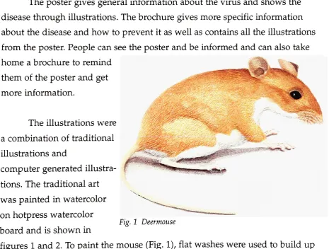
masked out.The
lungs
(Fig.
2)
werepainted
in
the
same manner asthe
mouse.The bodies
wereflat
washeswith
the
bronchi
masked out.Later
these
weredry
brushed in
as well asthe
capsules and
fissures. The
lung
painting
wasdeliberately
left
flat
and unfinished.
It is
the
backdrop
for
some ofthe
computer work.
The
computer work wasdone
in Adobe
Illustrator
andAdobe
Photoshop
.
For the
lung
illustration,
the
watercolorlungs(Fig.
2)
were scannedin
Figa2
Lungs,
healthy
anddiseased
and used as a
background.
Sketches
of alveoli were scannedin
at150
dpi
andIllustrator
was usedto
make paths ofthe shapes,
as atthe
time
this
wasthe
most efficientuseof
the
availabletechnology.
These
paths wereimported into
Photoshop
and saved asworking
paths.These
paths werethen
filled
withcolor
from
the
painting
andmanipulated
to
createthe
alveolicoming
out ofthe
lungs. The
airbrush
tool
was usedto
paintthe
alveoli and give
them
a "3-D"feeling.
Some
ofthe
paths ofthe
cellswere stroked
to
make cellborders
and a soft-edged, adjusted
paintbrush gave
them
nuclei.The
outer paths were
filled
with shadowcolors and moved
into
positionfor
the
cast shadows.The
excess parts ofthe
shadows were erasedto
meetthe
edges ofthe
lung
tissue.
All
ofthe
individual
elements were
done
on separatelayers
andthen
flattened into
onefinal layer
(Fig.
3).
A
copy
wasenlarged
for
the
poster and another was reducedfor
the
brochure
maintaining
150 dpi.
r? u j.u u* t- a * 11 Fi8-
4 Graphic
of spray
bottle
Each
ofthe
graphics as inFig.
4
wereoriginally
pencil sketches scanned at
150
dpi into Illustrator
wheretheir
shapes werecreated.
Each
sketch wastraced
using
the
pentool
to
make a shape ofthe
sketch.
The
shape wasfilled
with solidblack. A
copy
ofthis
shape was scaledto
fit
ontop
ofthe
black
shape andfilled
with white.The
edges ofthe
whiteobjectwere
then
manipulatedto
revealthe
black
underneath and givethe
appearanceof
the
original sketchwithits
variance ofline
weight.These
solid objects werethen
imported into
Photoshop
wherethe
white areas were selected andfilled
with
the
appropriate colors.These
werethen
copied and resizedkeeping
the
same
dpi
to
fit
eitherthe
poster orbrochure.
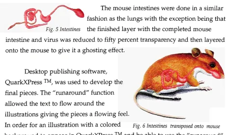
The
mouseintestines
weredone
in
a similar [image:10.612.118.540.58.213.2]fashion
asthe
lungs
withthe
exceptionbeing
that
Fig. 5 Intestines
the
finished layer
withthe
completed mouseintestine
andvirus was reducedto
fifty
percenttransparency
andthen
layered
onto
the
mouseto
giveit
aghosting
effect.Desktop
publishing
software,
QuarkXPress
,
was usedto
develop
the
final
pieces.The
"runaround"
function
allowed
the text
to
flow
aroundthe
illustrations
giving
the
pieces aflowing
feel.
In
orderfor
anillustration
with a coloredfig.
e Intestines transposed
ontomouse
[image:10.612.76.537.453.727.2]10
function,
aclipping
path was createdin
Photoshop
that
allowedthe
illustration
to
appear withoutthe
surrounding
white space .The
QuarkXpress
document
with
the
appropriateillustrations
savedin
a special graphicsfile
werethen
saved
to
zip
disks
andtaken to
a printer.The
printers provedto
be
an educationin
and ofthemselves.
The
illustrations
weretoo
large
and complexfor
them
to
handle. Resolutions
had
to
be dropped
to
anincredibly
low 80 dpi
for
the
lungs
and mouse and100 dpi
for
the
graphics.The
clipping
pathshad
to
be
redoneby
filling
the
white spacearound
the
illustrations
with amatching
background
color and a more simplepath
drawn
aroundthe
illustration in
orderfor
the
printerto
readit.
Even
withall
the
precautionstaken
and allthe
fine
adjustmentsmade,
the
final
printouthad
some problems.Colors
weren't quite rightandbecause
the
poster andbrochure
weredone
ondifferent
printersthey
didn't
match exactly.Overall,
CO
bo
XI
CO
2
J-lo
M-4
IH
O
> feO ex 0 T3 3 0) 0 Mh cu
o o o X x 13 J-l T3
6
o 03 a> o u M-H 5-t 4-t o> (d 1/1 4^ a> 03o P* -*T ' o u i to 3 -a X> 0 -4-CO X C 03 u X> O 4-' 03 a> t-i
S
OJ J-l 03 4-> OJ J-l c c CO 3 o c to "11 a>03 two a. Ui *C co
g
*03
s
td G coP
"3
a> to 3 O x; -4-J > os
OJ > 03 c 03 0) j-u c o o to u <y T3 Dh C & T3 a 00 03 C o3S
X a 03 u t>0 3 O C OJ u a> >, Hs
o OS a in to o u aj x; oto
X
toTJ
u to OJ QJ 5-1
3
j-i to
3 03 > pH 03 CX <U
o > u to Ui
OJ -M 3 to a .ttj)w s J-< o 0> H-l c
O o <u >>
"0 4-
>-3 -3 X> 4-' 03 03 a; "d CD to oJ
to o -d CO at
3
M-H
t1 to
X! -5 at x; to -^ 03 M-< I I G <U 03 3 Dh tO X
CO j_ >_. _ >- TO
o a> u
ex x to
x C OJ
^
a> J-J',y <
OJ u CD
1 1 1 1 1 1 1 1
i
1 1 1
1 1 It
1 1 1 1 1 1
1 1
1
1
1 1 1 1 1 1
1
1 1 1 1 1
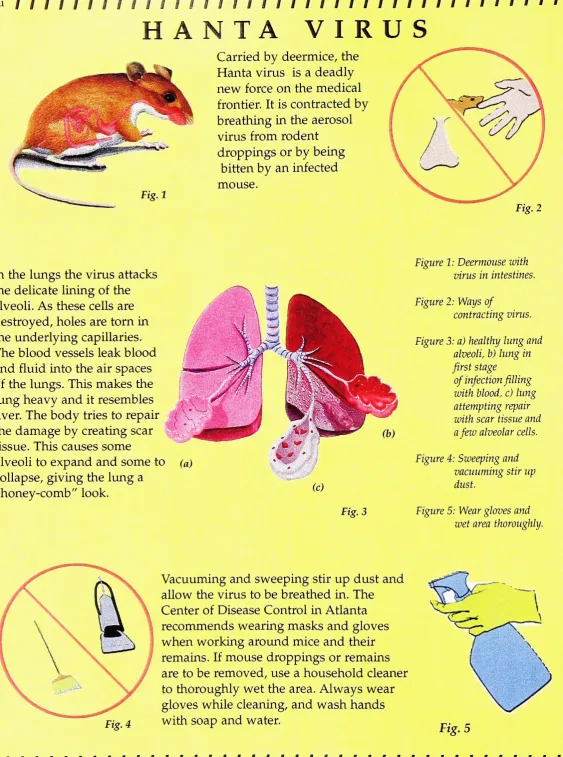
HANTA
VIRUS
Carried
by
deermice,
the
Hanta
virusis
adeadly
new
force
onthe
medicalfrontier. It
is
contractedby
breathing
in
the
aerosol virusfrom
rodentdroppings
orby being
bitten
by
aninfected
mouse.
In the
lungs
the
virus attacksthe
delicate
lining
ofthe
alveoli.As
these
cells aredestroyed,
holes
aretorn
in
the
underlying
capillaries.The
blood
vesselsleak blood
and
fluid into
the
air spaces ofthe
lungs. This
makesthe
lung heavy
andit
resemblesliver.
The
body
tries to
repairthe
damage
by
creating
scartissue.
This
causes somealveoli
to
expand and someto
collapse,
giving
the
lung
a"honey-comb"
look.
(0
[image:13.612.20.584.11.769.2]Fig, 3
Figure 1:
Deermousewith virusin
intestines.Figure 2: Ways
of
contracting
virus.Figure 3:
a)healthy
king
and alveolib)
lung
in
first
stageof
infection
filling
with
blood,
c)lung
attempting
repair with scartissueanda
few
alveolar cells.Figure 4:
Sweeping
andvacuuming
stirup
dust.
Figure
5: Wearglovesandwet area thoroughly.
Fig. 4
Vacuuming
andsweeping
stirup dust
andallow
the
virusto
be breathed
in.
The
Center
ofDisease Control in
Atlanta
recommends
wearing
masks and gloveswhen
working
around mice andtheir
remains.If
mousedroppings
or remains areto
be
removed,
use ahousehold
cleanerto
thoroughly
wetthe
area.Always
weargloves while
cleaning,
and washhands
withsoap
and water.Fig. 5
Xlll
Bibliography
CDC,
Public
Health
Service,
U.S.
Department
ofHealth
andHuman
Services,
"Morbidity
andMortality
Weekly
Report"Vol. 45/No.l4
(1996)
Eisenberg,
John
E,
"New World Rats
andMice."
The
Encyclopedia
of
Mammals,
Ed.
David W.
Macdonald,
1987.
640-643.
Hjelle, Brian,
M.D.
"Hantavirus,
withemphasis onFour
Corners
Hantavirus"
http://www.uct.ac.za/depts/microbiology/hanta.html.
10 Nov.
1996
Ing,
Roy,
M.D.
"Preventing
Hantavirus
Disease"