NATIONAL INSTITUTE OF SIDDHA
Chennai - 47THE TAMIL NADU DR. M.G.R. MEDICAL UNIVERSITY, CHENNAI - 600 032
A STUDY ON
IYA ERAIPPU
(DISSERTATION SUBJECT)
For the partial fulfillment of the
requirements to the Degree of
DOCTOR OF MEDICINE (SIDDHA)
BRANCH I- MARUTHUVAM DEPARTMENT
.
CERTIFICATE
This is to certify that I have gone through the dissertation
submitted by Dr. S.BHAVANI, a student of final M.D.Siddha, branch I,
Maruthuvam department, National Institute of Siddha, Chennai - 47 and the
dissertation work “A study on Iya Eraippu” has been carried out by the
individual only. The dissertation does not represent or reproduced the
dissertation submitted and approved earlier.
Place: Chennai – 47. Professor and Head of the Department
Date: Branch I, Maruthuvam department,
National Institute of Siddha,
INTRODUCTION
The siddha system of medicine is our heritage knowledge on
health care, a treasure of indigenous medicine. Siddha system of
medicine is being practiced from the birth of our Tamil language. It
has its own lot of numerous specialities & incomparable to other
systems of medicine. Inspite of the great & spectacular advances
in modern medical sciences in the war against diseases &
disorders the mankinds rely on traditional system of medicine and
seeks the usage of medicinal plants & their products for curing
ailments.
Herbal medicine believes that the ailments results only in
case of internal uncleanliness and tries to root them. Siddha
system of medicine excels in the manner in which it deals with the
human body and its diseases. It tends to lay emphasis in
prevention of diseases rather than curing it. It also encourages one
to maintain his health by paying attention to balance one’s life
through diet and lifestyle. In enables one to understand how to
create balance of body, mind and soul according to one’s own
individual constitution and how to make life style changes to regain
the balance that they have lost in their physical living and maintain
the vital force essential for leading successful life.
In siddha system of medicine the tridoshas namely, vatha,
pitha and kapha are essential constituents of the living body which
‘caph;f;fhjhuk; caph;jhnjdTk;
Kg;gphpthfp Kf;FzkZfp
cliyAk; capiuAk; Nkhk;gpf;fhj;J
tUnkd; KJkiw tFf;Fe; JzpNg”
In healthy persons, the tridoshas maintain proper functioning
of all the organs and tissues depending upon individual needs.
Siddha system of medicine lays great emphasis on early changes
caused by disturbances of doshas in the whole body, as a result
of excessive turnover of these substances and it is at these stages
that one can prevent the development of disease in a full fledged
manner.
Siddha system of medicine is also known for its simple
principle.
‘czNt kUe;J> kUe;Nj czT”
Which is also said by Hippocrates as
“Let thy food be medicine
And the medicine be thy food”
The two words food & medicine denote the same meaning.
In health it is taken as food and taken as medicine in ill- health
The formation of six tastes from five elements
(panchabuthas) & their role in siddha medicinal formulations &
principles are well known.
It is of paramount importance to acquire international
acceptance of our own siddha system of medicine is of utmost
importance & demanding in this current situation. This brings the
need for scientific investigation of herbs used in siddha system
using the modern parameters & methods of study like
phytochemistry, biochemical analysis, pharmacological study &
clinical trial. These all steps, one or the other will give us new
insights and understanding into the rationale for the use of these
AIM
The drug Ilavangathi choornam chosen from siddha literature
is to be studied in 50 Iya Eraippu patients by open clinical trial for
the duration of one month. The results and observations have to
be documented by means of standard statistical calculations. By
this the efficacy of a siddha drug which in unrevealed, yet is to be
OBJECTIVES
1. The Primary objective of this study is to do an open clinical
trial on Iya Eraippu noi affected individuals with the trial drug.
Drug- Ilavangathi choornam 4 g t.d.s. with honey, after food.
Taken from the literature Sigicharathnadeepam.
2. To make a detailed study of various siddha literatures like
Yugi vaidya chinthamani 800, Agasthiyar 2000, Theraiyar
Vagadam, Jeevaratchamirtham, etc.
3. To analyse the correlation of etiology, classification, signs &
symptoms of Iya Eraippu noi in siddha aspect with Bronchial
asthma in modern aspects.
4. To have an idea about the incidence of the disease with
regard to age, sex, socio-economic status, occupation, family
history and paruvakalam.
5. To study how the disease Iya Eraippu Noi alters the normal
condition under the headings mukkutram, poripulangal,
seven udalthathukkal, Neerkuri & Neikuri and Envagai thervu
especially, Naadi Nadai.
6. To make a detailed clinical evaluation of the disease by a
careful examination on etiology, signs & symptoms,
complaints, past history of patients to allergic disease,
sinusitis, or occupation, treatment and prognosis during the
7. To utilize the possible modern evaluatory equipment and lab
investigation to conduct the clinical trial.
8. To evaluate the biochemical, phytochemical &
pharmacological studies of trial drug to establish the efficacy.
REVIEW OF LITERATURES
SIDDHA
The disease Bronchial asthma can be correlated in Siddha
as Iya Eraippu or Mandara Kasam. Here, we can discuss on
various author’s view on Eraippu noi, beginning from their etiology,
classification, prodormal signs & symptoms, diseased state,
prognosis of disease, etc.
Etiology:
According to Yugi vaidya chinthamani – 800,
‘Ntfpd;w tjpfkhk; GifapdhYk;
kPWfpd;w ghzj;jhy; kpFf;Fe;jhNd
ghzj;jhy; gukhf;fpdp kpFf;ifahYk;
ghu khkprq;fs; Grpf;ifahYk;
jhzj;jhw; rQ;rhuk; jtph;f;ifahYk;
rhpglh gjhh;j;jq;fs; Grpj;jjhYk;
jPzj;jhw; GrpahkypUf;ifahYk;
Nrapioahh; NkypigQ; rpijtjhYk;
khzj;jhy; khJf;f kiltjhYk;
• Smoke and fumes
• Excessive intake of cold & hot drinks
• Due to disturbances in digestion
• Excessive intake of non- vegetarian food
• Consumption of improperly cooked food
• Not being generous
• Excessive sexual indulgence
According to siddhar Kaiyezhuthu pirathi,
‘fhy ngUf;FzTg; nghUs; jz;zPh; khwy;
fUjpUky; kpfy; the;jp Fsph;e;j fhw;W
khy; nra;J ehs;NjhWk; tUe;Jk; fha;r;ry;
ke;jd Kaph;epiyapy; mbfs; jhf;fy;
VyrPjNgjp tplghz;L Giffs;
,yfpa ney;yhjp kzpr; RizAl; nry;yy;
Nky; topapy; rpythpD kpiug;ghk; NehA
NkTnkd Kdpth;fs; tpsk;gpdNu”
• Excessive intake of food at inappropriate time
• Change in drinking water
• Cough
• Vomiting
• Cold air
• Fever
• Anaemic due to toxic substances
• Fumes and smoke
• Pollens from grasses etc., any of the above are the
Causes which may induce asthma
According to Jeevarakshamirtham
• Excessive cough
• Intake of substances which promotes vatham
• Diarrhoea & vomiting
• Anameic due to toxic substances
• Exposure to cold climate & cold air
• Trauma to vital organs.
The modern science gives atopic etiology for Bronchial
asthma, which our siddhars have also defined as an important
cause which was referred in Agasthiyar Guru Naadi Sasthiram.
‘cw;wpLk; cyfj;NjhUf;F cWgy tpahjpnay;yhk;
kw;wpLk; Fzq;fs; jd;id gfh;e;Jiu nra;a Ntz;by; xj;jpLk; rd;duhYk; clYapUsthYk;
mj;jpkh kiyapy; thOk; khKdp tFj;jjhNk”
So as the Thirumoolar says,
Prodormal signs & symptoms
A siddha maruthuva Kaiyezhuthu pirathi says, about the prodormal
signs and symptoms as
‘khh;gpy; tpyhtpuz;by; kw;WkpU nehpapy; Nrh;e;J
typj;jy; jpzwy; m‡jhy; %r;R
cg;gy; tapw;wpy; cUJNt Kw;Fwpahr;
nra;A kpiug;G Neha;f;fpjidr; Nrh;”
• Pain in the chest
• Pain in the intercostal region
• Dyspnoea
• Distention of abdomen
• Followed by asthmatic attack
Theraiyar, says in his Theraiyar vagadam as follows
‘te;jpLk; nts;Nshf;fhsk; thaJ jpj;jpg;ghFk;
nehe;jpLk; gplhpkz;il ke;jKk; kpisg;gpNdhh;f;Fk; Ke;jNt jiyjh ndhe;J rhPu KfKq; Fj;Jk;
fe;juj; njhz;il ehrp fufud;WlNd Jk;ky;”
• Nausea
• Sweet taste in the mouth
• Pain in the occipital region
• Indigestion
• Headache
• Irritation in throat, nasal region
• Sneezing
• Sneezing is followed by dry cough
In siddha maruthuvam, asthma is also compared to
mandarkasam or kulir Erumal Noi.
‘jhdhd J}aNjhh; ehrp jd;dpy;
ryNeha;ePh; jhd; tpOe;J Jk;g Yz;lhk; khdhd khh;G neQ; rilj;J %r;R
tYthfg; ghk;GNghy; rPw yhFk; fhdhd fz;lnkhU KfKq; fhJq;
fhakJq; frpthfp tpah;it ahFk; Vdhd ,UknyhL Nfhiof; fk;ky;
,iug;ghF ke;jhu fhrkhNk.”
• Rhinitis
• Sneezing
• Constriction of chest like sensation
• Breathing sound is like snake snarling
• Sweating over face & body
• Cough with little expectoration
• Dyspnoea are the signs & symptoms of mandara kasam
‘ke;jhu fhrk; kiokg;G khh;G neQ;R
je;jhd kPis jdpailj;j Fj;jpUky; Jk;gy; typ ehrpePh; Rukpisg; ghrkjp
- Mandara Kasam occurs due to rain, dyspnoea followed by
constriction of chest like sensation, non – productive cough,
sneezing, rhinitis, sometimes fever.
Classification:
According to siddha maruthuram, Eraippu noi is classified as:-
1. Vali Eraippu
2. Iya Eraippu
3. Iyavali Eraippu
4. Mukutra Eraippu
5. Mel Nokku Eraippu
Following are the signs & symptoms -
1. Vali Eraippu:
Consumption of food those are not easily digestible.
Wandering in hot sun.
Consuming tubers like potato.
All the condition above stated will cause vitiation of vali
dosha & together with it, if the person is physically debilitated may
result in difficulty in breathing in & out.
• A sensation of vaccum in the chest.
• This condition does not give much grief to the patient.
• This condition can be easily curable.
2. Iya Eraippu:
• Consumption of food causing Kapha dosha.
• Due to rain and cold air vitiated kapha reaches head
results in nasal block, rhinitis followed by constriction of
chest difficulty in inspiration and expiration.
• If expectoration is possible with cough, the difficulty in breathing will get relieved.
• Otherwise, the patient gets dyspnoeic, wheezing.
• Patient is unable to lie down supine.
• Sweating in forehead, discoloration of face, anxious look
due to dyspnoeic state, cold peripheries, dryness of
mouth, severe dyspnoea, unable to lie down supine.
• These are the signs & symptoms characterstic of Iya
eraippu or ‘Thamakka Swasam’.
3. Iya Vali Eraippu:
In this diseased state, both Iyam & vali dosha together get
deranged and results in following signs & symptoms-
• Vali dosha gets deranged and along with Udanan, causes
difficulty in inspiration & expiration.
• Also causes constipation & oliguria, distention of abdomen.
• Dryness of mouth, redness of eyes, sweating, state of
delirium & giddiness.
4. Mukutra Eraippu:
The tridoshas together gets deranged and alters the normal
state of udanan, abanan, vyanan & samanan results in emaciation
of seven udalthathukkal thus resulting in eraippu noi worsening the
patient’s condition.
• Before setting in wheezing attack causes severe dysponea
and anxiety.
• The act of expiration is compared to breathe out by a big
cow.
• Constriction of chest, syncope, constipation, Oliguria,
distention of abdomen, myalgia, delirious state, sweating in
forehead.
• This condition is known as ‘Maha swasam’.
5. Melnokku Eraippu:
Above four types of Eraippu being refractory to treatment,
leads to chronic state called ‘melnokku eraippu’.
This occur due to altered state of udanan causing difficulty in
expiration, dyspnoea, protrusion of eyeball, dryness of mouth,
unable to lie from supine, air hunger by opening his mouth.
At this state immediate treatment should be given, otherwise
patient face gets cyanosed, keeps his mouth open gasping for
In Jeevarakshamirtham, the disease is classified into 5 types
1. Soothira Swasam
2. Thamakka Swasam
3. Vitchinna Swasam
4. Maha Swasam
5. Oorthuva swasam
Sarabendra vaidya muraigal classifies swasa noi as 7 types
1. Maha swasam
2. Oorthuva swasam
3. Chinna swasam
4. Thamaka Swasam
5. Soothira swasam
6. Pirantha maha swasam
7. Santha maha swasam
Vaidya sara sangraham classifies as
1. Manthara Eraippu
2. Swasa kasam
3. Pachai udambi ledutha swasakasam
According to Anubhava vaidya devaragasiyam the disease is
1. Arpa Swasam
2. Thamaraka swasam
3. Vitchinna swasam
4. Maha swasam
5. Oorthurva swasam
Agasthiyar 2000 says about Mandarkasam as
‘ke;jhufhrk; te;jhy; thq;fpLQ; RthrNkyh
apj;jhu nka;r;RuNk fhZkpisj;jpL kpUky; nkj;JQ; re;jhTlk;G jiyAlk;G jsutypf;F kpisg;ghFk;
ape;jhJlk;G neQ;RKfk; gj;jptypf;Fk; gz;gpJNt”
‘,f;fz kjNdnlhf;f tpUkY kpisg;Gz;lha; rpfnfd neQ;rpfl;br; rPwpL kpWfpg; gw;wp gf;fke; jhue; jd;dpy; gw;wpa fhrkhNk”
• Dyspnoea
• Fever
• Cough
• Myalgia, headache
• Pain in the chest
• Coughs, dyspnoea, constriction of chest are the symptoms
follows before an asthmatic attack.
‘Ja;aNjhh; ehrpjd;dpy; Jk;kY kpfTz;lhfp
neha;A ePuha; tpOe;J neQ;R NehTl dPiothq;F ma;apd;ke; jhufhrj; jltpJjhNd ePNfs;
nra;Akh Kdpth; nrhd;d FzkpJ njhpe;J nfhs;Ns”
• Sneezing
• Rhinitis
• Constriction of chest
• Cough, dyspnoea
In Athmaratchamirtha vaidya sarasangiragam,signs &
symptoms of mandara kasam is stated as follows
• Itching sensation over face & ear
• Sneezing, rhinitis
• Cough, dyspnoea
• Pain in the chest & intecostal region
• These symptoms are aggravated during winter season
MUKKUTRA VERUPADUGAL – SIDDHA PATHOLOGY
In Siddha system of medicine the manifestations of all
diseases are the result of derangement of tridoshas – vatha, pitha
and Kapha.
Noi Naadal Noi muthal Naadal:
‘njhz;ilnad;gpd; re;jpepwTk; Nrhw;Wg;ig ehrpjiy Xz;nlhba thkj; Jsgpj;jj; - njhz;zPh;r; Rug;gp apurjhJ Rj;jepzk; ehTk;
jukhd itaj; jplk;”
-kUj;Jt jdpg;ghly;
From the above quotation,it is clear that the stomach is one
among the sites of kapha. The function of kapha in gastro intestinal
tract is to tolerate the thirst and appetite which results in anorexia
and indigestion. It produces `amam’ in the stomach which in turn
also vitiates kapha. Thus the vitiated kapha is responsible for the
respiratory disorder.
This is also clearly understood by the following phrase.
‘fgj;jpidad;wp fhr Rthrq; fhzhJ”
- Njiuah;
Excess of kapha in the respiratory organ affects the
terminal point of respiration produces gasping and labored
breathing.
Few authors say, Eraippu noi is caused by deranged vatha.
It was admitted because the obstruction of air in the respiratory
tract is abnormal. Excessive consumption of vatha promoting diet
induces the pitha. This vitiated pitha produces more heat & this
further reaches the head resulting in rhinitis, heaviness of head
and neck & sneezing. This is indicated by the following quote-
‘gpj;jk; kpFe;jhy; <is ,UkYk; ngyj;J epw;Fk; cw;wpLk; Iaehb Xq;fpNa Jbj;J epd;why; gw;wpLk; ,Uky; <is gjwpNa ,iug;Gz;lhfp
nkj;jNt Nfhio tha; kpFjpg; gLk;”
Naadi Nadai:
‘Kg;gpzp kUtp KdpT nfhs; Fwpg;igj; jg;gh jwpAk; jd;ikAk; thj gpj;jitak; gphpitA kitjhk; Vwpapwq;fp ,ize;Jf; fye;Jkhwp
khwptUk; nraw;ifahw; gpzp Neh;ikawpe;J ePl;L kUe;Nj rPhpajhnkdr; nrg;Gth; rpj;jNu”
‘%d;wpnyhd; Wah;e;jij Kd;duwpe;J Ke;jpajid nahopj;jpL kUe;jpL jzpAk; Nehapd; je;jpukpJNt
From the above quotes, Naadi examination is the best
method to elucidate the patient’s condition to do appropriate
treatment. Also the three humors vatha, pitha & kapha together
from the Naadi & their variation from regular function results in
diseased state.
The various types of Naadi Nadai in the disease Iya Eraippu:
The Manifestations of Mandara kasam due to deranged Iya
Naadi:
As per siddha Maruthuvam, the primary cause of this
disease is due to deranged kapha, which plugs the airways and
results in difficulty in breathing.
‘jhdKs;s Nrj;J ke;jhdpypfpy; ntg;G rakPisapUky; ke;jhu fhrk; <dKWQ;re;jp tplNjNjhlk; tpf;fy;
apUj;Nuhfq;fug;ghd; tpuz Njhlk; khdizaPh; #iy jpus; tpahjp tPf;fk;
tUQ;rj;jp Rthrk; neQ;rilg;G J}f;fk; Vd KWq;fhkhiy ghz;L Nrhig
VO Ruq;fs; gyJf;Fk; tpl Kz;lhNk”
‘IaNk fjpj;j NghjwpaNt nghUky; fhZk; ieAke;jhufhrk; esph; Fsph; tpf;fy; rj;jp> nra;Akh %h;r;rilg;G jPjW fhrNuhfk;
‘tplq;fpa ita Nkypiug; Ngw;wpLk;
jlq;fpapUkpLe;jd; tpyh tpuz;Lk; NehFk;
jpU%yh; ‘cw;wpLk; Iaehb Xq;fpNa Jbj;J epd;why;
gw;wpLk; <is gjwpNa ,iug;Gz;lhf;fp nkj;jNt Nfhio tha; kpFjpg;gLk;”
m.F
Iyapitha Thontha Naadi
‘,lkhd Nrj;Jkj;jpy; gpj;jehb
vOe;jZfpy; tplKlNd tPf;fKz;lhk; jplkhd Fsph; fha;r;ry; kQ;rs; NehTe;
Njfj;jpYisr;rypisg; gpUky; the;jp tplkhd neQ;rilg;G Rthrk; tpf;fy;
ntFRuKk; ehtwl;rp ghz;L Nuhfk; mlkhd Ftisuj;j kjprhue;jhd;
mZfp ntFgy Neha;f;Fj; jlq;fz;lhNa”
According to few siddhars school of throught, the primary cause of
the disease is due to deranged vali dosha, as it affects the free flow
of air through the airways for breathing.
Mandara Kasam due to vatha Iya thodam:
‘ghq;fhd thjj;jpy; Nrj;Jk ehbg;
ghprpj;jhy; jpkph; NkT Kisr;ryhFk; jPq;fhd ,UkYld; re;ep Njhlk;
thq;fhj <is ke;jhufhrk;
typAlNd GwtPr;RAs; tPr;R tPf;fk; Xq;fhZr;Ru KlNd Rthrfhrk;
cz;lhFk; ntF Neha;f;F KWjpjhNd”
Due to ushnam with deranged Iyam causes swasakasam as
per sathaga Naadi
‘fjpg;ghd Nrj;Jkj;jpYl;bzq; $by; fye;j FzQ;rakpUky; Rthrfhrk;”
Vayu with deranged Kapha also can cause swasakasam
says sathaga Naadi
‘njhe;jpj;j Nrj;Jkj;jpy; thA $bj; njhlh;e;j Fd;kk; neQ;rilg;G Rthrfhrk;”
Seethalam with deranged Iyam causes swasam
‘milthd Nrj;Jkj;jpy; rPjsk; gw;wpy;
mZfpdhy; Rthrkilg;Gapisg;G %h;r;ir..”
Dietary recommendations & restrictions for Kapha
constitution & patients by siddhars-
‘Ntis kzj;jf;fhsp nkd;rPij rf;uth;j;jp
gPis triy Rf;F ngz;Rzq;fs; - Ntisapiy nre;jsph; fisf;fPiu nra;th; fgNjfh; epjk;
te;jdp Azj;jhd; kfpo;e;J”
‘fj;jhp Nga;g;Gly; tiu apUghfy; gUq;fsh fz;lfhhp
Mj;jpf; fha;fSk; tUf;ifkh gaw;iw fiuahy; gPh;f;fUk; gpQ;RNth; nkha;j;j #uzq; fjypj; jz;Lfisg; g+Kypq;fp KUf;fUk;Gk;
mj;jp g+rpdpf; fhaUs;sp ts;spAq; fgj;Njhh;f; fhzkhNk”
PHYSIOLOGICAL ANATOMY
The organs of respiration consist of the respiratory passages
and the lungs. The respiratory passage consists of the nasal
cavities, pharynx, larynx, trachea, bronchi & bronchioles. The
terminal divisions of the bronchioles open into the gas exchange
unit, the alveoli.
Tracheo – bronchial Tree:
The trachea is a tubular structure about 10 cm long and 1.5
cm in diameter. It begins at the lower border of the larynx and
entering the thorax, it divides into two branches-the right & left
bronchi and each bronchus enters the corresponding lung at the
hilum. The lumen of the trachea is kept patent by a number of
C-shaped rings of cartilage which are deficient posteriorly. The
gaps in the posterior wall are bridged by fibroelastic tissue and
smooth muscle. The mucous membrane of the trachea is lined by
ciliated columnar epithelium.
The Lungs:
The lungs, one on either side, are large cone shaped spongy
structures which occupy most of the thoracic cavity.
They are comparatively light because of their content of air
The lungs contain a high proportion of elastic tissue. This
elasticity is responsible for most of the expiratory force in quiet
respiration. Each lung lies free in its own pleural cairty attached
only to the mediastinum by its root.
The substance of the lung is formed by the numerous
branches of the respiratory tract which form the bronchial tree, and
several million airspaces which form the bulk of the lung; vascular,
lymphatic, nervous and connective tissue form a smaller portion.
Lobes of lungs:
The left lung is divided into two lobes by deep oblique
fissures. It extends into the lung almost to the hilus, and separates
the inferior and superior lobes. The superior lobe forms the apex
and anterior margin of the lung. The inferior lobe makes up the
diaphragmatic and the greater part of the posterior surfaces.
The right lung is divided by a similar oblique fissure. This
separates the superior and middle lobes from the inferior lobe. A
second horizontal fissure extends from the anterior margin
horizontally backwards to meet the oblique fissure in the mid
axillary line. This fissure separates the wedge - shaped middle
lobe from the superior lobe.
Bronchi and Bronchioles:
Each main bronchus after entering the lungs divides into
lobe of the lung. Each secondary bronchus or lobar bronchus
divides into segmental bronchi supplying a bronchopulmonary
segment of which there are ten in the right & eight in the left lung.
The segmental or tertiary bronchi further divide several
times, with progressive reduction in the length and diameter, giving
rise to bronchioles with a diameter of 1 mm or less. The
bronchioles branch further, the smallest subdivision being the
terminal bronchiole with a diameter of about 0.5 mm. It has been
estimated that the number of divisions from the tracheal bifurcation
to the terminal bronchiole is 16. As branching occurs with a
reduction in the diameter of each succeeding division. The total
cross sectional area increases enormously from 2.5 cm2 in the
trachea to over 10,000 cm2 at the end of the alveolar ducts. The
bronchial tree upto including the terminal bronchiole is purely a
conducting pathway for the passage of air. Respiratory gas
exchange does not occur in this region and this portion is referred
to as the ‘anatomical dead space’.
Gas exchange apparatus:
The terminal bronchiole divides into the respiratory
bronchiole. The respiratory brochioles gives rise to a number of
short passages called the alveolar ducts. These open into wider
alveolar sacs, on the walls of which are located the pulmonary
alveoli. Some alveoli are present in respiratory bronchioles, but
BRONCHIAL SMOOTH MUSCLES:
The bronchial smooth muscles are present in the
submucosal coat, appears in histological section as two helical
tracts which run in opposite direction along the bronchial tree. They
are innervated by the vagus and sympathetic nerves. Stimulation
of the vagus causes contraction of the bronchial muscles resulting
in broncho constriction. Bronchodilatation results from stimulation
of sympathetic nerves, which relax the smooth muscles
Blood vessels:
The branches of the pulmonary artery distribute venous
blood to the lungs. There is a branch of the pulmonary artery to
each lobe, bronchopulmonary segment, and lobule of the lung. The
terminal capillaries lie in the walls of the alveoli and respiratory
bronchioles where gaseous exchange takes place between the
blood and the air.
Pulmonary function tests:
Pulmonary function tests are very useful for evaluation of
lung functions in respiratory disorders. Being physiological tests,
they can only indicate whether the disease process has caused an
impairment of function; they may not be able to detect early stages
of the disease in which function has not been appreciably reduced.
They also cannot make a specific clinical diagnosis. But they give
system, and indicate the nature and extent of functional
disturbance in disorders associated with pulmonary impairment
and disability. Serial measurements are useful in following the
course of disease, evaluating therapy and determining prognosis.
TEST FOR VENTILATOR CAPACITY:
The simplest tests of dynamic ventilatory function are tests of
forced expiration. A spirometer is used for these tests, and the
procedure is called spirometry. Nowadays, computerized
spirometers are available which give a print out of the data, as well
as the predicted values.
PULMONARY VENTILATION AND LUNG VOLUMES:
Pulmonary ventilation is the process by which fresh air from
atmospher is drawn into the lungs during inspiration on an
approximately equal volume of air is expelled during the
subsequent expiration. Being a dynamic process, it is best
described as the rate of movement of volume of air. However, it is
convenient to define at this stage the quantities of gas in the lungs
at different levels of the respiratory act. These can be expressed in
terms of lung volumes and lung capacities.
LUNG VOLUMES:
There are four lung volumes which do not overlap with one
1. TIDAL VOLUME (TV):
It is the volume of air inspired or expired during one
respiratory cycle. TV in adults is about 500 ml during quiet
breathing.
2. INSPIRATORY RESERVE VOLUME (IRV):
It is the maximum volume of air that can be inspired by a
forced inspiration after a normal inspiration. It is about 1400 – 2200
ml in males and 1000-1800 ml in females.
3. EXPIRATORY RESERVE VOLUME (ERV):
It is the maximum volume of air that can be expired by a
forced expiration after a normal expiration. It is about 1000- 1800
ml in males and 600 – 1200 ml in females.
4. RESIDUAL VOLUME (RV):
It is the volume of air that remains in the lungs at the end of
maximal expiration. It is about 1100 ml in males and 1000 ml in
females.
LUNG CAPACITIES:
Four lung capacities are recognized and each includes two
INSPIRATORY CAPACITY (IC):
It is the maximum volume of air that can be inspired by
forced inspiration after a normal expiration. It is about 1800 – 2000
ml in men and 1400 – 2200 ml in women.
VITAL CAPACITY (VC):
It is the maximum volume of air that can be expelled by a
forced expiration after a maximum inspiration.
Functional Residual capacity (FRC):
It is the volume of air remaining in the lungs at the end of a
normal expiration. The FRC is physiologically very important. If
there were no FRC and the lungs were completely emptied during
each respiratory cycle, the alveolar PO2 & PCO2 will vary widely
during breathing and will interfere with diffusion of respiratory
gases.
Total lung capacity (TLC)
It is the volume of air contained in the lungs at the end of a
BRONCHIAL ASTHMA
Asthma is defined as a chronic inflammatory disorder of the
airways, characterized by reversible airflow obstruction causing
cough, wheeze, chest tighteness and shortness of breath.
Inflammation of the bronchial wall involving eosinophils, mast cells
and lymphocytes, together with the cytokine and inflammatory
products of these cells, induces hyper-responsiveness of the
bronchi so that they narrow more readily response to a wide range
of stimuli. Narrowing of the airways is usually reversible, but in
some patients with chronic asthma the bronchal wall inflammation
may lead to irreversible obstruction of airflow.
PREVALENCE AND ETIOLOGY:
Asthma is a very common disease with immense social
impact. Bronchial Asthma occurs at all ages but predominantly in
early life. About one half of cases develop before age 10, and
another third occur before age 40. In childhood, there is a 2:1 male
/ female preponderance, but the sex ratio equalizes by age 30.
From an etiologic standpoint, asthma is a heterogenous
disease and genetic (atopic) and environmental factors, such as
viruses, occupational exposures and allergens are the factors
results & provokes asthma.
Atopy is the single largest risk factor for the development of
or family history of allergic diseases such as rhinitis, urticaria and
eczema, with positive wheal and flare skin reactions to intradermal
injection of extracts of airborne antigens with increased levels of
IgE in the serum.
A significant fraction of patients with asthma present with no
personal or family history of allergy, with negative skin tests and
with normal serum levels of IgE.
These patients are said to have idiosyncratic asthma or
non- atopic asthma.
PATHOGENESIS:
Asthma results from a state of persistent subacute
inflammation of the airways. The physiologic and clinical features
of asthma derive from interaction among the resident and
infiltrating inflammatory cells of the airway surface epithelium,
inflammatory mediators and cytokines. The cells thought to play
important parts in the inflammatory response are mast cells,
eosinophils, lymphocytes and airway epithelial cells. The roles of
neutrophils. Macrophages and other cellular constituents of the
airways are less well defined.
Pathphysiology:
The pathophysiologic hallmark of asthma is a reduction in
secretions. The net result is an increase in airway resistance, a
decrease in forced expiratory volumes and flow rates,
hyperinflation of the lungs & thorax increased work of breathing,
alterations in respiratory muscle function, changes in elastic recoil,
abnormal distribution of both ventilation pulmonary blood flow &
altered blood gas concentrations when patient presents for therapy
the forced expiratory volume (FEV1) or peak expiratory flow rate
(PEFR) is typically & 40% of predicted.
Hypoxia is a universal finding during acute exacerbations but
frank ventilatory failure is relatively uncommon. Most individuals
with asthma have hypocapnia and a respiratory alkalosis. The
presence of metabolic acidosis in the setting of acute asthma
signifies severe obstruction. Cyanosis is a very late sign.
Therefore, in patients with suspected alveolar hypoventilation,
arterial blood gas tension must be measured.
Risk factors for asthma:
1. Genetic susceptibility:
Asthma and atopy show clear indications of genetic
susceptibility, the frequency of disease in family members is
greater than in the population as a whole and is greater in identical
2. Allergen exposure:
Natural allergen exposure induces asthma and airway
hyper-responsiveness. Allergens & other substances liable to provoke
attacks of asthma are pollens, mite’s in house dust, animal dander,
etc.
3. Environment and Air pollution:
Environmental causes of asthma are usually related to
climatic conditions that promote the concentration of atmospheric
pollution and antigens the air pollutants known to have this effect
are ozone, nitrogendioxide and sulfur-dioxide. In these
circumstances, although the general population can develop
respiratory symptoms, patients with asthma and other respiratory
diseases tend to be more severely affected.
4. Occupational factors:
Occupation related asthma is a significant health problem,
and acute and chronic airway obstruction have been reported to
follow exposure to a large number of compounds used in many
types of industrial processes.
In general, the agents can be classified into high molecular
weight compounds, which are believed to induce asthma through
immunologic mechanisms and low molecular weight agents can
COMMON OCCUPATIONAL CAUSES OF ASTHMA
High Molecular weight
compounds
Low Molecular weight
compounds
1. Wood and vegetable
dusts
Eg. Those of Oak, grain
flour, castor bean, green
coffee bean, gum acacia &
tragacanth, etc.
1. Metal salts eg., platinum
chrome, nickel, vanadium,
etc.
2. Pharmaceutical agents –
e.g. antibiotics, piperazine,
etc.
2. Industrial chemicals &
plastics
Eg. Toluene diisocyanate,
ethylene diamine
3. Biologic enzymes
Eg., Laundry detergents,
pancreatic enzymes,
Bacillus subtilis, etc.
3. Formalin- Hospital
workers
4. Animal & insect dusts,
serum and secretions
laboratory animals,
prawns, oyster crab, bees,
etc.
5. Infections:
Respiratory infections are the most common of the stimuli
actively and chronically destabilize asthma, and they are perhaps
the only stimuli that can produce constant symptoms for weeks.
The mechanism by which viruses induce exacerbations of asthma
may be related to the production of T-cell derived cytokines that
potentiate the infiltration of inflammatory cells into already
susceptible airways. 85% of asthma attacks in children and 44% in
adults were induced by upper respiratory tract infections, of which
the great majorities were caused by rhinoviruses.
6. Exercise:
Exercise is a very common precipitant of acute episodes of
asthma. Typically, the attacks follow exertion and do not occur
during it. This stimulus differs from other naturally occurring
provocations, such as antigens, viral infections and air pollutants in
that it does not evoke any long term sequelae.
7. Emotional stress:
Psychological factors can worsen or ameliorate asthma.
Changes in airway caliber seem to be mediated through
modification of vagal efferent activity. The extent to which
psychological factors participate varies from patient to patient and
in the same patient from episode to episode.
Patients with asthma can be categorized by whether their
symptoms are intermittent or persistent, and by the severity of their
mild asthma – intermittent or persistent can develop severe
asthma.
1. Mild intermittent asthma – symptoms occur less than weekly
with normal or near normal lung function between episodes.
2. Mild persistent asthma – symptoms occur more than weekly
but less than daily with normal or near normal lung function
between episodes.
3. Moderate persistent asthma – symptoms occur daily with
mild to moderate variable airflow limitation.
4. Severe persistent asthma – symptoms occur daily and
interfere with normal activities. There is frequent noctumal
waking and moderate to severe variable airflow limitation.
5. Severe asthma – severe distressing symptoms prevent
sleep. Severe airflow limitation responds poorly to inhaled
bronchodilators and can be life threatening
Clinical Features:
The symptoms of asthma consist of a traid of dyspnoea,
cough and wheezing. In its most typical form all three symptoms co
exist. At the onset of an attack, patients experience a sense of
constriction in the chest, often with a non-productive cough.
Respiration becomes audibly harsh, wheezing in both phases of
respiration becomes prominent, expiration becomes prolonged &
patients frequently have tachypnoea, tachycardia and mild systolic
hypertension. If the attack is severe or prolonged, there may be a
loss of adventitial breath sounds, and wheezing becomes very high
and a paradoxical pulse often develops. These two signs are
extremely valuable in indicating the severity of the obstruction.
The end of an episode is frequently marked by a cough that
produces thick, stringy mucus, which often takes the form of casts
of distal airways (curschmann’s spirals) and, when examined
miroscopically, often shows eosinophils and charcot-leyden
crystals.
Lung function tests:
Measurements of respiratory function may provide valuable
information. First, in conjunction with the clinical assessment and
to help establish a diagnosis. Secondly, they will help indicate the
severity of the condition. Thirdly, serial measurements over time
will show changes indicating disease progression or, alternatively,
a favorable response to treatment.
Forced expired volume in the first second:
The forced expired volume is defined as the volume expired
in the first one second during forced vital capacity exhalation. This
is measured from the analysis of the forced expiratory spirogram.
In normal subjects, FEV1 is more than 80% of FVC values of FEV1
are reduced in both central and peripheral airways obstruction. In
patients with reduced lung volumes, FEV1 expressed as % FVC
Peak expiratory flow rate (PEFR):
PEFR may be simply measured using equipment such as the
Wright peak expiratory flow meter. The machines are cheap &
portable and give the reading of the peak flow rate on a dial or a
linear scale. This is defined as the maximum expiratory flow rate
sustained for atleast 20 milliseconds during the forced expiratory
manouvre.
Variability in PEFR of greater than 15-20% in a single day or
from day to day is very suggestive of asthma. Most normal
subjects demonstrate less than 10% variation in PEFR over a 24 h
period. Similarly PEFR may be used as an index of response to
treatment in asthma. Finally, PEFR is the most convenient
measurement for use in the diagnosis of exercise induced asthma,
where a fall in PEFR of greater than 15% following exercise is
considered diagnostic.
Normal values are 400-600 L / min in young mean and 300 –
400 L / min in young women. Serial recordings of PEFR are useful
in distinguishing patients with chronic asthma from those with fixed
MATERIALS AND METHODS
PROTOCOL
AN OPEN CLINICAL TRIAL OF ILAVANGATHI CHOORNAM FOR THE TREATMENT OF IYA ERAIPPU (BRONCHIAL
ASTHMA)
1. BACKGROUND
Eraippu noi has been classified into five types in Siddha
system. Iya Eraippu is one among them. It correlates with the classical signs and symptoms of Bronchial asthma.
In manuscripts, the etiology of the disease Iya Eraippu or
Mandara Eraippu is stated as
"ñ‰î£ó M¬óŠ¹ õO ñ¬ö °O˜
ªð£¼Àí¾ õ¼ W› 裟Á
õ‰î è£K¼ðQJ™ õO ïó‹H¡
õNòîQ™ ñ£ø£è„ ªê¡
Á‰î£¬õòˆ¬î ïQ ¶‡®
òî¡ ªî£N¬ô Iè «õ£ƒè„ ªêŒ¶
º‰î£ù î¬ô ªî£‡¬ì JìºKf
õ¼ˆîªôù ªñ£N‰îó¡«ø"
Drug Ilavangathi choornam mentioned in Siddha literature Sigicha Rathna Deepam under Choorna Vilambam is indicated specifically for Eraippu Noi. The efficacy of this drug is not
documented. Therefore, we propose to carry out an open clinical
trial to estimate the efficacy of the drug.
1. AIMS
a) Primary aim
To estimate the efficacy of Ilavangathi choornam in the treatment of Iya Eraippu.
b) Secondary aim
To find out the side- effects of the drug, if any.
3. POPULATION & SAMPLE
The population consists of patients with Iya Eraippu (Peak Expiratory Flow Rate > 200 L / minute) satisfying the inclusion &
exclusion criteria mentioned below. The sample consists of Iya Eraippu patients attending the O.P.D of the Ayodthidoss Pandithar Hospital of the National Institute of Siddha, Tambaram Sanatorium,
and Chennai-47.
4. SAMPLE SIZE
The trial size will be 50 patients.
5. INCLUSION CRITERIA
1. Age 12 to 80 yrs.
2. Willing to be admitted in the Hospital for 30 days or willing
to attend the O.P.D. once in 10 days.
6. EXCLUSION CRITERIA
1. Smoking
2. Alcohol consumption
3. History of epilepsy or ischaemic heart disease
4. Pregnancy
5. Lactation.
7. WITHDRAWAL CRITERIA
1. Acute severe asthma
2. Occurrence of any serious illness
8. TRIAL DRUG AND DURATION
Drug – Ilavangathi choornam- 4 g t.d.s. with honey, after food.
Duration of trial treatment – 30 days
10. TESTS AND ASSESSMENTS
a. CLINICAL ASSESSMENTS
1. Dyspnoea
2. Cough
3. Wheezing
4. Sense of constriction of chest
5. Type of breathing
6. Accessory muscles of respiration
7. Hoarseness
8. Inability to sleep
11. Vocal resonance
12. Vocal fremitus
SIDDHA ASPECT- ENVAGAI THERVU
1. Naa
2. Niram
3. Mozhi
4. Vizhi
5. Malam
6. Moothiram-Neerkuri & Neikuri
7. Meikuri (Sparisam)
8. Naadi (Kaikuri)
a) INVESTIGATIONS
1. Blood test- TC, DC, ESR, Hb, RBC, Sugar (F/PP)
2. Urine test – Albumin, Sugar & Deposits
3. Motion test-Ova, Cyst.
11. CONDUCT
Iya Eraippu patients satisfying inclusion and exclusion criteria will be admitted to the trial. Informed consent will be
obtained from the patients.
Routine investigations will be carried out before and after the
by the doctor. For out - patients the trial drug will be issued for 10
days at a time. They will be advised to visit the O.P.D once in 10
days. At each visit they will be clinically assessed.Also, they will be
advised to bring back unconsumed drugs and return them during
their subsequent visit.
12. FORMS
Form I - Selection Proforma – It is used before admission
of the patients to the trial.
Form II -Assessment Proforma- It is used once in 10 days
during treatment.
13. ANALYSIS
The change in the PEFR before and after treatment will be
analyzed using Paired t-test.
For the changes observed in the proportion of patients with
RESULTS & OBSERVATION
Observations were made during the course of dissertation
work with regard to the following features.
1. Age & sex distribution
2. Socio – economic status
3. Occupational reference
4. Personal habit & diet
5. Past history & family history
6. Distribution of thinai
7. Kalam
8. Paruvakalam
9. Reference to mukutram
10. Envagai thervu
11. Neerkuri & Neikuri
12. Recording PEFR (L/min), ESR (mm/hr), Eosinophil count
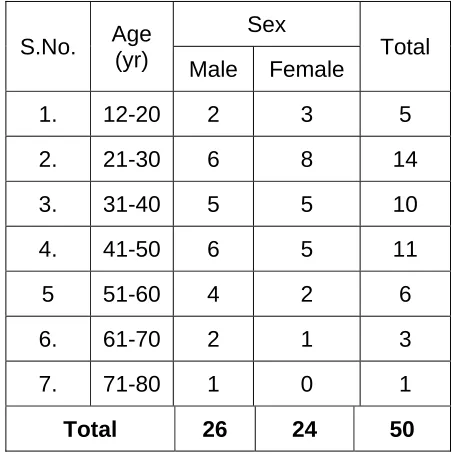
Table No.1
Age – Sex Distribution of 50 Iya eraippu patients, NIS, Chennai – 47, 2007
S.No. Age
(yr)
Sex
Total Male Female
1. 12-20 2 3 5
2. 21-30 6 8 14
3. 31-40 5 5 10
4. 41-50 6 5 11
5 51-60 4 2 6
6. 61-70 2 1 3
7. 71-80 1 0 1
1
OCCUPATIONAL REFERENCE
PIE DIAGRAM NO. 1
Housewife 34%
Miscellaneous 50%
Painter 4%
Farmer 8%
PAST HISTORY & FAMILY HISTORY
BAR DIAGRAM NO.1
50
24
8
0 5 10 15 20 25 30 35 40 45 50
DISTRIBUTION OF THINAI
PIE DIAGRAM NO.2
Kurinji 8%
Neithal 76% Marutham
PARUVAKALAM
PIE DIAGRAM NO.3
Munpanikaalam 80%
Pinpanikaalam
MUKUTRAM
PIE DIAGRAM NO.4
Kapha 87%
Pitha 2% Vatha
TABLE NO.2
OBSERVATIONS IN 50 IYA ERAIPPU PATIENTS, NIS, CHENNAI – 47, 2007.
S.
No. Date
O.P. /
I.P. No. Name
Age / sex
family h/o
H / o sinisutis /allergic PEFR Initial (L/min) final
1. 20.11.2006 S8703 Rajeshkannan 14/M - - 270 360
2. 20.11.2006 S8781 Mankali 49/M - - 440 450
3. 20.11.2006 S8839 Mangai 30/F + + 280 400
4. 22.11.2006 S9736 Durairaj 50/M - + 210 150
5. 22.11.2006 S9671 Ramasamy 75/M - + 200 150
6. 23.11.2006 S9994 Ragamathulla 32/M - - 400 390
7. 23.11.2006 T114 Nesam 70/F - - 210 200
8. 24.11.2006 T402 Mahalingam 59/M + + 150 180 9. 27.11.2006 T1611 Anandakumar 20/M + - 330 440 10. 28.11.2006 T2263 Aruldoss 31/M + + 400 480 11. 28.11.2006 T2276 Ashok 28/M - + 300 350 12. 29.11.2006 T2759 Prakash 25/M - - 320 350 13. 29.11.2006 T2732 Poongodi 32/M - + 320 320 14. 30.11.2006 T3084 Ranganayaki 57/M - + 320 350 15. 02.12.2006 T3920 Paramasivam 42/M - + 210 170
16. 03.12.2006 T4035 Balu 60/M - - 210 330
17. 04.12.2006 T4409 Abarna 26/F - + 200 240 18. 07.12.2006 T5941 Karthick 24/M - - 450 480 19. 07.12.2006 T5864 Chellam 42/F + + 220 260 20. 08.12.2006 T6183 Jayashree 40/F - + 250 370 21. 12.12.2006 T8021 Hariharan 21/M - - 220 370 22. 13.12.2006 T8443 Pushpa 40/F - - 300 360 23. 15.12.2006 T9189 Chandika 43/F + + 250 260 24. 20.12.2006 U1210 Bathuru 49/F + + 270 300 25. 21.12.2006 U1683 Murali 32/M - - 290 380 26. 21.12.2006 U1716 Karthikeyan 29/M + - 340 360 27. 22.12.2006 U2075 Shanthi 43/F - - 300 360
28. 23.12.2006 U2215 Dillibabu 34/M - - 400 470
S.
No. Date
O.P. /
I.P. No. Name
Age / sex
family h/o
H / o sinisutis /allergic PEFR Initial (L/min) final
31. 28.12.2006 U4923 Selvi 32/F - - 250 250
32. 29.12.2006 U5284 Cathirin 40/F - + 380 390 33. 29.12.2006 U5154 Sathyaseelan 19/M - - 420 440
34. 29.12.2006 U5066 Raja 42/M - - 220 260
35. 30.12.2006 U5447 Parveen 25/F + - 250 330
36. 02.01.2007 U6434 Sudha 54/F - + 250 350
TABLE NO.3
RESULTS OF OBJECTIVE PARAMETERS BEFORE AND AFTER TREATMENT OF 50 IYA ERAIPPU TREATMENT, NIS, CHENNAI – 47,
2007.
Sl. No. Eosinophil count (%) ESR (mm / hr)
Before After Before After
1. 8 6 12 12
2. 2 3 12 8
3. 2 2 20 12
4. 4 3 20 12
5. 5 4 4 4
6. 2 2 7 4
7. 2 3 8 7
8. 4 4 24 20
9. 6 5 22 22
10. 6 5 8 12
11. 6 5 16 8
12. 8 10 16 6
13. 4 5 8 8
14. 4 2 12 22
15. 2 2 8 10
16. 8 10 8 10
17. 10 10 12 16
18. 2 2 12 16
19. 2 8 4 6
20. 6 6 16 12
21 4 3 6 5
22. 4 3 24 12
23. 6 4 4 4
Sl. No. Eosinophil count (%) ESR (mm / hr)
Before After Before After
25 4 3 12 10
26. 4 3 14 6
27. 4 3 58 24
28 4 4 22 14
29 4 4 28 20
30. 6 3 14 8
31. 12 6 12 16
32. 8 2 28 22
33. 6 4 24 18
34. 4 4 22 25
35. 6 4 12 8
36. 4 4 16 12
37. 4 4 12 10
38 6 4 8 6
39. 2 2 12 6
40. 10 6 22 16
41. 10 5 18 8
42. 2 4 10 14
43. 4 3 12 10
44. 4 2 12 14
45 4 3 12 8
46. 10 6 8 12
47. 8 6 20 16
48. 3 2 12 8
49. 4 4 16 12
THE T – TEST ON PAIRED OBSERVATIONS FOR OBJECTIVE PARAMETERS
To make comparisons on the basis of paired observations on
an individual e.g., before & after treatment, t - test is done. In such
situations, the difference between the two observations must, on
average, be zero, under the Null hypothesis; the t-test described
above has therefore frequent practical application. Here, using
t-test the values of PEFR (L / min), ESR (mm / 1 hr), and Eosimophil
count (%) are calculated for testing the significance.
We could see the following data in Table No 4.
The mean value of PEFR, before treatment in 50 Iya eraippu
patients is 285.4 L/ min after treatment the mean value for the
same is 328.4 L / min the difference in the means is 43. L / min. To
test the significance of the difference paired t-test is done. The
calculated paired‘t’ – test value is 4.559 L / min. In consequence,
the quantity above is compared with P–value of 5% significance
level. Hence, the null hypothesis is rejected, since the calculated
value is greater then P < 0.05 (2.010) significance level. Therefore,
the trial drug shows considerable improvement in patients which is
statistically significant.
The mean value of ESR, before & after treatment in 50 Iya
eraippu patients is 14.9 (mm / hr) & 11.7 (mm / hr) respectively.
The difference in the means is 3.2 (mm / hr). To test the
significance of the difference paired t-test is done. The calculated‘t’
value is 3.810 mm / hr. This quantity is compared with P value of
the P < 0.05 (2.010) significance level, Null hypothesis is rejected.
Hence, the trial drug is considered statistically significant.
The mean value of Eosmophil count, before and after
treatment in 50 Iya eraippu patients included in clinical trial is
5.06% and 4.14% respectively. The difference in the means is
0.92%. The significance of the difference is calculated by paired
t-test. The calculated t-value is 3.159% compared with P < 0.05
(2.010). Hence, the Null hypothesis is rejected. The trial drug is
[image:59.612.105.542.428.606.2]considered to be statistically significant.
TABLE NO. 4
RESULTS OF STATISTICAL ANALYSIS OF SOME OBJECTIVE PARAMETERS BEFORE AND AFTER TREATMENT OF 50 IYA ERAIPPU
PATIENTS, NIS, CHENNAI – 47, 2007
S.No. Parameter Mean Difference Paired ‘t’ test value P Value Significance of the difference Before treatment After Treatment
1. PEFR (L /
min) 285.4 328.4 43 4.559
<
0.05
2.010
Statistically
significant
2. ESR – 1
Hr (mm) 14.9 11.7 3.2 3.810
<0.05
2.010
Statistically
significant
3. Eosinophil
(%) 5.06 4.14 0.92 3.159
<0.05
2.010
Statistically
TABLE NO. 5
RESULTS OF SUBJECTIVE PARAMETERS BEFORE AND AFTER TREATMENT OF 50 IYA ERAIPPU PATIENTS, NIS, CHENNAI – 47
S.No. Wheezing Constriction
of chest Dyspnoea Cough
B A B A B A B A
1. + − − − + − + −
2 + − + − + + + +
3. + − + − + + + +
4. + − + − + _ + −
5. + + + + + + _ −
6. + + + _ + + + +
7. + _ + + + _ + −
8. + + + − + _ + +
9. + − − − + + + +
10. + − − − + _ + +
11. + _ + − + _ + −
12. + + _ − + + + +
13. + − + − + + + +
14. + − + − + + + −
15. + − + − + + + −
16. + + + + + + + +
17. + + − − + + + +
18. + − + − + _ + +
19. + − + + + + + −
20. + − + − + − + −
21 + + + + + + + +
22. + − + − + _ + −
23. + − + − + _ + −
S.No. Wheezing Constriction
of chest Dyspnoea Cough
26. + + − − + + + +
27. + + + + + + + +
28. + − − − + + + −
29. + − + − + + + −
30 + − − − + _ + +
31. + _ + − + + + +
32. + _ _ − + − + +
33. + _ + − + − + +
34. + + _ − + + + +
35. + − + − + − + +
36. + − + + + − + −
37. + − + − + − + −
38. + − + + + + + +
39. + − − − + _ + −
40. + + + + + _ + −
41. + _ + − + _ + −
42. + + + − + + + −
43. + − − − + − + −
44. + + + + + + + +
45. + − + + + + + +
46. + − + + + + + +
47. + − + _ + + + +
48. + + + + + + + +
49. + + − − + − + −
50. + + + − + + + +
Note: B - Before treatment
THE CHI – SQUARE TEST FOR SUBJECTIVE PARAMETERS
Data collected in the field of medicine is often qualitative
here for e.g., the presence or absence of a symptom like cough,
wheezing, dyspnoea and constriction of chest is taken.
Comparisons between 2 or more proportions, and the test of
significance employed for such purposes is called the chi-square
test.
The chi -square test is designed to examine whether a series
of observed numbers in various categories of the data are
consistent with the number expected in those categories on some
specific hypothesis called Null hypothesis. By comparing the
computed value with the tabulated value, the probability that the
difference may have arisen from chance is obtained, this is called
chi-square test. This test is very useful test of significance which is
used here for symptoms like cough, wheezing, dyspnoca &
constriction of chest in patients of Bronchial asthma.
Observed data is represented in Table No. 6.
In all, among 50 Iya eraippu patients before treatment cough
is present as 86% & after treatment as 14%. The difference in
treatment is 72% for the chi-square test, x2 value is calculated as
15.05. On referring to tabulated values of the chi – square
distribution with 1 degree, freedom, it is found that the probability
corresponding to a chi – square value of 15.05 is greater than P <
In the 50 Iya eraippu patients underwent clinical trial with
drug Ilavangathi choornam, wheezing was present 86% in before
treatment and 14% in after treatment. The difference in treatment
is 72%. x2 value is calculated as 28.03 On referring the tabulated
values of the chi-square distribution with 1 degrees of freedom it is
found value of 28.03 is more than P < 0.05 (3.84) level. Hence, null
hypothesis is rejected and the cure is statistically significant.
The patient with dyspnoea before treatment is 76% and after
treatment is 24%. The difference in the treatment is 52%. x2 value
is calculated as 12.89. On referring to tabulated values of the chi –
square distribution with 1 degrees of freedom it is found that the
probability corresponding to x2 – value of 12.89 is greater than P <
0.05 (3.84) level. Hence, Null hypothesis is rejected & the trial drug
is proved to be efficacious.
In all 50 Iya eraippu patients of asthma, the patient suffered
with constriction of chest is 60% before treatment and 40% after
the treatment. The difference is 20%. The x2 value is calculated, on
referring to tabulated values of chi-square distribution with 1
degrees of freedom. It is found that the probability corresponding
to x2 value of 15.058 is greater than P < 0.05 (3.84) Hence, Null
hypothesis is rejected & the trial drug shows statistically significant
TABLE NO.6
RESULTS OF STATISTICAL ANALYSIS OF SOME SUBJECTIVE
PARAMETERS BEFORE AND AFTER TREATMENT OF 50
IYA ERAIPPU PATIENTS, NIS, CHENNAI – 47, 2007
S.No. Parameter
Percentage present
Difference Paired x2 P value Significance of the difference Before treatment After treatment
1. Cough 86% 14% 72% 15.05 <0.05
(1d.f) 3.84
Statistically significant
2. Wheezing 86% 14% 72% 28.03 <0.05
1 (d.f) 3.84
Statistically Sgnificant
3. Dyspnoea 76% 24% 52% 12.89 <0.05
1 (d.f) 3.84 Statistically significant 4. Constriction of chest
60% 40% 20% 15.058 <0.05
1 (d.f) 3.84
DISCUSSION
The present study brings on record, the work done on Iya
Eraippu by clinical trial of Ilavangathi choornam with well defined
protocol.
The present work was carried out on Iya Eraippu
representing various views of its aetiology, classification,
prodormal signs, signs & symptoms, Noi Naadal Noi mudal naadal,
Naadinadai & also Dietary, Pranyamam & Yogasana prescriptions.
The Iya Eraippu patients are studied with regard to their past
history of Ovammai & Peenisam, (allergic rhinitis, sinusitis & family
history of asthma). This is represented by bar diagram (1)
In the Iya Eraippu patients admitted to clinical trial, age & sex
distribution presented by Table No.1 & occupational history,
distribution of thinai, incidence in paruvavakalam, derangement of
mukuttram are studied and represented by pie diagram (1,2,3,4)
respectively.
The observation on objective parameters like peak expiratory
flow rate (PEFR) & laboratory investigation particularly, Eosinophil
count and ESR (mm / hr) are calculated by statistical method and
tested for significance. This is presented in Table No.4.
The observation on subjective parameters such as cough,
wheezing, dyspnoea and constriction of chest are also analysed
The preliminary biochemical analysis were carried out and
presented.
The preliminary photochemical analysis were carried out with
dichloromethane solvent and presented.
The acute toxicity study of drug Ilavangathi choornam is
conducted on albino rats & recorded.
The trial drug is subjected to study on guinea pig for
pharmacological activity – anti spasmodic effect & kymograph is
SUMMARY AND CONCLUSION
The open clinical trial of dry Ilavangathi choornam is
conducted in 50 patients of Iya Eraippu at Ayothidoss Pandithar
hospital of the National Institute of Siddha, Tambaram Sanatorium,
Chennai - 47.
Among the fifty patients, the 50% of patients were having
history of allergic rhinitis, 24% suffer from sinusitis & 4% with
family history.
The patients were belonging to Neithal thinai are the most as
to 76%.
The incidence of disease, Iya Eraippu looking with respect to
paruvakalam showed about 80% suffer mostly during Munp
anikaalam (December - Feburary).
The derangement of Kapha in Iya Eraippu patients is about
87% vatha, 11% & pitha about 2% only.
In the 90% of the Iya eraippu patients neikuri is `muthu’ i.e.
kabha neer.
The biochemical analysis of trial drug showed the presence
of chloride.
The phytochemical analysis of the trial drug revealed the
presence of carbohydrates & glycosides, phenolic compounds,
The pharmacological analysis of trial drug in guinea pig
showed potent anti-spasmodic activity.
The trial drug is estimated non-toxic at a high dose of 2000
mg/kg of body weight in albino rats.
This compound trial drug is formulated in such a way as to
alleviate any digestive disturbances and also to prevent from any
ill-effects due to excessive kaarpu suvai.
The trial drug had showed no adverse effects when given to
the patient during the trial period.
The trial drug has been able to relieve cough and enhance
expectoration, thereby relieving dyspnoea in Iya eraippu patients.
The objective parameters-PEFR (L/min), ESR (mm/hr),
eosinophil count (%) all measured are proven statistically p<0.05
significant.
The subjective parameters-cough, wheezing, dyspnoea,
and constriction of chest showed significant improvement after the
PREPARATION OF TRIAL DRUG – ILAVANGATHI CHOORNAM
The drug Ilavangathi Choornam is choosen for trial from the
Siddha literature Sigicharathna deepam indicated for Iraippu noi.
The constituents of trial drug include-
1. ,ytq;fk;
2. ,ytq;fg;gl;il
3. rjFg;ig
4. fUQ;rPufk;
5. Vyk;
6. jdpah
7. rPufk;
8. jhsprgj;jphp
9. rpWNjf;F
10. jpg;gpyp %yk;
11. nrt;tpak;
12. rlhkhQ;rpy;
13. [hjpf;fha;
14. [hjpgj;jphp
15. Xkk;
16. Nfh\;lk;
17. FNuhrhzp Xkk;
18. tha;tpsq;fk;
19. khrpf;fha;
21. rKj;jpug;gok;
22. rpWehfg;g+
23. rpw;wuj;ij
24. Rf;F
25. kpsF
26. jpg;gpyp
27. nghd;KRl;il Nth;
28. tpyhkpr;R Nth;
29. kfug;g+
Each of the above constituents are cleansed & purified, taken
as same quantity then made into fine powder. The dosage is given
as 4 gm three times a day (½ Njhyh ,UNtis)
As per T.V. Sambasivapillai dictionary Vol.5 Page No.634 & 635
kfug;g+ is known as nte;jak;.
Also, by gr;rpiy - ghpghi\ mfuhjp Page 104
& %ypif kUj;Jt mfuhjp Page 197 kfug;g+ is nte;jak;.
Properties of trial drug:
The trial drug Ilavangathi choornam consists of 29 drugs.
Most of its constitutents are having pungent taste (Kaarpu suvai).
Least among them belongs to bitter and astringent taste (Kaippu &
thuvarppu suvai).
We all know, that siddha system of medicine is based on five
A maruthuva thanipadal quotes the formation of six tastes
from five elements.
‘kz;ZlNd Gdy; jPf;fhy; Kiwahfr; Nrh;e;jpl;lhy; tUkh kpdpg;Gj;
jpz;zkpyk; Jth;g;gpurk; rjhfjpNah lhh;jPtpd; jplkh Kiwg;Gk;
vz;zwpa frg;G Kz;lhk; jz;zPhpy; jzypidg;ghy; vOkh Kth;g;G
cz;zhpa mWRitapd; rpwg;gpnjDq; FUrpj;jh; ciuj;j kiwNa”
kz; + ePh; - ,dpg;G(Sweet)
kz; + jP - Gspg;G(Sour)
kz; + fhw;W - Jth;g;G(Astringent)
ePh; + jP - cth;g;G(Salt)
fhw;W + jP - fhh;g;G(Pungent)
fhw;W + ntsp - ifg;G(Bitter)
Maruthuva thanipaadal quotes the action & character of
Kaarpu, Kaippu & Thuvarppu suvai as below.
fhh;g;G Ritapd; nra;if :
fhh;g;G - Fzk; :
‘njhz;ilapy; Yz;lhk; kpz;L nra; gpzpfs; mopj;jpLk; ehb ehs; milg;gpidf;fow;wpNa..”
• The pungent taste stimulates appetite.
• Cures the diseases of throat.
• Destroys the vitiated kapha .
Jth;g;G Ritapd; ,ay;G :
‘fl;LtJrw;Wf; fufug; ghf;FtJ
jpl;lkha; Njhw;gjdQ; nra;tJ - kl;bw; nfhOg;GePh; ky;Fq; nfhOg;Gk; tul;ly;
njhopyhe; Jth;g;Gr; Ritf;F”
Astringent controls the excessive discharge of faeces, urine &
bleeding.
nra;if:
‘nghy;yh itak; khw;Wk;.. Fsph;e;j Jth;g;gpd; Ntiy” ifg;gpd; gz;Gk; nra;ifAk;
It the astringent changes the decreased appetite due to various
reasons.
It changes the vitiated kapha.
‘NtW fhuzk; tpisj;j t+z; ntWg; Nghl;L kpay;gh Naw;f tpUk;ghr; Ritahk; gpj;jika tpfw;gq;...
tha;ePUwy; mow;rpAk; jzpf;Fk; nka;ePh;f; frpitAe; jbiaAe; jbAk;
Cz;ryk; kyQ;ryk; epzryk; vd;gpDs; twl;Lk;....”
The astringent taste changes the hatred towards food.
It converts the vitiated pitha & Kapha.
It controls the hypersecretions of glands in the body.
Considering the actions & characters of Kaarpu, Kaippu &
Thuvarpu suvai, the drug is formulated in such a way that the
adverse effect of excessive kaarpu suvai is counteracted by
Kaippu & thuvarpu suvai. Also, the kaarpu & thuvarpu suvai
corrects the deranged deepakini in the body & produces good
appetite. Both the kaarpu & thuvarpu suvai has the ability to control
excessive secretion, as here, it will be appropriate to say the
sputum production & expectorant. Hence Kaarpu suvai, by its
inherent property antagonizes kapha by having fire as its
component.
Generally kapha disease patient’s complaints of reduced
appetite or anorexia. In Iya eraippu patients their condition gets
worse if they suffer from dyspnoea particularly after food due to
improper digestion.Siddhars had given us plethora of drugs, mostly
combination of herbs as compound drugs.The drug Ilavangathi
choornam is also one among those formulation.Many drugs being