A Dissertation on
“TO ASSESS THE EFFICACY OF ANTERIOR APPROACH
TO BOTH FEMORAL AND SCIATIC NERVE VIA A
SINGLE SKIN SITE INJECTION TECHNIQUE vs
CLASSICAL TWO SITE INJECTION TECHNIQUE USING
NERVE LOCATOR FOR LOWER LIMB SURGERY”
Submitted to the
THE TAMILNADU DR. M.G.R. MEDICAL UNIVERSITY In partial fulfilment of the requirements
For the award of degree of M.D. (Branch-X)
ANAESTHESIOLOGY
GOVERNMENT STANLEY MEDICAL COLLEGE & HOSPITAL THE TAMILNADU DR. M.G.R. MEDICAL UNIVERSITY,
CHENNAI, TAMILNADU
CERTIFICATE
This is to certify that this dissertation entitled “TO ASSESS THE EFFICACY OF ANTERIOR APPROACH TO BOTH FEMORAL AND SCIATIC NERVE VIA A SINGLE SKIN SITE INJECTION TECHNIQUE vs CLASSICAL TWO SITE INJECTION TECHNIQUE USING NERVE LOCATOR FOR LOWER LIMB SURGERY”, Submitted by DR.GIRISH.B.K to the faculty of ANAESTHESIOLOGY, The Tamil Nadu Dr. M.G.R. Medical University, Chennai, in partial fulfillment of the requirement in the award of degree of M.D. Degree, Branch - X (ANAESTHESIOLOGY), for the April 2013 examination is a bonafide research work carried out by him during the period of November 2011 to May 2012 at Government Stanley Medical College and Hospital, Chennai under our direct supervision and guidance of Dr. KUMUDHA LINGARAJ, Professor, Department of Anaesthesiology at Stanley Medical College, Chennai.
Prof. Dr .KUMUDHA LINGARAJ,
M.D., D.A.,
Professor, Guide,
Department of Anaesthesiology, Stanley Medical College and Hospital, Chennai – 600 001.
Prof. Dr. P. CHANDRASEKAR, M.D., D.A., Professor and H.O.D.
Department of Anaesthesiology Stanley Medical College
and Hospital, Chennai – 600 001.
Prof . Dr. GEETHA LAKSHMI, M.D, Ph.D Dean
CERTIFICATE
This is to certify that the dissertation presented herein by
“TO ASSESS THE EFFICACY OF ANTERIOR APPROACH TO
BOTH FEMORAL AND SCIATIC NERVE VIA A SINGLE SKIN
SITE INJECTION TECHNIQUE vs CLASSICAL TWO SITE
INJECTION TECHNIQUE USING NERVE LOCATOR FOR
LOWER LIMB SURGERY”, Dr.GIRISH.B.K., is an original work done
in the Department of Anesthesiology, Government Stanley Medical College
and Hospital, Chennai in partial fulfillment of regulations of the Tamilnadu
Dr. M.G.R. Medical University for the award of degree of M.D.
(Anesthesiology) Branch X, under my supervision during the academic period
2010-2013.
Prof.Dr GEETHA LAKSHMI, M.D.,Ph.D. Dean
Stanley Medical College, Chennai – 600 001.
Prof. Dr. P. CHANDRASEKAR, M.D., D.A., Professor and H.O.D.
Department of Anaesthesiology Stanley Medical College
DECLARATION
I, Dr .GIRISH.B.K , Solemnly declare that the dissertation “TO ASSESS THE EFFICACY OF ANTERIOR APPROACH TO BOTH FEMORAL AND SCIATIC NERVE VIA A SINGLE SKIN SITE INJECTION TECHNIQUE vs CLASSICAL TWO SITE INJECTION TECHNIQUE USING NERVE LOCATOR FOR LOWER LIMB SURGERY”, is a bonafide work done by me during the period of November 2011 to May 2012 at Government Stanley Medical College and Hospital, under the expert supervision of Dr.P. CHANDRASEKAR, M.D, D.A. Professor and Head of Department Of Anaesthesiology, Government Stanley Medical College, Chennai.
This thesis is submitted to The Tamil Nadu Dr .M.G.R. Medical University in partial fulfilment of the rules and regulations for the M.D. degree examinations in Anaesthesiology to be held in April 2013.
Chennai-1 Dr .GIRISH.B.K.
ACKNOWLEDGEMENT
I am deeply indebted to Dr .P. CHANDRASEKAR. MD DA.,
Professor and Head, Department of Anaesthesiology, Stanley Medical
College, Chennai for the able guidance, inspiration and encouragement
rendered at every stage of this study.
I express my gratitude to Dr. KUMUDHA LINGARAJ MD DA.,
Professor of Anesthesiology, for her able assistance and guidance in doing
this project.
I extend my thanks to Dr. MATHAN KUMAR, MD DA.,
Dr. PONNAMBALA NAMASIVAYAM MD DA DNB., Dr. N.KRISHAN
MD DA, AND Dr. SELVA MANI MD DA Professors of Anaesthesiology
for their valuable advice and encouragement to conduct this study.
I am also thankful to Dr. SARAVANA KUMAR MD,
Dr.P.MAHENDRAN DA, who has guided me and other Assistant Professors
of Department of Anesthesiology, for their kind cooperation for helping me in
My profound thanks to Prof Dr. GEETHALAKSHMI MD., Ph.D.,
Dean, Stanley Medical College and Hospital, chennai for permitting to utilize
the clinical materials of this hospital in the completion of my dissertation.
I thank MR.ALBERT.C, Msc, who helped me in arriving at the
statistical conclusions of this study. My sincere thanks to all those post
graduates who helped me during this study period. I thank the staff nurses
and theatre personnel, Government Stanley Medical Hospital for their
cooperation and assistance.
I gratefully acknowledge my patients who gave their consent and
CONTENTS
S.NO. TITLE PAGE NO.
1. INTRODUCTION 1
2. AIM OF THE STUDY 3
3. HISTORY 4
4. ANATOMICAL CONSIDERATIONS AND TECHNIQUES
6
5. PHYSIOLOGY OF PERIPHERAL NERVE STIMULATOR
24
6. PHARMACOLOGY 29
7. REVIEW OF LITERATURE 42
8. MATERIALS AND METHODS 47
9. OBSERVATION AND RESULTS 62
10. DISCUSSION 79
11. SUMMARY 84
12. CONCLUSION 86
ANNEXURES (a) BIBLIOGRAPHY
(b) PROFORMA
(c) MASTER CHART
1
INTRODUCTION
Peripheral nerve blocks have been increasingly recognized as the
technique of choice for providing anaesthesia and postoperative
analgesia. These techniques have evolved as the sole technique for
surgery 2, 3 and are also used as a treatment modality in acute and
chronic pain management.
There technique avoids the complications of general anaesthesia,
also where general anesthesia and other modes of anesthesia cannot be
given12.
The peripheral nerve blockade provides better hemodynamic
conditions and also provides good post-operative pain relief 3, 16. When
the sciatic nerve block is combined with femoral nerve block the entire
lower extremity can be blocked providing surgical anesthesia and good
post operative pain relief. Several techniques are known to block sciatic
nerve. The blocks to the lower limb can be performed in different
positions. The positions may be supine 2, 3, lateral 13 or prone 23.
2
Lower extremity operations requires blockade of two nerves
and since the change of the patient position will be difficult and painful
in trauma or arthritis in old age, the method of block needs to consider
the change of position of patient and joint mobility. For surgery below
knee in trauma patients, who cannot change the position from supine to
prone or lateral position, the anterior approach to both sciatic and
femoral nerve is indicated.
Anterior approach is easy to perform in supine position with
minimal discomfort to patient especially in case of trauma, as no change
of position is required. Single skin needle injection also causes minimal
discomfort to patients 7, 9.
My study attempts to evaluate the techniques of combined
femoral and sciatic nerve block through anterior approach by using
3
AIM
To assess the efficacy of combined anterior Approach to both
femoral and sciatic nerve via a single skin site injection technique vs.
classical two site injection technique using nerve locator for lower limb
4
HISTORY
1. “The doctrine of specific energies of the senses” – proclaimed
by Johannes P.Mueller in 1826 stated that it is the nerve that
determine what the mind perceives which opened up a new field
of scientific thought and research into nerve function.
2. Regional anaesthesia traces its origin to Dr.Carl Koller, a young
Viennese Ophthalmologist, who in 1884 employed a solution
cocaine for topical corneal anaesthesia in patients undergoing eye
surgeries.
3. In 1845 on 3rd June Sir France Rynd appointed surgeon to Meath
hospital delivered morphine solution to a nerve for relieving
intractable neuralgia – solution delivered by means of gravity
through cannula.
4. 1884 William Steward Halsted performed first documented
Brachial plexus anesthesia.
5. In 1911 Hirschel and Kulenkompff performed first percutaneous
5
6. Victor pauchet in 1914 published the textbook for Regional
anaesthesia – ‘L’ Anesthesie regionale” first book of its kind.
8. Gaston Labat in 1922 published the first edition of “Regional
anaesthesia – technique and clinical applications”.
9. In 1923 an American society o f regional anesthesia was found.
10. In 1973 Montgomery Raj first used nerve stimulator in clinical
practice.
11. Rosenblatt was the first to use catheters to provide continuous
6
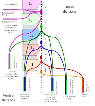
[image:13.612.170.457.175.422.2]ANATOMICAL CONSIDERATIONS
Figure 1: Anatomy of Lumbar plexus
The lumbar plexus present in lumbar region is part of
Lumbosacral plexus. It is formed by ventral divisions of the first four
lumbar nerves (L1 toL4) and from contributions from sub costal (T12).
L1 is frequently supplemented by T12 divides into upper and lower
branch. Upper branch gives illioinguinal and illiohypogastric nerves
7
L2, L3, L4 nerves divides into ventral and dorsal division. Ventral
division as obturator nerve. Dorsal division as femoral nerve and Lateral
[image:14.612.152.487.232.605.2]cutaneous nerve of thigh.
8
FEMORAL NERVE
Figure :3 Femoral nerve
Femoral nerve the largest branch of plexus appears at the lateral
margin of psoas major muscle and remains in the groove between psoas
major and iliacus muscle. At the base of femoral triangle the nerve lies
one finger breadth lateral to the femoral artery. In the femoral triangle
9 Anterior division gives muscular branches to
1) Pectinius
2) Sartorius
Posterior division gives muscular braches to
1) Quadriceps femoris
2) Cutaneous branch – saphenous nerve
Femoral nerve block will facilitate analgesia to anterior thigh,
also blocks flexor muscles of hip and knee extensors. 3-in-1 block will
block femoral, obturator and lateral femoral cutaneous nerve of thigh
from a single injection at the level of inguinal crease. In this block
femoral and lateral cutaneous will be blocked while obturator will be
often spared.
Femoral nerve block along with the sciatic nerve block will
provide excellent block for surgery on knee, leg and ankle. It provides
10
It is comparatively easy to block femoral nerve at the level of
inguinal crease. A needle directed lateral 1-2 cms to femoral artery with
upwards angulation. When nerve stimulator is used, check for the
contractions of quadriceps muscle, which produces movement of
patella, because femoral nerve supply quadriceps group of muscles.
20-25 ml of the local anaesthetic mixture is infiltrated to block the femoral
11
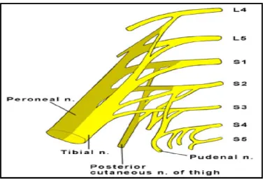
[image:18.612.125.504.112.369.2]SACRAL PLEXUS:
Figure 4 :Sacral plexus
Sacral plexus is formed from anterior rami of the spinal nerves
L4, L5, S1, S2, S3, and S4. Each anterior ramus divides into two
branches-anterior and posterior. Anterior branches supply to flexor
muscles of leg while the posterior branches supply extensor and
abductor muscles.
Sacral plexus provides motor and sensory nerves supply to
posterior part of thigh, most part of leg, whole of foot. It is part of
12 SCIATIC NERVE (L4, 5 S1, 2, 3)
It is the largest nerve in human being, around the size of thumb in
adult. It extends from lower spine to toes supplying most of muscles of
posterior thigh, leg and foot. The nerve originates in the lower spine as
nerve roots exit the spinal cord and extends all the way down the back
of the leg to the toes.
It is composed of two components, tibial and peroneal, enclosed
in common sheath made up of fibrous tissue. The nerve usually divides
into tibial and common peroneal nerve at apex of popliteal fossa.
The muscles supplied by sciatic nerve are as follows-
1) In the thigh-
semimembranosus, semitendinosus, biceps femoris and hamstring
portion of adductor magnus.
2) In the leg and foot tibial nerve will supply-
Gastrocnemius,soleus,plantaris , tibialisposterior,flexordigitorum longus
13
[image:20.612.143.485.169.690.2]In the leg and foot common peroneal nerve will supply-short head of biceps femoris, peroneus longus and peroneus brevis.
14
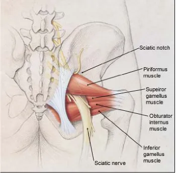
Relation:
From the pelvis the sciatic nerve leaves via greater sciatic
foramen below the piriformis and travels between greater trochanter and
ischial tuberosity. It then moves downwards along back of thigh,
dividing into tibial and common peroneal nerves at around apex of
[image:21.612.138.488.343.688.2]popliteal fossa.
15
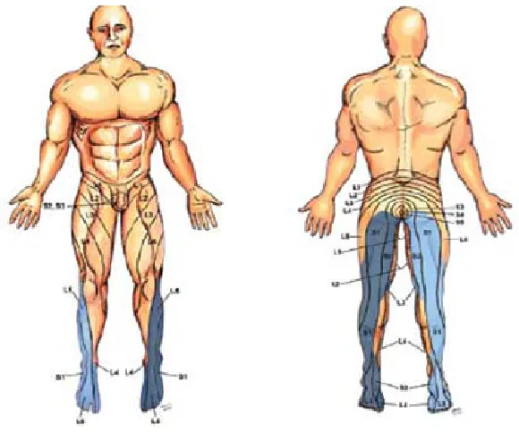
[image:22.612.178.468.128.370.2]SCIATIC NERVE BLOCK
Figure 7: Dermatomal distribution of sciatic nerve
It can be blocked by
1) Labat12 – the posterior approach-transgluteal approach
2) Mid-gluteal posterior approach
3) Gluteal Approach
4) Raj15 approach
5) Para sacral approach
16
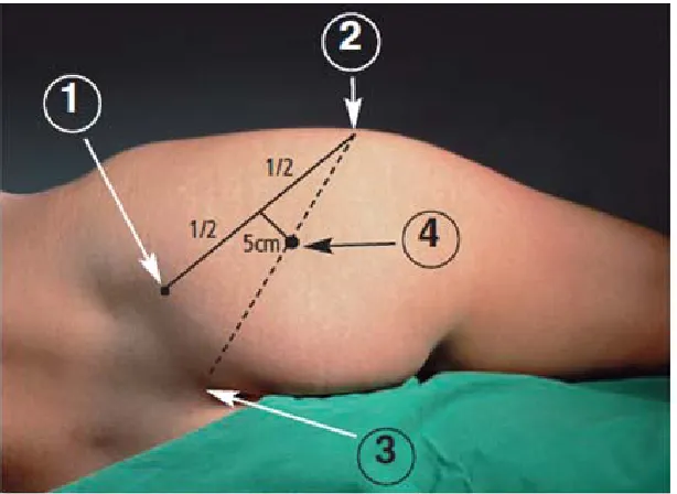
Transgluteal approach (according to Labat)
12, 37The patient is placed in lateral position with operative site above
and flexion given at knee joint of operative limb (SIMS position). The
three anatomical landmarks drawn are posterior superior iliac spine,
[image:23.612.161.468.311.535.2]greater trochanter and sacral hiatus.
17 Technique:
The three anatomical land marks namely Greater trochanter,
posterior superior iliac spine (PSIS) and sacral hiatus are located and
marked. A line is drawn between greater trochanter and PSIS. Another
line from Greater trochanter to sacral hiatus is drawn. A perpendicular
line is drawn from midpoint of line joining greater trochanter and PSIS
to the line joining greater trochanter and sacral hiatus. The point of entry
of needle is the intersection of the two lines as shown in above figure.
Insulated needle connected to the nerve stimulator is introduced
perpendicular to skin at that point of intersection.
Upon stimulation of sciatic nerve, contractions of the muscle
producing plantar flexion and inversion of the foot or toes due to tibial
nerve stimulation are observed. While the stimulation of common
peroneal nerve causes dorsiflexion and eversion of foot or toes. When
the motor response with current 0.5 mA is obtained, the local
anesthetics is injected carefully after repeated negative aspiration for
blood .Repeated aspiration is done with every 5ml injection of local
18
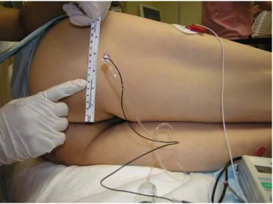
Mid-gluteal posterior approach
37Patient is positioned in lateral decubitus position with side of
block up. Slight flexion is given at both hips and knees. Buttocks are
placed such that it lies 90 degree to the bed. The only landmark for this
[image:25.612.122.513.270.563.2]technique is intergluteal sulcus
19
Technique
Regardless of patient gender and body habitus in adults, the
sciatic nerve runs 10cms parallel to the midline formed by intergluteal
sulcus. So in this approach patient is placed in lateral decubitus position
and 10 cms measured laterally from midpoint of intergluteal sulcus.
Because of easy visualization, inter gluteal sulcus is chosen. Usually the
10 cms line drawn will come immediately lateral to ischial tuberosity.
Using 15cms insulated needle with nerve locator sciatic nerve is
stimulated.
Upon good dorsiflexion, eversion and plantar flexion inversion at
0.5mA, local anesthetic drug is injected after repeated negative
20
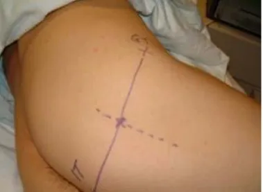
Gluteal Approach
32,37The Patient is positioned in sim’s position. The anatomical
landmarks are greater trochanter and ischial tuberosity. Here sciatic
[image:27.612.125.504.282.559.2]nerve is covered by gluteus maximus.
21 Technique
Greater trochanter and ischial tuberosity are marked. A line
joining these two points is drawn. The site of introduction of needle is
midpoint of the line joining greater trochanter and ischial tuberosity.
This point represents groove between two muscles namely biceps
femoris and vastus lateralis. An insulated needle of 15 cm is introduced
attached to nerve locator. After stimulating sciatic nerve at 0.5mA
current, local anaesthetic mixture is injected after repeated negative
22
[image:29.612.194.431.133.408.2]Ra
j Technique
15, 37Figure 11: raj approach
In this approach, sciatic nerve is blocked posteriorly in supine
position. Both hip and knee are flexed to 90º angle and foot of the
patient is held by assistant. Sciatic nerve becomes superficial by flexion
of hip.
Mark greater trochanter and ischial tuberosity. Introduce 15 cm
insulated needle attached to nerve locator from midpoint of line joining
23
Para sacral Technique
37Figure 12: Para sacral approach
This is one of the unique technique in which sciatic nerve is
blocked along with obturator nerve. Sciatic nerve is blocked most
proximally in this approach. Patient positioned in lateral decubitus
position.
Anatomical Landmarks marked are posterior superior iliac spine
(PSIS) and ischial tuberosity. A line is drawn between these two points.
10 – 15 cm insulated needle is inserted caudal posterior superior iliac
spine. At a depth of 6 – 8 cm needle advances through sciatic foramen
to block sciatic nerve. After obtaining dorsiflexion, inversion and
plantar flexion, eversion at 0.5 mA current, local anaesthetic mixture is
24
PHYSIOLOGY OF PERIPHERAL NERVE
STIMULATOR
Peripheral nerve stimulator
32, 37In 1912, Von Perthes first said about the use of peripheral nerve
stimulator. With increasing interest in regional anesthesia and for the
accurate localization of the peripheral nerve for blocks, the interest in
the peripheral nerve stimulator has increased.
Basic Electrophysiology
To understand electrophysiology the following two terms should
be well understood.
1) RHEOBASE- is the minimal threshold current required to
stimulate a nerve with a long pulse width.
2) CHRONAXIE- is the duration of the stimulus required to
stimulate at twice the rheobase .It measures the relative excitability of
25
It is possible to stimulate only motor nerves A-α without
stimulating the sensory A-δ and C fibers which causes pain. Even the
mixed nerve can be identified by evoking motor response without
causing discomfort to the patient.
Stimulation intensity is determined by coulomb’s law. A very
high current is needed when tip of the needle is far from nerve. Due to
high strength of current, it may cause pain and also causes systemic
effects to the patient. The stimulating current of <0.5mA is associated
with high success rates with peripheral nerve blocks using peripheral
nerve locator.
Needles
Insulated short and long beveled needles are used for nerve
blocks along with nerve locator. The positive electrode of nerve
locator is connected to a electrocardiogram electrode while negative to
the needle of the nerve locator. These needles come with different
26
Choosing the type of needle depends on type of patient adult or
paediatric patients. The insulated needles are available from size 2.5
cms to 15 cms. 2.5cms needle with 25 gauge may be used for paediatric
patients. 2.5 to 5 cms needle may be used for adult upper limb blocks,
[image:33.612.144.485.382.638.2]while 10 to 15 cms insulated needles are used for lower limb blocks.
27 Characteristics of an ideal PNS:
1. Constant current output-A particular current not the voltage
stimulates the nerve. Therefore, the current delivered by the
device should not vary with changes in the resistance of the
external circuits.
2. Digital display of the delivered current
3. Variable output control
4. Clearly identifiable polarity
5. Option for different pulses
6. A wide range of current output 0.1-5.0mA
28 Peripheral nerve stimulator settings:
MIXED NERVE
(most peripheral nerves)Current (dial) -1mA, Current duration-0.1ms, Frequency- 1-2Hz.
SENSORY NERVE (e.g.-Lateral femoral cutaneous and saphenous
nerves)
29
PHARMACOLOGY
32, 33, 34, 35.Pharmacological consideration:
a)Pharmacology of Bupivacaine:
BUPIVACAINE MOLECULAR STRUCTURE
Bupivacaine is an amide linked local anaesthetic. It is derived
from Mepivacaine and is very stable compound and may be autoclaved
repeatedly.
Pka is 8.1
MW- 288
Protein binding - 95%
Lipid solubility- 28
30
Approximate duration of action - 175mts
Peak time 0.17-0.5 hours
Peak concentration 0.8 microgram/ml
Toxic plasma concentration > 1.5micro gram /ml
Availability:
Ampoule - - 0.5% Bupivacaine hydrochloride with dextrose
(heavy) 4cc
Vials - 0.25% and 0.5% Bupivacaine hydrochloride 20cc
Dosage - Maximum dosage 3mg/kg body weight.
Pharmacokinetics:
It is rapidly absorbed from the site of injection, but the absorption
rate depends on both vascularity at the region and presence of
vasoconstrictors.
High lipid solubility of bupivacaine makes it easy for nerve and
vascular tissue penetration.80-95% of the absorbed bupivacaine binds to
31 Biotransformation:
Possible path ways of metabolism of bupivacaine include
aromatic hydroxylation and conjugation. Only the N-dealkylated
metabolite, N-desbutyl bupivacaine has been measured in blood (or)
urine after epidural (or) spinal anaesthesia. Alpha1 acid glycoprotein is
the most important plasma protein binding site of bupivacaine and its
concentration is increased by many clinical situations including post
operative trauma.
Excretion:
It is excreted through the kidney; 4-10% of the drug is excreted
unchanged.
Mode of Action:
Sodium Channel blockade:
It acts by non depolarization blockade at sodium channels. They
impede sodium ion access to the axon interior by occluding the
transmenbrane sodium channels thus delaying the process of
32 Toxicity:
Toxicity is related to plasma level of unbound drug and more
likely due to an inadvertent intravenous injection. Systemic toxicity
reactions primarily involve central nervous system and cardio vascular
system. The blood level required to produce central nervous system
toxicity is less than that required to produce circulatory collapse.
Central Nervous System Toxicity:
Initial symptom includes feeling of light headedness and
dizziness, followed by visual and auditory disturbances. Objective signs
are excitatory and include shivering, muscle twitching and tremor.
Ultimately generalized tonic, clonic seizures occurs.
Cardiovascular System Toxicity:
The rate of depolarization in fast conducting tissue of purkinje
fibres and ventricular muscle is decreased. The rate of recovery due to
bupivacaine induced block is slower than that of lignocaine. Very high
33
PHARMACOLOGY OF LIGNOCAINE
LIGNOCAINE MOLECULAR STRUCTURE
In 1943 Lofgren synthesized Lignocaine in Sweden. It is
chemically a tertiary amide, diethyl amino acetyl, 2, 6 xylidine
hydrochloride monohydrate. It is a local anaesthetic of moderate
potency and duration but of good penetrative powers and rapid onset of
action.
Lignocaine is the standard to which all other local anesthetics are
compared. It is currently the most widely used local anaesthetic. In
addition, it is a popular antiarrythmic. It can be given by almost any
34 Mechanism of action:
It acts by blocking the ion gated sodium channels and prevents
flow of sodium. Threshold potential is not reached and it prevents
propagation of nerve impulses. But resting membrane potential is not
altered. Lignocaine binds to the inner portion receptor (i.e. Sodium
channel) after entering the cell membrane.
Physiochemical properties:
Molecular weight 234
Weak base with a pka 7.6 – 7.8
Very stable, not decomposed by boiling, acids or alkalis
It is less lipid soluble than that of Bupivacaine,
Lipid solubility 2.9
Protein binding 70% to alpha-1acid glycoprotein.
Volume of distribution 91 litres
Clearance 0.95 litres /minute
Elimination half life 96 minutes
35 Pharmacokinetics:
Absorption:
It is absorbed from the site of application or injection into the
blood stream. Rate of absorption depends on the vasularity of the area
and use of epinephrine.
Metabolism:
Metabolised in liver by oxidative dealkylation to
monoethylglycine xylidide followed by hydrolysis of this metabolite to
xylidide. Metabolism is dependent on hepatic blood flow.
Monoethylglycine xylidide has 80% activity of the parent drug.
Xylidide has 10% activity of the parent drug.
Onset of action:
Rapid onset of action
Topical anaesthesia 5-10 mins
Conduction anaesthesia-For small nerves 5-10 mins,
for large nerves 10-15 mins.
36 Availability:
a) 2% lignocaine – plain – 30 ml vial –contains methyl and
propyl paraben as preservative
b) 2% ligcocaine (xylocard) without preservative – 50 ml vial for
intravenous use
c) 2% lignocaine ointment
d) 4% lignocaine with 1 in 200000 Adrenaline – 30 ml vial.
e) 4% lignocaine viscus
f) 4% lignocaine aqueous solution
g) 5% heavy 2 ml ampoules which contain 50 mg of lignocaine /
ml with 75 mg – 100 mg of dextrose
h) 5% lignocaine ointment
37 Pharmacodynamics:
Local actions:
Causes nerve blockade with loss of pain and temperature
sensation, touch, motor power and vasomotor tone in the region
supplied by the nerve.
Systemic actions:
Result of systemic absorption from the site of administration or
intravenous administration.
Cardiovascular system:
It has a stabilizing effect on the cell membranes of cardiac tissue.
Lignocaine depresses myocardial automaticity by antagonizing the
spontaneous phase IV depolarization and reduces the duration of
effective refractory period. Myocardial contractility and conduction
velocity are depressed at higher concentrations. These effects result
from direct cardiac muscle membrane changes (i.e.) cardiac sodium
channel blockade. It stabilizes the membrane of damaged and excitable
38 Central nervous system:
Produces a sequence of stimulation followed by depression.
Produces sedation on intravenous administration.
Intravenous administration decreases cerebral blood flow and
attenuates the rise in intracranial pressure that accompanies intubation.
Infusion of lignocaine is capable of reducing the MAC of volatile
anaesthetics by 40%.
Indications:
1. For infiltration block,
2. peripheral nerve blocks,
3. epidural ,spinal blocks,
4. topical anaesthesia ,
39
6. Antiarrythmic- Lignocaine is a class IB antiarrythmic. Used in (a)
Treatment of Ventricular tachyarrhythmia, (b) In digitalis
toxicity because it doesnot worsen AV
block (c) Arrhythmias following acute MI during cardiac surgery.
7. Suppresses noxious reflexes such as coughing & sympathetic
stimulations associated with endotracheal suctioning and
intubation.
8. Prevention or treatment of increases in intracranial pressure
during intubation due to its antitussive effect.
9. Reflex induced bronchospasm is also attenuated by iv
administration of lignocaine
10. Used as an antiepileptic agent IV
11. Used as an IV analgesic for certain chronic pain states
40 Contraindications:
Hypersensitivity
Should not be used with vasoconstrictor in digits of hand, feet and
penis
Stokes Adams syndrome
Severe degree of heart block
Doses:
Maximum recommended dose:
a) Plain - 3 mg / kg
b) With adrenaline- 7 mg / kg
c) For reflex suppression - 1.5 mg / kg IV.
41
Epinephrine
Epinephrine (adrenaline) is the prototype drug among the
sympathomimetics.
Functions
• Regulation of myocardial contractility, heart rate, vascular and
bronchial smooth muscle tone.
• Potentiates glandular secretions and metabolic processes.
• Agonist of α- adrenergic, β 1 and β2 receptors.
• Poorly lipid soluble hence lack of cerebral effects.
Uses
• Addition to local anaesthetic solution in order to decrease
systemic absorption and to prolong duration of action.
• Treatment of life threatening allergic reaction.
• During CPR as a very important therapeutic drug. Continuous
42
REVIEW OF LITERATURE
1.
Beck2 in 1963 described the anterior approach for sciatic nerve.He identified three points
1. Anterior superior iliac spine (ASIS)
2. Pubic tuberosity
3. Greater trochanter
He connected ASIS to pubic tuberosity and draw a line parallel to
it From greater trochanter. From the midpoint of first line he drew a
perpendicular line to the second line. From the point of intersection of
the second line with the perpendicular line, he gave sciatic nerve block2.
2.
Chelly7 JE, Delaunay L. did a comparative study on Sciatic nerve block. He did study 22 patients with differentconcentration of mepivacaine. He draw a line connecting inferior border
of ASIS to the superior angle of pubic symphysis. A perpendicular line
dividing the first line was drawn and extended 8 cm downwards. The
needle was passed perpendicularly and identification of sciatic nerve
43
Landmarks are written in 1.3minutes (0.5 to 2 minutes),
sciatic nerve was identified in 2.5 minutes (1.2 to 5 minutes) using nerve
locator.Onset of sensory nerve block of both common peroneal nerve
and tibial nerve distribution was achieved at a mean of 15 minutes (5 to
30 minutes). Rapid onset was seen in patients given only mepivacaine
than combination of mepivacaine and ropivacaine (10 minutes [5 to 25
minutes] vs 20 minutes [10 to 30 minutes] with P < 0.05). Duration of
block was 4.6 hours (2.5 to 5.5 hours) with mepivacaine alone, while
along with ropivacaine, duration of block was 13.8 hours (5.2 to 23.6
hours) with P < 0.057.
3. Pierre Pandin 3and Nathalie Vancutsem et al conducted a
random study in which 119 patients with ASA status I and II posted for
below knee surgery. Fifteen ml and thirty ml of 0.5% ropivacaine
solution were injected near femoral and sciatic nerve through anterior
approach. Landmarks were drawn in 1.7 min. Average procedure time
was 4.2 minutes. Femoral nerve block was successful in 89.9%, while
sciatic nerve block in 94.9%. Motor block in tibial (97.4%), common
peroneal (85.7%) and femoral (98.3%), and nerve regions (modified
Bromage’sscale > 2)4.
44
They concluded that combined anterior approach for both sciatic
and femoral nerve by a single skin needle entry is reliable and easy for
lower limb surgery in supine position.
4. Alain C. Van Elstraete11 and Claude Poey et al conducted a
study with the new landmark for anterior approach, using inguinal
creaseand femoral artery and the effect of external rotation of the leg
during this technique. The sciatic nerve identified twicein 20 patients
scheduled for ankle or foot surgery.
First leg in neutral position and then in externally rotated position.
Point of needle insertion is 2.5 cms below the inguinal crease and 2.5
cms medial to femoral artery. 15 cms insulated needle was directed
postero-laterally with 10° to 15° to vertical plane. At a depth of 10.6 ±
1.8 cms sciatic nerve detected with leg in the neutralposition and 10.4
± 1.5 cms with leg in externally rotated position . The anatomic
landmarks were identified in 28 ± 15 seconds and 26 ± 14 seconds in
neutral and externally rotated position respectively. Sciatic nerve was
located in 79 ± 53 seconds and 46 ± 25 seconds for neutral and
externally rotated positions respectively, with P < 0.006. Their
conclusion was inguinal crease and femoral artery are reliable land
marks and the optimal position of leg is externally rotatedposition for
45 5. Jerry D. Vloka9 and Admir conducted a study in 10 lower
limbs of 5 cadavers. They studied the effects of rotation of leg in
locating the sciatic nerve by anterior approach. They studied the leg
rotation effect on needle plane need to reach the sciatic nerve. They
studied with legs in neutral, 45 degree in internal and external rotation.
In 80% attempts in neutral position, the needle was not fully advanced
due to obstruction by lesser trochanter. Medial redirection of the needle
(10° to 15°) made the needle to pass lesser trochanter but tip of needle
was too medial to sciatic nerve. Internal rotationof leg made all needles
to pass lesser trochanter. Their conclusion was internal rotationof leg
will facilitate needle insertion for anterior approach to the sciatic nerve
block.
6.
Fuzier13 R, Albert N et al. conducted a comparative study between anterior approach and lateral approach with the patient in the
supine position. 59 patients posted for traumatic lower limb surgery
were studied for anterior vs lateral approach to sciatic nerve block.
Using 15 cm insulated needle with nerve locator sciatic nerve was
identified. 0.75% of 25 ml ropivacaine was injected. The mean
procedure time was 4.9 ± 4.0 minutes in anterior and 6.1 ± 6.9 minutes
in lateral group. The sciatic nerve was located at a mean depth of 107 ±
46
success rate was same in both groups (77% in anterior and 79% in
lateral group) but the failure rate was higher in the anterior approach
(86% in anterior and 33%in lateral approach, with p<0.05).
7. Marty21L. Ericksen Jeffrey D. showed relationship of
sciatic nerve to the lesser trochanter using MRI in 20 patients in the
supine position. Study was done at lesser trochanter level and at 1 cms,
2cms, 3cms and 4cms inferior to the lessertrochanter. In 19 out of 20
patients, sciatic nerve was medial to femur at 4cms inferior to lesser
trochanter. In 13 of 20 patients sciatic nerve was lateral to femur
border at lesser trochanter level. In classic description of anterior
approach to sciatic nerve, needle is passed medially at the level of the
lesser trochanter. In contrast, in this study at 4 cm below lesser
trochanter, sciatic nerve was medial to femur in 19of 20 patients.
8.
Raghu S. Thota22, Ajay Aravind et al did study for
anterior approach using fluoroscopy for calcaneal fractures. Lesser
trochanter of femur was identified using C arm fluoroscopy and a small
amount of contrast material was passed through 22 gauge 150mm spinal
needle for needle placement confirmation. Then 20 mL of 0.25%
bupivicaine was injected. The anterior approach when combined with
47
MATERIALS AND METHODS
Study design:
This is a single blinded, prospective, randomized study. This
study was done at Government Stanley medical college and hospital,
Chennai during the period of November 2011 to May 2012. After
obtaining clearance from the Institutional Ethical Committee of the
Stanley Medical College, Chennai-1, a pilot study was done to calculate
the sample size.
A pilot study with a sample size of 5patients in each group was
done before the start of the study to decide on sample size. The sample
size calculated based on the formula given in monographers on statistics
and applied probability.
Where
48
Z1-β=0.842 (80% power)
D = difference between two means
S = S1+S2 / 2
On entering the values, the mean procedure time observed in the
pilot study for single skin site needle technique was 7.5min ± 3.2min,
and for classical two skin needle technique was 10.1±6.8 seconds.
N = 2 X (1.96 + 0.842)2 (3.2+6.8/2)2/ (10.1-7.5)2
N = 2 X 7.85 X (10/2)2/ (2.6)2
N = 15.7 X (5)2/ 6.76
N = 15.7 X 25/ 6.76
N = 58.06
Sample size taken as in each group. 60 patients were included
in each group. 120 patients of ASA I – II physical status who got
admitted for below knee surgical procedures were taken into
49
The 60 patients are divided into two groups as follows-
Group ONE – single needle technique
Group TWO – Classical two needles technique.
Inclusion criteria:
Age 20-60 YRS
Both sexes
ASA I-II undergoing surgery for both elective/emergency below
knee.
Exclusion criteria:
Pregnancy, psychiatric illness.
Infection at the puncture site
50
MATERIALS
The following materials were needed for the study:
i. Injection 2% lignocaine – 20 ml
ii. Injection 0.5% bupivacaine – 20 ml
iii. Injection adrenaline used at dosage of 5 microgram / ml
iv. Distilled water-20 ml
v. Injection midazolam at dosage of 20 microgram / kg
vi. Injection fentanyl at dosage of 1 microgram / kg
vii. One 20ml syringe for administration of local anaesthetic mixture
viii. Sterile towels and 4*4 gauge packs
ix. Sterile gloves, Marking pens and Surface electrodes
x. One 25g needle for skin infiltration
xi. Nerve stimulator - Stimuplex DigRc-B.Braun
xii. Needle - 22 gauge, 5 cms -long and 15cms, short bevelled
insulated needle (StimuplexDig, B.Braun)
xiii. ASA Standard monitors – pulse oxymetry (SPO2), non invasive
51
xiv. Appropriate size endotracheal tubes and laryngoscopes.
xv. Intravenous fluids, intravenous cannula and emergency drugs.
PROCEDURE:
Drugs: 20 ml of 2% Lidocaine with 5 microgram of adrenaline/ml,
20 ml of 5% bupivacaine and 20 ml of distilled water are kept
Ready.
After establishing an Intravenous line, patient was positioned
52
[image:59.612.205.411.141.408.2]Classic Anterior Approach of Sciatic Nerve Block
2:Figure 14: Land Marks of classic anterior approach
Anterior approach to sciatic nerve was developed to overcome
problem of patient positioning. The land marks for the classical
approach are ASIS, PUBIC TUBERCLE and GREATER
TROCHANTER of femur. First a line joining ASIS and pubic tubercle
is drawn. This represents the inguinal ligament. Another line joining the
53
the junction of lateral 2/3rd and medial 1/3rd a perpendicular line joins
which intersects at the 2nd line. Through the intersecting point anterior
approach of sciatic nerve performed.
With the help of the nerve stimulator the end point for locating
the nerve , either dorsi flexion or plantar flexion of the foot, which is
considered as the evoked motor response with 0.5mA as the lower limit
54 CLASSICAL TECHNIQUE FOR FEMORAL NERVE BLOCK
Anatomic Landmarks: Inguinal crease, femoral artery, anterior superior
[image:61.612.126.467.208.441.2]iliac spine.
Figure 15: Land mark for classical femoral block
Approach and Technique:
At the level of inguinal crease the femoral artery is palpated. A
22 gauge insulated needle is connected to a nerve stimulator. The point
of entry of needle is around 1 cms lateral to femoral artery at inguinal
crease. Needle is directed upwards at an angle of 45° angle to skin and
55
Avoid medial direction of the needle because of femoral artery
puncture, which is present medial to femoral nerve. The stimulation of
femoral nerve is identified by contraction of quadriceps and patellar
movement known as patellar dance. The presence of muscle contraction
with current of 0.5 mA is assessed. Then, inject 20 to 25 ml of local
anesthesia solution after careful aspiration, every 5 ml to prevent
56
[image:63.612.208.453.129.471.2]Anatomical Land marks for single needle technique
3:
Figure 16 : land mark for new single skin site approach for both femoral and sciatic nerve
Patient in the supine position
a) 1st line is drawn connecting ASIS and pubic tubercle
b) 2nd line drawn parallel to 1st line passing along the greater
57
c) The 1st line is divided into three equal parts. From the junction of
lateral 2/3rd and medial 1/3rd (point A) a perpendicular line drawn.
d) The point of intersection of second line by first line is point B.
e) The midpoint of the line AB (point C) IS point of needle entry for
giving block.
Under strict aseptic precautions local anesthetics solution was
prepared and all the equipments, needed like nerve stimulator, insulated
needle, syringe were kept ready.
First the femoral nerve was blocked using the nerve stimulator
.the needle advanced from point C, in direction of 45° cephalad
angulation with 10° medial angulation, until dancing of patella seen
due to stimulation of femoral nerve at a depth of 5–6 cm . Nerve
stimulator output initially 2mA current was used first to identify the
nerve and then gradually reduced to 0.5mA till the response is present.
The patella dance was considered as the evoked motor response or end
point of nerve location. When the response is present at 0.5mA 25ml of
the mixture of local anaesthetic solution injected after negative
58
The sciatic nerve block was done after the blockade of
femoral nerve with same point of needle entry; the needle was
withdrawn to the level of subcutaneous tissue, and then redirected at
10°lateral angle with 80° caudal angle. Initially 2mA Strength was
used to first identify the nerve. At a depth of 9–13 cms, the sciatic nerve
was identified with motor response related to the sciatic nerve –plantar
flexion or foot inversion is noted. Common peroneal nerve stimulation
will cause dorsiflexion and eversion of foot and tibial nerve stimulation
will cause plantar flexion with inversion of foot, also causes flexion of
toes. And gradually reduced to 0.5mA till the foot eversion (or) plantar
flexion movement noticed by moving the needle. When the plantar
movement is present with 0.5mA 35ml of the prepared local anaesthetic
solution was injected after careful aspiration.
Intensity of motor and sensory block was assessed every 5 min till
40 min after completion of procedure. Then the patient is assessed every
hour after the surgery till the motor and sensory function recovered to
know about the recovery time.
The scale used in this study to know intensity of motor block
59
The assessment of sensory block was done by PIN PRICK
sensation in the anterior part of thigh (for femoral nerve), dorsal aspect
of the foot (for common peroneal nerve) and plantar aspect of foot (for
tibial nerve). Only in patients having pin prick sensation completely
absent are considered as, complete sensory block.
To assess the intensity of motor block dorsiflexion and inversion,
plantar flexion and eversion was done by patient. Depending on
intensity, the block was classified into No block, partial block and
complete block. Only when femoral, tibial and common peroneal
muscles were blocked, the motor block was quantified as to complete
block.
Duration of sensory block is defined as time interval between
onset of block to the recovery of pain sensation. Duration of motor
block is defined as time interval between onset of block the recovery of
motor function. After confirming the adequacy of block, surgery was
allowed to proceed. Complications such as vascular injury, neurological
deficits, anaphylaxis and toxicity of the drug were taken into
60
PARAMETERS OBSERVED
PRIMARY OUTCOME MEASURES
:
Performance time- Time from insertion of needle to completion of
block
Onset time for sensory block (min) - Time from completion of block
to onset of complete sensory block.
A) Onset time for SENSORY femoral nerve block(min)-Time
from completion of block to loss of pinprick in anterior and
medial part of thigh, medial part of leg.
B) Onset time for SENSORY sciatic nerve(min)-Time from
completion of pin prick in posterior part of thigh, all part of the
leg except medial part.
Motor block
Complete block - No movement against gravity,
Partial block - Minor movements of toes possible,
No block - Normal range of movements present.
Duration of sensory blockade - Duration of time interval from
61 Duration of motor blockade- Duration of time interval from
Completion of block to return of full motor power.
Duration of analgesia
The pain score assessed by visual Analogue scale having 10cm
length numbered from 0 - 10. Patient was explained about visual
Analogue scale as 0 - No pain and 10 means the worst possible pain and
was asked the score in visual analogue scale.
The patient was observed every 30 minutes post operatively till
the motor block was reversed and thereafter hourly for 12 hrs. Time at
which VAS score is greater than 5 was noted and patient was given
intramuscular Tramadol as rescue analgesia. Duration of post operative
analgesia is the period of time after the surgery till the patient needed
analgesic, that is VAS score more than 5.
SECONDARY OUTCOME MEASURES:
Complications- Accidental vascular puncture, neurological deficit,
62
OBSERVATION AND RESULTS
STATISTICAL TOOLS
The information collected regarding all the selected cases were
recorded in a master chart. The observations were compiled and data’s
were expressed as mean± SD.
Quantitative analysis was compared with independent sample
student’s t test. Qualitative analysis was compared with chi-square test.
When using these tests to compare mean among two groups, p-value of
less than 0.05 was taken as significant.
All analyses were done using SPSS version 11.5 statistical
software.
GROUP ONE - SINGLE NEEDLE TECHNIQUE
GROUP TWO- TWO NEEDLE TECHNIQUE
The study was done with the patient’s age ranging from minimum
63
Table-1: Age Distribution
Group-ONE(60) Group- TWO(60)
Age distribution (in Years)
Number Percentage Number Percentage
20 -30 30 50.00 24 40.00
30 – 40 14 23.30 24 40.00
40 -50 12 20.00 10 16.70
50 – 60 4 06.70 2 03.30
Range 35 35
Mean ±Sd 33.12 ± 10.91 34.08 ± 8.73
t-value 0.54
Df 118
64 Sex Distribution:
Table-2 : Age distribution by Sex
Age
Distribution
Group-ONE(60) Group-TWO(60)
Male Female Male Female
N % N % N % N %
20 – 30 17 41.50 13 68.40 14 33.30 10 55.56
30 – 40 13 31.70 01 05.30 17 40.50 07 38.89
40 – 50 07 17.10 05 26.30 09 21.40 01 5.55
50 – 60 04 09.80 00 00.00 02 04.80 00 0
Range 35 9 35 9
Mean ± Sd 34.49±11.12 30.16±10.10 35.67 ± 9.26 30.39 ± 6.08
t-value 1.44 2.22
Df 58 58
65
Table-3 : Sex Distribution
Sex Group-ONE(60) Group-TWO(60)
Number Percentage Number Percentage
Male 41 68.30 42 70.00
Female 19 31.70 18 30.00
Total 60 100 60 100
Chi square 0.04
df 1
p-value 0.84 (Not Significant)
In group ONE, the number of males are 41 and females 19, while in
group TWO the number of males are 42 and females 18. Both the
66 ASA Class Distribution:
Table-4 : ASA Grade
ASA Grade Group-ONE(60) Group-TWO(60)
Number Percentage Number Percentage
I 44 73.30 47 78.30
II 16 26.70 13 21.70
Chi square 0.41
df 1
p-value 0.52 (Not Significant)
In group ONE 44 patients belongs to ASA 1 and 16 to ASA 2.While in
Group TWO 47 belongs to ASA 1 and 13 to ASA 2. Both the groups
67 Time for procedure:
The average time that is required for the performance of the block
in Group ONE was around 6.35 minutes. The time varied from 4minute
– 9 minutes. While in group TWO average time required is 14.91
minutes, with Time varied from 8 to 18 minutes. With p value 0.001,
there is statistically significant difference in procedure time with less
[image:74.612.120.511.364.595.2]time for new technique.
Table-5:Procedure time
Time (in Mints) Group-ONE(60) Group-TWO(60)
Number Percentage Number Percentage
4 – 5 09 13.80 2 3.30
5 – 6 19 32.80 2 3.30
6 –- 7 16 27.60 2 3.30
7 –-8 10 15.50 2 3.30
8 – 18 6 10.30 52 86.70
Mean ± sd 6.34 ± 1.21 14.91 ± 2.52
t-value 23.07
Df 108
68 Femoral Nerve Block:
The average onset time of sensory block in GROUP ONE is
11.53 minutes and the range varied from 5min to 23min. In case of
GROUP TWO is 8.74 minutes with range varying from 5min to 13
minutes. Since the p value is 0.0001 there is statistically significant
69
Table-6 Onset of Femoral Sensory Nerve Block
Time (in Mints)
Group-ONE (52) Group-TWO(51)
Number Percentage Number Percentage
5--7 6 11.50 06 11.50
7--9 10 19.20 25 50.00
9--11 13 25.00 17 32.70
11--13 5 09.60 03 05.80
13--15 8 15.40 00 0
15--17 5 09.60 00 0
17--19 3 05.80 00 0
19--21 1 01.90 00 0
21--23 1 01.90 00 0
Mean ± sd 11.53 ± 3.89 8.74 ± 1.52
t-value 4.82
Df 102
70 Sciatic Nerve Block:
In group ONE patients, the average onset time for sciatic sensory
block 21.91 minutes. The range varied from 15 to27 minutes. In group
TWO patients, the average onset of time was 29.23 with range varying
from 24 min to 36 minutes. Since the p value is 0.0001, there is
statistically significant difference with short onset time in new single
needle technique.
Out of the 60 cases, in 52 cases the sensory blockade was well
taken up. In 8 cases it was spared, 5 cases were due to failure of sciatic
nerve block and 3 due to femoral nerve block.
In case group B, 51 cases sensory block was taken well. In 9 cases
the block was spared. 6 cases were due to failure of sciatic nerve block
71
Table-7 : Onset of Sensory Sciatic Nerve block
Time (in Mints) Group-ONE (52) Group-TWO(51)
Number Percentage Number Percentage
15 --18 2 03.80 0 0
18 --21 21 40.40 0 0
21 -- 24 13 25.00 0 0
24 -- 27 16 30.80 14 26.90
27 -- 30 0 0 19 38.50
30 -- 33 0 0 09 17.30
33 -- 36 0 0 09 17.30
Mean ± sd 21.91 ± 3.07 29.23 ± 2.92
t-value 12.44
Df 102
72
Onset of motor blockade
In group A the average onset of motor blockade is 25 mins, with
range from 18 to 40 mins. In group B the average onset of motor
blockade is 31 mins, with range from 24 to 40 mins. There is no
statistically significantly difference between two groups even though
[image:79.612.115.510.336.611.2]new single technique has early onset of action.
Table-8 : Onset of Motor blockade
Time (in Mints) Group-ONE(52) Group-TWO(51)
Number Percentage Number Percentage
18--21 11 21.20 0 0
21--24 12 23.10 0 0
24--27 11 21.20 4 7.70
27--30 09 18.90 17 34.60
30--33 04 07.70 11 21.20
33--36 03 05.80 13 25.00
36--40 02 03.80 06 11.50
Mean ± sd 25.58 ± 5.09 31.57 ± 3.53
t-value 7.00
Df 102
73
Degree of motor blockade
Block was considered failed, if no pain relief to pinprick even
after 40 min after the performance of the block.
The block is considered under three degrees-
0- Complete motor blockade
1- Partial motor blockade
2- No motor blockade.
74
Table-9: Degree of Motor blockade
Degree Group-ONE(60) Group-TWO(60)
Number Percentage Number Percentage
0 44 73.30 41 68.30
1 08 13.30 10 16.70
2 08 13.30 09 13.30
Chi-square Value 0.58
df 2
75 In group ONE In group TWO,
Complete motor blockade - 73.3% Complete motor blockade-68.30%
Partial motor blockade- 13.3% Partial motor blockade-18.80%
No motor blockade- 12.3% No motor blockade- 13.30%
Since the chisquare test value is 0.58,there is no statistically
significant variation in both groups regarding degree of blockade.
[image:82.612.119.513.474.699.2]Post operative analgesia:
Table-10: Duration of Sensory Block
Time (in Hours) Group-ONE (52) Group-TWO (51)
Number Percentage Number Percentage
5—6 4 7.70 12 21.60
6—7 3 5.80 15 29.40
7—8 5 9.60 10 15.70
8—9 5 9.60 05 07.70
9—10 5 9.60 08 15.70
10—11 25 57.70 0 0
11—12 05 09.60 0 0
Mean ± sd 10.93 ± 3.78 7.03 ± 1.21
t-value 7.09
Df 102
76
In group ONE, The average time that post operative pain relief
after the surgery was around 10.93 hours. The time ranged from 5 to
12hours.
While in group TWO mean duration of pain relief was
7.03 hours. So the new technique will clearly provide long
77
Table-11: Duration of Motor Block
Time (in Hours) Group-ONE (52) Group-TWO (51) Number Percentage Number Percentage
4--5 1 1.90 15 28.20
5--6 2 3.80 16 30.80
6--7 8 15.40 17 34.60
7--8 5 9.60 3 5.80
8--9 20 38.50 0 0
9--10 3 5.80 0 0
10-11 10 19.20 0 0
11--12 1 1.90 0 0
12 - 13 2 3.80 0 0
Mean ± sd 8.01 ± 2.16 5.74 ± 1.03
t-value 6.89
Df 102
p-value 0.0001 (Significant)
The mean duration of motor blockade was 8 hours in newer
technique (group ONE) while in classic technique (group TWO) it is
5.74 hours. This is statistically significant with long duration of block,
78
Table-12 : Vascular puncture
Vascular Puncture Group-ONE (60) Group-TWO(60)
Number Percentage Number Percentage
Yes 3 5.00 5 8.30
No 57 95.00 55 91.70
Total 60 100 60 100
The incidence of vascular puncture in newer technique (group
A) was in 3 cases, while in classic study (group B) it was 5 cases. There
79
DISCUSSION
In my study, a regional technique- sciatic and femoral nerve were
blocked through anterior approaches by a single needle vs two needles
and peripheral nerve stimulator for below knee surgeries. It is a safe
technique; causes no disturbance to milieu interior3. In my study sciatic
and femoral nerve block was performed with patient in supine position,
through a single point of entry and compared with classical two needle
technique.
The land marks were drawn at an average time of 1.5 minutes.
The mean time to perform the block was 6.35 minutes in group ONE
and 14.9 minutes in group TWO. The performance time was shorter in
new single needle technique, with statistically significant difference. So
newer technique has advantage of short performance time. In Pierre
pandin3Et al study done for single needle approach, the performance
time4.2 minutes.
The average time for onset of femoral sensory block was 11.53
min in new group while in classical technique it was 8.74 minutes. The
80
sensory block. In Pierre pandin3 et al where the mean onset of femoral
sensory block was 15.4 minutes, correlating to my study.
The onset of sciatic sensory nerve block in new single needle
technique was 21.91 minutes while in classic technique was 29.23
minutes. The newer technique has statistically significant difference
with short onset of time.
The mean onset of motor blockade in new technique was 25.58
minutes while in classic technique was 31.57 minutes. Even though it is
not a statistically significant difference, but newer technique has early
onset in comparison to classical technique. This can be compared with
the original study by Pierre pandin3 et al in which onset of motor
81 Degree of motor blockade
Failure of 8 cases in new technique and 9 cases in classic
technique were noted. In both study the failure of femoral block was in
3 cases, while failure of sciatic block was 5 in newer and 6 in classic
study. There was no statistically significant difference regarding degree
of motor blockade between the two groups. In newer technique, the
success of femoral nerve block was 95% and sciatic was 91.7%.
The success rate of combined sciatic and femoral block in my
study was 91.7% and 95%. This is similar to that of the study conducted
by Pierre Pandin 3et al in 119 patients. In his study, the success of
femoral nerve block was 90% and sciatic nerve block was 95%.
Fuzier13 R, et al conducted a comparative study in 60 patients in
emergency lower limb surgeries. He concluded that the success rate of
both the anterior (80%) and lateral approach (85%) of the sciatic nerve
blockade was the same. In my study the success rate of sciatic nerve
82
In newer technique group, the mean duration of post operative
analgesia lasted for 10.93 hours, while in classic technique it was 7.02
hours. The difference between two groups were statistically significant
with prolonged period of analgesia in newer technique. In our hospital
inj.Tramadol was given as rescue analgesia when VAS score was more
than 5. In Pierre Pandin3 et al, the post mean duration of post operative
analgesia lasted for 10.6 hours, which is correlating to our study.
The mean duration of motor blockade was 8 hours in newer
technique while in classic technique it is 5.74 hours. This is statistically
significant with long duration of block, which is helpful for surgery of
long duration . In Pierre pandin 3et al the duration of motor blockade
was 8.2 hours, our study correlating to the original study by Pierre
pandin.
The incidence of vascular puncture in newer technique was in 3
cases, while in classic study it was 5 cases. There is no statistically
significant difference between the two groups.
This is the only alternative technique to neuraxial blockade in
83
The technique has many advantages, such as easy identification
of landmark, very low risk of vascular injury and lack of complications
such as total spinal or epidural Anesthesia.
This technique allows the patient to be placed in supine position
which over comes the disadvantage of shifting and positioning in trauma
patients with fracture of lower limb. This technique can also be used in
patients with reduced cardio pulmonary reserve. The other advantages
of this technique are short procedure time, early onset sciatic nerve
sensory block, prolonged duration of motor blockade, prolonged sensory
blockade with good post operative pain relief compared to the classic
84
SUMMARY
This randomised prospective study was done on 120 patients
satisfying the inclusion criteria and undergoing surgeries below knee
surgery. They were assigned into two groups, Group ONE and Group
TWO. Of which, 60 patients received newer single needle technique for
combined femoral and sciatic nerve block through anterior approach
using nerve locator in group ONE, and other 60 patients received
classical two needle technique block for combined femoral and sciatic
block through anterior approach using nerve locator in Group TWO.
The complete sensory block at the end of 40 minutes was seen in
52 patients of Group ONE and 51 patients of Group TWO.8 patients in
Group ONE and 9 patients in Group TWO were excluded from the
study due to lack of complete sensory block at end of 40 minutes.
Parameters observed were the performance time, onset of
femoral sensory block, onset of sciatic sensory block, onset of motor
block, duration of motor block, duration of post-op analgesia (duration
of sensory block), and block related complications like haematoma
85
The study shows that:
1. Procedure time was shorter in Group ONE compared to Group
TWO.
2. Onset of femoral sensory block was earlier in Group TWO
compared to Group ONE.
3. Onset of sciatic sensory block was earlier in Group ONE
compared to Group TWO.
4. Even though the Onset of motor block was earlier in Group ONE
compared to Group TWO, it was not statistically significant.
5. Degree of motor blockade was similar in both groups.
6. Duration of motor block was longer in Group ONE compared to
Group TWO.
7. Duration of post – operative analgesia was longer in Group ONE
compared to Group TWO.
8. Complications- there were no significant complications like
86
CONCLUSION
In my study, it is found that the newer anterior approach using
single skin needle technique for performing sciatic and femoral nerve
blocks is easy and reliable. It is an alternative to subarachnoid block for
below knee surgeries, especially in patients with trauma and in patients
with reduced cardio pulmonary reserve. It has the advantage of short
procedure time, prolonged sensory and motor blockade with good post
87
BIBLIOGRAPHY
1. Winni