DISSERTATION ON
STUDY OF TESTOSTERONE LEVELS IN MEN WITH
TYPE 2 DIABETES MELLITUS
Submitted to
THE TAMIL NADU DR. M.G.R. MEDICAL UNIVERSITY
In partial fulfilment of the regulationsfor the award of the degree of
M.D. BRANCH - I
GENERAL MEDICINE
MADRAS
CHENGALPATTU MEDICAL COLLEGE AND GOVERNMENT
GENERAL HOSPITAL, CHENGALPATTU
THE TAMIL NADU DR. M.G.R. MEDICAL UNIVERSITY
CHENNAI, INDIA
CERTIFICATE
This is to certify that the dissertation entitled “
STUDY
OF
TESTOSTERONE LEVELS IN MEN WITH TYPE 2 DIABETES
MELLITUS”
is a bonafide work done by Dr. P.ASIR JULIN, post graduate
student, Department of Medicine, Chengalpattu Medical College and
Government General Hospital, Chengalpattu in partial fulfillment of the
University Rules and Regulations for the award of MD Branch – I General
Medicine, under my guidance and supervision, during the Academic period
from April 2010 to April 2013.
Prof. K.E ARUMUGAM, M.D.,
Professor of Medicine,
Department of Medicine,
Chengalpattu Medical College,
Government General Hospital,
Chengalpattu.
Prof. P. R THENMOZHI VALLI, M.D.,
The Dean
Chengalpattu Medical College
Government General Hospital,
DECLARATION
I
solemnly
declare
that
this
dissertation
entitled
“
STUDY
OF
TESTOSTERONE LEVELS IN MEN WITH TYPE 2 DIABETES
MELLITUS”
was done by me at Chengalpattu Medical College and
Government General Hospital during 2010-2013 under the guidance and direct
supervision of
Prof. K.E ARUMUGAM, M.D.,
Professor of Medicine,
Department of Medicine, Chengalpattu Medical College and Government
General Hospital, Chengalpattu. This dissertation is submitted to the Tamil
Nadu Dr. M.G.R. Medical University towards the partial fulfillment of
requirements for the award of M.D. Degree in General Medicine (Branch-I).
Place: Chengalpattu
(DR. P.ASIR JULIN)
Date:
ACKNOWLEDGEMENT
At the outset, I thank
Prof. P. R THENMOZHI VALLI, M.D.,
Dean,
Chengalpattu Medical College and Government General Hospital,
Chengalpattu for having permitted me to use the hospital data for the study.
I am very much thankful to
Prof. R. JEGANATHAN, M.D.,
Medical
Superintendent, Chengalpattu Medical College and Government General
Hospital, Chengalpattu for permitting me to carry out my study.
I am indebted to
Prof. K.E ARUMUGAM, M.D.,
Professor of Medicine,
Department of Medicine, Chengalpattu Medical College and Government
General Hospital, Chengalpattu, for his support and his painstaking efforts
and guidance in scrutinizing the study.
I thank
Prof. VASUMATHI M.D.,
Professor of Medicine, Department of
Medicine, Chengalpattu Medical College and Government General Hospital,
Chengalpattu for her guidance throughout the study.
I thank
Dr. DHARMARAJ M.D., Dr. RAVI SHANKAR M.D.,
Assistant
Professors of Medicine, Chengalpattu Medical College and Government
General Hospital, Chengalpattu for their contributions to the study.
I thank all my professional colleagues for their support and valuable
contributions and criticisms.
STUDY OF TESTOSTERONE LEVELS IN MEN WITH TYPE 2
DIABETES MELLITUS
ABSRACT
BACKGROUND
Diabetes Mellitus, one of the most common non- communicable diseases, is
a chronic metabolic disorder which is heralded by a number of micro-vascular and
macro-vascular complications which are well known of. But the other
endocrinological perspective of diabetes is often not given much importance. Most
diabetics are associated with low levels of testosterone. Yet this aspect is not often
investigated mainly because hypotestosteronemia is usually attributed to the aging
process rather than diabetes itself. Moreover the symptoms of hypotestosteronemia
are mostly non-specific, further making the diagnosis more elusive. The purpose of
our study is to bring forth this association between type 2 diabetes mellitus and
hypotestosteronemia.
METHODOLOGY
We started the study with the selection of cases and controls. We selected
100 diabetic men from our Diabetology OPD, Chengalpattu Medical College who
fitted into the inclusion criteria as our cases. As controls, we selected subjects
without history of diabetes mellitus whose FBS was less than 126 mg/dl on two
occasions, from our male Medical OPD. Anthropometric measurements like
height, weight, waist and hip circumferences were measured and BMI and Waist
Hip ratio were calculated. Total cholesterol was measured. Total testosterone in the
early morning (8:00–10:00 AM) was measured using CLIA (Chemiluminescence
enzyme immunoassay) method. The average serum testosterone levels were
analysed. The diabetic patients were further grouped, based on the duration of
diabetes, BMI, cholesterol levels and presence or absence of associated risk factor
and analysed.
RESULT
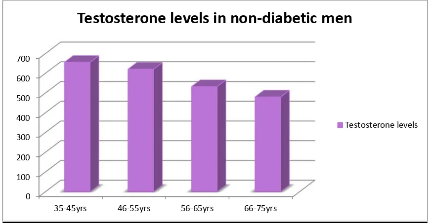
It was observed that on comparing the average total testosterone level
between the two groups, we found that, there was a significant decrease in the
testosterone levels in the diabetics compared to that of the non-diabetics in each
age group. We also found that as the age progress there is a steady decline in the
serum levels of testosterone in both the groups. But the fall in the testosterone
values is much more significant in the diabetics.
Statistical analysis was made and the P values were calculated by the student
T- test. The association between the diabetic and non-diabetics in each age group
was found to be significant with a P-value of <0.05. Thus the decrease in the serum
level of testosterone is found to be significant.
As the second part of our study we compared the serum testosterone levels
among the diabetic men, by separating them into smaller groups, using multiple
parameters such as duration of diabetes, BMI, Waist Hip ratio, cholesterol levels,
associated other risk factors. We were able to derive an inverse relation between
serum testosterone levels and most of these parameters. Thus we also evaluated the
effects of multiple risk factors on diabetic men and testosterone levels.
CONCLUSION
Thus, through this study we demonstrate the association between type 2
diabetes mellitus and testosterone. We conclude that, there is a significant
reduction in the levels of serum testosterone in diabetic men as compared to that of
as the age increases the reduction in the testosterone levels are more significant in
the diabetic group as compared to that of the non-diabetic men. Thus from our
study we propose that age factor alone is not the cause of hypotestosteronemia in
diabetes men.
From the analyses of different factors like BMI, cholesterol and other risk
factors we have shown the influence of multiple factors on the levels of
testosterone. Thus, in diabetic patients, hypotestosteronemia is a common entity.
We suggest the measurement of testosterone in patients with diabetes, as the
replacement of testosterone can have multiple benefits in diabetic patient to
prevent diabetic complication and cardiovascular diseases. Testosterone level can
further predict the development of type 2 diabetes mellitus, the utilization of this
aspect of testosterone in future, is not too far.
KEY WORDS
CONTENTS
SL.NO TITLE PAGE NO
1. INTRODUCTION 8
2. AIMS & OBJECTIVES 10
3. REVIEW OF LITERATURE 11
4. MATERIALS AND METHODS 61
5. OBSERVATIONS AND RESULTS 64
6. DISCUSSION 78
7. CONCLUSION 81
8. LIMITATION 82
ANNEXURES
ABBREVIATIONS
PROFORMA
MASTER CHARTS
BIBLIOGRAPHY
PATIENT CONSENT FORM
DIGITAL RECIEPT
83
85
90
98
124
125
INTRODUCTION
Testosterone a 19-carbon steroid secreted by the testis (Leydig cells) is the
primary circulating androgen in the male human. Testosterone is essential for
health and well-being and its levels decreases with aging. Men with type 2
diabetes mellitus have low testosterone levels, but this concept has not received
much attention because of the fact that both type 2 diabetes and
hypotestosteronemia are associated with aging. But studies have shown that
young men with type 2 diabetes mellitus do suffer from low testosterone levels.
Testosterone plays a vital role in the metabolism of glucose and lipid.
Decreased levels of serum testosterone often have adverse effects on the
metabolic profile. Epidemiological studies have shown that there is a direct
relationship between the serum testosterone levels and insulin sensitivity, and
that hypotestosteronemia is associated with an increased risk of type 2 diabetes
mellitus. It is also found that there is an inverse correlation between serum
testosterone and fasting insulin levels in men irrespective of age and obesity.
The mitochondrial dysfunction is the key factor leading to insulin resistance in
men.
hypotestosteronemia should be an element in the definition of metabolic
syndrome, since low levels of testosterone are associated with or predict the
development of metabolic syndrome and of diabetes mellitus.
In men with decreased serum levels of testosterone and type 2 diabetes
mellitus, C-reactive protein concentration have found to be decreased which
leads to increased risk of developing atherosclerosis and coronary heart disease
independent of the risk associated with diabetes mellitus alone.
Further testosterone replacement suggests that there is a shift of fat mass to
lean body mass. Both the systolic and diastolic blood pressure reduced and the
mean plasma glucose levels declined. There was a significant improvement in
lipid profiles. There was also an initial significant decrease in levels of liver
enzymes. Additionally, there was a marked reduction in the C-reactive protein
levels.
Further perspective is the healing of the diabetic ulcers with testosterone
AIMS & OBJECTIVES
To evaluate the level of serum testosterone in men with type 2
Diabetes Mellitus and to compare it with that of non-diabetic men
of the same age.
Effect of multiple parameters like obesity, hypertension etc. on
the serum testosterone levels in men with type 2 Diabetes
REVIEW OF LITERATURE
Though the effects of testosterone have been known since ancient times,
it was Ernest Laqueur [1] who first used the term testosterone (T) in 1935 when
he isolated it from the bull testes. It is derived from the German word
Testosteron (1935), coined from a presumed combined form of L. testis
"testicle" (testis) + ster(ol) (steroid) + chemical ending -one.[2]
HISTORICAL MILESTONES:
The researches done on testosterone dates back to the 1800 [3]. Since
antiquity, medicine men and scientists had the knowledge that castration of the
testes leads to the loss of the vitality of men and animals. A German
physiologist, Arnold Berthold [4], in 1849 experimented with four roosters,
where he removed the testes in two roosters and transplanted it into the
abdominal cavity of the other two [5]. The castrated roosters became fat and lazy
which is a sign of testosterone loss. The two with grafted testes remained a
rooster with bright red combs. [6]
Ernest Laqueur received the Nobel Prize [7] in 1935 for isolating the
testosterone molecule. It was found in 1951 that, testosterone will produce a
positive nitrogen balance and can increase the lean muscle mass [8]. This forms
disease [9] by synthesis of protein. With positive nitrogen balance there is
increased protein synthesis which leads to easy repair of the bodily structures
when damaged [10]. In addition, testosterone also induces the production and
synthesis of the protein enzymes which are useful for the structure and
functioning of the organs.
It was in 1935 that the newer era of modern testosterone therapy began,
when Aldolf Butenandt [11] and Leopold Ruzicka [12] synthesized testosterone
chemically. Oral forms of testosterone were found ineffective and 17-alkylated
derivatives of testosterone which was used orally became obsolete later due to
its side effects. So other methods of testosterone delivery were tried. A buccal
testosterone tablet [13] was introduced which adheres to the buccal mucosa and
by dissolving slowly released testosterone. Implants of crystalline testosterone
were also inserted in the subcutaneous tissue. The other form was as
testosterone gels which were opted for its ease in application. During 1950 and
60’s researches intensified to aid modifications in the testosterone preparations
[14]. Orally effective T undecanoate was introduced during 1970’s. Transdermal
scrotal patches [15, 16] were invented in 1992. Non-scrotal transdermal patches [17]
were accessible then in 2000. The most recent preparations used in testosterone
replacement therapy are the injectable forms which includes testosterone
Though we are very much aware of the association of type 1 diabetes
mellitus with that of other endocrine disorders [19], that is the Polyglandular
Autoimmune (PGA) Syndromes which includes adrenal insufficiency, type 1
diabetes mellitus, autoimmune thyroid disease, hypophysitis, celiac disease,
atrophic gastritis, pernicious anemia, vitiligo, hypoparathyroidism, myasthenia
and thymoma, the association of type 2 diabetes with other endocrinological
abnormalities is not well established. Around early nineties studies involving
the association between type 2 diabetes mellitus and hypotestosteronemia
began.
Since 1960 researches were conducted on the role of testosterone on the
cardiac well-being, its association with other cardiovascular risk factors and
metabolic syndrome [20, 21]. The knowledge of low levels of serum testosterone
as a reason for the all-cause mortality has led way to further researches. By
many studies it is now proved that apart from attenuation of sexual symptoms
BACKGROUND:
Over the last few years, there have been several reports demonstrating
that men with type 2 diabetes mellitus (T2DM) have a higher prevalence of
hypotestosteronemia compared to that of normal population [24]. It has been
found that reduced testosterone level is associated with increased risk of
metabolic syndrome and diabetes [25]. In men, hypotestosteronemia has been
observed to be associated with obesity, upper body fat distribution, and
increased level of glucose and insulin [26]. Men with low serum testosterone
levels present with loss of sexual interest, decreased vigor, depression, reduced
bone strength, redistribution of body fat, easy fatigability. As these symptoms
are usually non- specific, hypotestosteronemia is frequently under diagnosed
[27]. Compared to normal population, individuals with hypertension, obesity,
hyperlipidemia and diabetes have hypotestosteronemia [28, 29]. The purpose of
this study, therefore, is to evaluate the serum level of testosterone in males with
TESTOSTERONE
:
Secretion:
The male gonad, testes secretes the male sex hormones, which are known
as the androgens [30]. This includes testosterone, dihydrotestosterone, and
androstenedione. Among these three hormones, testosterone is found to be
secreted in larger quantities and so it is considered to be the important testicular
hormone. It is to be noted that the testosterone eventually gets converted to
dihydrotestosterone which then acts on the target tissues.
The Interstitial cells of Leydig [31], which are polyhedral cells that lies as
clusters between the seminiferous tubules elaborates the hormone testosterone,
parallel that of the testosterone surge. They are found to be more numerous in
neonates in the initial months and then in the post pubertal adulthood period. In
the childhood period these Leydig cell are scanty and so there is no detectable
testosterone [32].
Chemistry [33]:
Testosterone, the principal testicular hormone is a steroidal compound, a
C19 steroid with an -OH group in the 17 position. Its molecular formula being,
C19H28O2.
Synthesis:
Testosterone is derived [34] from cholesterol, the major site of synthesis
being the Leydig cells of the testes. Adrenal cortex also shares its contribution
by the secretion of androstenedione which then gets further converted into
cholesterol into the mitochondria which need a transport protein. The initial step
begins with the conversion of cholesterol to pregnenolone when it is acted upon
by the side chain cleavage system. [35]. Pregnenolone is further converted into
testosterone by two pathways, either by the progesterone pathway or by the
dehydroepiandrosterone pathway. The array of enzymes which are involved
includes 3 beta hydroxysteroid dehydrogenase, 17 alpha hydroxylase and 17, 20
lyase, 17 beta hydroxysteroid dehydrogenase. Reduction of the adrostenedione
at the 17th carbon position results in the formation of testosterone.
Transport and Metabolism:
Testicular secretion of testosterone contributes to the major quantity of
the testosterone in the circulation. It constitutes around 90-95%. The rest is
contributed by the adrenal steroidogenesis and from the conversion of
androstenedione to testosterone in the periphery [36]. The most important
peripherally from testosterone by a reaction that is catalyzed by the enzyme 5
alpha reductase, although the testes secrete a very minor quantity of
dihydrotestosterone. In many tissues like the external genitalia, prostrate and
some parts of the skin, dihydrotestosterone is the active form of the hormone.
The production of dihydrotestosterone is around 400 micro grams as compared
to about 5 grams of testosterone which shows that the plasma concentration of
dihydrotestosterone is only one-tenth of that of testosterone concentration in
adult males.
Testosterone gets metabolized by two pathways. The first pathway which
involves oxidation is the major pathway. It occurs in many tissues including the
liver and the end product being ketosteroids which are usually less active than
testosterone. The second pathway involves reduction of the double bond and it
occurs in the target tissues. The end product is dihydrotestosterone which is
more potent than testosterone.
In male a small quantity of testosterone is converted into estradiol by
peripheral aromatization and this contributes to about 80% of the circulating
estradiol in men.
98% of the testosterone in the circulation is bound to protein the rest
remains unbound as free hormone and it is this unbound free form which is
biologically active. About 65% is bound to beta globulin also called the sex
plasma level of testosterone. Conditions producing a decrease in SHBG are
obesity, diabetes, administration of androgens, while aging and estrogen
produces an increase. About 33% of the testosterone is bound to albumin.
The plasma testosterone level (free and bound) is 300-1000ng/dl in adult
men and 30-70ng/dl in adult women.
Control of Testicular functions:
The secretion of testosterone is under the influence of the Luteinizing
hormone (LH) secreted by the anterior pituitary. LH acts on the Leydig cells [38]
and stimulates them to secrete testosterone. Excess testosterone in turn
suppresses the secretion of LH from the pituitary. Testosterone does not have
much influence on FSH (Follicle stimulating hormone) whereas another
hormone called inhibin which is secreted by the Sertoli cells decreases the FSH
concentration.
Steroid Feedback:
The current "working hypothesis" of the way the functions of the testes are
regulated involves the feedback control of the LH on the Leydig cells and by
FSH on the Sertoli cells. These in turn by the secretion of testosterone and
inhibin controls the secretion of LH and FSH respectively. These feedback
mechanisms are further under the control of GnRH secretion from the
GnRH apart from LH. Castration is followed by a rise in the pituitary content
and secretion of FSH and LH [39], and hypothalamic lesions prevent this rise. [40,
41].
Disorders involving the hypothalamus can lead to atrophy of the testes.
This hypothesis can be used upon clinically. Systemically administered
testosterone has not found to increase the concentration of androgen in the
local environment of the testes (a phenomenon which is necessary for normal
spermatogenesis to occur i.e. increased concentration of androgens around the
Sertoli cells) and this causes inhibition of LH secretion. Thus the ultimate
effect of administering testosterone systemically is to produce a decreased
sperm count. Thus is the use of testosterone as a method of contraception [42].
The main drawback is the need to use testosterone at very high doses, at with it
Mechanism of Androgen Action [43]:
The androgen receptor (AR) [44] belongs to the superfamily of receptors
which have highly conserved DNA binding domain in common-the nuclear
receptor super family. The long arm of the X chromosome [45] codes for the
androgen receptors.
Testosterone binds itself to an intracellular receptor and forms a
receptor-steroid complex. This complex then binds to the DNA in the nucleus leading to
transcription of various genes [46, 47]. Other mechanism of androgen action is
Basic Intracellular Mechanism of Action of Testosterone
Most of the effects of testosterone result basically from an increased rate
of protein formation in the target cells. This has been studied extensively in the
prostate gland, one of the organs that is most affected by testosterone. In this
gland, testosterone enters the prostatic cells within a few minutes after
secretion. Then it is most often converted, under the influence of the
intracellular enzyme 5 alpha-reductase [48], to dihydrotestosterone, and this in
turn binds with a cytoplasmic “receptor protein” [49]. This combination migrates
to the cell nucleus, where it binds with a nuclear protein and induces
DNA-RNA transcription. Once the DNA-RNAP gets activated, the amounts of the DNA-RNA
starts to rise, which then leads to a significant raise in the protein content of the
cells [50]. After several days, the quantity of DNA in the prostate gland also
increases and there is a simultaneous increase in the number of prostatic cells.
Testosterone stimulates production of proteins virtually everywhere in the body,
although more specifically affecting those proteins in “target” organs or tissues.
Thus testosterone leads to the development of the primary sexual characteristics
and those of secondary too. Recent studies suggest that testosterone, like other
steroidal hormones, may also exert some rapid, non-genomic effects that do not
require synthesis of new proteins. The physiological role of these non-genomic
Functions of Testosterone
In general, testosterone is responsible for the development of male body
configuration. Even during fetal life, the testes are stimulated by chorionic
gonadotropin from placenta to produce moderate quantities of testosterone
throughout the entire period of fetal development and for 10 or more weeks
after birth; thereafter, essentially no testosterone is produced during childhood
until about the ages of 10 to 13 years. Under the influence of the luteinizing
hormone from the anterior pituitary, the Leydig cells begin to secrete
testosterone. This increase in testosterone starts with the beginning of puberty
and continues secretion thereafter. As the age increases, the amount of
testosterone secretion decreases, declining progressively after the age of 50yrs
with the maximum fall being after the age of 80 yrs.
Functions of Testosterone during Fetal Development:
Testosterone begins to be elaborated by the male fetal testes at around
seventh week of intra uterine life [51]. It can be said that, the main impetus for
the development of a male fetus is the secretion of testosterone by the fetal
gonads, which is mediated by the presence of the Y-chromosome. Testosterone
also mediates the development of male internal genitalia and
dihydrotestosterone (its active metabolite) mediates the development of the
female fetus. By experimental studies it has been proved that when a large
amount of testosterone is injected into a pregnant animal, even if the fetus was a
female, it led to the development of male sex organs [52]. It was also found that
when the testes were castrated early from a male fetus it developed female sex
organs. These prove the essentiality of testosterone for the development of both
the internal and the external genitalia in a male fetus [53] and at the same time it
inhibits the development of female sex organs.
Effect of Testosterone to Cause Descent of the Testes. The testes usually
descend into the scrotum during the 8th to 9th month [54] of gestation during
which the testes begin secreting reasonable quantities of testosterone. If a male
child is born with undescended but otherwise normal testes, injection of
testosterone has been found to causes the descent of the testes normally.
Administration of gonadotropic hormones also, which stimulate the Leydig cells
of the newborn child’s testes to produce testosterone, can also cause the testes to
descend. Thus, the stimulus for descent of the testes is testosterone [55],
indicating that testosterone is an important hormone for male sexual
development during fetal life.
Functions of Testosterone after puberty:
Effect of Testosterone on Development of Adult Primary and Secondary
After puberty, the so long quiescent testes begin to secrete testosterone and
the testosterone concentration increases considerably. This leads to certain
changes in the external genitalia of the male i.e. enlargement of these organs.
There is also enlargement of the prostate gland and the seminal vesicles with
production of fructose by the seminal vesicles. The second important function
of testosterone during puberty is the emergence of secondary sexual
characteristics. These two are the very essential characters because it helps in
distinguishing a man physically. Testosterone also enhances the male psychic
effects.
Effect on the Distribution of Body Hair:
Testosterone causes the male pattern of hair growth over the body. These
usually are present:
(1) On the beard area with the development of moustache
(2) Over the chest
(3) Over the abdomen along the linea alba up to the pubic symphysis
(4) Also on the back along the shoulders
(5) Pubic hair growth. It can also produce hair growth anywhere in the
body but it generally decreases the scalp hair growth.
Baldness: As mentioned above, testosterone reduces the scalp hair growth
sole reason for baldness; it is the interplay of many factors especially genetic
factors [57] followed by excessive androgens [58].
Effect on the Voice:
The masculine type of voice is a harsh and low pitched voice. This
character to the voice is produced by testosterone. Testosterone makes the
laryngeal mucosal hypertrophy with resultant laryngeal enlargement. Around
puberty the voice become discordant initially which is termed “cracking” voice
[59], this is followed by a gradual change to voice of an adult male.
Testosterone Increases Thickness of the Skin and contribute to
Development of Acne:
Testosterone produces certain structural changes in the skin. Firstly it
increases the skin thickness of the whole body and produces ruggedness of the
tissues. Secondly it acts on the sebaceous glands and causes its growth and
increases the secretion of sebum from the sebaceous glands. This results in the
production of acne especially over the face [60]. Therefore, acne is a common
feature of male adolescence when the body is first becoming introduced to
increased testosterone. After several years of testosterone secretion, the skin
Testosterone Increases Protein Formation and Muscle Development [61]:
One of the most important male characteristics is the development of
increasing musculature after puberty, averaging about a 50 per cent increase in
muscle mass over that in the female. This is associated with increased protein
formation in the non-muscular parts of the body as well. Many of the changes in
the skin are due to deposition of proteins in the skin, and changes in the voice
also result partly from this protein anabolic function of testosterone. Because of
this great effect that testosterone and other androgens have on the body
musculature, synthetic androgens are widely used by athletes to improve their
muscular performance. This practice is to be severely deprecated because of the
fact that prolonged harmful effects of excess androgens, in relation to sports
physiology. Testosterone or synthetic androgens are also occasionally used in
elderly age as a “youth hormone” to improve muscle strength and vigor, but
with questionable results.
Testosterone Increases Bone Matrix and Causes Calcium Retention:
After the great increase in circulating testosterone which occurs during
puberty (or after prolonged injection of testosterone), the bones grow
considerably thicker [62] and deposit considerable additional calcium salts. Thus,
testosterone increases the total amount of bone matrix [63] and causes calcium
retention. The increase in bone matrix is believed to be a result of the general
response to the increased protein. Testosterone gives the male pelvis a
differentiating effect from that of the female pelvis thereby making it strong to
bear heavy weights. In the absence of testosterone, the male pelvis develops into
a pelvis that is similar to that of the female. Because of the ability of
testosterone to increase the size and strength of bones, as aging occurs it can
lead to decreased BMD and it is often used in older men to treat osteoporosis
[64]
. When greater amounts of testosterone (or any other androgen) are secreted
abnormally in the still-growing child, the rate of bone growth increases
markedly, causing a spurt in total body height. However, the testosterone causes
the epiphyses of the long bones to unite with the shafts of the bones at an early
age. Therefore, despite the rapidity of growth, this early uniting of the epiphyses
prevents the person from growing as tall. Even in normal men, the final adult
height is slightly less than that which occurs in males are castrated before
puberty.
Testosterone Increases Basal Metabolism [65]:
Injection of large quantities of testosterone increases the basal metabolic
rate by as much as 15 per cent. This increased rate of metabolism is possibly an
indirect result of the effect of testosterone on protein anabolism, the increased
quantity of proteins—the enzymes especially—increasing the activities of
Effect on Red Blood Cells:
When normal quantities of testosterone, is injected into a castrated adult,
the number of red blood cells per cubic millimeter of blood increases by about
15 to 20 per cent [66]. Also, an average man has about 700,000 more red blood
cells per cubic millimeter than an average woman. This difference is partly due
to the increased metabolic rate that occurs after testosterone administration
rather than to a direct effect of testosterone on red blood cell production.
Effect on Electrolyte and Water Balance:
Testosterone causes an increase in the distal tubular sodium reabsorption
by the kidneys. Testosterone also has such an effect, but only to a minor degree
when compared to that of the adrenal mineralocorticoids. Nevertheless, after
puberty, the blood and extracellular fluid volumes of the males in relation to
Normal Testosterone Levels in Men
[67]by Age (Healthy)
Measurements in SI Units (nmol/L)
Age Total
Test
Free
Test SHBG
25-34 21.38 0.428 35.5
35-44 23.14 0.356 40.1
45-54 21.02 0.314 44.6
55-64 19.49 0.288 45.5
65-74 18.15 0.239 48.7
75-84 16.32 0.207 51.0
85-100 13.05 0.186 65.9
Measurements in Conventional Units ( ng/dl), SHBG in (nmol/L)
Age Total
Test
Free
Test SHBG
25-34 617 12.3 35.5
35-44 668 10.3 40.1
45-54 606 9.1 44.6
55-64 562 8.3 45.5
65-74 524 6.9 48.7
75-84 471 6.0 51.0
85-100 376 5.4 65.9
The normal testosterone level in young healthy males is between 300 and
1000ng/dl. Testosterone level shows diurnal variations with lower value in the
CAUSES OF HYPOTESTOSTERONEMIA [68]
The causes of hypotestosteronemia are many to be enumerated. In a nutshell,
there are three main levels at which defect can occur, finally leading to
hypogonadism:
1. The level of the hypothalamus
2. The level of pituitary gland
3. The level of the testes.
When the defect lies in the hypothalamus or in the pituitary they are not
able to produce appropriate quantities of gonadotrophins to stimulate
adequate testicular secretions. Hence it is called hypogonadotrophic
hypogonadism. When the testes are at defect though there are adequate
quantities of gonadotrophins the testes is not able to secrete adequate
amounts of testosterone. Hence it is called hypergonadotropic
hypogonadism. In addition, SHBG also affects the serum testosterone
levels.
When the defect involve the gonads, the testes, then it is termed as
"primary hypogonadism or primary gonadal failure[69] "
When the pituitary gland or its control over testosterone regulation is at
When the defect lies high up in the hypothalamus, it is termed as
"tertiary hypogonadism [69]." The last two entities are together called as
hypogonadotrophic hypogonadism.
PRIMARY HYPOGONADISM OR PRIMARY GONADAL FAILURE
(TESTES):
A. Primary gonadal failure may lead to defect in spermatogenesis,
production and secretion of testosterone.
B. Chromosomal abnormalities: Klinefelter's syndrome [70], in which there is
an abnormal testicular development with reduced size of the testicles ad
decreased production of testosterone.
C. Anorchia syndrome [71]
D. Uncorrected cryptorchidism [72]
E. Acquired defects of the testes:
1) Trauma to the scrotum resulting in testicular injury
2) Torsion of testes leading to vascular insufficiency[73]
Viral orchitis may be caused by the mumps virus, group
B arbovirus echovirus, lymphocytic choriomeningitis
virus, most important of which is the mumps infection [74]
Other bacterial and parasitic infections.
4) HIV infection[74]
5) Tuberculosis and Lepromatous leprosy
6) Toxic causes [75]
Drugs: ketoconazole (inhibition of testosterone
synthesis), spironolactone (blockade of androgen action),
marijuana and digitalis (increased estrogen)
Chemotherapeutic agent: causes direct inhibition of
Cyclophosphamide,procarbazine and patients on MOPP
(mechlorethamine, oncovin, procarbazine, prednisone
Alcohol, when consumed in excess for prolonged
periods, decreases testosterone, independent of liver
disease or malnutrition.
Known environmental hazards include microwaves and
ultrasound and chemicals such as nematocide
7) Systemic illness: cirrhosis, chronic renal failure, sickle cell anemia,
acute febrile illness, celiac disease
8) Neurologic disorders: myotonic dystrophy [76], spinobulbar muscular
atrophy, and paraplegia
9) Androgen Insensitivity Syndromes
10) Aging [77]: serum levels of testosterone declines with aging. But the
available testosterone is still enough to carry out essential functions of
the body.
11) Hypotestosteronemia in women: Certain conditions in women cause
low levels of serum testosterone. These includes bilateral
oophorectomy due to any gynecological cause and premature ovarian
failure
SECONDARY AND TERTIARY HYPOGONADISM :
These are due to defect either at the level of the hypothalamus or the pituitary
gland resulting in decreased production of the gonadotrophins (LH/ FSH/
GnRH) and failure of gonadal stimulation.
1. Congenital Hypogonadotropic Disorders:
Kallman's syndrome [78], Laurence-Moon syndrome, adrenal hypoplasia
congenital, Prader-Willi syndrome.
2. Acquired Hypogonadotropic Disorders
Tumors of the pituitary gland [79]
Brain tumors
Sellar Mass Lesions
Tuberculosis
Infiltrative diseases like hemochromatosis [80] sarcoidosis and
histiocytosis X damages the pituitary gland
HIV and AIDS [74] affects the hypothalamus and pituitary
Anabolic steroid use
Obesity [81]
Hyperprolactinemia [83]
Severe Illness
Stress, Malnutrition
Normally, the conversion of testosterone to estrogen [82] occurs in the
adipocytes. In the presence of obesity, the amount of fat tissue mass that can act
as a site for this conversion increases and hence there is more aromatisation
reaction taking place thereby, decreasing the serum level of testosterone, due to
its excessive conversion in to estrogen in the adipocytes.
HYPOTESTOSTERONEMIA
DEFINITION [84]:
The American Association of Clinical Endocrinologists through detailed
studies have come forward with the definition for hypogonadism which is
defined as a serum level of testosterone <300ng/dl. [85, 86]. There is a diurnal
variation in the level of testosterone. So it is better to measure the testosterone
level in the morning rather than other timings i.e. around 8 a.m. in the morning
[87, 88]
In case the total testosterone levels are found to be subnormal before
reporting it is always necessary to confirm it by a second test. Clinically there
are certain indicators for the diagnosis of hypotestostreronemia, but most of
these symptoms are non-specific. Hypotestostreronemia [89] is characterized by
reduced sexual drive, reduced frequency of sexual intercourse or difficulty in
maintaining erections, reduced growth of beard, loss of muscle mass, increased
fat mass, decreased density of bone, decrease in testicular size, and enlargement
of breast tissue. But only less than 10% of patients with erectile dysfunction
whole, the entire symptomatology, to conclude as hypogonadism. Another
lacuna in this area is that unless the symptoms are very profound it almost
overlaps with the changes that occur in normal aging process.
TESTOSTERONE ASSAY:
When the word Total Testosterone (TT) is used, it means the measurement
of both the bound form of testosterone as well as the unbound, free form. This
total testosterone can be measured by a number of methods. Some of them are
the radio-immuno assay method, liquid chromatography tandem mass
spectrometry (LC-MS/MS) [90]. The more accurate method is the LC-MS/MS.
This method is done by using organic solvents for the purpose of extracting the
serum, followed by the isolation of testosterone from that of other steroids by
the method of high-performance liquid chromatography and mass spectrometry,
and finally to quantify the unique testosterone segments by mass spectrometry.
The advantage of using this LC-MS/MS is that it is an accurate and a sensitive
method for measuring even lower ranges of serum levels of testosterone. In
future it is the method of choice for the estimation of serum levels of
testosterone. Unlike many other hormone analyses, evaluation of testosterone
levels can be accurate with the average testosterone levels with even a single
sample but it must always be kept in mind that serum levels of testosterone
consistent with hypogonadism, the initial step is the measurement of total
testosterone in the morning sample using assay such as LC-MS/MS.
Measurement of Unbound Testosterone Levels [91, 92]
It is known that 98% of the plasma testosterone is bound to plasma
proteins, either to sex hormone binding globulin (which carries most of the
testosterone) or to albumin. The rest of the testosterone is called the unbound
fraction or the free form. It should be noted that this form is the active form.
This free form can be either calculated from the total testosterone levels and the
sex hormone binding globulin by a formula called free androgen index in which
ratio of total testosterone and the sex hormone binding globulin is obtained and
expressed as percentage. Another method used for measurement of free
testosterone is equilibrium dialysis method. Other method includes tracer
analogue methods and though the results by this method are inaccurate it’s a
more convenient method. Bioavailable testosterone is the term used to indicate
the unbound free testosterone plus testosterone which is bound loosely to the
albumin; this bioavailable testosterone is measured by the ammonium sulfate
precipitation method. In elderly male and in patients in whom conditions
associated with SHBG alterations are suggested, utilizing the equilibrium
method for the direct measurement of free testosterone can be useful in
HCG Stimulation Test
There are two methods to perform an hCG stimulation test. [93] It can be
performed either by administrating hCG as a single dose or as multiple doses
and then recording the levels of testosterone. In the first method 1500–4000 IU
of hCG is administered intramuscularly single dose. This is followed by
measuring the level of serum testosterone at the baseline and then on 24, 48, 72,
and 120 hours after injecting hCG. The second method is that of giving 1500
units of hCG on three successive days amounting to three hCG doses. Serum
testosterone levels are analyzed at least 24 hours since the last dose. The
expected response to this hCG stimulation test is that, in an adult male, there
should be a doubling of the testosterone concentration. This test is of main
purpose in children, especially prepubertal aged. A response of testosterone
levels to >150 ng/dL in a prepubertal child shows that testicular tissue is
present. When there is no response to this test, it shows that either the testicular
tissue is absent or there is a functional defect in Leydig cells. Another test
which is useful in the evaluation of undescended testes is the measurement of a
substance called MIS, Mullerian inhibiting substance, secreted by the Sertoli
LOW TESTOSTERONE IN TYPE 2 DIABETES
Men suffering from type 2 diabetes mellitus are found to have a low serum
testosterone levels on comparison with men who do not have a previous history
of diabetes mellitus. It has also been found that there exists an inverse
relationship between serum testosterone and HbA1c concentrations [95]. A
number of Meta-analytic studies have shown that there is a profound
hypotestosteronemia in men with type 2 diabetes mellitus [96]. Conversely in
men suffering from hypotestosteronemia the risk of developing type 2 diabetes
mellitus is high. About 30-50% of the diabetic men have found to have
associated hypotestosteronemia. Recent researches prove that testosterone
replacement therapy has been found to be beneficial in men with low
testosterone levels with associated type 2 diabetes mellitus or metabolic
syndrome [97].
Experimental studies showed that testosterone has found to afford
protection to the endothelium of the blood vessels thereby maintaining the
integrity of the vasculature. Thus hypotestosteronemia leads to endothelial
dysfunctions and its sequelae, irrespective of the presence of any other risk [98].
Further lower serum levels of testosterone concentrations can cause stiffness of
the arteries in men suffering from type 2 diabetes mellitus [99]. This appears to
be one of the main contributing factors for the development of cardiovascular
In men with type 2 diabetes mellitus presence of hypotestosteronemia is
common entity but it still remains a hidden epidemic. This is in turn associated
with insulin resistance. As the symptoms of hypogonadism are almost always
nonspecific, it goes unnoticed unless profound. Some of the symptoms in
diabetic men pointing towards testosterone deficiency include decreased libido,
impaired mood, and low performance scale [100]. There appears to be a direct
association between hypotestosteronemia and development of cardiovascular
risk factors. Furthermore there are evidences suggestive of an inverse
relationship between hypotestosteronemia and insulin resistance, which in turn
is strongly associated with the development of complications of diabetes
mellitus both micro vascular and macro vascular[101]. Prospective studies have
proved that in patients with decreased levels of total testosterone (TT) who have
associated insulin resistance there is a higher risk for the development of type 2
diabetes mellitus in future [102]. This is further proved by the fact that
testosterone administration shows improvement in insulin sensitivity.
As it was explained previously, SHBG is the major transporter protein of
testosterone in the circulation. Another important fact which must be kept in
mind is that a low serum concentration of SHBG can by itself be associated
with the risk of developing type 2 diabetes mellitus irrespective of whether
serum testosterone concentration is decreased or within the normal ranges. [103]
conditions it acts as a confounding factor. Estimation of the free testosterone
values rather than the total testosterone would useful under such conditions [104].
The prevalence of hypogonadism is much more common than expected in
patients with type 2 diabetes mellitus. This association is proved to be present
independent of certain important factors such as the duration of diabetes, the
status of the glycemic control, presence of micro or macro vascular
complications and whether associated with other risk factor or not. Either alone
or as a part of metabolic syndrome diabetes mellitus has a definitive association
with diabetes mellitus.
Another important aspect in the evaluation of hypogonadism is aging.
Testosterone concentration steadily declines after the age of 50 yrs. The total
testosterone levels fall around 0.5 to 2% each year to reach a maximum fall
around 80yrs. In diabetic patients there is a more profound decline in the
After analyzing a number of studies we are able to conclude that
hypotestosteronemia is per se associated with an increase in all-cause mortality
irrespective of whether associated with diabetes or metabolic syndrome [105].
Low testosterone levels associated with or without type 2 diabetes mellitus can
be used as a predictor for the development of cardiovascular risks and
complications irrespective of the presence or absence of other cardiovascular
risk factors. Hypotestosteronemia and cardiovascular disease are associated
with common risk factors like central obesity, redistribution of fat,
inflammatory state and coagulation abnormalities [106]. Also in patients
presenting with hypogonadism and cardiovascular disease (CVD) the mortality
rate appears higher when compared to men without hypotestosteronemia [107].
Testosterone supplementation has been done to evaluate for the effects on
the glycaemic status of men with type 2 diabetes mellitus and hypogonadism
through many studies. But none of the studies has proven to give any conclusive
results. Half of the studies showed no specific effect on the glycaemic status
while half of the studies showed a definitive decrease in the glycaemic control
with testosterone administration [108].
Testosterone and Insulin resistance:
Evidences from many data have proved that hypotestosteronemia is
mechanism which leads to this resistance was made by many molecular studies
of which the most convincing was the alterations in fatty acid metabolism,
probably due to reduced expression of the genes which are necessary in
oxidative metabolism. Similarly it has also been proved that testosterone
improves the insulin sensitivity directly. Thus testosterone replacement has been
found to increase the insulin sensitivity. Also it was found that in such men who
were receiving testosterone supplementation for hypogonadism, when the
treatment was stopped abruptly for around 15 days the insulin sensitivity
decreased though without much alterations in the body composition. This is an
evidence to show that testosterone has a direct control over sensitivity of insulin
[109]. But some reports have shown that in elderly males by bringing the
testosterone levels into normal range, there has been a favorable effects on the
fat distribution and hence composition thus improving the insulin sensitivity,
but they do not have any beneficial effect on the triglyceride metabolism [110].
But by experimental studies it has been proved that the effect of testosterone on
insulin resistance was an acute phenomenon and the do not produce any
immediate effect on the body composition. At the same time, experimentally, by
reducing the estradiol concentration and by increasing the testosterone levels for
a period of 7 days, it has reduced the insulin resistance with beneficial effects
on the triglyceride metabolism, and has improved the postprandial
glucose-dependent insulinotropic polypeptide (GIP) release [111]. Thus the association
common mechanism of increasing the proinflammatory factors, decreasing the
insulin sensitivity thus produces endothelial dysfunction which are the essential
factors leading to the development of cardiovascular diseases and erectile
dysfunction (ED).
Pathogenesis of insulin resistance:
Present concepts throw light on the contributions of mitochondria in the
pathogenesis of the development of insulin resistance. Studies on animal
models have confirmed that, in insulin resistance there is a decreased expression
of OXPHOS genes in skeletal muscle along with the down regulation of
peroxisome proliferator–activated receptor-γ coactivator (PGC-1α) [112]. In
proposing the model for demonstrating the pathogenesis of insulin resistance it
was found that there was a reduction in the peroxisome proliferator–activated
receptor-γ coactivator expression in the presence of physical inactivity in
genetically susceptible subjects. The decreased expression of this single protein
led to the decreased transcription of certain genes involved in the metabolism of
fatty acids, leading to accumulation of triglycerides in the cells of the striated
muscles, decreased expression of mitochondrial genes with the decreased
efficacy of oxidative phosphorylation and resulting in insulin resistance
ultimately. Experimental reproduction of hypogonadism has proved to show
of testosterone. There are not much data to explain the direct effect exerted by
testosterone on insulin.
Another mechanism which is claimed in the development of insulin
resistance is that of reduced glycogen synthetase activity [114] in experimental
rats with low testosterone levels. This reduced activity of the glycogen
synthetase [115] is computed to be the cause of the triglyceride accumulation in
the myocytes intracellularly. The genetic model of type 2 diabetes mellitus with
obesity is the Otsuka Long Evans Fatty (OLETF) [116] rat, confirms the effect of
testosterone on the mitochondrial functions. These rats having low levels of
androgens showed a decreased expression of peroxisome proliferator–activated
receptor-γ coactivator and beta 3 receptor, androgen receptors present in the
adipocytes. Further there is an increased expression of uncoupling protein 1
(UCP-1) and up regulation of its genes. All these lead to decreased peripheral
utilization of energy, its accumulation as fats and therefore obesity [115]. When
these OLETF rats were supplemented with testosterone (DHEA) for 2 weeks, it
caused many positive effects in the form of improved insulin sensitivity,
reduction of weight, decrease in FFA, and leptin concentration, all due to a
considerable increase in the expression of UCP-1, β3AR, and PGC-1α [116].
It requires further experimentations and analysis in to the expression of
genes in the skeletal muscle with testosterone replacement. If the expression of
surely testosterone has a bright future in the prevention and treatment of type 2
diabetes mellitus in men with hypogonadism.
Testosterone and Metabolic syndrome:
Presently there have been many studies proving the correlation of
metabolic syndrome and hypotestosteronemia. Such men have a reduced
concentration of TT, FT, and SHBG [117]. This syndrome which includes an
alteration in a number of metabolic parameters has an increased risk of
mortality more so when the patient do have associated low levels of
testosterone. There is a resulting proinflammatory state which leads to
endothelial dysfunction and other complications like cardiovascular disease and
ED. On the other hand, hypotestosteronemia can be used as a predictor for the
future development of syndrome X and type 2 diabetes mellitus.
Low levels of serum total testosterone cause redistribution of fat and
further accumulation of intra-abdominal fat thus leading to central obesity [118].
Independent of the age factor or the presence of obesity a low testosterone
levels are associated with the development of metabolic syndrome.
Experimentally suppression of testosterone in men caused an increase in the
body fat [119]. The same effect occurs when there is a decrease in the
concentration of sex hormone binding globulin. Since obesity is found to
testosterone SHBG is a confounding factor in defining the correlation between
hypotestosteronemia and obesity [120].
There are a number of factors which determine the presence of
hypotestosteronemia or low levels of sex hormone binding globulin. These
include age, race, and obesity. But hypotestosteronemia is associated with type
2 diabetes mellitus independent of these factors or the diagnostic criteria of
diabetes [121]. Studies also prove that TT, FT, and sex hormone binding globulin
independently can be used in the prediction of the development of type 2
diabetes. [122] There is an emerging interest on SHBG as an independent
positive predictor of diabetes mellitus. But some recent studies have proved that
free testosterone cannot be useful for this purpose [124]. As mentioned above
SHBG is a confounding factor for the association of hypotestosteronemia and
type 2 diabetes mellitus and since most cases of diabetes are associated with
obesity, obesity also remains confounding to establish this relationship.
Compared to non-diabetic men reduced free testosterone concentrations are
more prevalent in diabetic men [123]. Also, low SHBG was found to be a strong
independent predictor of type 2 diabetes mellitus [124]. Finally, in prospective
studies, androgen deprivation therapy either using bilateral orchidectomy or
gonadotropin-releasing hormone agonist in older men with prostate cancer is
Pathobiology of low testosterone in type 2 diabetes
Adipocyte is biologically active tissue secreting a number of substances
cytokines and chemokines. Some of them include adipokines, tumor necrosis
factor (TNF)-α, interleukin (IL)-6, IL-1β, VEGF, transforming growth factors,
plasminogen activator inhibitor-1, and angiotensinogen [126]. All of these factors
are mediators in inflammation. Thus there is an inflammatory state which is set
up in the body milieu. When the amount of the adipose tissue increases as it
occurs in obesity, there is a chronic proinflammatory state set up in the body
which results in increased concentration of inflammatory mediators as well as
FFA and an increased estrogens output from the adipose tissue, which is
obtained by its conversion from testosterone. This state of chronic inflammation
leads to alteration in the blood mechanics with stiffness of the vasculature and
endothelial dysfunction. All these together results in systemic inflammation and
contributes to the development of metabolic syndrome and type 2 diabetes
mellitus as well as hypotestosteronemia.
The proposed mechanism of how obesity and inflammation promotes
insulin resistance is through the release of excess of FFA. This in turn through
the nuclear factor-κB pathways produces augmented synthesis of TNF-α,
interleukin 6 and MCP-1. The former factor causes increased lipolysis and
of insulin. Other mechanism of actions of these inflammatory mediators is an
increased expression of the adhesion molecules in the endothelium of the blood
vessels and also in its smooth muscles mediated by TNF-α. Apart from this
TNF-α also causes a dysregulation between the vasoconstriction mediated by
ET-1 and the vaso relaxtion induced by nitric oxide ultimately favouring
vasoconstriction; similarly CRP, though nonspecific, a marker of inflammation
IL-6 is increased through IL-6 mediated enhanced hepatic synthesis.
Adipokines, [127] another important mediator modulates chemotaxis and
adhesion of inflammatory cells especially monocytes and facilitates its
conversion in to macrophages.
Normally the conversion of testosterone to estrogen [83] occurs in the
adipocytes because the enzyme aromatase is located in the fat cells. In the
presence of obesity the amount of fat tissue mass that can act as a site for this
conversion increases and hence there is more aromatisation reaction taking
place thereby decreasing the serum level of testosterone due to its excessive
conversion in to estrogen in the adipocytes. This in turn activates the
hypothalamic E2 receptors and produces feedback inhibition of the gonads.
Thus aromatase inhibitors play a role in attenuating the symptoms of
Testosterone and atherogenesis:
Testosterone has an antithrombotic effect on the clotting mechanism.
There is an inverse relationship between the prothrombotic clotting factors,
fibrinogen and the tissue plasminogen activator inhibitor 1. It also has a direct
relationship with the plasminogen activator activity [129]. Thus the presence of
normal levels of testosterone concentrations is necessary for maintaining proper
blood homeostasis.
From experimental studies it has become evident that testosterone slows down
the atherogenesis [130]. Iatrogenic orchidectomy in mouse led to the development
of atherosclerotic plaques in the aorta. Another important point to be noted is
that, irrespective of the lipid profile, testosterone slows down the progress of
atherogenesis [131] and the further development of cardiovascular risk. By
inhibition of the inflammatory mediators testosterone reverses prothrombotic
environment. Testosterone reduces the early progression of atherosclerosis by a
major mechanism by which it gets converted to estradiol in the walls of the
blood vessel by the reaction of aromatization. This can be proved by the
increase in the progression of atherosclerosis in patients on aromatase
inhibitors.
Testosterone Levels and Cardiovascular Disease (CVD)
It was initially presumed that high levels of testosterone produce adverse
effects on the heart and the vascular system. But latter there was a consensus
that high normal range of testosterone level did not produce any risk to the
cardiovascular system. Presently it has been found through many meta-analyses
that lower levels of testosterone are associated with a stronger positive
correlation to the development of adverse cardiovascular effects. The most
important effects by which hypotestosteronemia can induce atherosclerosis and
hence coronary artery disease includes alteration of hemostatic mechanics
towards a prothrombotic as well as a fibrinolytic state, increased insulin
resistance along with an adverse lipid profile [132]. It has also been proven by
experimental studies that castration of the testes resulted in increased risk of
Apart from decreasing the adverse cardiac effects testosterone also has
many beneficial effects on the heart. Testosterone has a strong vaso-relaxing
effect on the coronary vasculature [133]. This effect is caused by testosterone
induced calcium channel blockade in the smooth muscles of the blood vessels.
This action of testosterone has been used to raise the threshold of angina in men
with hypotestosteronemia thereby decreasing the development of angina [134].
Testosterone is also beneficial in the treatment of chronic congestive heart
failure by increasing the functional capacity and by improving the insulin
sensitivity. [135]
With the knowledge of the available studies it can be thus concluded that
for optimal cardiovascular health it is always necessary to maintain the level of
plasma testosterone about the mid-range of the normal values. It has also been
shown that testosterone levels both below or above the normal values pertaining
the particular age has found to raise the cardiovascular risks especially [136].
Thus is the need for maintaining a normal testosterone level to prevent
increased cardiovascular mortality.
In summary, hypotestosteronemia has a definitive risk on the
cardiovascular wellbeing. By producing adverse effects on various metabolic
parameters they increase the risk and create a pro inflammatory, prothrombotic
improvement that it produces in angina thereby reducing myocardial ischemia
[137]. Recent studies have shown that the use of testosterone administration in the
improvement of chronic heart failure [138]. Thus it has been found by recent
studies that the mortality rates in men from cardiovascular death are much more
in men with hypotestosteronemia rather than men with a normal range of serum
testosterone level.
TESTOSTERONE REPLACEMENT THERAPY:
The aim of testosterone replacement therapy is to produces symptomatic
relief in patients with hypogonadism. This is achieved by bringing the
testosterone levels to near normal with the help of exogenous synthetic
hormones. With the emergence of novel methods for the delivery of testosterone
these have become possible [139]. Some of the newer modes of testosterone
delivery include 1% testosterone gel, transdermal testosterone patch, buccal,
bioadhesive, T tablets, testosterone pellets, testosterone-inadhesive matrix patch
[140]
and depot intramuscular injections. Further moving one step ahead,
testosterone replacement in patients with type 2 diabetes mellitus who suffer
from hypotestosteronemia, there is multiplicity of benefits. Apart from relief of
symptoms pertaining to hypogonadism and stabilization of glycemic control, it
also has a positive effect on the following: reduction of blood pressure,
normalization of the lipid profile with reduction in the obesity and other
diabetic men, testosterone increases the insulin sensitivity in patients with
hypogonadism. Furthermore studies have proved that there is a marked
reduction in the insulin resistance in diabetic men with hypotestosteronism [142].
Similarly in patients with chronic congestive cardiac failure, it has been found
that insulin resistance is a common entity. Thus administration of testosterone in
such patients has decreased the insulin resistance. Thus it is evident that
testosterone improves the insulin sensitivity, irrespective of whether an
individual has glucose intolerance or not.
The important mediators involved in producing insulin resistance are
proposed to be the inflammatory cytokines i.e. interleukins 1 beta and 6, tumor
necrotic factor alpha etc. Testosterone has been found to decrease the synthesis
of these inflammatory mediators, thereby improving the insulin sensitivity. And
also organs involved in insulin resistance like the liver, adipose tissue and
muscles are affected by testosterone. By these mechanisms testosterone
produces a decline in insulin resistance.
Another important aspect of testosterone treatment is that it decreases body
fat and thereby the body mass index and waist and hip circumference in men
with hypogonadism, irrespective of whether they are obese or not [143]. These
parameter are one of the important markers for the development of metabolic
stroke. Similarly central obesity which is considered one of the markers for the
metabolic syndrome has been found to decrease after testosterone replacement.
Another factor, the Leptin is related to body fat amount. In patients with
metabolic syndrome and hypogonadism treatment with testosterone has shown
to decrease the leptin levels [144].
Testosterone also affects the lipid profile positively. This has been proved
by many studies undertaken in patients with metabolic syndrome. These studies
have shown that testosterone replacement have shown to produce a considerable
fall in the total cholesterol levels. It also produces a small though significant fall
in the LDL cholesterol [145]. But the effect of testosterone on the HDL
cholesterol is not predictable; it may show an increase, decrease or may even
remain unchanged. Some studies have also shown that after an initial reduction
in HDL cholesterol, it rises back to its previous values. The triglyceride levels
remain unchanged with testosterone therapy. Another important lipid fraction
which has been presently given more importance due to its strong positive
relation with coronary heart disease [146] is Lipoprotei