0022-538X/96/$04.0010
Copyrightq1996, American Society for Microbiology
Pathogenesis of Murine Enterovirus Myocarditis: Virus
Dissemination and Immune Cell Targets
KARIN KLINGEL,1,2* SIBYLLE STEPHAN,2MARTINA SAUTER,1ROLAND ZELL,1
BRUCE M. MCMANUS,3BURKHARD BU¨ LTMANN,1
ANDREINHARD KANDOLF1,2
Institute for Pathology, Department of Molecular Pathology, University of Tu¨bingen, D-72076 Tu¨bingen,1
and Max Planck Institute for Biochemistry, Department of Virus Research, Martinsried,2
Germany, and Department of Pathology and Laboratory Medicine,
University of British Columbia, Vancouver, Canada3
Received 20 March 1996/Accepted 16 August 1996
In order to identify organ and cellular targets of persistent enterovirus infection in vivo, immunocompetent mice (SWR/J, H-2q
) were inoculated intraperitoneally with coxsackievirus B3 (CVB3). By use of in situ hybridization for the detection of enteroviral RNA, we show that CVB3 is capable of inducing a multiorgan disease. During acute infection, viral RNA was visualized at high levels in the heart muscle, pancreas, spleen, and lymph nodes and at comparably low levels in the central nervous system, thymus, lung, and liver. At later stages of the disease, the presence of enteroviral RNA was found to be restricted to the myocardium, spleen, and lymph nodes. To characterize infected lymphoid cells during the course of the disease, enteroviral RNA and cell-specific surface antigens were visualized simultaneously in situ in spleen tissue sections. In acute infection, the majority of infected spleen cells, which are located primarily at the periphery of lymph follicles, were found to express the CD45R/B2201phenotype of pre-B and B cells. Whereas viral RNA was also detected in certain CD41helper T cells and Mac-11macrophages, no enteroviral genomes were identified in CD81 cytotoxic/suppressor T cells. Later in disease, the localization of enteroviral RNA revealed a persistent type of infection of B cells within the germinal centers of secondary follicles. In addition, detection of the replicative viral minus-strand RNA intermediate provided evidence for virus replication in lymphoid cells of the spleen during the course of the disease. These data indicate that immune cells are important targets of CVB3 infection, providing a noncardiac reservoir for viral RNA during acute and persistent myocardial enterovirus infection.
Enteroviruses of the family Picornaviridae, and in particular coxsackieviruses of group B (CVB), have been established as highly prevalent human pathogens. Clinically, enterovirus in-fections are known to be associated with a variety of acute and chronic forms of disease, including myocarditis, meningoen-cephalitis, hepatitis, and pancreatitis (36, 37, 42, 48). Myocar-dial enterovirus infections may be detected by in situ hybrid-ization in acute and chronic enterovirus-induced myocarditis, indicating the possibility of enterovirus persistence in the hu-man heart (7, 19). Such infections can also be observed in patients with dilated cardiomyopathy, a condition which may evolve from chronic myocarditis (22, 36). The discovery of possible persistent enterovirus infection of the human heart is supported by the finding, with different strains of immunocom-petent mice, that CVB3, typically a cytolytic virus, is capable of evading immunological surveillance in a host-dependent fash-ion, thus inducing a persistent type of heart muscle infection (27). Persistent myocardial infection is characterized by syn-thesis of viral plus- and minus-strand RNAs in approximately similar amounts. In contrast, during acute myocardial infec-tion, viral plus-strand RNA is synthesized in great excess from relatively low copy numbers of the minus-strand RNA inter-mediate (18, 27). Resistance to the development of persistent
heart muscle infection was found not to be linked to the H-2 haplotype of the host (27).
Experimental infections in vitro have suggested that CVB may replicate not only in cultured human heart muscle cells but also in human lymphoid cell lines for several months (20, 33). Viral persistence in cellular constituents of the immune system may have crucial pathogenetic consequences. The in-teraction of virus with cells of the immune system may con-tribute to the spread and dissemination of the infection within the host (1). Virus-replicating immunocompetent cells also have been shown to contribute to the development of various disorders of immune functions (14, 25, 35).
Although the pathogenesis of coxsackievirus infection has been studied extensively in the murine model (11, 17, 19, 24), so far it has been unclear whether constituents of the immune system itself represent possible targets for enterovirus infec-tion in vivo. We have addressed this quesinfec-tion in a multiorgan
study of CVB3-infected SWR/J (H-2q) mice, which develop a
typical pattern of persistent heart muscle infection sustaining ongoing inflammation (27). Here we show that CVB3 is indeed capable of infecting cells of the immune system in vivo. With the use of a double-labeling method for identifying infected lymphoid cells, patterns of CVB3 infection in splenic tissue during the acute and chronic phases of the disease were as-sessed and compared. The results indicate that in addition to heart muscle cells, predominantly B lymphocytes represent cellular targets of acute and persistent enterovirus infection.
* Corresponding author. Mailing address: Institute for Pathology, University of Tu¨bingen, Liebermeisterstr. 8, D-72076 Tu¨bingen, Ger-many. Phone: 49-7071-2984925. Fax: 49-7071-292258. Electronic mail address: Karin.Klingel@uni-tuebingen.de.
8888
on November 9, 2019 by guest
http://jvi.asm.org/
MATERIALS AND METHODS
Virus and animals. Four-week-old immunocompetent inbred mice (strain SWR/J; The Jackson Laboratory) were inoculated intraperitoneally with 105 PFU of transfection-derived CVB3 (Nancy strain) (21). Seventy infected and 21 noninfected animals were sacrificed at days 3, 6, 9, 12, 18, 28, and 42 postinfec-tion (p.i.). Ninety-five percent of infected SWR/J mice developed myocardial lesions. Isolation of infectious virus from heart muscles and spleens was success-ful up to 18 days p.i. In addition, 49 DBA/1 mice, which have been shown to be resistant to the development of chronic myocarditis, were inoculated with 105 PFU of CVB3 and sacrificed at various times.
Tissue preparation.Samples of aseptically removed tissues (brain, thymus, lung, liver, pancreas, kidney, skeletal muscle, heart, spleen, and mesenteric lymph nodes) were either quickly frozen in liquid nitrogen or fixed for 4 h at 48C by immersion in 1.5% paraformaldehyde–1.5% glutaraldehyde–0.1 M sodium phosphate buffer (pH 7.2) and embedded in paraffin. For in situ hybridization, serial paraffin tissue sections (6mm) were mounted on microscopic slides that had been cleaned in 10% Extran MA 01 (Merck) and coated with 3-aminopro-pyl-triethoxysilane (Sigma). Combined in situ hybridization-immunolabeling was performed on frozen tissue sections which were air dried at 378C and fixed for 10 min at 48C in acetone.
Preparation of hybridization probes.Recombinant CVB3 cDNA (21) contain-ing a full-length transcript of the viral genome was digested with KpnI and BamHI restriction endonucleases to generate a 6.2- and a 1.0-kb fragment, respectively. CVB3 cDNA fragments were purified twice by gel electrophoresis and radiolabeled by nick translation as described previously (19). Control DNA probes were prepared from the plasmid vector p2732B (19). The specific activity of probes was 53108dpm/mg of DNA. Preparation of35S-labeled RNA probes (specific activity, 109dpm/mg of RNA) for strand-specific detection of viral plus-or minus-strand RNA was carried out as described previously (27).
In situ hybridization.Pretreatment, hybridization, and washing conditions were as described previously (19, 27). Briefly, tissue sections were incubated for 24 h at 258C in hybridization buffer containing35S-labeled CVB3 cDNA probe (200 ng/ml) or plasmid vector control DNA probe (200 ng/ml) in 10 mM Tris-HCl (pH 7.4)–50% (vol/vol) deionized formamide–600 mM NaCl–1 mM EDTA– 0.02% polyvinylpyrrolidone–0.02% Ficoll–0.05% bovine serum albumin–10% dextran sulfate–10 mM dithiothreitol–200mg of denatured sonicated salmon sperm DNA per ml–100mg of rabbit liver tRNA per ml. Slides were washed as described previously (19) and then washed for 1 h at 558C in 23standard saline citrate. Myocardial slide preparations were autoradiographed (19) and counter-stained with hematoxylin&eosin. In situ hybridization with RNA probes was done as previously reported (27).
For quantitative evaluation of infected immune cells, in situ autoradiographs from hybridized spleen tissue sections obtained during acute and chronic myo-carditis were processed by means of an interactive automatic image analyzing system as described for heart muscle infection (27). Areas of hybridization-positive cells within follicles were compared with the total areas of spleen folli-cles and calculated as area fractions of infection.
Immunohistochemistry.For immunohistochemistry, tissue sections were washed in phosphate-buffered saline (PBS) for 10 min and incubated for 1 h at 258C with a panel of rat anti-mouse antibodies recognizing Mac-1 (macrophages and NK cells, clone M1/70 HL), Ly-2 (cytotoxic/suppressor T lymphocytes, clone 53-6.7), and L3T4 (helper T cells, clone H129.19) (all from Boehringer Mannheim); Thy-1.2 (pan-T cells) (Becton Dickinson); and CD45R/B220 (pre-B and B lym-phocytes, clone RA3-6B2) (Pharmingen). Controls with normal rat serum were run to exclude nonspecific staining. Immunohistochemistry was performed with biotinylated sheep anti-rat immunoglobulin G (Amersham) followed by the application of a streptABComplex/AP (DAKO) under the conditions described by the manufacturer. As a substrate for alkaline phosphatase, New Fuchsin (Merck) was used. All antisera were treated with 180 U of RNasin (Promega) per ml.
Concurrent immunohistochemistry and in situ hybridization.For processing of tissue sections in combined immunohistochemistry and in situ hybridization, a technique initially described by Brahic et al. (4) and Gendelman et al. (10) was adopted. Immunohistochemically stained frozen tissue sections were postfixed in 2% paraformaldehyde–1% glutaraldehyde in PBS and subjected to the in situ hybridization procedure described above. Tissue sections were counterstained with hematoxylin.
Reverse transcription-PCR (RT-PCR) amplifications.Total RNA from in-fected and noninin-fected heart muscle, spleen, and lymph nodes was extracted by boiling deparaffinized tissue in Tris-EDTA buffer for 10 min followed by phenol-chloroform-isoamyl alcohol extraction. Viral genomic RNA and the minus-strand RNA intermediate were reverse transcribed with avian myeloblastosis virus reverse transcriptase according to the supplier’s recommendations (AGS, Heidelberg, Federal Republic of Germany), using 0.2 mM primers specific for nucleotides 64 to 83 (59-CGGTACCTTTGTGCGCCTGT-39) or 541 to 521 (59-GTTCCGCTGCAGAGTTGCCCG-39) of CVB3, respectively (28). Enzy-matic amplification of cDNA was performed as a nested PCR on a Perkin-Elmer GeneAmp PCR System 9600 with two 30-cycle programs consisting of denatur-ation at 948C for 1 min, annealing at 568C for 30 s, and extension at 728C for 45 s. Each reaction mixture contained PCR buffer (Perkin-Elmer, Norwalk, Conn.), 1.5 mM MgCl2, 0.2 mM primers, 200mM deoxynucleoside triphosphates, and 2.5
U of Taq polymerase (Perkin-Elmer), to which was added 10ml of cDNA reaction mixture or 5ml of the first PCR product, respectively. The outer primers were specific to nucleotides 64 to 83 and 541 to 521 of CVB3 (amplification product, 478 bp); the inner primers were specific to nucleotides 181 to 200 (59-CCCCGGACTGAGTATCAATA-39) and 480 to 460 (59-CAGTTAGGATT AGCCGCATT-39) of CVB3 (amplification product, 300 bp) (28). As a control for successful extraction of RNA from diverse tissues, oligonucleotide sequences were chosen from the cDNA sequence of the glyceraldehyde-3-phosphate dehy-drogenase gene (47). Primers were specific to nucleotides 3932 to 3949 (59-A ATGCCTCCTGCACCACC-39) and 4355 to 4372 (59-ATGCCAGTGAGC TTCCCG-39) of human glyceraldehyde-3-phosphate dehydrogenase cDNA (amplification product for mRNA, 248 bp). The specificity of amplification prod-ucts was shown by automatic DNA sequencing (43).
RESULTS
Patterns of acute enterovirus organ infection.
Immunocom-petent SWR/J (H-2q) mice were used as a model of chronic
enterovirus myocarditis to study the distribution of CVB3 dur-ing the course of the disease. Figure 1 summarizes typical in situ hybridization patterns of various organ infections as ob-served during the acute phase of the disease (6 days p.i.). As expected, the heart muscle revealed extensive virus infection associated with the development of multifocal inflammatory lesions (Fig. 1A). In addition, high numbers of viral RNA-positive cells were visualized in lymphoid organs, such as the spleen and lymph nodes (Fig. 1B and C). The majority of autoradiographic signals in the spleen and lymph nodes are localized to immune cells at the periphery of germinal centers. In addition to the abundant viral RNA load of the white pulp, single CVB3 RNA-positive cells were found to be scattered in the red pulp of the spleen during acute infection (Fig. 1B). Macroscopically, CVB3 infection resulted in a massive spleen atrophy which was obvious from day 6 to 18 p.i. In the pan-creas, the hybridization pattern revealed a strong tropism of the virus for the exocrine pancreas during acute infection (Fig. 1D). Virus-induced cytopathic changes resulted in a rapid, widespread lysis of exocrine acinar cells, occasionally accom-panied by inflammatory infiltrates. Compared with these highly infected organs, low numbers of in situ positive cells were detected during the acute stage of infection in the lung (Fig. 1E), liver (Fig. 1F), and thymus (Fig. 1G). Hybridization of representative specimens from these tissues revealed single randomly dispersed CVB3 RNA-containing cells. The detec-tion of viral RNA in the thymus, lung, and liver was usually not associated with histopathologic lesions of these organs. Re-garding central nervous system tissue, a predilection of the virus for the olfactory bulb was observed in this model, with involvement of mitral and glomerular cell layers (Fig. 1H). No viral RNA-positive cells were detected in skeletal muscles or in kidneys of CVB3-infected SWR/J mice (data not shown).
Patterns of persistent enterovirus organ infection.During the course of the disease a continuous decrease in the numer-ical densities of viral RNA-positive cells was observed in all infected organs. Figure 2 represents typical examples of hy-bridized tissue samples obtained during the chronic phase of the disease (42 days p.i.). As reported previously (27), persis-tent enterovirus infection was consispersis-tently observed in the heart muscle. Figure 2A illustrates the typical pattern of per-sistently infected heart muscle infection as observed during chronic myocarditis. Persistently infected myocardial cells are localized mainly within foci of chronic inflammatory lesions. In addition to the myocardium, spleens (Fig. 2B) and lymph nodes (Fig. 2C) were found to be persistently infected. Impor-tantly, during the course of infection, a shift in the viral hy-bridization patterns was observed in lymphoid tissues. During acute infection, the majority of CVB3 RNA-positive lymphoid cells were found to be localized in the outer zones of the
on November 9, 2019 by guest
http://jvi.asm.org/
germinal centers (Fig. 1B and C). In contrast, in chronic in-fection, viral RNA-positive lymphoid cells were found to be concentrated in the middle of germinal centers (Fig. 2B and C). The quantitative evaluation of splenic infections as ob-tained by interactive automatic image analysis revealed that
compared with those during acute disease, the area fractions of infection in splenic follicles during chronic myocarditis are reduced by a factor of 15 to 30. From the number of
hybrid-ization-positive cells counted in follicle areas (n520 follicles),
[image:3.612.110.506.69.618.2]it can be estimated, according to basic stereological principles
FIG. 1. Organ targets of CVB3 infection during acute infection (6 days p.i.) as detected by in situ hybridization with35S-labeled CVB3 cDNA probes. The most extensive viral hybridization patterns were observed in the heart muscle (A), spleen (B), lymph nodes (C), and pancreas (D). In addition, single virus-positive cells were found to be distributed in other organs, such as the lung (E), liver (F), thymus (G), and brain (H), during acute disease. Magnifications,360 (A, B, C, and H),3130 (D, F, and G), and3250 (E).
on November 9, 2019 by guest
http://jvi.asm.org/
(50), that approximately 200 cells in the germinal center of secondary follicles harbor enteroviral RNA during chronic dis-ease. In contrast to persistently infected heart muscle, spleen, and lymph nodes, hybridization of the other organs, such as the pancreas (Fig. 2D), lung (Fig. 2E), liver (Fig. 2F), thymus (Fig. 2G), and brain (Fig. 2H), revealed an absence of infected cells
during chronic disease, demonstrating successful elimination of the virus from these tissues.
[image:4.612.110.506.67.618.2]Identification of infected immune cells of the spleen by con-current immunohistochemistry and in situ hybridization. CVB3-infected cells were identified in lymphoid tissue by using a technique which allows the simultaneous in situ detection of
FIG. 2. Patterns of CVB3 organ infection at later stages of disease. As demonstrated 42 days p.i. by radioactive in situ hybridization, virus infection was found to be restricted to cells of the myocardium (A) and lymphoid tissue of the spleen (B) and lymph nodes (C). No enteroviral genomes were visualized in pancreas (D), lung (E), liver (F), thymus (G), or brain (H). Magnifications,360 (H),3130 (B, C, D, F, and G), and3250 (A and E).
on November 9, 2019 by guest
http://jvi.asm.org/
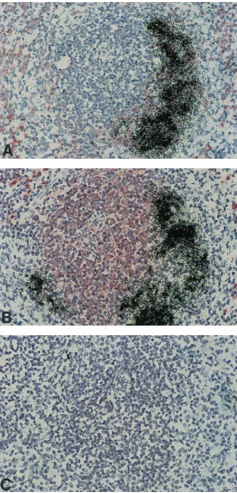
antigens and RNA in tissue sections (4, 10). Figure 3 illustrates the results of combined immunohistochemical staining and in situ hybridization of serial tissue sections of the spleen as observed during acute virus replication (6 days p.i.). The typ-ical in situ pattern of acute splenic infection following immu-nohistochemical labeling of B lymphocytes is demonstrated in Fig. 3A. The great bulk of autoradiographic signals due to in situ hybridization of viral RNA can be localized to
immuno-histochemically red-stained CD45R/B2201cells (pre-B and B
lymphocytes) which are located within the lymph follicle area. In B cells which are interspersed in the splenic marginal zones, usually no enteroviral sequences were observed. Hybridization of a consecutive tissue section of the same follicle revealed that the majority of T cells within the periarteriolar lymphatic
sheath which were stained by anti-Thy-1.21antibodies are not
infected (Fig. 3B). To exclude nonspecific labeling of lymphoid cells by radioactive hybridization probes following the immu-nohistochemical staining procedure, infected control tissue sections were incubated with normal rat serum and hybridized
with the nonrecombinant 35S-labeled plasmid vector p2732B
control DNA probe, demonstrating the specificity of concur-rent immunohistochemistry and in situ hybridization (Fig. 3C). In addition, hybridizations were performed with splenic tis-sue sections which were previously labeled by anti-L3T4 (help-er T cells) and Ly-2 (cytotoxic/suppressor T lymphocytes) anti-bodies. As demonstrated in Fig. 4A, during acute myocarditis, a portion of T-helper cells, which are localized within germinal centers of spleen follicles, was found to be labeled by the viral probe. In contrast, no viral genomes were detected in cytotoxic/ suppressor T cells (Fig. 4B).
To identify the nature of persistently infected lymphoid cells, the same cell-type-specific antibodies were used for simulta-neous visualization of viral RNA and gene products as per-formed for identification of acutely infected spleen cells. In situ hybridization of murine spleens during chronic myocarditis demonstrated that, in correspondence to acute infection,
en-teroviral RNA is present mainly in B2201B cells (Fig. 4C).
During persistent infection, all hybridization-positive B lympho-cytes were found to be located within the germinal center of splenic secondary follicles. In addition, when the macrophage-specific antibody Mac-1 was used for immunohistochemical labeling, enteroviral RNA-positive macrophages could be de-tected also at later stages of infection (Fig. 4D), indicating that, in addition to B cells, macrophages may harbor enteroviral genomes during chronic disease. However, in contrast to the case for acute infection, no viral genomes were detected in
anti-L3T41T-helper cells during chronic myocarditis.
Virus replication in immune cells. To examine whether CVB3 replicates in lymphoid tissue, spleens and lymph nodes of SWR/J mice and DBA/1 mice were investigated for the presence of viral plus-strand RNA as well as the replicative minus-strand RNA intermediate during the course of myocar-ditis by means of nested RT-PCR. As shown in Fig. 5, virus-specific amplification products reflecting viral plus- and minus-strand RNAs were detected in heart muscle and splenic tissues of SWR/J mice obtained during acute (6 days p.i.) and also during chronic (42 days p.i.) myocarditis. The observation that viral minus-strand RNA is rarely detected in lymph nodes of these animals after the acute stage of the disease is likely to be due to relatively low copy numbers of viral minus-strand RNA in lymphoid tissue, a phenomenon which was also confirmed by strand-specific in situ hybridization (data not shown).
Evidence for viral replication in immune cells during chronic myocarditis is further provided by the appearance of foci of cells in spleens (Fig. 2B) and lymph nodes (Fig. 2C) with strong signals for viral RNA-positive strands, indicating positive-strand RNA synthesis rather than mere trapping of single particles, which should result in diffuse single-cell signals.
In contrast to the findings for permissive SWR/J mice,
resist-ant DBA/1 (H-2q) control mice, which do not develop chronic
[image:5.612.62.296.69.555.2]myocarditis, were found to eliminate the virus regularly from heart muscle and lymphoid tissues during the course of acute infection.
FIG. 3. Identification of infected immune cells in spleens of acutely CVB3-infected mice (6 days p.i.). In situ hybridization for the detection of CVB3 RNA was performed following immunohistochemical labeling of frozen tissue sections with antibodies recognizing B cells (A) or T cells (B). No labeling of cells was observed when this spleen follicle was hybridized with35S-labeled, nonrecombi-nant plasmid vector p2732B control DNA after immunohistochemical staining with normal rat serum instead of the first antibody (C). Magnification,3130.
on November 9, 2019 by guest
http://jvi.asm.org/
DISCUSSION
Recent molecular studies have demonstrated that picorna-virus infections not only can induce a variety of acute diseases but also can result in persistent infections (27, 29, 31, 45). Enterovirus persistence in myocardial cells, as observed in permissive immunocompetent mouse strains with chronic CVB3 myocarditis, was found to be associated with restricted viral RNA and capsid protein synthesis (27). Since one of the key obstacles to viral persistence is the failure of the host’s immune system to eliminate the virus during acute infection (15, 39), the present study focused on the interplay established by coxsackievirus and the immune system itself.
Various organs of SWR/J mice were analyzed as cellular targets during the course of CVB3 infection. Radioactive in situ hybridization revealed a broad tissue tropism for CVB3, involving diverse lymphoid and nonlymphoid organs. During acute disease, widespread patterns of infection were detected in the heart muscle, pancreas, spleen, and lymph nodes. Where-as the virus wWhere-as found to be eliminated from most organs, including the thymus, lung, liver, and central nervous system, during the course of acute infection, lymphoid cells of the spleen and lymph nodes revealed the presence of viral ge-nomes during chronic myocarditis, in addition to persistently infected myocardial cells.
Infection of lymphoreticular tissues provides viruses with a potential way to avoid the immune system. The finding that
viruses may impair immunity by acute or persistent infection of immunocompetent cells in humans and animals has been de-scribed for a variety of viruses (1), e.g., Theiler’s murine en-cephalomyelitis virus (31), Aleutian mink disease parvovirus (2, 23), lymphocytic choriomeningitis virus (8, 46), lactate de-hydrogenase-elevating virus (3), human cytomegalovirus (38), and rabies virus (41). Previous in vitro investigations support the view that enteroviruses also can infect lymphoid cells. It was demonstrated that poliovirus is capable of replicating in human antigen-stimulated lymphocytes (52). In addition, there is evidence that one or more cell types within the human peripheral blood mononuclear cell populations support polio-virus replication (9). For CVB it has been shown that various cultured human lymphoid lines of B- and T-cell origin (33, 49), as well as a murine lymphoma cell line (6), are permissive for diverse serotypes.
In the present study we demonstrated a tropism of CVB3 for immune cells in vivo. Importantly, CVB3 was found to be capable of replicating in lymphoid cells as indicated by the detection of viral minus-strand RNA in splenic tissue during acute and chronic myocarditis by RT-PCR as well as by the recovery of infectious virus from spleens up to 18 days p.i. Since the nature of cells containing enteroviral RNA cannot be characterized with certainty on the basis of morphological cri-teria, a double-labeling method capable of identifying virus target cells in infected lymphoid tissues was used. By this
ap-FIG. 4. In situ hybridization analysis with radioactively labeled CVB3 cDNA probes of frozen spleen tissue sections obtained during acute disease after immunohistochemical staining of helper T cells (A) and cytotoxic/suppressor T cells (B). Whereas a minority of helper T cells were found to be infected, no viral RNA was observed in cytotoxic/suppressor T cells. During chronic disease, enteroviral sequences were detected primarily in B cells (C) but also in single macrophages (D). Magnifications,3160 (A, B, and C) and3490 (D).
on November 9, 2019 by guest
http://jvi.asm.org/
[image:6.612.64.555.69.399.2]proach, viral genomes were localized predominantly to B
lym-phocytes expressing the CD45R/B2201phenotype during both
acute and chronic myocarditis.
Spleens as well as lymph nodes are secondary lymphoid organs which have been demonstrated to play a major role in the antiviral immune response (40). As observed in CVB3-infected immunocompetent mice, spleens have been shown to be important for filtration of viral particles during acute infec-tion (34). As a result of the virus-immune cell interacinfec-tions in these lymphoid organs, a cascade of immune processes which involves a diversity of cell types is induced. Within the periar-teriolar lymphocyte sheaths of the white pulp, dendritic cells
present processed viral antigens to CD41and CD81cells (26).
In germinal centers of secondary follicles, viral antigens are presented from follicular dendritic cells to B cells which
un-dergo clonal selection. B cells may process antigens to CD41
cells, which in turn help B cells to develop into antibody-forming cells (12, 44). As observed by in situ hybridization, the virus had a high affinity for the splenic B-cell population in CVB3-infected mice. During acute infection, the majority of B cells that were positive for viral plus-strand RNA were local-ized in the outer zones of the splenic follicles, an area which is typical for trapping of viral particles. Consequently, when strand-specific in situ hybridization studies were performed, in addition to RT-PCR, positivity for the viral minus-strand RNA intermediate was observed in a minor subset of these lymphoid cells (data not shown). These results indicate that the great bulk of in situ-positive B lymphocytes during acute disease reflects a high viral load due to sequestration in addition to productive infection. Later in disease, infected B cells were found in the middle of germinal centers, which is the charac-teristic site for stimulated B lymphoblasts. Minus-strand RNA synthesis was consistently detected in splenic follicles by RT-PCR but rarely detected in mesenteric lymph nodes obtained during chronic infection. Notably, immunohistochemical
stain-ings of lymphoid tissues, using polyclonal antisera that have been raised against bacterially synthesized viral structural fu-sion proteins (51), provided evidence that single cells within viral RNA-positive germinal centers of secondary follicles from the spleen and mesenteric lymph nodes harbor viral gene products (data not shown). Persistently infected B cells might contribute to the long-term presence of memory B cells (13) and explain the presence of relatively high titers of anti-CVB3 immunoglobulin G antibodies in susceptible mice for many weeks (32). In addition, it is suggested that infected splenic B cells can support virus dissemination in the organism when they turn over to peripheral recirculating B cells (13).
In addition to the in vivo finding of infected B cells in enterovirus myocarditis, the present study implicates T lympho-cytes and macrophages as being infected. Whereas T-helper cells were shown to represent a subset of acutely infected cells,
CD81T cells were not found to contain viral genomes during
the course of the disease. Regarding the detection of viral RNA in macrophages, it should be considered that in situ detection of viral genomes may reflect phagocytosis of nonrep-licative viral material. However, there is evidence from in vitro experiments that a portion of cultured human monocytes can release newly synthesized CVB3 without exhibiting cytopathic effects (16). Viral replication in a small fraction of brain mac-rophages was also reported for infection with Theiler’s virus (30), a murine picornavirus responsible for a persistent demy-elinating infection of the central nervous system (5).
The results obtained in the present study support the view that infected immune cells represent a noncardiac reservoir for viral genomes, which could play an important role in dissem-ination of the virus and in maintenance of chronic disease. The model of persistently enterovirus-infected immune cells pre-sented here should prove to be useful to further elucidate viral and host-specific determinants which account for the different outcomes of enteroviral myocarditis in susceptible and resis-tant hosts.
ACKNOWLEDGMENTS
This work was supported by the Thyssen Foundation; the Deutsche Forschungsgemeinschaft (DFG), SFB 120, project A11; the Heart and Stroke Foundation of British Columbia and Yukon Territory; and a Max Planck Research Award (to R.K. and B.M.M.).
We thank Sandra Bundschuh and Gerd Janke for excellent technical assistance.
REFERENCES
1. Ahmed, R., and J. G. Stevens. 1990. Viral persistence, p. 241–265. In B. N. Fields and D. M. Knipe (ed.), Virology. Raven, New York.
2. Alexandersen, S., M. E. Bloom, and J. Wolfinbarger. 1988. Evidence of restricted viral replication in adult mink infected with Aleutian disease of mink parvovirus. J. Virol. 62:1495–1507.
3. Anderson, G. W., R. R. R. Rowland, G. A. Palmer, C. Even, and P. G. W. Plagemann.1995. Lactate dehydrogenase-elevating virus replication persists in liver, spleen, lymph node, and testis tissues and results in accumulation of viral RNA in germinal centers, concomitant with polyclonal activation of B cells. J. Virol. 69:5177–5185.
4. Brahic, M., A. T. Haase, and A. Cash. 1984. Simultaneous in situ detection of viral RNA and antigens. Proc. Natl. Acad. Sci. USA 81:5545–5448. 5. Brahic, M., W. G. Stroop, and J. R. Baringer. 1981. Theiler’s virus persists in
glial cells during demyelinating disease. Cell 26:123–128.
6. Cao, Y., and D. P. Schnurr. 1988. Persistent infection of YAC-1 cells by coxsackievirus B3. J. Gen. Virol. 69:59–65.
7. Easton, A. J., and R. P. Eglin. 1988. The detection of coxsackievirus RNA in cardiac tissue by in situ hybridization. J. Gen. Virol. 69:285–291. 8. Fazakerley, J. K., P. Southern, F. Bloom, and M. J. Buchmeier. 1991. High
resolution in situ hybridization to determine the cellular distribution of lymphocyte choriomeningitis virus RNA in the tissues of persistently in-fected mice: relevance to arenavirus disease and mechanisms of viral persis-tence. J. Gen. Virol. 72:1611–1625.
9. Freistadt, M. S., H. B. Fleit, and E. Wimmer. 1993. Poliovirus receptor on human blood cells: a possible extraneural site of poliovirus replication. Vi-rology 195:798–803.
FIG. 5. Gel electrophoresis of 300-bp amplification products of CVB3 plus-strand (1) and minus-strand (2) RNAs from the heart muscle, spleen, and lymph nodes of SWR/J mice by nested RT-PCR obtained during acute (6 days p.i.) and chronic (42 days p.i.) myocarditis. As positive controls for amplifiable RNA, RT-PCR with glyceraldehyde-3-phosphate dehydrogenase mRNA (248-bp fragment) was performed. H2O was used as a negative control.
on November 9, 2019 by guest
http://jvi.asm.org/
10. Gendelman, H. E., T. R. Moenck, O. Narayan, D. E. Griffin, and J. E. Clements.1985. A double labeling technique for performing immunocyto-chemistry and in situ hybridization in virus infected cell cultures and tissues. J. Virol. Methods 11:93–103.
11. Godeny, E. K., and C. J. Gauntt. 1987. In situ immune autoradiographic identification of cells in heart tissues of mice with coxsackievirus B3-induced myocarditis. Am. J. Pathol. 129:267–276.
12. Gray, D. 1993. Immunological memory. Annu. Rev. Immunol. 11:49–77. 13. Gray, D., and H. Skarvall. 1988. B-cell memory is short-lived in the absence
of antigen. Nature (London) 336:70–73.
14. Harper, M. E., L. M. Marselle, R. C. Gallo, and F. Wong-Staal. 1986. Detection of lymphocytes expressing human T-lymphotropic virus type III in lymph nodes and peripheral blood from infected individuals by in situ hy-bridization. Proc. Natl. Acad. Sci. USA 83:772–776.
15. Haywood, A. M. 1986. Patterns of persistent viral infections. N. Engl. J. Med. 315:939–948.
16. Henke, A., C. Mohr, H. Sprenger, C. Graebner, A. Stelzner, M. Nain, and D. Gemsa.1992. Coxsackievirus B3-induced production of tumor necrosis fac-tor-a, IL-1b, and IL-6 in human monocytes. J. Immunol. 148:2270–2277. 17. Herskowitz, A., L. J. Wolfgram, N. R. Rose, and K. W. Beisel. 1987.
Cox-sackievirus B3 murine myocarditis—marked strain differences in histopatho-logic features of early and late phase myocarditis. J. Am. Coll. Cardiol. 9:1311–1319.
18. Hohenadl, C., K. Klingel, J. Mertsching, P. H. Hofschneider, and R. Kan-dolf.1991. Strand-specific detection of enteroviral RNA in myocardial tissue by in situ hybridization. Mol. Cell. Probes 5:11–20.
19. Kandolf, R., D. Ameis, P. Kirschner, A. Canu, and P. H. Hofschneider. 1987. In situ detection of enteroviral genomes in myocardial cells by nucleic acid hybridization: an approach to the diagnosis of viral heart disease. Proc. Natl. Acad. Sci. USA 84:6272–6276.
20. Kandolf, R., A. Canu, and P. H. Hofschneider. 1985. Coxsackie B3 virus can replicate in cultured human foetal heart cells and is inhibited by interferon. J. Mol. Cell. Cardiol. 17:167–181.
21. Kandolf, R., and P. H. Hofschneider. 1985. Molecular cloning of the genome of a cardiotropic coxsackie B3 virus: full-length reverse-transcribed recom-binant cDNA generates infectious virus in mammalian cells. Proc. Natl. Acad. Sci. USA 82:4818–4822.
22. Kandolf, R., and P. H. Hofschneider. 1989. Viral heart disease. Springer Semin. Immunopathol. 11:1–13.
23. Kanno, H., J. B. Wolfinbarger, and M. E. Bloom. 1992. Identification of Aleutian mink disease parvovirus transcripts in macrophages of infected adult mink. J. Virol. 66:5305–5312.
24. Khatib, R., A. Probert, M. P. Reyes, G. Khatib, and J. L. Chason. 1987. A review. Mouse strain-related variation as a factor in the pathogenesis of coxsackievirus B3 murine myocarditis. J. Gen. Virol. 68:2981–2988. 25. Kim, W. K., Y. Tang, J. J. Kenny, D. L. Longo, and H. C. Morse. 1994. In
murine AIDS, B cells are early targets of defective virus and are required for efficient infection and expression of defective virus in T cells and macro-phages. J. Virol. 68:6767–6769.
26. King, P. D., and D. R. Katz. 1990. Mechanisms of dendritic cell function. Immunol. Today 6:206–211.
27. Klingel, K., C. Hohenadl, A. Canu, M. Albrecht, M. Seemann, G. Mall, and R. Kandolf.1992. Ongoing enterovirus-induced myocarditis is associated with persistent heart muscle infection: quantitative analysis of virus replica-tion, tissue damage, and inflammation. Proc. Natl. Acad. Sci. USA 89:314– 318.
28. Klump, W. M., I. Bergmann, B. C. Mu¨ller, D. Ameis, and R. Kandolf.1990. Complete nucleotide sequence of infectious coxsackievirus B3 cDNA: two initial 59uridine residues are regained during plus-strand RNA synthesis. J. Virol. 64:1573–1583.
29. Koide, H., Y. Kitaura, H. Deguchi, A. Ukimura, K. Kawamura, and K. Hirai. 1992. Viral genomic detection in the hearts of C3H/He mice with experi-mental coxsackievirus B3 myocarditis by gene amplification using the poly-merase chain reaction. Jpn. Circ. J. 56:148–156.
30. Levy, M., C. Aubert, and M. Brahic. 1992. Theiler’s virus replication in brain macrophages cultured in vitro. J. Virol. 66:3188–3193.
31. Lipton, H. L., G. Twaddle, and M. L. Jelachich. 1995. The predominant virus antigen burden is present in macrophages in Theiler’s murine encephalomy-elitis virus-induced demyelinating disease. J. Virol. 69:2525–2533. 32. Lodge, P. A., M. Herzum, J. Olszewski, and S. A. Huber. 1987.
Coxsackievi-rus B3 myocarditis. Acute and chronic forms of the disease caused by dif-ferent immunopathogenic mechanisms. Am. J. Pathol. 3:455–463. 33. Matteucci, D., M. Paglianti, A. M. Giangregorio, M. R. Capobianchi, F.
Dianzani, and M. Bendinelli.1985. Group B coxsackieviruses readily estab-lish persistent infection in human lymphoid cell lines. J. Virol. 56:651–654. 34. Matteucci, D., A. Toniolo, P. G. Conaldi, F. Basolo, Z. Gori, and M. Bendi-nelli.1985. Systemic lymphoid atrophy in coxsackievirus B3-infected mice: effects of virus and immunopotentiating agents. J. Infect. Dis. 151:1100– 1108.
35. McChesney, M. B., and M. B. A. Oldstone. 1987. Viruses perturb lymphocyte functions: selected principles characterizing virus induced immunosuppres-sion. Annu. Rev. Immunol. 5:279–304.
36. McManus, B. M., and R. Kandolf. 1991. Evolving concepts of cause, conse-quence, and control in myocarditis. Curr. Opin. Cardiol. 6:418–427. 37. Melnick, J. L. 1990. Enteroviruses: polioviruses, coxsackieviruses,
echovi-ruses, and newer enteroviechovi-ruses, p. 549–605. In B. N. Fields and D. M. Knipe (ed.), Virology. Raven, New York.
38. Minton, E. J., C. Tysoe, J. H. Sinclair, and J. G. P. Sisson. 1994. Human cytomegalovirus infection in monocyte/macrophage lineage in bone marrow. J. Virol. 68:4017–4021.
39. Oldstone, M. B. A. 1989. Viral persistence. Cell 56:517–520.
40. Raviola, E. 1986. The immune system, p. 406–478. In W. Bloom and D. W. Fawcett (ed.), A textbook of histology. The W. B. Saunders Co., Philadel-phia.
41. Ray, N. B., L. C. Ewalt, and D. L. Lodmell. 1995. Rabies virus replication in primary bone marrow macrophages and in human and murine macrophage-like cell lines: implications for viral persistence. J. Virol. 69:764–772. 42. Rotbart, H. A., J. P. Kinsella, and R. L. Wassermann. 1990. Persistent
enterovirus infection in culture-negative meningoencephalitis: demonstra-tion by enzymatic RNA amplificademonstra-tion. J. Infect. Dis. 161:787–791. 43. Smith, L. M., J. Sanders, R. J. Kaiser, P. Hughes, C. Dodd, C. R. Connel,
S. B. H. Kent, and L. E. Hood.1986. Fluorescence detection in automated DNA sequence analysis. Nature (London) 321:674–679.
44. Szakal, A. K., M. H. Kosco, and J. G. Tew. 1989. Microanatomy of lymphoid tissue during humoral immune responses: structure function relationships. Annu. Rev. Immunol. 7:91–109.
45. Tam, P. E., A. M. Schmidt, S. R. Ytterberg, and R. P. Messner. 1991. Viral persistence during the developmental phase of coxsackievirus B1-induced murine polymyositis. J. Virol. 65:6654–6660.
46. Tishon, A., P. Southern, and M. B. A. Oldstone. 1988. Virus-lymphocyte interactions. II. Expression of viral sequences during the course of persistent lymphocytic choriomeningitis virus infection and their localization to the L3T4 lymphocyte subset. J. Immunol. 140:1280–1284.
47. Tokunaga, K., Y. Nakamura, K. Sakata, K. Fujimori, M. Ohkubo, K. Sawada, and S. Sakiyama.1987. Enhanced expression of a glyceraldehyde-3-phosphate dehydrogenase gene in human lung cancers. Cancer Res. 47: 5616–5619.
48. Tracy, S., N. M. Chapman, B. M. McManus, M. A. Pallansch, M. A. Beck, and J. Carstens.1990. A molecular and serologic evaluation of enteroviral involvement in human myocarditis. J. Mol. Cell. Cardiol. 22:403–414. 49. Vuorinen, T., R. Vainionpa¨a¨, H. Kettinen, and T. Hyypia¨.1994.
Coxsack-ievirus B3 infection in human leukocytes and lymphoid cell lines. Blood 3:823–829.
50. Weibel, E. R. 1979. Stereological methods: practical methods for biological morphometry. Academic Press, San Diego, Calif.
51. Werner, S., W. M. Klump, H. Scho¨nke, P. H. Hofschneider, and R. Kandolf. 1988. Expression of coxsackievirus B3 proteins in Escherichia coli and gen-eration of virus-specific antisera. DNA 7:307–316.
52. Willems, F. T. C., J. L. Melnick, and W. E. Rawls. 1969. Replication of poliovirus phytohemagglutinin-stimulated human lymphocytes. J. Virol. 3: 451–457.