0022-538X/96/$04.0010
Copyrightq1996, American Society for Microbiology
Novel DNA Binding Specificities of a Putative
Herpesvirus bZIP Oncoprotein
ZHENG QIAN,1PETER BRUNOVSKIS,1LUCY LEE,2PETER K. VOGT,3
ANDHSING-JIEN KUNG1*
Department of Molecular Biology and Microbiology, School of Medicine, Case Western Reserve University,
Cleveland, Ohio 441061; USDA Avian Disease and Oncology Laboratory, East Lansing, Michigan 488232;
and Division of Oncovirology, Department of Molecular and Experimental Medicine,
Scripps Research Institute, La Jolla, California 920373
Received 6 May 1996/Accepted 18 July 1996
Marek’s disease virus is a highly oncogenic herpesvirus that can cause T lymphomas and peripheral nerve demyelination in chickens.meq, a candidate oncogene of Marek’s disease virus, encodes a basic leucine zipper (bZIP) transcription factor which contains a large proline-rich domain in its C terminus. On the basis of its bZIP structural homology,meqis perhaps the only member of thejun-fosgene family completely viral in origin. We previously showed that Meq’s C-terminal domain has potent transactivation activity and that its bZIP domain can dimerize with itself and with c-Jun also. In an effort to identify viral and cellular targets of Meq, we have determined the optimal binding sites for Meq-Jun heterodimers and Meq-Meq homodimers. By a PCR-based approach using cyclic amplification of selected targets, Meq-Jun heterodimers were found to optimally bind tetradecanoylphorbol acetate response element (TRE) and cyclic AMP response element (CRE) consensus sequences. This result was consistent with the results of our previous functional analysis implicating Meq-Jun heterodimers in the transactivation of the Meq promoter through a TRE- or CRE-like sequence. Interestingly, Meq-Meq homodimers were found to bind two distinct motif elements. The first [GAGTGATG AC(G)TCATC] has a consensus which includes a TRE or CRE core flanked by additional nucleotides critical for tight binding. Methylation interference and mutational analyses confirmed the importance of the flanking residues. The sequences of a subset of TRE and CRE sites selected by Meq-Meq are closely related to the binding motif of Maf, another bZIP oncoprotein. The second putative Meq binding site (RACACACAY) bears a completely different consensus not shared by other bZIP proteins. Binding to this consensus sequence also requires secondary structure characteristics associated with DNA bending. CACA motifs are known to promote DNA curvature and function in a number of special biological processes. Our results lend further weight to the increasing importance of DNA bending in transcriptional regulation and provide a baseline for the identifi-cation of Meq-responsive targets.
Meq is a basic leucine zipper (bZIP) protein encoded by Marek’s disease virus (MDV), a herpesvirus that causes rapid onset of T-cell lymphomas in chickens (5). Meq has been detected in all tumor samples and established MDV-trans-formed T-cell lines examined thus far (16). Consistent with being unique to oncogenic (serotype I), but not nononcogenic (serotypes II and III) vaccine strains, recent evidence has as-cribed a role for Meq in maintaining the transformed pheno-type of MDV tumor cell lines (57). How Meq contributes to the oncogenic process is presently unclear. However, insofar as Meq shares a structural organization with the other bZIP on-coproteins, Jun, Fos, and Maf (see below), Meq could similarly deregulate the expression of growth-related genes in the trans-formation process.
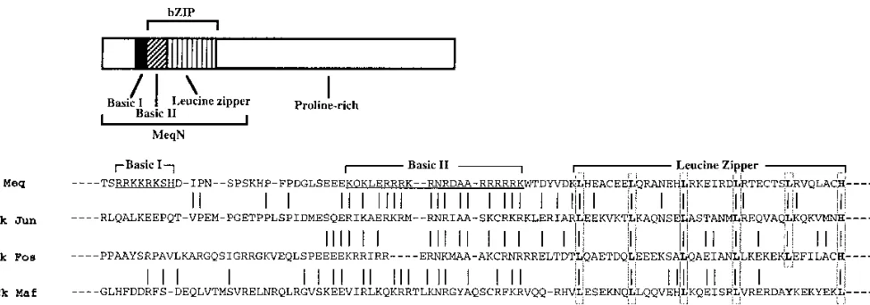
Meq protein consists of an N-terminal bZIP domain and a C-terminal proline-rich domain. Adjacent to the leucine zip-per, the Meq basic region has 65% homology with the basic regions of Jun (30) and Fos (11). Like that of the Jun-Fos subfamily of bZIP proteins, the Meq leucine zipper (LZ) do-main exhibits a heptad spacing of five leucines and a C-termi-nal histidine. Such a close kinship places Meq within the im-mediate family of Jun- and Fos-like bZIP proteins (21). Other
structural features of Meq, however, are quite different from those of Jun and Fos. Meq has an additional basic region N terminal to its bZIP domain, possibly affecting its DNA bind-ing or nuclear translocation properties. The regulatory C ter-minus of Meq is completely different from that of Jun or Fos. The unusually long proline-rich domain resembles that of the repression domain of Wilms’ tumor suppressor protein (4, 13) and contains a number of consensus binding sites for signal transduction proteins containing SH3 domains (25, 42). We recently reported that the Meq C-terminal region contains a transactivation domain that can activate the expression of Gal4 reporter constructs when fused to the Gal4 DNA binding do-main (38). We also showed that Meq and Jun can heterodimer-ize and bind DNA with affinity levels higher than that of either homodimer and can together bind and transactivate an AP-1-like motif (GTCATGCATGACGT) present in the Meq pro-moter region. Aside from these observations, very little is known about the genes that are regulated by homodimers of Meq or heterodimers of Meq and other bZIP proteins.
As a first step in identifying Meq target genes, we have characterized the optimal DNA binding motifs for Meq-Jun heterodimers and Meq-Meq homodimers. The cyclic amplifi-cation of selected targets (CASTing) approach (56) allows a sensitive and nonbiased selection of sequences bound to pro-tein complexes from a pool of random sequence oligonucleo-tides. Using this PCR-based methodology, we report a number of interesting DNA binding properties that are associated with
* Corresponding author. Mailing address: Department of Molecular Biology and Microbiology, Case Western Reserve University School of Medicine, 10900 Euclid Ave., Cleveland, OH 44106. Phone: (216) 368-6655. Fax: (216) 368-3055.
7161
on November 9, 2019 by guest
http://jvi.asm.org/
Meq. Whereas Meq-Jun was found to bind tetradecanoylphor-bol acetate response element (TRE; TGAGTCA) and cyclic AMP response element (CRE; TGACGTCA) core-containing sequences [RTGAC(G)TCAY, where R is a purine and Y is a pyrimidine], Meq-Meq homodimers were found to bind two distinct motifs. One motif has an extended TRE or CRE se-quence [GAGTGATGAC(G)TCATC]; and the other has a RACACACAY consensus sequence. These binding properties were validated by gel retardation assays using wild-type (wt) and mutant oligonucleotide competitors, by methylation inter-ference analysis, and by transient transfections utilizing con-structs containing the sequence motifs selected by CASTing. Interestingly, the novel RACACACAY core was found to be necessary but not sufficient for DNA binding; a yet-to-be-determined local structure contributes in binding to these sites. The information presented in this paper should enable us to search for the functional targets of Meq in MDV and cellular genomes and provides more insights into the role Meq may play in MDV oncogenesis.
MATERIALS AND METHODS
Determination of the optimal binding sites of the Meq homodimer and Meq-Jun heterodimer.MeqN, a Meq C-terminal truncation mutant (Fig. 1) (previ-ously referred to as MeqbZIP [38]) was purified by nickel chelate affinity chro-matography as previously described (38). Bacterially expressed c-Jun was from Promega (Madison, Wis.). A double-stranded probe with 25 random nucleotides in its center, 59-GCTTAAGCTTGCTCTAGAAN25GAATTCCATGGTACCC TA-39, was32P-labeled with polynucleotide kinase, added to purified protein(s) in a buffer containing 25 mM HEPES (N-2-hydroxyethylpiperazine-N9 -2-ethane-sulfonic acid; pH 7.9), 100 mM KCl, 0.5 mM MgCl2, 1 mg of bovine serum albumin per ml, 10% glycerol, 5 mM dithiothreitol, and 0.1 mg of poly(dI-dC) per ml and incubated at 378C for 30 min prior to electrophoresis on a 4% nondenaturing polyacrylamide–Tris–glycine gel run at 48C for 2 h at 250 V. The gel was then wrapped in Saran Wrap and exposed to X-ray film. The shifted bands corresponding to Meq-Meq and Meq-Jun were excised with razor blades, and their bound DNAs were eluted in 50ml of double-distilled H2O in 378C overnight. One to two microliters of the eluate was then amplified in a PCR with primer 1, 59-GCTTAAGCTTGCTCTAGA-39, and primer 2, 59-TAGGGTACC ATGGAATTC-39. The amplified products were then32P labeled with polynu-cleotide kinase and used in the next round of selection. After four to five rounds of selection, the amplified products were digested withEcoRI andXbaI and cloned into pBluescript (Stratagene, La Jolla, Calif.) and pUC18dh (43). The latter plasmid was a generous gift from Ed Stavnezer (Case Western Reserve University). Sequencing reactions were performed with a Sequenase kit (United States Biochemical Corp., Cleveland, Ohio).
Gel retardation assays.Gel retardation assays were performed essentially as described above with the following modifications. Probes from the individual clones were labeled with Klenow after the plasmids were digested withEcoRI
andXbaI. The mixtures were passed through spin columns and loaded onto 10 to 15% nondenaturing polyacrylamide gels. The labeled inserts were excised and eluted in double-distilled H2O following overnight incubation at 378C. The eluates were extracted with phenol-chloroform and ethanol precipitated prior to their ad-dition (about 10,000 cpm) to the gel retardation assays. Synthesized oligonucle-otide probes were constructed by labeling one strand with [g-32
P]ATP and polynucleotide kinase, annealing the strand to the opposite strand, and then elongating with Klenow. When competition or antibody supershift assays were performed, appropriate amounts of unlabeled oligonucleotides or antibodies were added to the reaction mixtures prior to the addition of the protein(s). Band inten-sities were quantitated with a radioanalytic system (Ambis Co., San Diego, Calif.). Methylation interference assay.Methylation interference assays were per-formed to identify the DNA contacts in the protein-DNA complexes. DNA probes were labeled on one strand with Klenow, digested with a second enzyme, and then methylated with 1ml of dimethyl sulfate (DMS) in 50 mM sodium cacodylate–1 mM EDTA (pH 8.0) at room temperature for 2 to 5 min. The methylated probes were isolated from a nondenaturing 10 to 15% polyacryl-amide gel, eluted, extracted with phenol-chloroform, ethanol precipitated, and dissolved in water prior to being used in scaled-up gel retardation assays. Both bound and unbound probes were excised from the nondenaturing polyacryl-amide gel and eluted with water overnight at 378C. The probes were then extracted with phenol-chloroform, ethanol precipitated, and dissolved in 10% piperidine. The cleavage reactions were carried out at 958C for 30 min prior to lyophilization of the reaction products in a speed vac. The dried pellets were dissolved in 100ml of water and lyophilized twice more. The final products were dissolved in loading buffer (80% formamide, 1 mM EDTA, 0.1% xylene cyanol, 0.1% bromophenol blue), heated at 958C for 5 min, and loaded onto 10 to 15% sequencing gels.
Cells, plasmids, and transfections.Double-stranded oligonucleotides corre-sponding to Meq response element (MERE) I binding motifs [MERE I (CRE), TCAGAGTGATGACGTCATCAC, or MERE I (TRE), TCAGAGTGATGA CTCATCAC] and a MERE II binding motif (AATTCAAAAACACATAACA TTCGTATATATTCTGA) were synthesized, phosphorylated at their 59ends with polynucleotide kinase, and cloned into the bluntedSalI site of pBLCAT2 (26). Plasmids with multiple-insert copies in both orientations were identified by DNA sequencing. The Meq expression plasmid used in these experiments was Meq/pECE, in which the Meq open reading frame was cloned into the
HindIII-XbaI site of the vector pECE (38).
Chicken embryo fibroblasts (CEF) were maintained in 1:1 Dulbecco modified eagle medium (low glucose)-M199 medium with 2% chicken serum and 3% fetal bovine serum. Transfections were carried out by the calcium phosphate method (6) with 2mg of reporter plasmids, 10mg of Meq/pECE, and 2 ng of a cytomeg-alovirus enhancer-driven luciferase plasmid (pCMV-luc) as an internal control. The cells were lysed 48 to 60 h later with reporter lysis buffer (Promega Corp.). Chloramphenicol acetyltransferase (CAT) assays were performed by the phase-extraction method as described previously (45). Luciferase assays were per-formed with a luciferase assay kit (Promega) and a Berthold luminometer.
RESULTS
[image:2.612.65.552.69.240.2]DNA binding site selection. (i) Optimal Meq-Jun binding sites. We previously showed that Meq can readily dimerize
FIG. 1. Meq structure and bZIP sequence comparison of Meq, Jun, Fos, and Maf. Lines indicate identical or highly conserved residues between Meq and other bZIP proteins. The basic regions I and II of Meq are underlined; conserved leucine and histidine residues in the leucine zipper regions are boxed. Bacterially expressed MeqN, which was used in the selection of Meq-Jun and Meq-Meq binding sites and in gel retardation assay and methylation interference assay experiments, is ameq
mutant with the majority of its proline-rich domain removed. Ck, chicken.
on November 9, 2019 by guest
http://jvi.asm.org/
with c-Jun and that Meq-Jun heterodimers exhibit stronger binding to an AP-1-like site than either Meq-Meq or Jun-Jun homodimers. This association appears functionally relevant, since Meq-Jun can upregulate a natural MDV promoter con-taining this site to a level significantly higher than that achieved by a Meq or Jun homodimer (38). This site was uncovered in a search for potential regulatory elements present in the Meq promoter; it represents only one of the potential Meq-Jun binding sites present in the MDV genome. To systematically identify optimal binding motifs for Meq-Jun, we employed a modified CASTing approach (56) using purified Meq and c-Jun proteins. MeqN represents the N-terminal 168 amino acids of Meq, including the bZIP domain, fused to a T7 epitope tag at its N terminus and a 6His tag at its C terminus. Because of MeqN’s shorter length relative to full-length c-Jun, MeqN-Jun heterodimers can be readily resolved from their homodimer counterparts on nondenaturing polyacrylamide gels (38).
MeqN was overexpressed in Escherichia coliand purified to
about 85% homogeneity by nickel chelate affinity chromatog-raphy. A highly complex pool of double-stranded oligonucle-otides containing a 25-base degenerate core sequence with flanking restriction sites was first mixed with a preincubated MeqN- and c-Jun-containing fraction; the resulting DNA-pro-tein complexes were then electrophoretically separated in a gel retardation assay. The MeqN–c-Jun complexes will be referred to as Meq-Jun for simplicity. In this assay, the ternary com-plexes formed between the DNA probe and Jun, Meq-Meq, or Jun-Jun have distinct mobilities (see below). The specific Meq-Jun band was excised; its DNA was then eluted and subjected to PCR amplification. After four rounds of oli-gonucleotide selection and PCR amplification, a highly re-stricted pool of oligonucleotides with strong affinities to Meq-Jun appeared. At this stage, the oligonucleotides bound to Meq-Jun were purified, cut with restriction enzymes, and
cloned into theEcoRI andXbaI sites of both pBluescript and
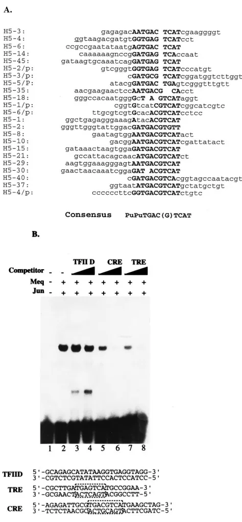
pUC18dh. The latter is a modified pUC18 vector which facil-itates a white-to-blue selection of insert-containing clones (43). Our two-vector cloning strategy was designed to eliminate any cloning bias favoring certain inserts in a given vector. Of the 23 different clones subjected to sequence analysis (Fig. 2A), all were found to contain a common core motif present in TREs and CREs. The selected motifs were critical in DNA binding as
demonstrated by competition analyses using TRE (59-CGCTT
GATGAGTCATGCCGGAA-39) or CRE (59-AGAGATTGC
GTGACGTCATGAAGCTAG-39) consensus
oligonucleo-tides. These oligonucleotides could effectively compete with the binding to the selected oligonucleotides (Fig. 2B, lanes 5 to
8), whereas a control TFIID consensus (59-GCAGAGCATA
TAAGGTGAGGTAGG-39) could not (Fig. 2B, lanes 3 and 4).
Immediately flanking the TRE or CRE core, purine residues (especially adenine) were favored on one end and pyrimidine residues (especially thymine) were favored on the other. No other flanking sequence preferences were observed. Similar DNA binding specificities were previously noted for Jun-Fos and Jun-Jun. These results indicate that TRE and CRE motifs represent determinants most favorable for Meq-Jun DNA binding. Our previously described Meq-Jun binding to a TRE-or CRE-like site (TGACGT) in the promoter region of Meq is consistent with these new findings and lends further validity to our CASTing approach.
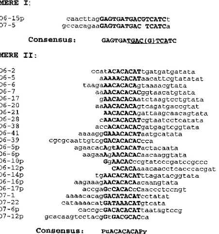
(ii) Optimal Meq-Meq binding sites.We next applied the CASTing approach to identify optimal binding sites for Meq-Meq homodimers. After five rounds of selection, strong DNA binding was observed. The selected oligonucleotide pool com-plexing with Meq-Meq was cloned and sequenced as before. Two distinct sequence motifs were identified. We shall refer to
[image:3.612.316.553.64.576.2]these two motifs as MERE I and II. MERE I represents a minor population with a palindromic TRE- or CRE-like motif as the core. MERE II carries a consensus ACACACA se-quence. Among 100 or so distinct clones analyzed, 2 display a MERE I motif (Fig. 3, top). The remaining clones all contain
FIG. 2. Characterization of Meq-Jun binding sites. (A) Sequences of selected Meq-Jun binding sites. The designation of each clone is shown at the left. Consensus nucleotides are capitalized and in boldface type. (B) Competition assay for Meq-Jun binding. The pooled Meq-Jun-selected oligonucleotides were labeled with [g-32P]ATP and used as probes; unlabeled competitor oligonucle-otides containing TFIID, CRE, or TRE consensus binding sites were added at a 50- or 100-fold excess. The competitor oligonucleotide sequences are shown below the gel; boxed nucleotides highlight the consensus TRE or CRE sites. Bacterially expressed and purified MeqN and c-Jun were used in binding assays under conditions as described in Materials and Methods.
on November 9, 2019 by guest
http://jvi.asm.org/
MERE II motifs, similar to the representative 22 sequences shown at the bottom of Fig. 3.
MERE I, the TRE- or CRE-like motif.Given that Meq has a basic region similar to Jun and Fos, it is perhaps not surpris-ing that the TRE and CRE motifs recognized by Jun-Jun and Jun-Fos (39, 44) are also recognized by Meq-Meq. On the other hand, our previous binding studies revealed weak affinity between Meq homodimers and TRE or CRE consensus probes. Despite exhaustive screening, only 2 of the 100 clones analyzed were found to contain a TRE or CRE consensus that could efficiently mediate Meq-Meq DNA binding. However, these two clones were found to share eight additional bases flanking the TRE- or CRE core [GAGTGATGAC(G)TCAT C], extending the palindrome by two more bases (GA and TC). The sequences of the remaining clones, other than those of the primers, were different, indicating the independent origins of these two clones. It would appear unlikely that two indepen-dent, randomly selected clones would have the same flanking sequences unless they were important in establishing high-affinity binding to Meq-Meq. Further support for this view will be presented later.
MERE II, the ACACACA motif.MERE II represents the predominant fraction of the selected motifs. The consensus sequence is RACACACAY. There seems to be an AT-rich sequence following this motif, but its distance from the con-sensus varies. This binding cannot be effectively inhibited by the core ACACACA sequences, nor can the core motif alone bind a Meq-Meq homodimer at any detectable level (data not shown). Thus, while the ACACACA residues are the only recognized common sequences of these nucleotides, other structural features must also be required for the high-affinity binding of this group. In light of MERE II’s shorter length compared with that of MERE I (9 residues versus 15 to 16 residues), it is perhaps not surprising that only 2 of the 100
clones analyzed were found to contain the MERE I motif. On the basis of the sizes of the two consensus sequences and the
random probability of selecting either one (429versus 4215or
4216), the original oligonucleotide pool would have had about
4,000-fold more potential MERE II-containing oligonucleo-tides than MERE I-containing oligonucleooligonucleo-tides. To further probe the nature of DNA binding between a Meq-Meq ho-modimer and the above-described oligonucleotides, we per-formed methylation interference and nucleotide substitution analysis of MERE I and II.
Methylation interference analysis of MERE I.Methylation interference assays were performed to identify the G residues involved in Meq-Meq binding to MERE I. In Fig. 4A, lanes 1 (free probe) and 2 (bound probe) show the protection patterns for O7-5, a probe containing a TRE core sequence. G at
position 11 (G11), G13, and G16 were the primary residues
protected by the Meq-Meq homodimer. G11and G13are at
the center of the TRE core, whereas G16 is in the flanking
region. Thus, Meq-Meq homodimers contact not only the TRE or CRE central core but also the flanking regions. For com-parison, we studied Jun-Jun and Meq-Jun binding to O7-5 (Fig. 4A, lanes 3 and 4). Jun-Jun homodimers (lanes 3 and 4)
were primarily found to protect the G11and G13residues in
the central TRE core and, to a much lesser extent, G16in the
flanking region. Similar results were obtained with the
Meq-Jun heterodimer (lanes 5 and 6), although G13was protected
to a lesser extent and G16was barely protected. Methylation
interference analysis yielded analogous results on the other side of the TRE core with the opposite strand of O7-5 (Fig.
4B). Here, the G25residue flanking the TRE site and the G22
residue within the TRE site were well protected by the
Meq-Meq homodimer; G25was only partially protected by Jun-Jun
(lane 4) and Meq-Jun (lane 6). These experiments suggest that Meq-Meq homodimers have the ability to contact additional sequences flanking the TRE or CRE core, protecting a region larger than that of either Jun-Jun or Meq-Jun.
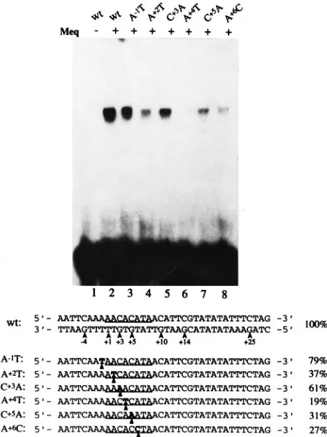
Nucleotide substitution analysis of MERE I.To further pin-point the nucleotides required for Meq-Meq binding, a series of oligonucleotide DNA probes were synthesized with different nucleotide substitutions in the MERE I site. The synthesized oligonucleotides were radiolabeled with polynucleotide kinase prior to their use in gel retardation assays. The wt and mutant MERE I probe sequences used in this analysis are shown in Fig. 5, along with their respective binding efficiencies (percent relative to the wt). Binding efficiency is defined as the percent-age of total counts bound to the probes, as measured by an Ambis radiation-quantitative system. The wt probe strongly
associated with Meq (lane 2). When the G29or G27
nucleo-tides were changed to thymidine (lane G29T or G27T), their
binding capacities were reduced to 64 and 51% of the wt level,
respectively. In the G29T-G27T double mutant, the binding
dropped to 32% of the wt level. Altering G25, a flanking G
residue protected by Meq-Meq, had an even more drastic
effect on binding; in this case G25T and G25A were found to
retain only 16 and 22% of the wt levels, respectively. As
ex-pected, changing G11, located in the center of the CRE core,
had the most severe effect on DNA binding. G11A was found
to bind to Meq-Meq at a barely detectable 5% capacity. We
also studied the importance of C16 (complementary to the
protected G16on the opposite strand) located in the 39
flank-ing sequence. C16and G25are symmetrically located relative
to the central CRE core. It is therefore not surprising that the
C16G mutant probe bound Meq-Meq at a level (14%) nearly
identical to that of the G25T mutant (16%). Significantly,
altering both residues (as in the G25T-C16G double mutant)
[image:4.612.68.294.73.314.2]led to a nearly background level (6% of the wt) of DNA
FIG. 3. Sequences of selected Meq-Meq binding sites. The designation of each clone is shown at the left. Consensus nucleotides for MERE I and MERE II are capitalized and in boldface type. Bacterially expressed and purified MeqN was used in the selection. The conditions used are described in Materials and Methods.
on November 9, 2019 by guest
http://jvi.asm.org/
binding, despite the presence of a consensus CRE site. These observations provide strong corroborating evidence that the flanking sequences are important in Meq-Meq binding to MERE I.
Methylation interference analysis of MERE II. MERE II motifs are characterized by a central ACACACA consensus without any obvious, commonly held flanking sequences. To demonstrate that the ACACACA consensus directly contacts Meq-Meq homodimers, we studied their methylation interfer-ence pattern. Since this assay relies on the presinterfer-ence of G residues, we analyzed the opposite strand of the O6-5 oligo-nucleotide bearing the complementary TGTGT consensus.
The results (Fig. 6) clearly illustrate that G13and G15within
the consensus were well protected by the Meq-Meq ho-modimer. As with the MERE I motif, Meq-Meq homodimers contact additional sequences flanking the central conserved
core (e.g., G110and G114).
Nucleotide substitution analysis of MERE II. Nucleotide substitution analysis was then carried out with the MERE II O6-5 oligonucleotide to determine critical residues involved in binding to Meq-Meq. When individual nucleotides within the central ACACACA core were changed (as described in the legend to Fig. 7), Meq-Meq binding was reduced to 19 to 61% of the wt levels (Fig. 7, lanes 4 to 8). By contrast, the
noncon-sensus A21T substitution in the flanking region reduced
bind-ing by only 21% (79% of the wt level). These results reinforce the notion that the selected consensus ACACACA is critical for complex formation between Meq-Meq homodimers and the MERE II site.
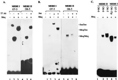
The differential binding specificities of MERE. It was ini-tially surprising to us that Meq-Meq homodimers recognize two completely different sequence motifs. To confirm that both MERE I and II ternary complexes contain Meq, we conducted antibody supershift experiments using a monoclonal antibody directed against the T7 gene 10 epitope tag fused to the N terminus of our Meq protein. Meq protein was incubated with MERE I or MERE II oligonucleotides in the presence (Fig. 8A, lanes 3 and 6) or absence (lanes 2 and 5) of monoclonal antibody against T7 gene 10 protein. The T7 antibody can clearly supershift both complexes, indicating Meq’s involve-ment in ternary complex formation with MERE I and II.
[image:5.612.126.487.67.418.2]We were also interested in the binding specificities of these motifs toward Meq-Jun and Jun-Jun dimers. Lanes 2 and 3 in Fig. 8B show that MERE I binds to both a Meq-Meq and Jun-Jun homodimer, as expected, given the presence of the TRE or CRE core in the motif. When equimolar amounts of Meq and Jun were incubated with MERE I, only the Meq-Jun heterodimer band was seen. This result is consistent with our
FIG. 4. Methylation interference analysis of the MERE I motif. (A) Results of an assay performed with O7-5 oligonucleotide labeled on the positive strand. Guanine nucleotides are numbered to the left of each gel; the residues which make up the consensus TRE sites are labeled. The lanes for free probes (lanes 1, 3, and 5) are labeled F; the lanes for bound probes are labeled B (lanes 2, 4, and 6). (B) Results of an assay performed with O7-5 oligonucleotide labeled on the negative strand. Lanes are labeled as described for panel A. In both sets of experiments, bacterially expressed MeqN and/or c-Jun was used. The assay conditions used are described in Materials and Methods.
on November 9, 2019 by guest
http://jvi.asm.org/
previous finding that Meq-Jun has a much higher affinity than either homodimer. By contrast, the ability to bind MERE II is unique to the Meq-Meq dimer (lane 6); neither Jun-Jun (lane 8) nor Meq-Jun (lane 7) forms a complex with this oligonucle-otide probe.
In addition to the differential binding of Meq-Jun and Jun-Jun toward MERE I and II, a structural difference in the binding of Meq-Meq to these two MEREs was noted. This is shown in Fig. 8C with equal-length oligonucleotide probes representing MERE I and II. The Meq-Meq–MERE II com-plexes (lanes 2 and 3) consistently migrate more slowly than the MERE I complexes (lanes 3 and 4), indicating significant conformational differences between these two types of complexes.
MERE-mediated transactivation by Meq-Meq. To see whether Meq-Meq can promote the transactivation of MERE-linked reporter genes, a MERE I (CRE) motif (TCAGAGT
GATGACGTCATCAC), a MERE I (TRE) motif (TCAGAG TGATGACTCATCAC, and a MERE II motif (AATTCAAA AACACATAACATTCGTATATATTCTGA) were cloned upstream of a CAT-encoding gene in a minimal promoter background, either in the same orientation as the CAT-en-coding gene [creating the CAT reporter constructs MEREI
(CRE)1CAT, MEREI(TRE)1CAT, and MEREII1CAT] or
in the opposite orientation [MEREI(CRE)2CAT, MEREI
(TRE)2CAT, and MEREII2CAT]. Themeqgene was cloned
into an expression plasmid, pECE, utilizing a simian virus 40
early promoter (38). The MERE2CAT plasmids were
cotrans-fected with Meq/pECE into CEF (a natural MDV host cell line) (Fig. 9). With each motif, there was a moderate (1.8- to 4.2-fold) but reproducible transactivation of the
[image:6.612.66.286.64.470.2]CAT-encod-FIG. 5. Gel retardation assay with wt and mutant MERE I probes. The materials and conditions used in this assay are described in Materials and Meth-ods. The filled dots on the wt sequence represent the guanine residues protected in the methylation interference assays above. The CRE consensus is underlined for each of the probes. Nucleotide substitutions are in boldface type and marked by filled triangles. Percentages at the right refer to the proportion of mutant probes bound to Meq (compared with that of the wt). The values were quantified by an Ambis radioanalysis system.
FIG. 6. Methylation interference assay for detection of the MERE II motif. A double-stranded oligonucleotide corresponding to clone O6-5, along with bacterially expressed MeqN, was used. Guanine nucleotides are numbered to the left; the residues which make up the consensus MERE II site are indicated. The lanes for free and bound probes are labeled F and B, respectively.
on November 9, 2019 by guest
http://jvi.asm.org/
[image:6.612.357.484.64.520.2]ing gene by Meq. Although we cannot rule out a possible contribution by endogenous Jun (as in Meq-Jun) to MERE I-mediated transactivation in CEF, the level of C-Jun activity in CEF was generally considered to be low (50). On the other
hand, the transactivation of MEREII2CAT is likely to be
solely attributed to the presence of Meq-Meq homodimers. These data are the first to demonstrate that Meq-Meq ho-modimers have transactivation potential.
DISCUSSION
MDV is perhaps the most potent oncogenic herpesvirus. The exceptionally short latency of tumor induction by MDV is in stark contrast to those of other herpesviruses and is rather similar to those of acute retroviruses. Like acute retroviruses, MDV may carry a direct-acting oncogene(s). Meq was origi-nally uncovered as a putative oncogene by its persistent ex-pression in all MDV-transformed tumor and T-cell lines (16). Meq is a bZIP protein with a structural organization resem-bling a fusion between Jun-Fos oncoproteins and the Wilms’ tumor 1 (WT-1) suppressor protein. Recent results have shown that overexpression of Meq can lead to transformation of fi-broblasts and can block apoptosis induced by serum starvation (25a) and that blocking Meq expression by antisense DNA reverses the transformed phenotypes of MDV (57). Among the
oncogenes of acute retroviruses, Jun, Fos, and Maf are bZIP transcriptional regulators (8, 17, 23, 24, 46). While there is at least one other herpesvirus protein with a functional bZIP domain, to our knowledge, Meq is the only one within the immediate family of Jun and Fos. We have recently shown that the C-terminal domain of Meq is a potent transactivator (38). Given its structural similarity to Jun-Fos and its ability to transactivate, Meq most likely exerts its transforming function by perturbing the pattern of cellular gene expression. In addi-tion to transformaaddi-tion, many DNA tumor virus oncoproteins important in transcriptional regulation are also important reg-ulators of viral DNA replication. Two examples are simian virus 40 large T antigen (19, 32, 47, 53) and Epstein-Barr virus EBNA-1 (27, 41, 49, 58). Similarly, Meq could also interact with MDV promoters and/or origins of replication. As a first step in understanding the mechanisms by which Meq modu-lates viral and cellular gene expression, we sought to identify optimal DNA binding motifs of Meq-Jun and Meq-Meq.
Previously, we showed that Meq-Jun avidly binds a TRE or
CRE hybrid motif present in the promoter of themeqgene and
transactivates reporter genes linked to this motif (38). Using a random oligonucleotide selection strategy, we found that a TRE or CRE motif was invariably contained in every clone selected for its ability to bind Meq-Jun heterodimers. These results, coupled with our previous observation that TRE and CRE core oligonucleotides alone are sufficient to gel shift Meq-Jun heterodimers, suggest that the TRE or CRE core is both necessary and sufficient for binding to Meq-Jun. The Meq-Jun binding specificity therefore resembles that of the Jun-Jun homodimer. However, because of the much higher affinity of Meq-Jun for TRE or CRE than that of the Jun homodimer, Meq may augment the regulatory function of Jun and modify the pairing of Jun to other bZIP proteins.
Interestingly, our analysis revealed two distinct DNA bind-ing motifs for Meq-Meq homodimers, MERE I and II. MERE I is represented by two independent clones, both of which carry a TRE or CRE core along with common flanking sequences. The rarity of clones carrying this motif coupled with the fact that TRE or CRE core oligonucleotides fail to bind Meq homodimers with any significant affinity tends to underscore the importance of the common flanking sequences. In support of this notion, nucleotide substitution analysis revealed that Meq-Meq bound not only the TRE or CRE core but also flanking sequences. The latter were critical in maintaining high-affinity binding (Fig. 5). In addition, the footprint of the Meq-Meq homodimer extends into the flanking region to a much greater extent than that of Meq-Jun or Jun-Jun (Fig. 4). Intriguingly, the sequence of MERE I is rather similar to that of the recognition motif for the Maf oncogene, TGCTGAC (G)TCAGCA (18, 20). Maf belongs to the Nrl or NF-E2 bZIP subfamily (33) and its basic region shows 53% identity with that of Meq (Fig. 1). Maf-responsive elements are found in the promoters of several growth-related genes, including those of the epidermal growth factor receptor (29), human interleukin 6 (40), and p53 (1). Such genes could represent potential targets for Meq. In the case of Maf, it was similarly shown that sequences flanking the TRE or CRE core were essential for high-affinity binding and that an auxiliary domain of Maf was responsible for this binding (18). We note that in Meq, there is an additional basic region N terminal to the bZIP domain. It is possible that this domain participates in DNA binding; further work is in progress.
[image:7.612.60.296.70.385.2]Perhaps the most striking result of the work reported here is the identification of MERE II as the Meq homodimer binding motif. This motif (RACACACAY) bears no resemblance to the TRE or CRE core, a signature binding motif for many
FIG. 7. Gel retardation assay with wt and mutant MERE II probes. Arrows on the wt sequence indicate the guanine residues analyzed in the methylation interference assay (Fig. 6). The first purine (in this case an adenosine) in the consensus is named residue11. The MERE II consensus for each of the probes is underlined. Nucleotide substitutions are in boldface type and marked by filled triangles. The percentages at right are measures of relative binding efficiencies and were calculated as described in the legend to Fig. 5.
on November 9, 2019 by guest
http://jvi.asm.org/
bZIP proteins. The flanking sequences are generally rich in adenines or thymines, but no stringent consensus could be identified. Even though our approach utilized gel-purified Meq-DNA complexes and did not depend on immunoprecipi-tation, we were initially concerned that the detected binding could be due to contaminating protein. However, the following observations suggest that the detected binding is Meq specific. First, when the T7 epitope-tagged Meq was used, a T7 mono-clonal antibody could supershift the MERE II–Meq-Meq com-plex (Fig. 8A); Meq-specific antibodies can also supershift the same complex (data not shown). Second, Meq prepared from different sources (bacteria, chicken cells, and by in vitro trans-lation) can all bind to MERE II. Finally, a Meq mutant with a deletion of the basic region (of the bZIP domain) failed to shift the MERE II oligonucleotide (unpublished results).
[image:8.612.109.501.69.334.2]As described above, MERE II motifs contain an ACAC ACA core flanked by sequences with no obvious homology. On the basis of methylation interference and nucleotide substitu-tion analyses, the ACACACA residues were characterized as crucial to this type of binding. However, the ACACACA se-quence alone was not sufficient in sustaining tight binding (unpublished results). The most straightforward interpretation of the above-noted data is that a Meq-Meq homodimer rec-ognizes an ACACACA motif in the context of a conformation determined by additional flanking sequences. Indeed, we cal-culated that all of the MERE II sequences that strongly bind Meq have characteristically high bending angles (16a). The CACA motif is known to be anisotropically flexible (28), and it can promote local kinks and bends (12, 52). It has been impli-cated in a number of special biological processes, such as microsatellite-associated instability (31, 37, 51), V(D)J joining
FIG. 8. The differential binding specificities of MEREs. (A) Antibody supershift patterns of MERE I and MERE II probes. MeqN, together with a T7 monoclonal antibody (Ab) which recognizes the epitope tag in MeqN, was used to bind O7-5 (MERE I) and O6-5 (MERE II) probes in the gel retardation assays. (B) Different binding specificities of MERE I and II toward Meq-Jun and Jun-Jun dimers. Gel retardation assays were performed with MERE I (O7-5) and II (O6-5) probes in conjunction with Meq-Meq, Meq-Jun, or Jun-Jun. The positions of the different DNA-protein complexes are labeled at the right. (C) Different gel-electrophoretic mobilities for DNA–Meq-Meq complexes with MERE I and MERE II probes. The MERE I and II probes of equal lengths (36 mer) were incubated with (1) or without (2) bacterially expressed MeqN. The gel retardation assay conditions used are described in Materials and Methods.
FIG. 9. Transactivation of MERE2CAT by Meq. The relative CAT activities for MERE I and II CAT reporter constructs were measured in CEF. For each set of experiments, a value of 1 corresponds to the level of CAT activity displayed by cells transfected with a reporter plasmid only (dotted bars); activities in the presence of Meq are represented by the hatched bars. pBLCAT2 is the parental reporter plasmid (26);1and2indicate the relative orientations of the cloned MERE sites with respect to that of the CAT gene. MEREI(CRE)1CAT and MEREI(CRE)2CAT each contain one copy of a MERE I (CRE) site (TCAG AGTGATGACGTCATCAC) upstream of the CAT-encoding gene; MEREI (TRE)1CAT and MERE(TRE)2CAT each contain two copies of a MERE I (TRE) site (TCAGAGTGATGACTCATCAC); MEREII1CAT and MEREII2CAT each contain three tandem copies of a MERE II site (AATTC AAAAACACATAACATTCGTATATATTCTAG) in the same position. Dif-ferences in transfection efficiencies were corrected by normalizing all CAT values against the level of an internal control reporter (pCMV-luc).
on November 9, 2019 by guest
http://jvi.asm.org/
[image:8.612.60.299.449.604.2]in immunoglobulin genes (14), duplication of humang-globin
genes (48), and transcription regulation of the mouseb-globin
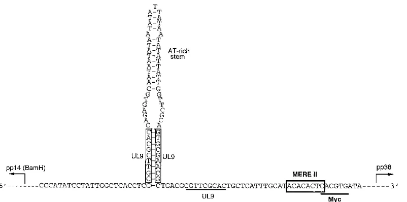
genes (9). Interestingly, a MERE II site was identified in the putative MDV origin of replication (3) (Fig. 10). A Meq ho-modimer can bind to this site in a CACA-dependent manner (3a). This raises the possibility of Meq modulating MDV DNA replication. Furthermore, this putative origin of replication overlaps the enhancer region of two divergently transcribed viral genes, the pp38- (7, 10) and the pp14-encoding genes of
theBamH gene family (15). Both genes have been implicated
in the MDV-mediated transformation process (34, 35, 57). Meq could therefore be involved in transcriptionally regulating these transformation-associated genes.
In summary, this study has led to the characterization of the DNA binding properties of a putative herpesvirus oncogene. On the basis of the binding motifs selected, Meq appears to be a versatile transcriptional regulator which has the potential to
engage itself in thejun-fosoncogene pathway, to substitute for
the Maf-encoding oncogene, and to regulate a unique set of genes. Our findings provide important leads in our understand-ing of Meq. We are now in a position to search for Meq’s functional targets in both the MDV and cellular genomes.
ACKNOWLEDGMENTS
We thank Juinn-Linn Liu for discussions and Ed Stavnezer and Keith Everiss for critical readings of the manuscript.
This work was supported by grants from the USDA (93-37204-9340 to L.L. and H.-J.K.), the NCI (CA46613 to H.-J.K.), and the Council for Tobacco Research (4034 to H.-J.K.).
REFERENCES
1.Bienz-Tadmor, B., R. Zakut-Houri, S. Libresco, D. Givol, and M. Oren. 1985. The 59region of the p53 gene: evolutionary conservation and evidence for a negative regulatory element. EMBO J.4:3209–3213.
2.Blackwell, T. K., L. Kretzner, E. M. Blackwood, R. N. Eisenman, and H. Weintraub.1990. Sequence-specific DNA binding by the c-Myc protein. Science250:1149–1151.
3.Bradley, G., M. Hayashi, G. Lancz, A. Tanaka, and M. Nonoyama.1989. Structure of the Marek’s disease virusBamHI-H gene family: genes of putative importance for tumor induction. J. Virol.63:2534–2542. 3a.Brunovskis, P., and H.-J. Kung.Unpublished data.
4.Call, K. M., T. Glaser, C. Y. Ito, A. J. Buckler, J. Pelletier, D. A. Haber, E. A. Rose, A. Krai, H. Yeger, W. H. Lewis, C. Jones, and D. Housman.1990. Isolation and characterization of a zinc finger polypeptide gene at the human chromosome 11 Wilms’ tumor locus. Cell60:509–520.
5.Calnek, B. W., and R. L. Witter.1991. Marek’s disease, p. 342–385.InB. W. Calnek (ed.), Diseases of poultry, 9th ed. Iowa State University, Ames. 6.Chen, C., and H. Okayama.1987. High-efficiency transformation of
mam-malian cells by plasmid DNA. Mol. Cell. Biol.7:2745–2752.
7.Chen, X., P. J. Sondermeijer, and L. F. Velicer.1992. Identification of a unique Marek’s disease virus gene which encodes a 38-kilodalton phospho-protein and is expressed in both lytically infected cells and latently infected lymphoblastoid cells. J. Virol.66:85–94.
8.Chiu, R., W. J. Boyle, J. Meek, T. Smeal, T. Hinter, and M. Karin.1988. The c-fos protein interacts with c-jun/AP-1 to stimulate transcription of AP-1 responsive genes. Cell54:541–552.
9.Cowie, A., and R. M. Myers.1988. DNA sequences involved in transcrip-tional regulation of the mouseb-globin promoter in murine erythroleukemia cells. Mol. Cell. Biol.8:3122–3128.
10. Cui, Z., L. F. Lee, J.-L. Liu, and H.-J. Kung.1991. Structural analysis and transcriptional mapping of the Marek’s disease virus gene encoding pp38, an antigen associated with transformed cells. J. Virol.65:6509–6515. 11. Curran, T., and N. M. Teich.1982. Candidate product of the FBJ murine
osteosarcoma virus oncogene: characterization of a 55,000-dalton phospho-protein. J. Virol.42:114–122.
12. Donlan, M. E., and P. Lu.1992. Transcriptional enhancer related DNA sequences: anomalous1H NMR NOE crosspeaks. Nucleic Acids Res.20: 525–532.
13. Gessler, M., A. Poustka, W. Cavenee, R. L. Neve, S. H. Orkin, and G. A. P. Bruns.1990. Homozygous deletion in Wilms’ tumors of a zinc-finger gene identified by chromosome jumping. Nature (London)343:774–778. 14. Hesse, J. E., M. R. Lieber, K. Mizuuchi, and M. Gellert.1989. V(D)J
recombination: a functional definition of the joining signals. Genes Dev. 3:1053–1061.
15. Hong, Y., and P. M. Coussens.1994. Identification of an immediate-early gene in the Marek’s disease virus long internal repeat region which encodes a unique 14-kilodalton polypeptide. J. Virol.68:3593–3603.
16. Jones, D., L. Lee, J.-L. Liu, H.-J. Kung, and J. K. Tillotson.1992. Marek’s disease virus encodes a basic-leucine zipper gene resembling the fos/jun oncogenes that is highly expressed in lymphoblastoid tumors. Proc. Natl. Acad. Sci. USA89:4042–4046.
16a.Kahn, J., and Z. Qian.Unpublished data.
17. Kataoka, K., K. Igarashi, K. Itoh, K. T. Fujiwara, M. Noda, M. Yamamoto, and M. Nishizawa.1995. Small Maf proteins heterodimerize with Fos and may act as competitive repressors of the NF-E2 transcription factor. Mol. Cell. Biol.15:2180–2190.
[image:9.612.112.513.70.269.2]18. Kataoka, K., M. Noda, and M. Nishizawa.1994. Maf nuclear oncoprotein recognizes sequences related to an AP-1 site and forms heterodimers with both Fos and Jun. Mol. Cell. Biol.14:700–712.
FIG. 10. Bidirectional promoter at the MDV origin of replication. The sequence was adopted from Bradley et al. (3). The three UL9 recognition sites are indicated. UL9 is an MDV-encoded DNA-binding protein involved in catalyzing DNA polymerase binding to the origin of replication (54, 55). The consensus MERE II is also boxed; underlined is a Myc-recognition site (CACGTG) (2, 36). The characteristic AT-rich stem-loop present in the replication origins of several herpesviruses is shown. This region also represents a bidirectional promoter for pp14 (15) and pp38 (7, 10). pp14 belongs to theBamHI gene family (22). The transcriptional directions of pp14 and pp38 are indicated.
on November 9, 2019 by guest
http://jvi.asm.org/
19. Keller, J. M., and J. C. Alwine.1985. Analysis of an activatable promoter: sequences in the simian virus 40 late promoter required for T-antigen-mediatedtransactivation. Mol. Cell. Biol.5:1859–1869.
20. Kerppola, T. K., and T. Curran.1994. A conserved region adjacent to the basic domain is required for recognition of an extended DNA binding site by Maf/Nrl family proteins. Oncogene9:3149–3158.
21. Kouzarides, T., and E. Ziff.1989. Behind the Fos and Jun leucine zipper. Cancer Cells1:71–76.
22. Kung, H. J., and M. Nonoyama.1995. Two gene families of Marek’s disease virus (MDV) with a potential role in tumor induction in chickens. Int. J. Oncol.6:997–1002. (Review.)
23. Kurschner, C., and J. I. Morgan.1995. Themafproto-oncogene stimulates transcription from multiple sites in a promoter that directs Purkinje neuron-specific gene expression. Mol. Cell. Biol.15:246–254.
24. Lech, K., K. Anderson, and R. Brent.1988. DNA-bound fos proteins activate transcription in yeast. Cell52:179–184.
25. Li, N., A. Batzer, R. Daly, V. Yajnik, E. Skolnik, P. Chardin, D. Bar-Sagi, B. Margolis, and J. Schlessinger.1993. Guanine-nucleotide-releasing factor hSos1 binds to Grb2 and links receptor tyrosine kinases to Ras signalling. Science363:85–88.
25a.Liu, J.-L., and H.-J. Kung.Unpublished results.
26. Luckow, B., and G. Schutz.1987. CAT constructions with multiple unique restriction sites for the functional analysis of eukaryotic promoters and regulatory elements. Nucleic Acids Res.15:5490.
27. Lupton, S., and A. J. Levine.1985. Mapping genetic elements of Epstein-Barr virus that facilitate extrachromosomal persistence of Epstein-Epstein-Barr vi-rus-derived plasmids in human cells. Mol. Cell. Biol.5:2533–2542. 28. Lyubchenko, Y. L., L. S. Shlyakhtenko, E. Appella, and R. E. Harrington.
1993. CA runs increase DNA flexibility in the complex oflCro protein with the OR3 site. Biochemistry32:4121–4127.
29. Maekawa, T., F. Imamoto, G. T. Merlino, I. Pastan, and S. Ishii.1989. Cooperative function of two separate enhancers of the human epidermal growth factor receptor proto-oncogene. J. Biol. Chem.264:5488–5494. 30. Maki, Y., T. J. Bos, C. Davis, M. Starbuck, and P. K. Vogt.1987. Avian
sarcoma virus 17 carries thejunoncogene. Proc. Natl. Acad. Sci. USA 84:2848–2852.
31. Merlo, A., M. Mabry, E. Gabrielson, R. Vollmer, S. B. Baylin, and D. Sidransky.1994. Frequent microsatellite instability in primary small cell lung cancer. Cancer Res.54:2098–2101.
32. Myers, R. M., D. C. Rio, A. K. Robbins, and R. Tjian.1981. SV40 gene expression is modulated by the cooperative binding of T antigen to DNA. Cell25:373–384.
33. Nishizawa, M., K. Kataoka, N. Goto, K. T. Fujiwara, and S. Kawai.1989. v-maf, a viral oncogene that encodes a “leucine zipper” motif. Proc. Natl. Acad. Sci. USA86:7711–7715.
34. Peng, F., G. Bradley, A. Tanaka, G. Lancz, and M. Nonoyama.1992. Isola-tion and characterizaIsola-tion of cDNAs fromBamHI-H gene family RNAs associated with the tumorigenicity of Marek’s disease virus. J. Virol.66: 7389–7396.
35. Peng, F. Y., J. Donovan, S. Spector, A. Tanaka, and M. Nonoyama.1993. Prolonged proliferation of primary chicken embryo fibroblasts transfected with cDNAs from the BamHI-H gene family of Marek’s disease virus. Int. J. Oncol.3:587–591.
36. Prendergast, G. C., and E. B. Ziff.1991. Methylation-sensitive sequence-specific DNA binding by the c-Myc basic region. Science251:186–189. 37. Pykett, M. J., M. Murphy, P. R. Harnish, and D. L. George.1994.
Identifi-cation of a microsatellite instability phenotype in meningiomas. Cancer Res. 54:6340–6343.
38. Qian, Z., P. Brunovskis, F. J. Rauscher III, L. F. Lee, and H. J. Kung.1995. Transactivation activity of Meq, a Marek’s disease herpesvirus bZIP protein
persistently expressed in latently infected transformed T cells. J. Virol.69: 4037–4044.
39. Rauscher, F. J., III, P. J. Voulalas, B. R. Franza, Jr., and T. Curran.1988. Fos and Jun bind cooperatively to the AP-1 site: reconstitution in vitro. Genes Dev.2:1687–1699.
40. Ray, A., S. B. Tatter, L. T. May, and P. B. Sehgal.1988. Activation of the human “b2-interferon/hepatocyte-stimulating factor/interleukin 6” promoter by cytokines, viruses, and second messenger agonist. Proc. Natl. Acad. Sci. USA85:6701–6705.
41. Reisman, D., and B. Sugden.1986. Transactivation of an Epstein-Barr viral transcriptional enhancer by the Epstein-Barr viral nuclear antigen 1. Mol. Cell. Biol.6:3838–3846.
42. Ren, R., B. J. Mayer, P. Cicchetti, and D. Baltimore.1993. Identification of a ten-amino acid proline-rich SH3 binding site. Science259:1157–1161. 43. Richmond, C.1992. Ph.D. thesis. University of Cincinnati, Cincinnati. 44. Ryseck, R.-P., and R. Bravo.1991. c-JUN, JUN B, JUN D differ in their
binding affinities to AP-1 and CRE consensus sequences: effect of FOS proteins. Oncogene6:533–542.
45. Seed, B., and J. Y. Sheen.1988. A simple phase-extraction assay for chlor-amphenicol acetyltransferase activity. Gene67:271–277.
46. Setoyama, C., R. Frunzio, G. Liau, M. Mudry, and B. de Crombrugghe.1986. Transcriptional activation encoded by the v-fos gene. Proc. Natl. Acad. Sci. USA83:3213–3217.
47. Shortle, D. R., R. F. Margolskee, and D. Nathans.1979. Mutational analysis of the simian virus 40 replicon: pseudorevertants of mutants with a defective replication origin. Proc. Natl. Acad. Sci. USA76:6128–6131.
48. Slightom, J. L., A. E. Blechl, and O. Smithies.1980. Human fetalGg- and Ag-globin genes: complete nucleotide sequences suggest that DNA can be exchanged between these duplicated genes. Cell21:627–638.
49. Sugden, B., and N. Warren.1989. A promoter of Epstein-Barr virus that can function during latent infection can be transactivated by EBNA-1, a viral protein required for viral DNA replication during latent infection. J. Virol. 63:2644–2649.
50. Suzuki, T., Y. Hashimoto, H. Okuno, H. Sato, H. Nishina, and H. Iba.1991. High-level expression of human c-jun gene causes cellular transformation of chicken embryo fibroblasts. Jpn. J. Cancer Res.82:58–64.
51. Thibodeau, S. U., G. Bren, and D. Schaid.1993. Microsatellite instability in cancer of the proximal colon. Science260:816–818.
52. Timsit, Y., E. Vilbois, and D. Moras.1991. Base-pairing shift in the major groove of (CA)ntracts by B-DNA crystal structures. Nature (London)354: 167–170.
53. Tooze, J.1980. DNA tumor viruses. Cold Spring Harbor Laboratory, Cold Spring Harbor, N.Y.
54. Weir, H. M., J. M. Calder, and N. D. Stow.1989. Binding of the herpes simplex virus type 1 UL9 gene product to an origin of viral DNA replication. Nucleic Acids Res.17:1409–1425.
55. Weir, H. M., and N. D. Stow.1990. Two binding sites for the herpes simplex virus type 1 UL9 protein are required for efficient activity of the oriS repli-cation origin. J. Gen. Virol.71:1379–1385.
56. Wright, W. E., M. Binder, and W. Funk.1991. Cyclic amplification and selection of targets (CASTing) for the myogenin consensus binding site. Mol. Cell. Biol.11:4104–4110.
57. Xie, Q., A. S. Anderson, and R. W. Morgan.1996. Marek’s disease virus (MDV) ICP4, pp38, andmeqgenes are involved in the maintenance of transformation of MDCC-MSB1 MDV-transformed lymphoblastoid cells. J. Virol.70:1125–1131.
58. Yates, J. L., N. Warren, and B. Sugden.1985. Stable replication of plasmids derived from Epstein-Barr virus in various mammalian cells. Nature (Lon-don)313:812–815.