0022-538X/96/$04.0010
Copyrightq1996, American Society for Microbiology
Mutagenesis of the Coxsackie B3 Virus 2B/2C Cleavage Site:
Determinants of Processing Efficiency and Effects on
Viral Replication
FRANK J. M.VANKUPPEVELD,* PATRICK J. J. C.VAN DENHURK, JAN ZOLL,
JOCHEM M. D. GALAMA,ANDWILLEM J. G. MELCHERS
Department of Medical Microbiology, University of Nijmegen, Nijmegen, The Netherlands
Received 1 April 1996/Accepted 6 August 1996
The enterovirus 2B/2C cleavage site differs from the common cleavage site motif AxxQ2G by the occurrence
of either polar residues at the P1* position or large aliphatic residues at the P4 position. To study (i) the
putative contribution of these aberrant residues to the stability of precursor protein 2BC, (ii) the determinants
of cleavage site specificity and efficiency of 3Cpro
, and (iii) the importance of efficient cleavage at this site for
viral replication, a mutational analysis of the coxsackie B3 virus (CBV3) 2B/2C cleavage site (AxxQ2N) was
performed. Neither replacement of the P1*asparagine with a serine or a glycine nor replacement of the P4
alanine with a valine significantly affected 2B/2C cleavage efficiency, RNA replication, or virus growth. The introduction of a P4 asparagine, as can be found at the CBV3 3C/3D cleavage site, caused a severe reduction in 2B/2C cleavage and abolished virus growth. These data support the idea that a P4 asparagine is an unfavorable residue that contributes to a slow turnover of precursor protein 3CD but argue that it is unlikely that the aberrant 2B/2C cleavage site motifs serve to regulate 2B/2C processing efficiency and protein 2BC
stability. The viability of a double mutant containing a P4 asparagine and a P1*glycine demonstrated that a
P1*residue can compensate for the adverse effects of an unfavorable P4 residue. Poliovirus (or poliovirus-like)
2B/2C cleavage site motifs were correctly processed by CBV 3Cpro
, albeit with a reduced efficiency, and yielded viable viruses. Analysis of in vivo protein synthesis showed that mutant viruses containing poorly processed 2B/2C cleavage sites were unable to completely shut off cellular protein synthesis. The failure to inhibit host translation coincided with a reduced ability to modify membrane permeability, as measured by the sensitivity to the unpermeant translation inhibitor hygromycin B. These data suggest that a critical level of protein 2B or 2C, or both, may be required to alter membrane permeability and, possibly as a consequence, to shut off host cell translation.
Enteroviruses contain a positive-strand RNA genome of 7.5 kb in length which encodes a single polyprotein. This
polypro-tein is processed by three virus-encoded propolypro-teinases, 2Apro,
3Cpro, and 3CDpro (Fig. 1A), into the structural P1 capsid
proteins and the nonstructural P2 and P3 proteins that are involved in viral RNA (vRNA) replication (reviewed in
refer-ence 50). Proteinase 2Apro cleaves between the P1 and P2
regions cotranslationally (45). The capsid proteins are
pro-cessed intransby proteinase 3CDpro(26, 51). Processing of the
nonstructural proteins by proteinase 3Cproyields both the final
cleavage products (2A, 2B, 2C, 3A, 3B, 3C, and 3D) and relatively stable processing intermediates (2BC, 3AB, and 3CD) (22). These precursor proteins have functions in vRNA replication that are distinct from those of their cleavage prod-ucts. Protein 3AB is the membrane-bound precursor that de-livers VPg (3B) to the membranous replication complex (42–
44). Furthermore, protein 3AB stimulates 3Dpolactivity and
the autocatalytic processing of 3CDproto 3Cproand 3Dpol(30,
34, 37). Protein 3CD not only is a proteinase but also is in-volved in the formation of a ribonucleoprotein complex at the
59end of the RNA, which is required for the initiation of viral
positive-strand RNA synthesis (2, 3, 23). Protein 2BC has been proposed to play a role in the induction of the membrane
vesicles at which viral positive-strand RNA synthesis occurs (7). Bienz et al. showed that in poliovirus (PV)-infected cells in which polyprotein processing was partially inhibited, the for-mation of these vesicles always coincided with the production of protein 2BC (8). Consistent with this finding, Barco and Carrasco demonstrated that the expression of PV protein 2BC, but not that of protein 2B or 2C, either individually or in combination, induced the formation of membrane vesicles in yeast cells (5). The finding that protein 2C alone can induce vesicles in human cells (1, 14) may have been hampered by the use of vaccinia viruses, which modify vesicular traffic them-selves.
The production and stability of proteins 2BC, 3AB, and 3CD require a finely tuned regulation of the temporal processing at different cleavage sites in the polyprotein. Proteolysis may be regulated by the occurrence of less favorable recognition
se-quences. In PVs, all 3Cpro-mediated cleavages occur between
glutamine-glycine (Q-G) dipeptide pairs. Additional determi-nants in substrate recognition, however, have been proposed because the PV polyprotein contains four Q-G amino acid pairs that are not cleaved and cleavage sites other than Q-G, as identified in other enteroviruses (15, 27, 32), were also
cleav-able by PV protein 3Cpro (17). The amino acid at the P4
position (i.e., the fourth residue proximal to the Q-G cleavage site), which is in most cases an alanine, seems to be a major determinant of the efficiency of cleavage. Data that support the role of the P4 amino acid in substrate recognition have been obtained by mutagenesis of infectious PV cDNA clones (9). The importance of the P4 amino acid for the stability of
pre-* Corresponding author. Mailing address: Department of Medical Microbiology, University of Nijmegen, P.O. Box 9101, 6500 HB Nijmegen, The Netherlands. Phone: 31 24 3614356. Fax: 31 24 3540216. Electronic mail address: [email protected].
7632
on November 9, 2019 by guest
http://jvi.asm.org/
cursor protein 3CDprohas been demonstrated by Pallai et al.
(36), who found that a synthetic peptide containing the
au-thentic PV 3C/3D cleavage site (TxxQ2G) was resistant to
hydrolysis by 3Cpro, whereas a peptide containing a
substitu-tion of the threonine by an alanine was efficiently cleaved. Other determinants that may affect substrate processing are the recognition of secondary or tertiary structures and the accessibility to a potential cleavage site. Analysis of structural data suggests that cleavage sites must be correctly displayed at the surface in a flexible turn configuration at the end of an
a-helix orb-sheet to be recognized efficiently (4, 35, 36).
Pro-cessing at the 3A/3B cleavage site seems to be determined by structural rather than sequence-specific determinants. Lama et al. found that only membrane-associated protein 3AB, not
solubilized 3AB, could be cleaved by 3Cpro, indicating that
either the recognition or exposure of the 3A/3B cleavage site, which lies in close proximity to the membrane-binding domain of protein 3A, is dependent on a hydrophobic environment (30).
Alignment of the 3Cpro cleavage sites of the enterovirus
nonstructural proteins (Fig. 1B) shows that nearly all
entero-viruses contain the sequence AxxQ2G (P4-P3-P2-P1-P19) at
their 2A/2B, 2C/3A, 3A/3B, and 3B/3C junctions. The 3C/3D cleavage sites differ from this sequence by the occurrence of unfavorable residues at the P4 position, which seem to be
involved in the stability of protein 3CDpro (see above). It is
remarkable that also all 2B/2C cleavage site motifs are
aber-rant from the sequence AxxQ2G. Coxsackie B virus
(CBV)-like viruses contain polar asparagine or serine residues at the
P19 position rather than a neutral glycine residue. PV-like
viruses, on the other hand, contain at the P4 position either a valine or an isoleucine, residues which are heavily branched at
the b-carbon, rather than an alanine, which has only a small
methyl group as its side chain. The only virus that contains the
sequence AxxQ2G at its 2B/2C junction, PV type 2 strain
Sabin, differs from all other enteroviruses by the occurrence of a negatively rather than a positively charged residue at its P2 position. The occurrence of aberrant sequence motifs at the 2B/2C cleavage site of all enteroviruses may indicate a regula-tory role in the kinetics of 2B/2C processing and, thereby, in the stability of protein 2BC. To test this possibility and to gain more insight in the determinants of the 2B/2C cleavage site specificity and processing efficiency, eight mutant coxsackie B3 virus (CBV3) cDNAs were constructed and the effects of the mutations on in vitro polyprotein processing were analyzed. To understand the importance of efficient cleavage at this site for viral replication, the effects of the mutations on virus viability and growth, RNA replication, and in vivo protein synthesis were assayed.
MATERIALS AND METHODS
Cells and viruses. Virus propagations and vRNA transfections were per-formed with Buffalo green monkey (BGM) cells. Plaque assays were perper-formed with Vero cells. The cells were grown in minimal essential medium (MEM) supplemented with 10% fetal calf serum. After infection, cells were fed with MEM containing 3% serum. After transfection, MEM containing 10% serum was added to the cells. Viruses were titrated in 96-well plates with BGM cell monolayers as described previously (46). Virus titers were calculated by the FIG. 1. (A) Schematic structure of the 7.5-kb single-stranded RNA genome of CBV3 containing protein VPg at 59end of the nontranslated region (NTR) and a polyadenylate tract at the end of the 39nontranslated region. The polyprotein-encoding region is shown boxed. The proteinase that is responsible for cleavage at a particular cleavage site (indicated by an arrow) is shown below the polyprotein. (B) Alignment of amino acid sequences that occur at the P4-P3-P2-P1/P19of the 2A/2B, 2B/2C, 2C/3A, 3A/3B, 3B/3C, and 3C/3D cleavage sites of enteroviruses for which complete sequences of the P2 and P3 regions are known. The enteroviruses are divided into a CBV-like subgroup and a PV-like subgroup. Enteroviruses with distinct amino acid sequences are shown apart. Only sequence motifs that differ from the most frequently occurring motif AxxQ/G are shown. Abberant amino acids are in boldface. Abbreviations: ECHO, echovirus; CAV, coxsackie A virus; SVDV, swine vesicular disease virus; PV1(S) and PV1(M), PV type 1 strains Sabin and Mahoney, respectively; PV2(S) and (L), PV type 2 strains Sabin and Lansing, respectively; PV3(S) and PV3(L), PV type 3 strains Sabin and Leon, respectively; EV70, enterovirus type 70; BEV1, bovine enterovirus type 1.
on November 9, 2019 by guest
http://jvi.asm.org/
[image:2.612.64.539.78.346.2]method of Reed and Muench and expressed in 50% tissue culture infective dose (TCID50) values (40).
Site-directed mutagenesis.Oligonucleotide-directed site-specific mutagenesis was performed with subgenomic phagemid pALTCB3/2080-4947 (46), using the Altered Sites in vitro mutagenesis system (Promega) according to the manufac-turer’s recommendations. The nucleotide sequences of the synthetic oligonucle-otides (Isogen Bioscience, Maarssen, The Netherlands) are 59-CTTAAGCCAG CTATTGCCTTGGCGTTCAGCCAT-39(mutation 2B/2C-19G), 59-CTTAAGC CAGCTATTTGGTTGGCGTTCAGCCAT-39 (2B/2C-19P), 59-CTTAAGCCA GCTATTCGATTGGCGTTCAGCCAT-39(2B/2C-19S), 59-ATTGTTTTGGCG TTCGACCATAGGGATTCCGTA-39(2B/2C-4V), 59-ATTGTTTTGGCGCTC ATTCATAGGGATTCCGTA-39(2B/2C-4N), 59-CTTAAGCCAGCTATTGCC TTGGCGCTCATTCATAGGGATTCCGTA-39(2B/2C-4N;19G), 59-CTTAAG CCAGCTATTGCCTTGGCGTTCGACCATAGGGATTCCGTA-39 (2B/2C-4V;19G), and 59-CTTAAGCCAATTGCCTTGGCGGATCACCATAGGGAT GATTCCGTAATA-39(2B/2C-4V;3I;19G). Each of these oligonucleotides creat-ed a novel endonuclease restriction site. Clones carrying the desircreat-ed mutation were identified by restriction enzyme analysis. The nucleotide sequence around the 2B/2C junction of these clones was verified by dideoxy-chain termination sequencing of plasmid DNA, using primer 59-CCATTCAATGAATTTCTG-39 (nucleotides 4117 to 4134). From the mutant clones, theSpeI (position 3837)-to-BssHII (position 4238) fragment was cloned in plasmids pCB3/T7, which contains a full-length cDNA of CBV3 Nancy behind a T7 RNA polymerase promoter (28), and pCB3/T7-LUC, a pCB3/T7-derived construct that contains the luciferase gene in place of the capsid coding region (46).
In vitro translation reactions.SalI-linearized plasmid DNA (0.5 mg) was transcribed and translated in a single reaction, using T7 TNT rabbit reticulocyte lysate (Promega) supplemented with HeLa cell initiation factors (kindly pro-vided by J. Flanegan, University of Florida) and Tran35
S-label (a mixture of [35
S]methionine and [35
S]cysteine; ICN). Labeled proteins were analyzed by sodium dodecyl sulfate-polyacrylamide gel electrophoresis (SDS-PAGE) as de-scribed previously (47).
Transfection of cells with RNA transcripts.Eighty percent confluent BGM cell monolayers were transfected with 5mg of full-length copy RNA transcripts, generated with T7 RNA polymerase fromSalI-linearized cDNA templates, using the DEAE-dextran method as described previously (46). After transfection, the cells were grown at either 33 or 368C. When virus growth was observed, the cultures were incubated until cytopathic effect was complete. Otherwise, the cultures were subjected to three cycles of freezing and thawing, and 200ml was passaged to fresh BGM monolayers, which were incubated for another 3 (338C) or 5 (368C) days.
Sequence analysis of 2B/2C junction of mutant viruses.vRNA extraction, cDNA synthesis, and amplification by PCR using forward primer 59-GCAATG GAACAGGGAGTGAAGGACTATGTGGA-39(nucleotides 3733 to 3765) and reverse primer 59-TTGGGATGGCGCGCTCTGCTC-39 (nucleotides 4231 to 4251) were performed as described previously (46). The resulting PCR products were purified from low-melting-point agarose, and the nucleotide sequence around the 2B/2C junction was determined as described above.
Plaque assays and single-cycle yield analysis.Plaque assays were carried out with confluent monolayers of Vero cells on 10-cm2
dishes in six-well plates as described previously (46). To determine the virus yield in a single replicative cycle, 100% confluent BGM cell monolayers (53106
cells) were infected with virus at an MOI of 1 TCID50per cell and grown at either 33, 36, or 398C. At 8 h postinfection, viruses were released by three cycles of freezing and thawing and the virus titer was determined.
Analysis of positive-strand RNA synthesis.BGM cell monolayers were trans-fected with 0.5mg of T7 RNA polymerase generated full-length copy RNA transcripts of the mutant pCB3/T7-LUC plasmids as described above. At 1, 4, 6, 8, and 10 h posttransfection, the cells were lysed and the luciferase production was measured as described previously (46).
Analysis of viral protein synthesis in vivo.BGM cell monolayers were infected with virus at an MOI of 25 for 30 min at room temperature. After infection, cells were supplied with new medium and incubated at 368C. At several times postin-fection, protein synthesis was monitored by labeling with 10mCi of Tran35
S-label in methionine- and serum-free MEM (Gibco). After 30 min, the medium was discarded, the cells were lysed in cold lysis buffer, and labeled proteins were analyzed by SDS-PAGE as described previously (46).
In experiments in which the permeability of cells to hygromycin B was assayed, the same procedure was followed except that cells were incubated in methionine-and serum-free MEM, in the presence or absence of 500mg of hygromycin B (Sigma) per ml, for 15 min prior to the addition of 10mCi of Tran35
S-label to the medium.
To determine the [35
S]methionine incorporation, sample lanes were cut from the gel and measured in a liquid scintillation counter following SDS-PAGE and autoradiography.
RESULTS
Construction and description of CBV3 2B/2C cleavage site
mutants. Substitution mutations at the 2B/2C cleavage site
were generated by oligonucleotide-directed mutagenesis and
introduced in infectious CBV3 cDNA clone pCB3/T7 and sub-genomic replicon pCB3/T7-LUC. The mutants fall into three categories. Within the first group are four mutants that were
constructed to examine the importance of the aberrant P19or
P4 residues that occur at the enterovirus 2B/2C cleavage site
(Fig. 1B). For this, the P19 asparagine was replaced with a
glycine (mutation 2B/2C-19G), a proline (2B/2C-19P), or a
serine (2B/2C-19S) or the P4 alanine was replaced with a valine
(2B/2C-4V). The second group consists of two mutants that were constructed to examine the effect on 2B/2C cleavage efficiency of a P4 asparagine, as can be found at the 3C/3D cleavage site of CBV-like viruses. The P4 asparagine was in-troduced either alone (2B/2C-4N) or, in order to create a site
that resembles the 3C/3D cleavage site, together with a P19
glycine (2B/2C-4N;19G). The third group consists of two
mu-tants that were engineered to examine the cross-species
sub-strate specificity of CBV3 3Cpro. In one mutant, a valine was
introduced at the P4 position and a glycine was introduced at
the P19position to create a motif with P4, P1, and P19residues
that occur at the 2B/2C junction of most PV-like viruses (2B/
2C-4V;19G). The other mutant contained an additional
re-placement of the P3 glutamic acid with a isoleucine
(2B/2C-4V;3I;19G) to create a P4 to P19sequence that is identical to
that found at a PV 2B/2C junction (PV type 3).
In vitro polyprotein processing of 2B/2C cleavage site
mu-tants.The effect of the mutations on the 3Cpro-mediated
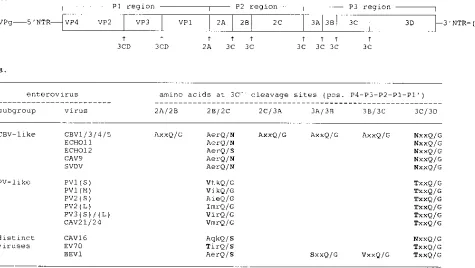
pro-cessing at the 2B/2C cleavage site was studied by in vitro translation of copy RNA transcripts of the mutant pCB3/T7 plasmids. Figure 2A shows the protein patterns generated after 3 h. This figure shows that some of the mutations affected the efficiency of 2B/2C processing, but not the 2B/2C cleavage site specificity, as shown by a correct migration of protein 2C. Remarkably, no accumulation of protein 2BC or any other potential precursor protein was observed. None of the muta-tions affected any of the other proteolytic processing events in the viral polyprotein, as shown by the normal production of other viral proteins, including protein 2A (Fig. 2C), to levels similar to those produced by wild-type RNA. Protein 2B (11 kDa) could not be visualized because it migrated in the heavily overloaded globin spot.
To determine the relative efficiency of 2B/2C cleavage, au-toradiograms from three independent translation experiments were analyzed by densitometric scanning and the ratio of pro-tein 2C/3CD was calculated (to correct for variations in total protein yield). The amount of protein 2C produced by RNA
transcripts carrying mutations 2B/2C-19G and 2B/2C-19S was
similar to that produced by wild-type pCB3/T7 RNA. Mutation
2B/2C-19P completely abolished 2B/2C cleavage. Even after
prolonged exposure of the gels, no protein 2C could be de-tected. Mutation 2B/2C-4V slightly impaired 2B/2C cleavage, and approximately 85% of the wild-type protein 2C amount was produced. The introduction of asparagine residues at the P4 position (single mutation 2B/2C-4N and double mutation
2B/2C-4N;19G) reduced 2B/2C cleavage efficiency to about
10% of the wild-type level. Mutations 2B/2C-4V;19G and 2B/
2C-4V;3I;19G reduced the cleavage efficiency to about 25% of
that of the wild type.
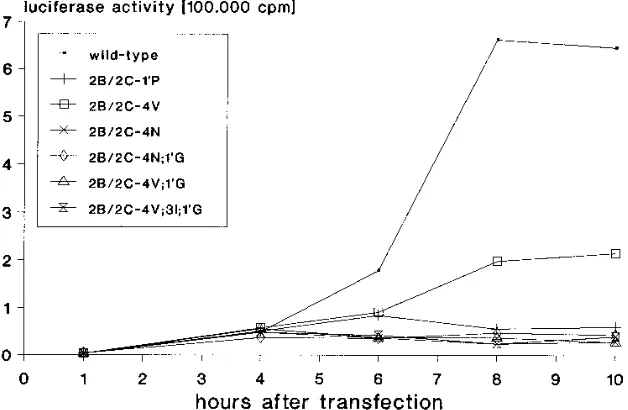
Effects of 2B/2C cleavage site mutations on RNA replication. To study the effects of the mutations on positive-strand RNA replication, BGM cells were transfected with copy RNA tran-scripts of wild-type and mutant pCB3/T7-LUC constructs, and the luciferase activity was measured at several times posttrans-fection. Figure 3 shows that none of the mutations interfered with the initial increase in luciferase activity (between 1 and 4 h) that reflects translation of the input RNA (46). However, some mutations interfered with the second increase in
on November 9, 2019 by guest
http://jvi.asm.org/
erase activity, which occurs from the fourth hour and reflects the replication of the input RNA and subsequent translation of the newly synthesized RNA strands (46, 47). Replicons
carry-ing mutations 2B/2C-19G and 2B/2C-19S, which had virtually
no effect on 2B/2C cleavage, displayed an increase (about 10-to 15-fold) in luciferase activity similar 10-to that of wild-type pCB3/T7-LUC (data not shown). Mutation 2B/2C-4V, which caused a minimal reduction in 2B/2C cleavage, reduced RNA replication and yielded an increase in luciferase accumulation that was only fourfold. Mutations that either abrogated (2B/
2C-19P) or severely reduced (2B/2C-4N, 2B/2C-4N;19G, 2B/
2C-4V;19G, and 2B/2C-4V;3I;19G) 2B/2C cleavage caused
se-vere defects in RNA replication, as shown by the absence of an increase in luciferase activity above the initial translation level. Effects of 2B/2C cleavage site mutations on virus viability
and growth.The effects of the mutations on virus viability were
determined by transfection of BGM cells with copy RNA tran-scripts from the mutant pCB3/T7 plasmids. For each mutation, two independently made constructs were tested. Four trans-fections were performed with RNA from each construct. After
transfection, two cell cultures were incubated at 338C and two
were incubated at 368C. Viruses were obtained consistently
upon transfection of cells with RNA transcripts carrying
mu-tations 2B/2C-19G, 2B/2C-19S, 2B/2C-4V, 2B/2C-19G;4N, 2B/
2C-19G;4V, and 2B/2C-19G;3I;4V, albeit virus growth was
se-verely delayed with RNA transcripts carrying the three latter mutations. Sequence analysis demonstrated that all viruses had retained the introduced mutation at the 2B/2C junction. The
mutant viruses were named vCB3-2B/2C-19G,
vCB3-2B/2C-19S, vCB3-2B/2C-4V, vCB3-2B/2C-4N;19G, vCB3-2B/2C-4V;
19G, and vCB3-2B/2C-4V;3I;19G, respectively. No virus growth
was observed in any of the cell cultures transfected with RNA
transcripts carrying mutation 2B/2C-19P. Upon transfection of
cells with RNA transcripts carrying mutation 2B/2C-4N, a re-vertant virus was isolated (see below).
Viral growth characteristics were examined by plaque assay and measuring the virus yield at 8 h postinfection in a
single-cycle infection (Table 1). vCB3-2B/2C-19G and
vCB3-2B/2C-19S showed wild-type growth characteristics. vCB3-2B/2C-4V
produced 16% of the wild-type virus yield and displayed a
small-plaque phenotype. vCB3-2B/2C-4N;19G and vCB3-2B/
2C-4V;19G produced about 2% of the wild-type virus yield and
exhibited a minute-plaque phenotype. Minute-plaque virus
vCB3-2B/2C-4V;3I;19G produced only 0.3% of the wild-type
virus yield. The relative virus yield of all mutants was the same
at 33, 36, and 398C.
Isolation of revertant virus vCB3-2B/2C-4S.Upon
transfec-tion of cells with RNA transcripts carrying mutatransfec-tion 2B/2C-4N, virus growth was observed in only one of the eight transfected cell cultures. Sequence analysis of the 2B/2C junction and the region surrounding this cleavage site showed that this virus contained a reversion of the introduced asparagine (AAU) to a serine (AGU). This mutant virus was named vCB3-2B/2C-4S. To examine whether this reversion mutation was sufficient to confer virus viability, mutation 2B/2C-4S was introduced in pCB3/T7, and BGM cells were transfected with RNA tran-scripts of the resulting plasmid. The systematic isolation of viruses carrying this mutation indicates that the reversion of the asparagine to serine at the P4 position of the 2B/2C cleav-age site was indeed sufficient to confer virus viability. This mutant virus displayed a minute-plaque phenotype and pro-duced about 2% of the wild-type virus yield in single-cycle infections at different temperatures.
To examine whether the viability of reversion mutation 2B/ 2C-4S correlated with an increased 2B/2C cleavage, the in vitro processing profiles of pCB3/T7 plasmids carrying mutations 2B/2C-4N and 2B/2C-4S were compared. Figure 2B demon-strates that the efficiency of 2B/2C cleavage was indeed in-creased by reversion mutation 2B/2C-4S; RNA transcripts car-rying mutation 2B/2C-4S generated about 25% of the wild-type protein 2C level, whereas RNA transcripts carrying mutation 2B/2C-4N produced only 10% of the wild-type protein 2C level.
In vivo protein synthesis of mutant viruses.The time course
and pattern of viral proteins synthesized in vivo was examined by infection of BGM cells with either wild-type virus or mutant
viruses at an MOI of 25 and pulse-labeling with [35
S]methi-onine at several times postinfection. The production of viral
proteins in cells infected with vCB3-2B/2C-19G and vCB3-2B/
2C-19S was similar to that in wild-type virus-infected cells (data
not shown). The protein patterns generated in cells infected with mutant viruses 2B/2C-4V, 2B/2C-4S,
vCB3-2B/2C-4N;19G, vCB3-2B/2C-4V;19G, and vCB3-2B/2C-4V;3I;
19G are shown in Fig. 4A. This figure shows that in wild-type
virus-infected cells, cellular protein synthesis was completely shut off at 4 h postinfection and that viral protein synthesis reached a maximum level at 5 h postinfection. In cells infected with small-plaque virus vCB3-2B/2C-4V, viral protein synthesis was delayed and reached a maximum level at 6 and 7 h
postin-FIG. 2. Effects of 2B/2C cleavage site mutations on polyprotein processing in vitro. In vitro translation reactions of RNA transcripts of the wild-type and mutant pCB3/T7 plasmids (A and C) and RNA transcripts carrying nonviable mutation 2B/2C-4N and reversion mutation 2B/2C-4S, which was identified at the 2B/2C junction of revertant viruses isolated from cells transfected with RNA transcripts carrying mutation 2B/2C-4N (B). RNA transcripts were synthesized and translated in a single reaction using T7 TNT rabbit reticulocyte lysate. Reactions were incubated for 3 h at 308C. [35S]methionine-labeled translation products were analyzed by SDS-PAGE on 12.5% (A and B) or 15% (C) poly-acrylamide minigels. An extract from cells infected with wild-type virus, labeled with [35S]methionine for 1 h at 4 h postinfection, was used as a marker (vivo).
on November 9, 2019 by guest
http://jvi.asm.org/
[image:4.612.65.284.66.379.2]fection, reflecting the reduced growth rate of this virus. Re-markably, cellular protein synthesis was reduced but not com-pletely shut off. This feature was also observed with
minute-plaque viruses vCB3-2B/2C-4S, vCB3-2B/2C-4N;19G, and
vCB3-2B/2C-4V;19G, which produced viral proteins in a large
background of cellular proteins. Even at 10 h postinfection, no complete inhibition of cellular translation was observed (data not shown). In cells infected with minute-plaque virus
vCB3-2B/2C-4V;3I;19G, virtually no shut off of host cell translation
occurred. For a comparison of the protein patterns, lysates of cells infected with the different mutant viruses were concur-rently electrophoresed on a single gel (Fig. 4B).
Ability of mutant viruses to increase plasma membrane
permeability.PV-infected cells show an enhanced permeability
to monovalent cations and other low-molecular-weight com-pounds, including the nonpermeant translation inhibitor hy-gromycin B, from the third hour postinfection (6, 12). It has been suggested that this membrane modification and the re-sulting alterations in ionic milieu may be involved in the shutoff of host cell protein synthesis (12).
To examine whether the failure of the CBV3 mutants to shut off cellular translation correlated with a decreased ability to modify membrane permeability, the sensitivity of translation to hygromycin B was assayed. Cells were infected with wild-type
virus, vCB3-2B/2C-4V;19G, or vCB3-2B/2C-4V;3I;19G and
pulse-labeled in the presence or absence of hygromycin B at 2, 4, 6, and 8 h postinfection. In wild-type virus-infected cells, hygromycin B reduced translation about 70% at both 4 and 6 h postinfection, time points at which cellular translation was indeed shut off and when only viral proteins were synthesized (Fig. 5). In contrast, hygromycin B had little effect on transla-tion in cells infected with the mutant viruses. No inhibitransla-tion of translation was observed at 4 and 6 h postinfection, when protein synthesis was suppressed to about 40%
(vCB3-2B/2C-4V;19G) and 55% (vCB3-2B/2C-4V;3I;19G) of the level
ob-served at 2 h postinfection. At 8 h postinfection, when
trans-lation was suppressed to about 20% (vCB3-2B/2C-4V;19G)
and 40% (vCB3-2B/2C-4V;3I;19G), some inhibition of
transla-tion (about 40%) by hygromycin B was observed in cells
in-fected with vCB3-2B/2C-4V;19G but not in cells infected with
vCB3-2B/2C-4V;3I;19G.
DISCUSSION
Enterovirus protein 2BC is a proteolytic processing interme-diate that exerts a specific function in vRNA replication (5, 8).
Protein 2BC is a poor substrate for 3Cpro and is only very
slowly processed into proteins 2B and 2C in vitro (20, 21, 34). The stability and function of this protein require a subtle reg-ulation of processing efficiency at the 2B/2C cleavage site. One mechanism to regulate the kinetics of proteolysis is the occur-rence of unfavorable residues at the scissile bond or positions
[image:5.612.150.464.75.280.2]FIG. 3. Effects of 2B/2C cleavage site mutations on RNA replication. RNA transcripts were synthesized from both mutant and wild-type luciferase replicon pCB3/T7-LUC. BGM cells were transfected with both mutant and wild-type RNA transcripts (0.5mg), and the luciferase activity was determined at the indicated time points posttransfection as described in Materials and Methods.
TABLE 1. Effects of mutations on 2B/2C cleavage efficiency, plaque size, and single-cycle virus yield
Mutation 2B/2C junctiona 2B/2C
cleavageb Plaque
sizec
Single-cycle virus yieldd
Log10 TCID50/ml
% of wt yield
None (wt) AerQ/N 111 wt 8.9
2B/2C-19G AerQ/G 111 wt 9.0
2B/2C-19P AerQ/P 2 Nonviable
2B/2C-19S AerQ/S 111 wt 8.8
2B/2C-4V VerQ/N 11 Small 8.1 15.8
2B/2C-4N NerQ/N 6 Nonviable
2B/2C-4S (revertant) SerQ/N 1 Minute 7.2 2.0
2B/2C-4N;19G NerQ/G 6 Minute 7.1 1.7
2B/2C-4V;19G VerQ/G 1 Minute 7.2 2.0
2B/2C-4V;3I;19G VirQ/G 1 Minute 6.4 0.3
aAmino acids at the P4-P3-P2-P1/P19
positions, respectively. Mutated amino acids are in boldface.
b
Relative efficiency of cleavage at the 2B/2C junction based on the densito-metric analysis of the cell-free cleavage assay shown in Fig. 2.
c
Relative plaque size at 96 h postinfection of confluent Vero cell monolayers. wt, wild type.
d
Confluent BGM cell monolayers were infected with virus at an MOI of 1 TCID50per cell and grown at 368C. At 8 h postinfection, viruses were released by freezing and thawing and the virus titer was determined by endpoint titration.
on November 9, 2019 by guest
http://jvi.asm.org/
surrounding this amino acid pair. The enterovirus 2B/2C
cleav-age site differs from the common motif AxxQ2G by the
oc-currence of either polar P19 residues or large alipathic P4
residues (Fig. 1B). We have constructed several CBV3 2B/2C cleavage site mutants (i) to examine the importance of these aberrant residues for the efficiency of 2B/2C cleavage and, thereby, the stability of protein 2BC, (ii) to gain more insight in the determinants of cleavage site specificity and efficiency of
CBV3 3Cpro, and (iii) to analyze the effects of alterations in the
2B/2C cleavage efficiency, leading to altered levels of proteins 2B and 2C, on virus viability and growth, vRNA replication, and protein synthesis in vivo.
The mutants that were generated fell into three groups. The first group was designed to investigate the importance of the
aberrant P19and P4 residues. Replacement of the P19
aspar-agine neither with a glycine (2B/2C-19G), which created the
common AxxQ2G motif, nor with a serine (2B/2C-19S)
af-fected 2B/2C cleavage, as shown by the production of wild-type levels of proteins 2BC and 2C. RNAs containing these mutant cleavage site motifs replicated efficiently and gave rise to vi-ruses that produced viral proteins and exhibited growth similar to that of the wild-type virus. These results suggest that it is
unlikely that the polar P19 residues occurring in CBV-like
viruses serve to regulate 2B/2C processing efficiency and, thereby, protein 2BC stability. This is irrespective of whether
all 2B and 2C proteins are produced exclusively bytrans
cleav-age of protein 2BC or whether a substantial amount of these
proteins are produced by successive cis-cleavage events from
larger precursors (e.g., 2BC-P3 or 2BC-3ABC). The latter pos-sibility should be considered given the relative inefficiency of
cleavage of 2BC in trans (20, 21, 34) and the finding that
insertion of internal ribosome entry site elements between nonstructural proteins or mutations at specific cleavage sites may interfere with processing at upstream located sites (11,
50). The efficient cleavage of 3Cproat a Q-S dipeptide pair and
the wild-type growth of these mutant viruses are consistent with the occurrence of this residue at the 2B/2C cleavage site of several other enteroviruses (Fig. 1B) and also with the wild-type growth of PVs containing a Q-S dipeptide pair at their 3C/3D cleavage sites (27). The relative efficient 2B/2C processing observed at sites containing a P4 valine (2B/2C-4V) argues that it is also unlikely that the large alipathic P4 amino acids occurring in PV-like viruses are unfavorable residues that provide the stability of protein 2BC. On the basis of these results, we propose that alternative determinants such as inef-ficient recognition of secondary and tertiary protein structures surrounding the cleavage site or decreased accessibility of
3Cproto the cleavage site contribute to the stability of protein
2BC and that a conformational change is required to improve recognition or exposure of the cleavage site. Such a change
[image:6.612.66.288.67.530.2]FIG. 4. (A) Time course of appearance of viral proteins after infection of BGM cells with wild-type recombinant virus (vCB3 wt) and viruses carrying 2B/2C cleavage sites mutations 2B/2C-4V, 2B/2C-4S, 2B/2C-4N;19G, 2B/2C-4V; 19G, and 2B/2C-4V;3I;19G. BGM cells (23105) were infected at an MOI of 25 TCID50per cell and incubated at 368C. At the indicated times postinfection, the cells were washed and incubated for 30 min in methionine- and serum-free medium containing 10mCi of Tran-35S-label as source of [35S]methionine. La-beled proteins were analyzed by SDS-PAGE on a 12.5% polyacrylamide minigel. (B) Comparison of labeled proteins in lysates of cells infected with wild-type vCB3 (labeled at 5 h postinfection) and the mutant viruses (labeled at 7 h postinfection) shown in panel A.
FIG. 5. Ability of mutant viruses to increase the plasma membrane perme-ability to the unpermeant translation inhibitor hygromycin B. BGM cells were infected with either wild-type virus or mutant virus vCB3-2B/2C-4V;19G or vCB3-2B/2C-4V;3I;19G at an MOI of 25 TCID50per cell. Pulse-labeling and analysis of proteins were performed as described in the legend to Fig. 4 except that the cells were incubated in methionine- and serum-free MEM for 15 min in the presence (1) or absence (2) of 500mg of hygromycin (hyg) B per ml prior to the addition of 10mCi of Tran35S-label to the medium.
on November 9, 2019 by guest
http://jvi.asm.org/
[image:6.612.319.553.520.658.2]may be induced by an interaction with a specific target (e.g., membrane or another protein). The observation of Molla et al. that the addition of protein 3AB significantly enhanced
prote-olysis of PV protein 2BC by 3Cproin vitro lends support to such
a hypothesis (34).
The introduction of a P19 proline (2B/2C-19P) completely
abolished 2B/2C cleavage. The nonviability of this mutation may be due to an adverse effect on the function of protein 2BC in vRNA replication. However, it seems more likely that effi-cient vRNA replication requires the production of either pro-tein 2B or propro-tein 2C or both. The activities that have been ascribed to protein 2B, i.e., inhibition of protein secretion and permeabilization of the plasma membrane (16), and protein 2C, which is endowed with nucleoside triphosphatase and RNA binding activities (31, 41), however, can also be fulfilled by protein 2BC (16, 41). The requirement for either mature protein 2B or 2C suggests that one of these proteins, or both, exerts a yet unidentified function in vRNA replication that cannot be fulfilled by protein 2BC. Alternatively, protein 2BC may be not be able to exert these activities when engaged with other viral proteins.
In the second group of mutants, the effect of P4 asparagine on 2B/2C cleavage and viral replication was examined. This polar residue has been identified at the P4 position of the 3C/3D cleavage site of CBV-like viruses. Introduction of this residue (2B/2C-4N) reduced the efficiency of 2B/2C cleavage to about 10% of that of the wild type. This finding provides experimental evidence for a role of this polar residue in deter-mining a slow turnover rate of protein 3CD, analogous with the role of the unfavorable polar threonine occurring at the P4 position of the PV 3C/3D cleavage site (36). The nonviability of this mutation argues that critical levels of protein 2B or 2C, or both, are required for efficient virus growth. Alternatively, mutation 2B/2C-4N may abolish a function of protein 2B in vRNA replication. The mutation did not completely abolish vRNA synthesis, as shown by the isolation of a revertant virus carrying a P4 serine. The viability of this reversion mutation is most probably due to the increase in 2B/2C cleavage efficiency (to about 25% of that of the wild type) and, as a consequence, the production of increased levels of proteins 2B and 2C.
These data fit well with the crystal structure of 3Cproof human
rhinovirus 14, a picornavirus closely related to enteroviruses (31). It was found that the S4 substrate pocket of this protein-ase is small and hydrophobic and that it best accommodates small and hydrophobic amino acids. Any change in the P4 position from a small alipathic to a polar residue results in strong energetic constraints. Our data suggest that these con-straints are more severe with a polar residue containing a large side chain (asparagine) than with a small polar residue (serine). Impaired processing efficiency was also observed in PV genomes that contained a serine, threonine, or glutamic acid residue at the P4 position of the cleavage site between two genetically engineered VPg coding units (11).
In contrast to nonviable mutation 2B/2C-4N, simultaneous
introduction of a P4 asparagine and a P19glycine (2B/2C-4N;
19G) yielded viable viruses. That a P19 residue (i.e., the first
amino acid of protein 2C) can intramolecularly compensate for a nonviable mutation at the P4 position (i.e., amino acid 96 of protein 2B) makes it unlikely that the nonviability of mutation 2B/2C-4N was due to an impaired function of protein 2B in vRNA replication. It seems unlikely that the rescuing effect of
the P19glycine is due to an effect on the function of protein 2C
in vRNA replication, because viruses carrying a P19asparagine
(wild type) or a P19 glycine (2B/2C-19G) replicated equally
well. Two alternative explanations must be considered. First,
the P4, P1, and P19 residues may play a synergistic role in
determining the cleavage site conformation, and the
compen-sating effect of the P19glycine may be due to a more efficient
2B/2C cleavage. All enterovirus 2B/2C cleavage sites contain
small amino acids at either the P4 position, the P19position, or
both. Cleavage sites carrying mutation 2B/2C-4N contain
as-paragine residues at both the P4 and P19positions. The
simul-taneous occurrence of such large P4 and P19 residues may
interfere with the cleavage site conformation and, as a conse-quence, the recognition of the substrate or its accessibility to
the active center of 3Cpro. However, no profound differences in
processing efficiency at sites carrying mutations 2B/2C-4N and
2B/2C-4N;19G were observed. Densitometric scanning of
au-toradiograms, however, may not be sensitive enough to detect minor quantitative differences. It therefore cannot be excluded
that the additional introduction of a P19glycine causes a subtle
enhancement of 2B/2C cleavage, leading to levels of proteins 2B and 2C that are sufficient to enable virus growth. Another possibility is that mutation 2B/2C-4N disrupts the structure and, as a consequence, the function of precursor protein 2BC
in vRNA replication, and that the compensating effect of a P19
glycine is due to stabilization of the protein conformation. The third group consisted of two mutants that were
engi-neered to examine the ability of CBV3 3Cproto process PV (or
PV-like) 2B/2C cleavage sites. Cleavage sites carrying P4, P1,
and P19residues that occur at the 2B/2C cleavage site of most
PV-like viruses (2B/2C-4V;19G) or P4 to P19residues that are
identical as those found at the 2B/2C cleavage site of PV type
3 (2B/2C-4V;3I;19G) were correctly processed by CBV3 3Cpro,
albeit with a reduced efficiency (about 25% of that of the wild type). These findings are consistent with the impaired cleavage of P2 proteins observed with a chimeric PV polyprotein
con-taining CBV3 protein 3Cpro(15). The cross-species substrate
specificity of CBV3 3Cproconfirms the existence of both
con-formational and sequence-specific cleavage determinants. The
finding that double mutation 2B/2C-4V;19G reduced cleavage
efficiency to a much greater extent than each of the single mutations did supports the proposed synergistic action of the
P4, P1, and P19residues in determining the cleavage site
con-formation (see above). The P3 residue seems to be of less importance for the cleavage site conformation, as mutations
2B/2C-4V;19G and 2B/2C-4V;3I;19G had similar effects on
cleavage efficiency. The finding that mutation 2B/2C-4V;3I;
19G reduced virus growth to a greater degree than mutations
that caused reductions in cleavage efficiency that were similar
(2B/2C-4V;19G and 2B/2C-4S) or even more severe
(2B/2C-4N;19G) suggests that the P3 glutamic acid is a determinant of
the structure and function of protein 2B, or 2BC, in vRNA synthesis rather than of 2B/2C cleavage efficiency. This view is
consistent with structural data for human rhinovirus 14 3Cpro,
which suggested that the side chain groups of residues at the P5 and P3 positions are pointing away from the active center of
3Cpro(31).
Analysis of the viral protein synthesis in vivo showed that none of the minute-plaque viruses containing poorly processed 2B/2C cleavage sites was capable to completely inhibit cellular protein synthesis. A reduced but significant amount of cellular proteins was continuously synthesized in cells infected with
viruses carrying mutations 2B/2C-4S, 2B/2C-4N;19G, and 2B/
2C-4V;19G, while viruses carrying mutation 2B/2C-4V;3I;19G
caused virtually no shutoff of host cell translation. The simul-taneous synthesis of viral and cellular proteins late in infection is remarkable and has, to our knowledge, not been demon-strated previously. It is unlikely that this situation is due to the reduced growth rate of these viruses, since it was not observed with other CBV3 mutants that exhibited a similar or an even more severe decrease in growth (47, 48). The mechanism by
on November 9, 2019 by guest
http://jvi.asm.org/
which enteroviruses shut off host cell translation is still de-bated. According to the traditional view, the shut off of cellular
translation relies only on the protein 2Apro-mediated cleavage
of the 220-kDa component (p220) of eucaryotic initiation fac-tor 4F (18, 19, 29). The integrity of p220 seems to be required for the translation of cellular mRNAs but not for the initiation of translation at vRNA, which occurs by internal entry of
ribosomes to internal ribosome entry site elements in the 59
nontranslated region (25, 38). However, several reports have described that substantial levels (25 to 45%) of cellular protein synthesis can take place in cells in which all p220 has been degraded (10, 24, 39). It has therefore been suggested that the p220 cleavage is necessary but not sufficient to completely inhibit host cell protein synthesis and that a second event, which requires RNA replication, is required to block cellular translation. The permeabilization of the plasma membrane and the resulting influx of sodium ions have been suggested as potential second events. High concentrations of sodium ions are inhibitory to host mRNA translation but not to the trans-lation of vRNAs (12). The reversal of the shutoff and the continuous synthesis of cellular proteins that occur in sodium-free medium are further indicative for a role of sodium ions in the inhibition of cellular protein synthesis (13).
Recently, it has been shown that of the PV nonstructural proteins, protein 2B has the highest intrisic capacity to modify the plasma membrane permeability to hygromycin B in human cells (16). This capacity was found to be conserved in CBV3
protein 2B (49), which contains a cationic amphipathica
-he-lical motif that is required for vRNA replication and that is typical for so-called lytic polypeptides (47). In this study, we have shown that the failure of mutant coxsackieviruses con-taining poorly processed 2B/2C cleavage sites to completely shut off host cell translation coincided with a reduction of
levels of mature proteins 2B and 2C, but not of protein 2Apro,
and a reduced ability to modify the plasma membrane perme-ability. These data suggest that the permeabilization of the plasma membrane by protein 2B may indeed be the second event required for the shutoff of cellular translation. Reduced levels of protein 2B may be responsible for a poor permeabi-lization of the plasma membrane and, as a consequence, a reduced influx of sodium ions. The failure to increase the intracellular sodium concentration could account for the main-tenance of host cell translation, the reduction in viral protein synthesis, and the reduced virus yield. However, it should be emphasized that normal levels of protein 2A were demon-strated in vitro but not yet in vivo. Furthermore, as it remains to be established that an increased membrane permeability contributes to the inhibition of cellular translation, it cannot be excluded that the accumulation of either protein 2B or 2C, or both, is required for another, yet unidentified process that is necessary to fully suppress cellular protein synthesis. In addi-tion, the mutations introduced near the 2B/2C cleavage site may affect not only the relative amounts of 2BC, 2B, and 2C but also their functions. Additional studies on the functions of the P2 region proteins are required to shed more light on the possible participation of these proteins in the shutoff of host cell protein synthesis.
REFERENCES
1.Aldabe, R., and L. Carrasco.1995. Induction of membrane proliferation by poliovirus proteins 2C and 2BC. Biochem. Biophys. Res. Commun.206:64– 76.
2.Andino, R., G. E. Rieckhof, P. L. Achacoso, and D. Baltimore.1993. Polio-virus RNA synthesis utilizes an RNP complex formed around the 59end of viral RNA. EMBO J.12:3587–3598.
3.Andino, R., G. G. Rieckhof, and D. Baltimore.1990. A functional ribonu-cleoprotein complex forms around the 59end poliovirus RNA. Cell63:369– 380.
4.Arnold, E., M. Luo, G. Vriend, M. G. Rossmann, A. C. Palmenberg, G. D. Parks, M. J. H. Nicklin, and E. Wimmer.1987. Implications of the picorna-virus capsid structure for polyprotein processing. Proc. Natl. Acad. Sci. USA 84:21–25.
5.Barco, A., and L. Carrasco.1995. A human virus protein, poliovirus protein 2BC, induces membrane proliferation and blocks the exocytic pathway in the yeastSaccharomyces cerevisiae. EMBO J.14:3349–3364.
6.Benedetto, A., G. B. Rossi, C. Amici, F. Belardelli, L. Cioe, G. Carruba, and L. Carrasco.1980. Inhibition of animal virus production by means of trans-lation inhibitors unable to penetrate normal cells. Virology106:123–132. 7.Bienz, K., D. Egger, and T. Pfister.1994. Characteristics of the poliovirus
replication complex. Arch. Virol. Suppl.9:147–157.
8.Bienz, K., D. Egger, Y. Rasser, and W. Bossart.1983. Intracellular distribu-tion of poliovirus proteins and the inducdistribu-tion of virus-specific cytoplasmic structures. Virology131:39–48.
9.Blair, W. S., and B. L. Semler.1991. Role for the P4 amino acid residue in substrate utilization by the poliovirus 3CD proteinase. J. Virol.65:6111– 6123.
10. Bonneau, A., and N. Sonenberg.1987. Proteolysis of the p220 component of the cap-binding protein complex is not sufficient to complete inhibition of host cell protein synthesis after poliovirus infection. J. Virol.61:986–991. 11. Cao, X., and E. Wimmer.1996. Genetic variation of the poliovirus genome
with two VPg coding units. EMBO J.15:23–33.
12. Carrasco, L., L. Pe´rez, A. Irurzun, J. Lama, F. Martı´nez-Abarca, P. Ro-drı´guez, R. Guinea, J. L. Castrillo, M. A. Sanz, and M. J. Ayala.1993. Modification of membrane permeability by animal viruses, p. 283–305.InL. Carrasco, N. Sonenberg, and E. Wimmer (ed.), Regulation of gene expres-sion in animal viruses. Plenum Press, New York.
13. Castrillo, J. L., A. Lo´pez-Rivas, and L. Carrasco.1987. Effects of extracel-lular cations on translation in poliovirus-infected cells. J. Gen. Virol.68:325– 333.
14. Cho, M. W., N. Teterina, D. Egger, K. Bienz, and E. Ehrenfeld.1994. Membrane rearrangement and vesicle induction by recombinant poliovirus 2C and 2BC in human cells. Virology202:129–145.
15. Dewalt, P. G., M. A. Lawson, R. J. Colonno, and B. L. Semler.1989. Chimeric picornavirus polyproteins demonstrate a common 3C proteinase substrate specificity. J. Virol.63:3444–3452.
16. Doedens, J. R., and K. Kirkegaard.1995. Inhibition of cellular protein secretion by poliovirus proteins 2B and 3A. EMBO J.14:894–907. 17. Dougherty, W. G., and B. L. Semler.1993. Expression of virus-encoded
proteinases: functional and structural similarities with cellular enzymes. Mi-crobiol. Rev.57:781–822.
18. Ehrenfeld, E.1982. Poliovirus-induced inhibition of host-cell protein synthe-sis. Cell28:435–436.
19. Etchison, D., S. C. Milburn, I. Edery, N. Sonenberg, and J. W. B. Hershey. 1982. Inhibition of HeLa cell protein synthesis following poliovirus infection correlates with the proteolysis of a 222,000 Da polypeptide associated with eukaryotic initiation factor 3 and a cap binding protein complex. J. Biol. Chem.258:7236–7239.
20. Giachetti, C., S.-S. Hwang, and B. Semler.1992.cis-acting lesions targeted to the hydrophobic domain of a poliovirus membrane protein involved in RNA replication. J. Virol.66:6045–6057.
21. Ha¨mmerle, T., C. U. T. Helen, and E. Wimmer.1991. Site-directed mutagen-esis of the putative catalytic triad of poliovirus 3C proteinase. J. Biol. Chem. 266:5412–5416.
22. Hanecak, R., B. L. Semler, C. W. Anderson, and E. Wimmer.1982. Proteo-lytic processing of poliovirus polypeptides: antibodies to polypeptide P3-7c inhibit cleavage at glutamine-glycine pairs. Proc. Natl. Acad. Sci. USA79: 3973–3977.
23. Harris, K. S., W. Xiang, L. Alexander, W. S. Lane, A. V. Paul, and E. Wimmer.1994. Interaction of poliovirus polypeptide 3CDprowith the 59
and 39termini of the poliovirus genome. J. Biol. Chem.269:27004–27014. 24. Irurzun, A., S. Sanchez-Palomino, I. Novoa, and L. Carrasco.1995.
Mon-ensin and nigericin prevent the inhibition of host translation by poliovirus, without affecting p220 cleavage. J. Virol.69:7453–7460.
25. Jang, S. K., M. V. Davies, R. J. Kaufman, and E. Wimmer.1989. Initiation of protein synthesis by internal entry of ribosomes into the 59nontranslated region of encephalomyocarditis RNA in vivo. J. Virol.63:1651–1660. 26. Jore, J., B. de Geus, R. J. Jackson, P. H. Pouwels, and B. E. Enger-Valk.
1988. Poliovirus protein 3CD is the active protease for processing of the precursor protein P1 in vitro. J. Gen. Virol.69:1627–1636.
27. Kean, K. M., N. Teterina, and M. Girard.1990. Cleavage specificity of the poliovirus 3C protease is not restricted to Gln-Gly at the 3C/3D junction. J. Gen. Virol.71:2553–2563.
28. Klump, W. M., I. Bergman, B. C. Mu¨ller, D. Ameis, and R. Kandolf.1990. Complete nucleotide sequence of infectious coxsackievirus B3 cDNA: two initial 59uridine residues are regained during plus-strand RNA synthesis. J. Virol.64:1573–1583.
29. Kra¨usslich, H. G., M. J. H. Nicklin, H. Toyoda, D. Etchison, and E. Wimmer. 1987. Poliovirus proteinase 2A induces cleavage of eucaryotic initiation factor 4F polypeptide p220. J. Virol.61:2711–2718.
30. Lama, J., A. V. Paul, K. S. Harris, and E. Wimmer.1994. Properties of
on November 9, 2019 by guest
http://jvi.asm.org/
purified recombinant poliovirus protein 3AB as substrate for viral protein-ases and as co-factor for RNA polymerase 3Dpol
. J. Biol. Chem.269:66–70. 31. Matthews, D. A., W. W. Smith, R. A. Ferre, B. Condon, G. Budahazi, W. Sisson, J. E. Villafranca, C. A. Janson, J. E. McElroy, C. L. Gribskov, and S. Worland.1994. Structure of human rhinovirus 3C protease reveals a trypsin-like polypeptide fold, RNA-binding site, and means for cleaving precursor polyprotein. Cell77:761–771.
32. Mirzayan, C., R. Ingraham, and E. Wimmer.1991. Specificity of the polio-viral proteinase 3C towards genetically engineered cleavage sites in the polio-viral capsid. J. Gen. Virol.137:1159–1163.
33. Mirzayan, C., and E. Wimmer.1994. Biochemical studies on poliovirus polypeptide 2C: evidence for ATPase activity. Virology199:176–187. 34. Molla, A., K. S. Harris, A. V. Paul, S. H. Shin, J. Mugavero, and E. Wimmer.
1994. Stimulation of poliovirus proteinase 3Cpro
-related proteolysis by the genome linked protein VPg and its precursor 3AB. J. Biol. Chem.269: 27015–27020.
35. Nicklin, M. H., K. S. Harris, P. V. Pallai, and E. Wimmer.1988. Poliovirus proteinase 3C: large-scale expression, purification, and specific cleavage ac-tivity on natural and synthetic substrates in vitro. J. Virol.62:4586–4593. 36. Pallai, P. V., F. Burkhardt, M. Skoog, K. Schreiner, P. Bax, K. A. Cohen, G.
Hansen, D. E. H. Palladino, K. S. Harris, M. J. Nicklin, and E. Wimmer. 1989. Cleavage of synthetic peptides by purified poliovirus 3C proteinase. J. Biol. Chem.264:9738–9741.
37. Paul, A. V., X. Cao, K. S. Harris, J. Lama, and E. Wimmer.1994. Studies with poliovirus polymerase 3Dpol
. J. Biol. Chem.269:29173–29181. 38. Pelletier, J., and N. Sonenberg.1988. Internal initiation of translation of
eukaryotic mRNA directed by a sequence derived from poliovirus RNA. Nature (London)334:320–325.
39. Pe´rez, L., and L. Carrasco.1992. Lack of direct correlation between p220 cleavage and the shut-off of host translation after poliovirus infection. Vi-rology189:178–186.
40. Reed, L. J., and H. Muench.1938. A simple method of estimating fifty per
cent endpoints. Am. J. Hyg.27:493–497.
41. Rodriguez, P. L., and L. Carrasco.1993. Poliovirus protein 2C has ATPase and GTPase activities. J. Biol. Chem.268:8105–8110.
42. Semler, B. L., C. W. Anderson, R. Hanecak, L. Dorner, and E. Wimmer. 1982. A membrane-associated precursor to poliovirus VPg identified by immunoprecipitation with antibodies directed against a synthetic heptapep-tide. Cell28:405–412.
43. Takeda, N., R. J. Kuhn, C. F. Yang, T. Takegami, and E. Wimmer.1986. Initiation of poliovirus plus-strand RNA synthesis in a membrane complex of infected HeLa cells. J. Virol.60:43–53.
44. Takegami, T., B. L. Semler, C. W. Anderson, and E. Wimmer.1983. Mem-brane fractions active in poliovirus RNA replication contain VPg precursor polypeptides. Virology128:33–47.
45. Toyoda, H., M. J. H. Nicklin, M. G. Murray, C. W. Anderson, J. J. Dunn, F. W. Studier, and E. Wimmer.1986. A second virus-encoded proteinase involved in proteolytic processing of poliovirus polyprotein. Cell45:761–770. 46. van Kuppeveld, F. J. M., J. M. D. Galama, J. Zoll, and W. J. G. Melchers. 1995. Genetic analysis of a hydrophobic domain of coxsackie B3 virus protein 2B: a moderate degree of hydrophobicity is required for acis-acting function in viral RNA synthesis. J. Virol.69:7782–7790.
47. van Kuppeveld, F. J. M., J. M. D. Galama, J. Zoll, P. J. J. C. van den Hurk, and W. J. G. Melchers.1996. Coxsackie B3 virus protein 2B contains a cationic amphipathic helix that is required for viral RNA replication. J. Vi-rol.70:3876–3886.
48. van Kuppeveld, F. J. M., and W. J. G. Melchers.Unpublished data. 49. van Kuppeveld, F. J. M., W. J. G. Melchers, K. Kirkegaard, and J. R.
Doedens.Submitted for publication.
50. Wimmer, E., C. U. T. Helen, and X. Cao.1993. Genetics of poliovirus. Annu. Rev. Genet.27:353–436.
51. Ypma-Wong, M. F., P. G. Dewalt, V. H. Johnson, J. G. Lamb, and B. L. Semler.1988. Protein 3CD is the major poliovirus proteinase responsible for cleavage of the P1 precursor. Virology166:265–270.