Dissertation on
A COMPARATIVE STUDY ON THE CLINICAL
PROFILE AND OUTCOME OF ST-ELEVATION
MYOCARDIAL INFARCTION AMONG
DIABETIC AND NONDIABETIC SOUTH INDIAN
PATIENTS
Submitted to
THE TAMILNADU DR. M.G.R. MEDICAL UNIVERSITY CHENNAI
In partial fulfillment of the regulations
For the Award of the Degree of
M.D GENERAL MEDICINE
(BRANCH-I)
KILPAUK MEDICAL COLLEGE
CHENNAI
CERTIFICATE
This is to certify that “A COMPARATIVE STUDY ON THE
CLINICAL PROFILE AND OUTCOME OF ST-ELEVATION
MYOCARDIAL INFARCTION AMONG DIABETIC AND
NONDIABETIC SOUTH INDIAN PATIENTS” is bonafide work
done by Dr. SURESH DAVIS, post graduate student, Department
of General Medicine, Kilpauk Medical College, Chennai 10 under
my guidance and supervision in fulfillment of regulations of The
Tamilnadu Dr. M.G.R. Medical university for the award of M.D.
Degree Branch I, Part II (General medicine) during the academic
period from March 2004 to March 2007.
Prof. S.R. SAKUNTHALA, M.D., Professor and Head
Department of General Medicine Kilpauk Medical College Chennai 10.
Dr. A THIAGAVALLI KIRUBAKARAN, M.D.
The Dean
ACKNOWLEDGEMENT
First and foremost I would like to thank Dr. Thiagavalli
Kirubakaran, Dean, Kilpauk Medical College for permitting me to
use the resources and clinical material of this hospital .
I thank Dr. S.R. Shakunthala, Professor and Head of
department of internal medicine for granting me the permission to
conduct this study. I also thank Dr. K. S. Sai Kumar retired
Professor of department of internal medicine, and assistants Dr.
Rajeshekar, Dr. Jayakumar, Dr. Venkateshvarlu for their
priceless support and guidance .
I also thank Dr. S. Ramasamy, Dr. A. Joseph Navaseelan
and Dr. Kulothungan for the support extended to me.
I am grateful to Dr. Dhanapal Professor Cardiology for his
valuable advice and help given to me for this research study.
I am grateful to Mr. Padmanabhan Statistician and
researcher Indian Council for Medical Research, my fellow post
graduates and house surgeons for their invaluable support.
Last but not the least I am grateful to the
electrocardiography technician, and the numerous support and
CONTENTS
Chapter No. Title Page No.
1 Introduction 1
2 Aims and Objectives 3
3 Review of Literature 4
4 Materials and Methods 27
5 Observation and Analysis 34
6 Discussion 49
7 Conclusion 51
8 Summary 52
9 Bibliography
Abbreviations
Proforma
INTRODUCTION
Heart disease was thought to be associated with diabetes as
early as 1883, when Vergeley recommended testing the urine of
patients with angina for glucose1. However, as more patients with
diabetes survived following the discovery of insulin and
improvements in the treatment of renal failure and infection, there
was a marked increase in morbidity and mortality from
cardiovascular disease.
Diabetes mellitus is a strong risk factor for cardiovascular
disorders, including coronary heart disease2,3. In previous studies,
diabetes mellitus has been diagnosed in 10 to 24% of patients with
acute myocardial infarction4,5,6. Furthermore, the age-adjusted
prevalence of diabetes among patients with acute myocardial
infarction has increased significantly over the past two decades. A
true increase in diabetes, improved documentation in medical
records and longer survival of diabetic patients are all factors
underlying this increase 7.
Patients with diabetes and myocardial infarction were older3
,8, 9 ,10, 11 ,12,13 and more likely to be females14. They present more
with anterior wall myocardial infarction, to receive thrombolysis
later and to have triple-vessel coronary artery disease14. They have
more severe coronary artery disease11 and poor left ventricular
for mortality after myocardial infarction3,10,11,15 and a two to
fourfold increase in mortality due to coronary artery disease was
noted among diabetics16. American Heart association AHA has
recently stated that “diabetes is a cardiovascular disease”17.
Most studies3,6,10,17 concur that atrioventricular block is more
frequent among diabetic than nondiabetic patients, the occurrence
of ventricular tachycardia or fibrillation has been less consistent.
Some reports16, 18 suggest that this arrhythmia is more common
among patients with diabetes. However, in a recent observational
study19, ventricular fibrillation occurred substantially less
frequently among diabetic patients treated with glibenclamide
AIMS AND OBJECTIVES
1. To study the influence of diabetes on the age of occurrence of
STEMI.
2. To study the gender distribution among Diabetic and
Nondiabetic patients with STEMI.
3. To study the incidence of painless STEMI in Diabetic and
Nondiabetic patients.
4. To study the influence of diabetes on the principal region of
myocardial involvement in STEMI.
5. To study the influence of diabetes on complications of STEMI.
6. To evaluate the influence of diabetes on in-hospital mortality
REVIEW OF LITERATURE
ST-ELEVATION MYOCARDIAL INFARCTION
DEFINITION
Epidemiological reports from the World Health Organization
and American Heart Association beginning in the late 1950s
required the presence of at least two of the following : characteristic
symptoms, electrocardiographic changes, and a typical rise and fall
in biochemical markers for the diagnosis of myocardial infarction20.
This epidemiological approach was then generally adopted in
routine clinical practice, although the rigor with which clinicians
apply the electrocardiographic and biochemical criteria for
infarction varies considerably.
Advances in the techniques for diagnosing MI, especially the
introduction of assays for cardiac-specific troponins, were the
impetus for a consensus document published jointly by the
European Society of Cardiology and the American College of
Revised Definition of Myocardial Infarction MI
Criteria for acute, evolving, or recent MI
Either one of the following criteria satisfies the diagnosis for an acute, evolving, or recent MI :
1. Typical rise and gradual fall troponin or more rapid rise and fall CK-MB of biochemical markers of myocardial necrosis with at least one of the following :
a. Ischemic symptoms
b. Development of pathologic Q waves on the ECG reading
c. ECG changes indicative of ischemia ST-segment elevation or depression
d. Coronary artery intervention e.g., coronary angioplasty
2. Pathological findings of an acute MI Criteria for established MI
Either of the following criteria satisfies the diagnosis for established MI :
1. Development of new pathological Q waves on serial ECG readings. The patient may or may not remember previous symptoms. Biochemical markers of myocardial necrosis may have normalized, depending on the length of time that has passed since the infarct developed.
2. Pathological findings of a healed or healing MI
The revised definition of MI has important implications not
only for clinical care of patients but also for tracking
Coronary thrombus in the Anterior descending coronary artery
Composition of Plaques
The atherosclerotic Plaque is composed primarily of fibrous
tissue of varying density and cellularity with superimposed
thrombus. Calcium, lipid-laden foam cells, and extracellular lipid
each constitutes 5 to 10 percent of the remaining area. The
total occlusion, located in infarct-related vessels, are generally
more complex and irregular than those in vessels not associated
with STEMI. Histological studies of these lesions often reveal
plaque rupture or erosion. Coronary arterial thrombi responsible
for STEMI are approximately 1 cm in length in most cases, adhere
to the luminal surface of an artery, and are composed of platelets,
fibrin, erythrocytes, and leukocytes. The composition of the
thrombus may vary at different levels: a white thrombus is
composed of platelets, fibrin, or both, and a red thrombus is
composed of erythrocytes, fibrin, platelets, and leukocytes24. Early
thrombi are usually small and non-occlusive and are composed
predominantly of platelets.
Plaque fissuring and disruption
In atherosclerotic plaques prone to disruption, there is an
increased rate of formation of metalloproteinase enzymes such as
collegenase, gelatinase, and stromelysin that degrade components
of the protective interstitial matrix25. These proteinases can be
elaborated by activated macrophages and mast cells that have been
shown to accumulate in high concentration at the site of
atheromatous erosion and plaque disruption in patients who died of
STEMI25. Examination of specimens from atherectomy reveals a
much higher content of macrophages and tissue factor in patients
with unstable angina or STEMI compared with patients with
vulnerable or high-risk plaques, stresses induced by intraluminal
pressure, coronary vasomotor tone, tachycardia cyclic stretching
and compression, and disruption of nutrient vessels combine to
produce plaque disruption at the margin of the fibrous cap near an
adjacent plaque –free segment of the coronary artery wall shoulder
region of plaque27. A number of key physiological parameters such
as systolic blood pressure, heart rate, blood viscosity, endogenous
tissue plasminogen activator t-PA activity, plasminogen activator
inhibitor-1 PAI-1 levels, plasma cortisol levels, and plasma
epinephrine levels that exhibit circadian and seasonal variations
are increased at times of stress. They act in concert to produce a
heightened propensity to plaque disruption and coronary
thrombosis, yielding the clustering of STEMI in the early morning
hours, and especially in the winter and after natural disasters28.
DIABETES AND CARDIOVASCULAR MORTALITY
Rancho Bernardo Study29, which followed subjects aged 40 to
79 for 14 years found that death rates were increased in subjects
with diabetes, the risk factor-adjusted relative odds were 3.3 in
women and 1.9 in men. Factors associated with an increase in
mortality rates among those with diabetes include male gender,
black race, longer duration of diabetes, and insulin use30. Coronary
artery and cerebrovascular disease, accounts for 65% of all deaths
patients with type 1 diabetes have similar causes of death,
including CAD and renal failure 31 32.
Life expectancy is shortened, with diabetic males living, on
average, 9.1 years less and diabetic females living 6.7 years less
than their nondiabetic counterparts33. Haffner and colleagues
examined the mortality among 1,000 persons with type 2 diabetes
and 1,300 subjects without diabetes and found that the mortality of
those with diabetes was similar to that for those without diabetes
who had a myocardial infarction MI34. These data suggest that
caregivers should treat individuals with type 2 diabetes as if they
had experienced an MI. Mukamal et al.35. Studied 1,935 patients
hospitalized with an acute MI and found that the mortality among
those with diabetes in the short-term period was similar to that of
the patients without diabetes who had an MI previously and twice
that of patients without diabetes who had suffered their first acute
coronary event. Malmberg et al.36 evaluated the findings of the
OASIS Organization to Assess Strategies for Ischemic Syndromes
registry and found that patients with diabetes hospitalized for
unstable angina or non-Q-wave MI had the same long-term
morbidity and mortality as patients without diabetes with
established cardiovascular disease.
Over the past three decades, there have been significant
decreases in cardiovascular mortality in the United States.
lagged well behind that in the general population37. The death rate
among nondiabetic men with CAD decreased by 36.4% as compared
to a decrease of 13.1% for diabetic men, and the death rate among
nondiabetic women decreased by 27% as compared to an increase of
23% among diabetic women 37.
PREVALENCE AND RISK FACTORS FOR CORONARY
ARTERY DISEASE IN TYPE 1 DIABETES
Long-term follow-up of patients with type 1 diabetes has
demonstrated that the first cases of clinically manifest CAD occur
late in the third decade or in the fourth decade of life regardless of
whether diabetes developed early in childhood or during late
adolescence. CAD risk increases rapidly after the age of 40, and by
the age of 55 years, 35% of men and women with type 1 diabetes die
of CAD32 compared with 8% of those without diabetes. Women with
type 1 diabetes lose most of the inherent protection from CAD
observed in women without diabetes32,38,39. The occurrence of severe
coronary artherosclerosis before the age of 55 in a subset of
patients with type 1 diabetes regardless of whether diabetes
developed in childhood or adolescence suggests that diabetes
mainly accelerates the progression of early atherosclerotic lesions
that commonly occur, even in the absence of diabetes, at a young
Patients with type 1 diabetes followed from the onset of
microalbuminuria developed CAD eight times more frequently than
patients without microalbuminuria40. Krolewski et al 31 reported
that the risk of development of CAD in patients with persistent
proteinuria was 15 times higher than the risk among those without
proteinuria. Microalbuminuria in type 1 diabetes is therefore not
only a marker for renal disuse but also a potent marker of CAD
risk.
Prevalence and Risk Factors for Coronary Artery Disease in
Type 2 Diabetes
Type 2 diabetes increases relative risk of cardiovascular
disease two to fourfold compared with the risk in the general
population41,42,43,44. The increase in cardiovascular risk is
particularly high in women. The protection against atherosclerosis
in premenopausal women is almost completely lost in women with
diabetes45,46.
Traditional risk factors play an important role in the
development of atherosclerosis in subjects with diabetes, the rate of
cardiovascular mortality and morbidity in persons with diabetes
exceeds by 50% the rate predicted by these risk factors.
Many of these patients with type 2 diabetes have several risk
factors for CAD. The term metabolic syndrome was first used by
including hypertension, dyslipidemia, hyperglycemia, and insulin
resistance. The National Cholesterol Education Program Adult
Treatment Panel III (ATPIII) guidelines for cholesterol
management in 2001 recognized that the metabolic syndrome is a
collection of the risk factors mentioned above, as well as abdominal
obesity 48.
PATHOPHYSIOLOGY OF DIABETIC CARDIOVASCULAR
COMPLICATIONS
Insulin levels, Insulin resistance and Hyperglycemia
Insulin resistance that is present many years or more before
the clinical onset of overt diabetes is associated with other
atherogenic risk factors, such as hypertension, lipid abnormalities,
and a procoagulant state49,50,51,52,53,54,55. Several studies have shown
an inverse correlation between insulin sensitivity and
atherosclerosis56,57,58. The Bruneck Study database suggest59 that
these risk factors are present in 84% of patients with type 2
diabetes. Thus, an increased prevalence of CAD is apparent in
patients with impaired glucose tolerance42,44,60 and in those with
newly diagnosed type 2 diabetes61,62. The duration of insulin
resistance among hyperglycemic and diabetic individuals probably
contributes to the development of atherosclerosis. However no
obvious association between the extent or severity of macrovascular
has been found, most likely because the duration of insulin
resistant is often unknown.
Another possibility is that the serum insulin level and not
insulin resistance has direct cardiovascular effects. Despres and
colleagues64 followed 2,000 diabetic men without clinically overt
CAD for 5 years and found that those who had a cardiovascular
event had serum insulin levels that were 18% higher than those in
controls.
Serum glucose levels may be an important risk factor for
cardiovascular disease. Andersson and Svardsudd65 demonstrated
that fasting serum glucose levels are independently related to
all-cause and cardiovascular mortality. The San Antonio Heart Study66
showed similar findings for subjects in the highest quartile of
fasting glucose levels, who had a 4.7 times greater risk of
cardiovascular disease than did those in the first two quartile levels
combined.
The direct relationship between glucose levels and
cardiovascular disease also is seen in patients with type 1 diabetes.
A 1% increase in levels of glycosylated hemoglobin doubled the
increase in cardiovascular disease 67. Several studies have shown a
direct relationship with the serum glucose levels on clinical events,
including MI and strokes, with glucose levels ranging from an
effect of serum glucose levels on clinical events may be due in part
to a direct effect on the vasculature, as evidenced by a similar
direct relationship of serum glucose levels to the intima-media
thickness of the carotid (as a marker for the presence and degree of
atherosclerosis). The Atherosclerosis Risk in Communities (ARIC)
study demonstrated that fasting glucose tolerance was directly
related to carotid wall thickness in individuals free of symptomatic
cardiovascular disease71.
The level of chronic hyperglycemia, as determined by
measurements of glycosylated hemoglobin, may also be an
independent risk factor for coronary heart disease, particularly in
women72,73. Recent prospective studies demonstrated that
microalbuminuria in patients with type 2 diabetes is also an
independent predictor of increased cardiovascular mortablity74, 75.
Insulin resistance may play an important role as a risk factor in the
development of diabetic cardiovascular disease. Hyper-insulinemia
may be the mechanism by which the effect of hyperglycemia results
in atherosclerosis. Insulin level is elevated patients with the
metabolic syndrome. The possibility that insulin resistance could
result in an increase in cardiovascular disease was first
demonstrated in population studies that showed an association
between fasting insulin levels and cardiovascular mortality
56,76,77,78. The relationship of insulin levels and cardiovascular
showed the effect of insulin on various possible mediators for the
development of atherosclerosis, specifically the increase in PAI-I
and the mitogenic effect on smooth muscle cells in vitro79.
Dyslipidemia
An important mechanism for the development of diabetic
atherosclerosis is dyslipidemia. The central feature of diabetic
dyslipidemia is increased levels of VLDL due both to increased
production of VLDL and to decreased catabolism of triglyceride-rich
lipoproteins, including chylomicrons. The increase in hepatic
production of VLDL occurs in response to increased delivery of fatty
acids from (a) decreased free fatty acid uptake from the striated
muscle and (b) increased delivery of the free fatty acids from the
increased adipose tissue associated with central obesity.
The increase in triglyceride-rich lipoproteins accumulates not
only because of increased VLDL production but also because of
decreased catabolism of triglyceride lipoproteins. Lipoprotein
lipase, which plays an important role in the metabolism of
triglyceride-rich lipoproteins and in particular chylomicrons, is
decreased in uncontrolled type 2 diabetes.
The increased level of triglyceride-rich lipoproteins provides
This promotes the flux of cholesterol from HDL particles, which
results in decreased HDL levels, a common finding in type 2
diabetes.
One mechanism of the protective effect of HDL against
atherosclerosis may be its ability to prevent oxidation of LDL.
There may be qualitative differences in HDL from patients with
poorly controlled diabetes that may make it a less effective
antioxidant than HDL from normal individuals80.
The dyslipidemia of diabetes is not characterized by marked
elevations of LDL, there are differences in the LDL type found in
patients with type 2 diabetes. Specifically, the LDL is smaller and
denser than typical LDL particles81, have a greater tendency to
undergo oxidation, which accelerates the atherosclerotic process.
Increased Oxidative Stress in Diabetes
There is recent evidence that increased oxidative stress in
diabetes contributes to the development of diabetic complications.
This increased stress may be due in part to the decreased
availability of antioxidants such as ascorbic acid, vitamin E, uric
acid, and glutathione. In addition, there may be an increase in lipid
peroxidation products and superoxide anion products, which may
Increase in oxidative stress may be the result of several
pathways, including advanced glycation end product (AGE)
production; small, dense LDL formation; altered polyol activity; or
imbalance in the redox state 85. The activation of this polyol
pathway is due to the conversion of glucose to sorbitol via aldolase
reductase, which has been associated with microvascular
complications 86,87.
Advanced Glycation End Products in Diabetes
AGEs occur as a result of the nonenzymatic glycation of both
lipids and proteins. Initially, a labile covalent bond develops
between the aldehyde of the glucose molecule and the amino acid
side chain on both sugars and lipids. Specifically, glucose is
covalently bound mainly to lysine residues in proteins, forming
fructose-lysine residues. This reaction results in the development of
a Schiff base, which, in turn, undergoes another chemical reaction
to form a ketoamine, termed an Amadori product. These products
result in cumulative oxidative damage to proteins. These products
include CML88 and pentosidine89. The increased levels of
pentosidine and CML correlate with the severity of diabetic
complications, including nephropathy, retinopathy, and vascular
disease. One such Amadori product is glycated (or glycosylated)
glycemic control in diabetic patients. Since both free-radical
oxidation and glycation are involved, these substances are also
called glyoxidation products.
AGEs cross-link to the proteins composing the extracellular
matrix and vascular basement membrane, which results in reduced
solubility and decreased enzymatic digestion 90,91. AGE formation
also prevents proper assembly of basement proteins, thereby
altering their function. This in turn may alter the ability of cells to
bind to their substrates.
Enhanced glycation, oxidation, and glyoxidation of
lipo-proteins have been postulated as a possible cause for the
development of diabetic macrovascular disease. Certainly there are
increased levels of AGE-modified LDL-apoprotein and LDL-lipid in
persons with diabetes relative to levels in persons without diabetes
92. This would suggest that even in the face of similar glycemic
control and other cardiovascular risk factors, the development of
diabetic vascular complications would depend on differences of
oxidative stress as well as on the tissue level of antioxidants.
Thrombosis and Fibrinolysis in Diabetes
Plaque disruption with overlying thrombosis is a major cause
of acute coronary syndromes, including MI, sudden death. There
heightened platelet reactivity, increased procoagulant activity, and
decreased antithrombotic and fibrinolytic activity.
The platelets of diabetic individuals appear to have an
increased adherence to the vessel wall and increased circulating
platelet mass93. Platelet aggregometry studies that measure in
vitro platelet reactivity have demonstrated increased aggregation
of platelets in response to the agonists ADP, collagen, and thrombin
and even spontaneous aggregation of platelets without any
agonist94,95,96,97,98. Assessment of platelet reactivity in vivo by
measurement of blood or urine metabolites released from activated
platelets such as thromboxane B2 has shown increased reactivity
relative to that of normal healthy controls 94,95.
Patients with diabetes have increased concentrations of
fibrinogen, von Willebrand factor, and factor VII 99,100,101. The level
of serum fibrinogen correlates with the levels of proinsulin and
insulin in the blood 102.
Several reports indicate that the activity of antithrombotic
factors, including protein C and antithrombins, are decreased in
subjects with diabetes, which further potentiates the
hypercoagulable state 103,104,105,106.
Fibrinolysis is also impaired in individuals with diabetes,
particularly those with type 2 diabetes 107,108. This impairment may
counteracts the action of native tissue plasminogen activator (t-PA)
to induce fibrinolysis. PAI-1 is elevated not only in resting states
but also in response to physiologic stimuli. The serum level of PAI-1
may be elevated as a result of several factors, including elevated
serum levels of insulin, lipids, and glucose109.
Endothelial Function and Diabetes
Alterations in endothelial function may play an important
role in the development of diabetic complications.
The vascular endothelium has been shown to be important in
modulating blood cell-vessel wall interaction, regulating blood flow,
angiogenesis, lipoprotein metabolism, and vasomotion. An
important mediator in maintaining vascular homeostasis is
endothelium-derived relaxing factor (EDRF)110 which has since
been found to be nitric oxide111. The release of nitric oxide activates
soluble guanylate cyclase, resulting in the formation of cyclic
guanosine monophosphate (cGMP), which, in turn, activates
cGMP-dependent protein kinases, resulting in relaxation of vascular
smooth muscle112,113,114,115. Alterations in the expression, release, or
activity of EDRF may play an important role in the initiation and
progression of both micro-and macrovascular disease. Several
studies have shown that endothelial-dependent vasodilator
function is impaired in patients with type 1 diabetes without
that in patients with type 2 diabetes, who have an impairment of
both endothelial – dependent and endothelial-independent (smooth
muscle) vasodilator function 117,118.
Acute hyperglycemia impairs endothelial-dependent
vasodilatation in both macro-and microvessels119. Insulin also may
play a role. Insulin results in vasodilatation due in part to nitric
oxide production. Glucose-clamp experiments with insulin infusion
have shown that subjects with type 2 diabetes have little
improvement in endothelial-dependent vasodilatation relative to
that in subjects without diabetes119. As stated previously, there
appears to be an increase in oxygen-derived free radicals in the
diabetic state. Several studies have shown that high doses of
vitamin C can improve endothelial-dependent vasodilatation in
patients with both type 1 and type 2 diabetes120,121. Intensive lipid
lowering by Statin therapy does not improve vasoreactivity in
patients with type 2 diabetes, suggesting that mechanisms other
than dyslipidemia are responsible for endothelial dysfunction 122.
Silent Ischemia
The propensity of patients with diabetes to present with
either silent or unrecognized MI is well established123,124. Atypical
symptoms such as confusion, dyspnoea, fatigue, or nausea and
vomiting were the presenting complaint in 32% to 42% of patients
without diabetes123,125. Several groups have reported that the
detection of silent ischemia by various noninvasive techniques,
including treadmill exercise testing126,127, ambulatory holter
monitoring128 and exercise thallium scintigraphy129,130,131,132, is
more common in patients with diabetes than in those without
diabetes. This finding, however, is not supported by all
studies133,134.
A plausible explanation for painless infarction and ischemic
episodes in patients with diabetes is autonomic neuropathy with
involvement of the sensory supply to the heart. In autopsies of
patients with diabetes who died of silent MIs, typical diabetic
neuropathic changes were found in the intracardiac sympathetic
and parasympathetic fibers135, and several studies correlated
abnormalities in autonomic function in patients with silent
ischemia126,128,130,136. The anginal perceptual threshold-the time
from the onset of myocardial ischemia (assessed by ST segment
depression) to the onset of chest pain during exercise testing -is
prolonged in patients with diabetes compared with those without
diabetes. This delay in the perception of pain may be related to the
impairment of autonomic nervous function136.
Acute Coronary Syndromes in Patients with Diabetes
Acute ischemic events represent a major cause of death in the
higher mortality than nondiabetic patients both in the acute phase
and on long-term follow-up. Numerous studies have shown that
in-hospital mortality rates from MI in patients with diabetes are 1.5
to 2-fold higher than in patients without diabetes137,138,139,140.
Diabetes remains an independent predictor for a poor prognosis in
the thrombolytic era. In the Thrombolysis and Angioplasty in
Myocardial Infarction (TAMI) trials, the in-hospital mortality rate
was nearly twice as high in patients with diabetes, with more
congestive heart failure and twice the rate of clinically recognized
reinfarction 137. In the Global Utilization of Streptokinase and
Tissue Plasminogen Activator for Occluded Coronary Arteries
(GUSTO-I) trial, mortality at 30 days was highest among patients
with diabetes treated with insulin (12.5%) compared with patients
with diabetes not treated with insulin (9.7%) and nondiabetic
(6.2%) patients (p<0.001)141. Similar results have been reported
from the other large studies142,143,144. Diabetes is also a risk factor
for cardiogenic shock in the setting of acute ischemic syndromes145.
Overall, despite the overall improvement in survival from an acute
MI with thrombolysis, the in-hospital mortality rates in patients
with diabetes remain 1.5 to 2 times higher than in patients without
diabetes 141,144.
This increased in-hospital mortality among patients with
diabetes with acute MI is due predominantly to an increase in the
in the incidence of reinfarction, infarct extension, and recurrent
ischemia have also been reported 138,139,140,147,148.
Studies using serial determinations of total creatine kinase
activity146,147 radionuclide ventriculography149, or echocardiography
have found no evidence that patients with diabetes sustain more
extensive infarctions than their nondiabetic counterparts150. Thus,
congestive heart failure and cardiogenic shock are more common
and more severe in subjects with diabetes than would be expected
from the size of the index infarction144,146,147,149,151,152. The
observation that clinical manifestations of heart failure occur in
patients with diabetes despite a modest decrease in left ventricular
ejection fraction (EF) led to the suggestion that preexisting
diastolic dysfunction is a major culprit in the congestive
symptoms140. Indeed, subclinical diabetic cardiomyopathy, which is
characterized by diastolic dysfunction153, is likely to be an
important factor in this setting.
It should be emphasized, however, the reductions in both left
ventricular EF149,154 and the regional EF of the noninfarcted
myocardium137,149,153 have been well documented in patients with
diabetes following MI as compared with patients without diabetes.
For example, early angiography in the TAMI trials has
demonstrated worse ventricular function in the noninfarcted zone
The performance of the left ventricle following MI is
determined largely by the extent of coronary disease155 and the
quality of collateral circulation. Thus, the diffuse nature of
coronary atherosclerosis in diabetes may contribute to systolic
dysfunction of the noninfarcted myocardium. Moreover, a recent
study has shown that patients with diabetes have a reduced ability
to develop collateral blood vessels in the presence of CAD156, a
finding that also may explain the more frequent occurrence of
postinfarction angina and infarct extension139,140,148,150.
Patients with diabetes surviving MI also suffer higher late
mortality rates than patients without diabetes140,148,157,158,159. Late
mortality is related primarily to both recurrent MI and the
development of new congestive heart failure 144,150,158,159,160.
CONGESTIVE HEART FAILURE AND DIABETES
CAD is the most common cause of congestive heart failure in
the overall U.S.161, and diabetic 162 populations. Diffuse CAD can
lead to nontransmural infarction with patchy necrosis and
myocardial fibrosis, resulting in an impairment of systolic function.
Myocardial ischemia may result not only in systolic dysfunction but
also in diastolic dysfunction163,164. In the setting of an acute MI,
patients with diabetes have been reported in some studies to
develop heart failure up to 50% of the time165. The GUSTO-1 trial
with type 1 diabetes compared with 20% of those with type 2
diabetes and 15% of those in the nondiabetic group. This amounts
to the occurrence of heart failure almost twice as frequently in the
diabetic population relative to the nondiabetic population140.
Persons with diabetes are also almost twice as likely as those
without diabetes to develop heart failure as a result of an acute
coronary syndrome (7.2% vs. 3.8%).
The presence of heart failure in the diabetic population is
associated with a poorer long-term prognosis. This GUSTO-1 study
demonstrated that cardiac mortality at 30 days in the subjects with
type 1 diabetes was 12.5% compared with 9.7% in those with type 2
diabetes and 6.2% in those without diabetes. This poorer outcome
was not the result of a larger MI but may have been due to
response of the noninfarcted myocardium to the infarct. The MILIS
database demonstrated that the prognosis of patients with diabetes
was worse relative to that of patients without diabetes (4-year
cardiac mortality rates of 25.9% in those with diabetes and 14.5%
in those without diabetes), despite the presence of smaller infarcts
[as measured by peak creatine phosphokinase (CPK) or area under
the curve] and fewer Q-wave infarcts140. The GUSTO-1 study also
found that vessel patency after MI did not explain this worse
prognosis either, since there were similar degrees of infarct-related
patency at 90 minutes in the patients with or without
which is frequently found in subject without diabetes immediately
after MI, is often blunted in the patient with diabetes166. This may
Materials and
MATERIALS AND METHODS
Setting
A randomised comparative study and analysis with patients
drawn from Intensive coronary care unit.
Study Population
The study was conducted over a 2 year period from June 2004
to June 2006. Cases were drawn from Intensive coronary care unit,
Department of Cardiology, Kilpauk Medical College. Informed
consent was taken from all participants of the study. The clinical
profiles of patients admitted with ST elevation MI (STEMI) were
analyzed. The patients were grouped into diabetics and
nondiabetics based on their previous history of diabetes. A total of
2113 patients were initially chosen for the study. 671 were found to
be diabetic and the remaining 1442 were nondiabetic. On
application of exclusion criteria, 458 and 1036 patients were
excluded from the diabetic and nondiabetic group respectively.
Eight of the 113 diabetic patients and 14 among the 406
nondiabetic patients expired as a result of cardiogenic shock soon
after admission. 9 and 6 patients expired due to other complications
of acute myocardial infarction in the diabetic and nondiabetics
groups respectively. In the above patients blood sugar values and
Inclusion Criteria
Patients with STEMI evidenced by ECG and clinical
symptoms <24 hrs
Patients with age above 20 years were chosen for the study.
Exclusion Criteria
• Patients with previous history of hypertension.
• Patients with previous history of MI.
Data Collection and Assessment
Information collected consisted of basic data, including name,
sex, age and occupation. The nature of habitat, whether
sedentary lifestyle or manual labourer, was recorded. Main
presenting complaint at the time of admission was recorded
from the patients.
Previous history of diabetes was sought including the
duration and regularity of treatment. Patients meeting the
exclusion criteria were excluded from the study by detailed history
and clinical examination of all systems.
Vitals were recorded.
Cardiovascular examination of patients done and patients
Random blood sugar and ECG was taken at the time of
admission. The region of myocardial damage was identified with
the help of ECG.
Patient was then treated for the acute coronary event (viz.
thrombolysed or anticoagulated) Fasting blood sugar was sent the
following morning.
All complications including mortality following the
myocardial infarction during the stay in ICCU were noted.
Echocardiography was performed at the time of discharge
and ejection fraction calculated.
Statistical Methods
The data obtained from the study was analyzed for statistical
significance using the Students t test, Chi square and Fischer’s
Test. Values were expressed as mean and standard deviation.
Calculation was done using SPSS software with the assistance of
our college statistician and ICMR research worker
Reference Criteria Used In This Study
ST Elevation Myocardial Infarction
The ECG criteria for the diagnosis of STEMI as outlined in
the MILIS study are the presence of any one of the following in the
30 ms wide and 0.20 mv deep) in atleast two leads from any of the
following (a) leads II, III or AVF ;(b) leads V1 through V6 or (c)
leads I and AVL ;(2) new or presumably new ST-T segment
elevation(>0.10 mv measured 0.02 s after the J point in two
contiguous leads) or (3) a complete left bundle branch block in the
appropriate clinical setting 167.
Localization of Region of Myocardial Infarction in ECG
• Extensive Anterior wall (AWMI) – Reflected by typical
infarction pattern in standard lead I, AVL and all precordial
leads
• Anteroseptal wall (ASMI) – Reflected by infarction pattern in
leads V1 – V4.
• Anterolateral wall (ALMI) – Reflected by infarction pattern
in leads I, AVL &V4 – V6.
• Inferior wall (IWMI) – Reflected by infarction pattern in
Killip Classification
Based on clinical examination169
Class Definition
I No signs of pulmonary or venous congestion
II Moderate heart failure evidenced by rales at lung base, S3, Tachypnea or Signs of Rt. heart failure
III Severe heart failure, Pulmonary edema
IV Shock with systolic blood pressure <90mm Hg, Signs peripheral vasoconstriction, Peripheral cyanosis, Mental confusion. Oliguria
Ventricular Tachycardia
A series of 3 or more consecutive ventricular ectopic beats
that are recorded in rapid succession170.
Sustained Ventricular Tachycardia
Ventricular tachycardia that persists for >30s or requires
termination due to hemodynamic collapse171.
Ventricular Fibrillation
ECG showing completely irregular, chaotic and deformed
Complete Heart Block
Complete interruption of AV conduction evidenced by AV
dissociation and slow ventricular rate172.
Blood Glucose Estimation
Method : In this study we used GOD/POD method for
estimation of fasting and random blood sugar.
Principle : Glucose is oxidized by the enzyme Glucose
Oxidase (GOD) to give D-Gluconic acid and hydrogen peroxide.
Hydrogen peroxide in presence of enzyme Peroxidase (POD)
oxidizes phenol which combines with 4 aminoantipyrine to produce
a red colored quinonimine dye. The intensity of the color developed
is proportional to glucose concentration in the sample.
GOD
D- glucose +H2O+O2---> D – gluconic acid + H2O2
H2O2 +4 Amonoantipyrine + Phenol ---> Red
Quinonimine dye + H2
Reagents
1. Enzyme reagent
2. Buffer solution
Normal Values
Fasting : 70– 100 mg / dl – Normal
>100 – 125 mg/dl – Impaired fasting glucose
≥ 126 mg/dl – Diabetes mellitus
After 2 hours of injection of 75 gms glucose.
<140 mg/dl- Impaired Glucose tolerance test
Observation and
OBSERVATION AND ANALYSIS
PATIENTS FROM CARDIOLOGY OP
[image:41.612.116.510.298.536.2]Diabetic : 113 Non-diabetic : 406
TABLE – 1
DISTRIBUTION ACCORDING TO AGE
Diabetic Non Diabetic
Age
No. Percentage No. Percentage
≤ 30 0 0 7 1.72
31-40 16 14.16 58 14.30
41-50 22 19.47 121 29.80
51-60 36 31.86 115 28.32
61-70 31 27.43 70 17.24
> 70 8 7.08 35 8.62
Total 113 100% 406 100%
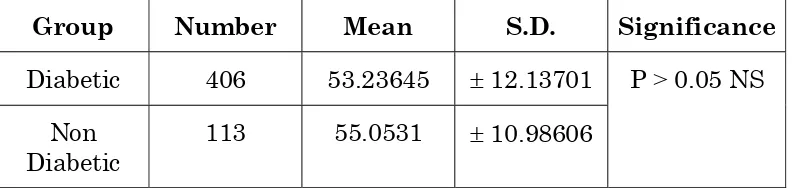
TABLE – 2
MEAN AND STANDARD DEVIATION
Group Number Mean S.D. Significance
Diabetic 406 53.23645 ± 12.13701
Non
Diabetic 113 55.0531 ± 10.98606
[image:41.612.117.514.623.717.2]0 5 10 15 20 25 30 35
P
e
rc
ent
age
£ 30 31-40 41-50 51-60 61-70 > 70
DISTRIBUTION ACCORDING TO AGE
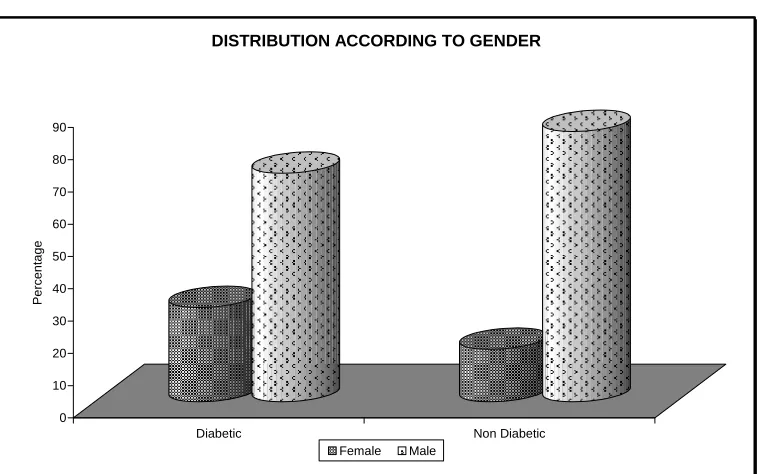
TABLE - 3
DISTRIBUTION ACCORDING TO GENDER
Diabetic Non Diabetic
Sex
No. Percentage No. Percentage
Female 33 29.20 66 16.25
Male 80 70.80 340 83.75
Total 113 100% 406 100%
0 10 20 30 40 50 60 70 80 90
Pe
rcen
ta
ge
Diabetic Non Diabetic
DISTRIBUTION ACCORDING TO GENDER
TABLE - 4
DISTRIBUTION ACCORDING TO OCCUPATION
Diabetic Non Diabetic
Occupation
No. Percentage No. Percentage
Manual
Labour 65 57.52 295 72.60
Sedentary 48 42.48 111 27.40
Total 113 100% 406 100%
0 10 20 30 40 50 60 70 80
Pe
rc
en
ta
ge
Diabetic Non Diabetic
DISTRIBUTION ACCORDING TO OCCUPATION
TABLE - 5
DISTRIBUTION ACCORDING TO THE PRESENCE OR
ABSENCE OF CHEST PAIN
Chest Pain Group
Present Absent
Total
Diabetic 90 23 113
Non Diabetic 392 14 406
χ2 = 38.08
P value < 0.001 (Significant)
Odds Ratio : 7.16
Non chest pain presentation was 7.16 times more common in
TABLE - 6
DISTRIBUTION ACCORDING TO THE DURATION OF DIABETICS
Duration Number Percentage
< 5 years 70 61.95
5-10 41 36.28
> 10 years 2 1.77
Total 113 100%
DISTRIBUTION ACCORDING TO THE DURATION OF DIABETICS
< 5 years 62% 10-May
36%
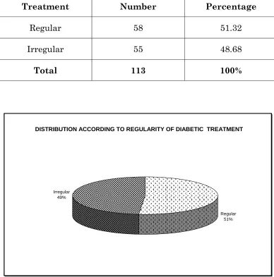
TABLE - 7
DISTRIBUTION ACCORDING TO REGULARITY OF DIABETIC TREATMENT
Treatment Number Percentage
Regular 58 51.32
Irregular 55 48.68
Total 113 100%
DISTRIBUTION ACCORDING TO REGULARITY OF DIABETIC TREATMENT
Regular 51% Irregular
TABLE - 8
MEAN AND STANDARD DEVIATION OF FASTING AND
RANDOM BLOOD SUGARS IN DIABETIC PATIENTS
Blood Sugar Mean SD
Fasting 175.61 ± 54.05
TABLE - 9
DISTRIBUTION ACCORDING TO KILLIP
CLASSIFICATION
Killip I II III IV Total
Diabetic 66 36 3 8 406
Non
Diabetic
371 19 2 14 113
χ2 = 80.07
P value <0.001 significant
Diabetic patients showed higher Killip class when compared
TABLE - 10
DISTRIBUTION ACCORDING TO THE WALL INVOLVED IN
MYOCARDIAL INFARCTION
Wall Diabetic Non Diabetic
AWMI 96 243
Others 17 163
Total 113 406
χ2 = 24.54
P value < 0.001 significant
Odds ratio : 3.79
Diabetic patients had 3.79 times anterior wall myocardial
infarction compared to non diabetic patients.
0 50 100 150 200 250
Nu
m
b
e
rs
Diabetic Non Diabetic
DISTRIBUTION ACCORDING TO THE WALL INVOLVED IN MYOCARDIAL INFARCTION
TABLE - 11
DISTRIBUTION ACCORDING TO EJECTION FRACTION
Diabetic Non Diabetic
Ejection
Fraction No. Percentage No. Percentage
≤ 40 9 8.82 2 0.51
41-50 44 43.14 23 5.88
≥ 51 49 48.04 366 93.61
Total 102 100% 406 100%
0 10 20 30 40 50 60 70 80 90 100
P
e
rc
ent
a
g
e
< 40 41-50 > 51
DISTRIBUTION ACCORDING TO EJECTION FRACTION
TABLE - 12
DISTRIBUTION ACCORDING TO THE PRESENCE OR
ABSENCE OF CARDIAC FAILURE
Congestive Cardiac failure Group
Present Absent Total
Diabetic 44 69 113
Non Diabetic 40 366 406
χ2 = 55.02
P value < 0.001 significant
Odds ratio : 5.83
Cardiac failure was 5.83 times more common in diabetics
compared to non diabetics.
0 50 100 150 200 250 300 350 400
Nu
mbe
r
Present Absent
DISTRIBUTION ACCORDING TO THE PRESENCE OR ABSENCE OF CARDIAC FAILURE
TABLE - 13
DISTRIBUTION ACCORDING TO THE PRESENCE OR
ABSENCE OF VENTRICULAR ARRYTHMIAS
Ventricular arrhythmias Group
Present Absent Total
Diabetic 7 106 113
Non Diabetic 6 400 406
χ2 = 8.04
P value = 0.0045 significant
Odds ratio : 4.40
Ventricular arrhythmias were 4.4 times more common in
diabetics compared to non diabetics.
0 50 100 150 200 250 300 350 400 Nu m b e rs Present Absent
DISTRIBUTION ACCORDING TO THE PRESENCE OR ABSENCE OF VENTRICULAR ARRYTHMIAS
TABLE - 14
DISTRIBUTION ACCORDING TO THE PRESENCE OR
ABSENCE OF COMPLETE HEART BLOCK
Complete Heart Block Group
Present Absent Total
Diabetic 8 105 113
Non Diabetic 2 404 406
An expected cell value is less than 5.
Fishers exact 2 tailed test used.
P = 0.00012 significant.
Complete Heart blocks were more common in diabetics when
compared with non diabetics in myocardial infarction.
0 50 100 150 200 250 300 350 400 450
Nu
mbe
rs
Present Absent
DISTRIBUTION ACCORDING TO THE PRESENCE OR ABSENCE OF COMPLETE HEART BLOCK
TABLE - 15
DISTRIBUTION ACCORDING TO MORTALITY
Group Dead Alive Total
Diabetic 17 96 113
Non Diabetic 20 38.6 406
χ2 = 13.67
P value < 0.001 significant
Odds ratio : 3.42
Mortality was 3.42 times more common in the diabetic
population compared to non diabetic.
0 10 20 30 40 50 60 70 80 90 100
Nu
mbe
rs
Dead Alive
DISTRIBUTION ACCORDING TO MORTALITY
TABLE - 16
DISTRIBUTION ACCORDING TO THROMBOLYSIS STATUS
Thrombolysis Group
Yes No Total
Diabetic 100 13 113
Non Diabetic 380 26 406
P value = 0.069 Not significant
0 50 100 150 200 250 300 350 400
Numb
er
s
Yes No
DISTRIBUTION ACCORDING TO THROMBOLYSIS STATUS
DISCUSSION
Out of the 519 cases of myocardial infarction in this study,
113 cases were diabetics and 406 cases were non diabetics.
There were 7 cases of myocardial infarction under the age of
30 years in the non diabetic group compared to none in the diabetic
group.
Between 31 to 50 years the incidence of myocardial infarction
was more common in the non diabetic group compared to diabetic
group.
Above the age of 50 years the incidence of myocardial
infarction was higher in the diabetic population. Even though there
was a slight increase in the age of occurrence of myocardial
infarction among the diabetic patients compared to nondiabetics, it
was not statically significant.
Percentage of female patients with myocardial infarction in
the diabetic group was 29.20% when compared to 16.25% in non
diabetic group. This is well in correlation with study of Stone P H
et al (14).
42.8% of the patients in the diabetic group had sedentary life
It was found that the incidence of painless myocardial
infarction were 7.16 times more common in the diabetic group
compared to non diabetics (P<0.001).
Most of the patients in the diabetic group were having a
duration of diabetes less than five years and only 50 percentage of
these diabetics were under regular treatment.
Diabetic group showed higher Killip class when compared to
non diabetic group. The incidence of congestive cardiac failure was
5.83 times to that of non diabetics (P<0.001). The incidence of
cardiogenic shock was also higher in the diabetic group. The
ejection fraction in diabetic group was very low compared to non
diabetics. 8.82% of diabetics had an ejection fraction less than 40%
when compared to only 0.51% in non diabetics.
Diabetic patients showed a higher incidence of anterior wall
myocardial infarction compared to non diabetics. They had 3.79
times more chance to have anterior wall myocardial infarction
compared to non diabetics (P<0.001).
The presence of complete heart blocks and life threatening
ventricular arrhythmias were also higher in diabetics. The
incidence of ventricular arrhythmias in diabetic group was 4.40
times that of non diabetic group.
The mortality due to myocardial infarction among diabetic
CONCLUSION
9 In this study, there was no significant difference in the age
of occurrence of myocardial infarction among diabetic and nondiabetic group.
9 Women with diabetes lost most of the inherent protection
against coronary artery disease when compared to non diabetics.
9 Painless myocardial infarctions were far more common in
diabetics compared to nondiabetics.
9 Anterior wall myocardial infarctions were common and
ejection fractions were consistently lower among diabetics.
9 Congestive cardiac failure and cardiogenic shock were more
common and severe in subjects with diabetes than non diabetics. This was more than to be expected from the size of the infarction.
9 Complications of myocardial infarction like life threatening
ventricular arrhythmias and complete heart blocks were far more common among diabetics compared to non diabetics.
9 In hospital mortality due to myocardial infarction in
SUMMARY
The present study was conducted over a period two years
among patients admitted with ST elevation myocardial infarction
in the cardiology department of Kilpauk Medical College. Patients
were divided into diabetic and non diabetic groups. With
application of inclusion as well as exclusion criteria 113 diabetics
and 406 nondiabetics were chosen for the study. Findings of the
study are as follows:
1. There was no significant difference in the age of occurrence of
myocardial infarction among diabetic and non diabetic group
(p value > 0.05).
2. The percentage of females in diabetic group were far more
compared to non diabetic group. (diabetic =29.20%, non
diabetic =16.25%).
3. The diabetic group had more sedentary life style compared to
non diabetics. (diabetic =42.48%, non diabetic =27.40% )
4. Painless myocardial infarctions were 7 times more common
in diabetic group. (odds ratio = 7.16 ) (P value < 0.001).
5. Most of the patients in the diabetic group were detected to
6. Diabetic patients with myocardial infarction showed a
preponderance to anterior wall compared to non diabetics.
(Odds ratio = 3.79).
7. Cardiac failure was about six times more common in the
diabetic group (Odds ratio = 5.83) (P<0.001).
8. Complications of myocardial infarctions were also common in
diabetic group (ventricular arrhythmia- odds ratio = 4.40,
complete heart block) (P<0.001).
9. Mortality due to myocardial infarction in diabetic group was
about four times that of non diabetic group. (odds ratio =
3.42)
10. No significant time difference in receiving thrombolysis was
BIBLIOGRAPHY
1. Vergely P. Del’ angine de poitrine dans ses rapports ave le
diabete Gaz Hebd Med Chir (Paris) (Series 2) 1883 ; 20 :
364-368.
2. Garcia MJ, McNamara PM, Gordon T, Kannel WB. Morbidity
and mortality in diabetics in the Framingham population:
sixteen year follow-up study. Diabetes 1974; 23 : 105–11.
3. Malmberg K, Ryden L. Myocardial infarction in patients with
diabetes mellitus. Eur Heart J 1988; 9 : 259–64.
4. Abbud ZA, Shindler DM, Wilson AC, Kostis JB. Effect of
diabetes mellitus on short- and long-term mortality rates of
patients with acute myocardial infarction: a statewide study.
Am Heart J 1995; 130 : 51– 8.
5. Partamian JO, Bradley RF. Acute myocardial infarction in
258 cases of diabetes: immediate and five-year survival. N
Engl J Med 1965; 273 : 456–65.
6. Czyzk A, Krolewski AS, Szablowska S, Alot A, Kopczynski J.
Clinical course of myocardial infarction among diabetic
patients. Diabetes Care 1980; 3 : 526–9.
7. Martin CA, Thompson PL, Armstrong BK, Hobbs MS, de
Klerk N.Long-term prognosis after recovery from myocardial
infarction: a nine year follow-up of the Perth Coronary
8. Smith JW, Marcus FI, Serokman R. Prognosis of patients
with diabetes mellitus after acute myocardial infarction. Am
J Cardiol 1984; 54 : 718 –21.
9. Jaffe AS, Spadaro JJ, Schechtman K, Roberts R, Geltman
EM, Sobel BE. Increased congestive heart failure after
myocardial infarction of modest extent in patients with
diabetes mellitus. Am Heart J 1984; 108 : 31–7.
10. Rytter L, Troelsen S, Beck-Nielsen H. Prevalence and
mortality of acute myocardial infarction in patients with
diabetes. Diabetes Care 1985; 8 : 230–4.
11. Molstad P, Nustad M. Acute myocardial infarction in diabetic
patients. Acta Med Scand 1987; 222 : 433–7.
12. Savage MP, Krolewski AS, Kenien GG, Lebeis MP, Christlieb
AR, Lewis SM. Acute myocardial infarction in diabetes
mellitus and significance of congestive heart failure as a
prognostic factor. Am J Cardiol 1988; 62 : 665–9.
13. Singer DE, Moulton AW, Nathan DM. Diabetic myocardial
infarction: interaction with other preinfarction risk factors.
Diabetes 1989; 38 : 350 –7.
14. Stone PH, Muller JE, Hartwell T, et al. The effect of diabetes
mellitus on prognosis and serial left ventricular function after
acute myocardial infarction: contribution of both coronary
artery disease and diastolic left ventricular dysfunction to the
15. Granger CB, Califf RM, Young S, et al. Outcome of patients
with diabetes mellitus and acute myocardial infarction
treated with thrombolytic agents. J Am Coll Cardiol 1993;
21:920 –5.
16. Barbash GI, White HD, Modan M, van de Werf F, and the
Investigators of the International Tissue Plasminogen /
Streptokinase Mortality Trial. Significance of diabetes
mellitus in patients with acute myocardial infarction
receiving thrombolytic agents. J Am Coll Cardiol 1993; 22 :
707–13.
17. Zuanetti G, Latini R, Maggioni AP, Santoro L, Franzosi MG,
on behalf of GISSI-2 Investigators. Influence of diabetes on
mortality in acute myocardial infarction: data from the
GISSI-2 study. J Am Coll Cardiol 1993; 22 : 1788 –94.
18. Soler NG, Bennett MA, Lamb P, Pentecost BL, Fitzgerald
MG, Malins JM. Coronary care for myocardial infarction in
diabetics. Lancet 1974; 1 : 475–7.
19. Lomuscio A, Vergani D, Marano L, Castagnone M, Fiorentini
C. Effects of glibenclamide on ventricular fibrillation in non–
insulin-dependent diabetics with acute myocardial infarction.
Coron Artery Dis 1994; 5 : 767–71.
20. Luepker RV, Apple FS, Christenson RH, et al : Case
definitions for acute coronary disease in epidemiology and
21. Aplert, JS, Thygesen K. Antman E. et al : Myocardial
infarction redefined : A consensus document of The Joint
European Society of Cardiology / American College of
Cardiology Committee for the redefinition of myocardial
infarction. J Am Coll Cardiol 36 : 959, 2000.
22. Newby LK, Alpert JS, Ohman EM et al : Changing the
diagnosis of acute myocardial infarction : Implications for
practice and clinical investigators. Am Heart J 144 : 957,
2002.
23. White HD : Things ain’t what they used to be : Impact of a
new definition of myocardial infarction. Am Heart J 144 :
933, 2002.
24. Fuster V. Corti R, Fayad ZA, et al: Integration of vascular
biology and magnetic resonance imaging in the
understanding of atherothrombosis and acute coronary
syndromes. J Thromb Haemost 1:1410,2003.
25. Libby P: Current concepts of the pathogenesis of the acute
coronary syndromes. Circulation 104:365,2001.
26. Ardissino D, Merlini PA, Ariens R. et al: Tissue – factor
antigen and activity in human coronary atherosclerotic
plaques. Lancet 349-769, 1997.
27. Malek AM, Alper SL, Izumo S: Hemodynamic shear stress
and its role in atherosclerosis. JAMA 282:2035, 1999.
28. Kloner RA, Leor J: Natural disaster plus wake-up time .A
29. Barrett-Connor EL, Cohn BA, Wingard DC, et al. Why is
diabetes mellitus a stronger risk factor for fatal ischemic
heart disease in women than in men? The Rancho Bernardo
Study (published erratum appears in JAMA 1991; 265 :
3249). JAMA 1991; 265 ; 627-631.
30. Gu K, Cowie CC, Harris MI. Mortality in adults with and
without diabetes in a national cohort of the U.S. population,
1971-1993. Diabetes Care 1998 ; 21 : 1138-145.
31. Krolewski, AS, Kosinski EJ, Warram JH, et al. Magnitude
and determinants of coronary artery disease in
juvenile-onset, insulin-dependent diabetes mellitus, Am J Cardiol
1987 : 750-755.
32. Krolewski, AS, Warram JH, Rand LI, et al. Epidemiologic
approach to the etiology of type I diabetes mellitus and its
complications. N Engl J Med 1987; 317 : 1390-1398.
33. Bale GS, Entmacher PS. Estimated life expectancy of
diabetics. Diabetes 1877; 26: 434-438.
34. Haffner SM, Lehtol S, Ronnemaa T, et al. Mortality from
coronary heart disease in subjects with type 2 diabetes and in
nondiabetic subjects with and without prior myocardial
infarction. N Engl J Med 1998; 339:229 – 234.
35. Mukamal KJ, Nesto RW, Cohen MC, et al. Impact of diabetes
on long-term survival after acute myocardial infarction:
comparability or risk with prior myocardial infarction.
36. Malmberg K, Kusuf S, Gerstein HC, et al. Impact of diabetes
on long-term prognosis in patients with unstable angina and
non-Qwave myocardial infarction: results of the OASIS
(Organization to Assess Strategies for Ischemic Syndromes)
Registry. Circulation2000; 102: 1014- 1019.
37. Gu K, Cowie Cc, Harris MI. Diabetes and decline in heart
disease mortality in U.S adults. JAMA 1999; 281:1291-1297.
38. Donahue RP, Orchard, TJ. Diabetes mellitus and
macrovascular. complications. An epidemiological
perspective. Diabetes Care 1992; 15 : 1141-1155.
39. Maser RE, Wolfson SK, Jr, Ellis D, et al. Cardiovascular
disease and arterial calcification in insulin-dependent
diabetes mellitus: interrelations and risk factor profiles.
Pittsburgh Epidemiology of Diabetes Complications Study. V.
Arterioseler Thromb 1991; 11: 958-965.
40. Jensen T, Borch-Johnsen K, Kofoed-Enevoldsen A, et al.
Coronary heart disease in young type 1 (insulin-dependent)
diabetic patients with and without diabetic nephropathy;
incidence and risk factors. Diabetologin 1987; 30 : 144-148.
41. Kannel WB, McGee DL. Diabetes and cardiovascular disease.
The Framingham Study, JAMA 1979; 241 : 2035-2038.
42. Jarrett RJ, McCartney P,Keen H. The Bedford survey : ten
year mortality rates in newly diagnosed diabetics, borderline
coronary heart disease in borderline diabetics. Diabetologia
1982 ; 22 : 79-84.
43. Jarrett RJ, Shipley MJ. Type 2
(non-insulin-dependent)diabetes mellitus and cardiovascular disease –
putative association via common antecedents; further
evidence from the Whitehall study. Diabetologia 1988 ; 31 :
737-740.
44. Fontbonne A, Eschwege E, Cambient F., et al.
Hypertriglycerdaemia as a risk factor of coronary heart
disease mortality in subjects with impaired glucose tolerance
or diabetes. Results from the 11-year follow-up of the Paris
Prospective Study. Diabetologia 1989; 32 : 300-304.
45. Nathan DM. Long-term complications of diabetes mellitus. N
Engl J Med 1993; 328 : 1676-1685.
46. Barrett-Connor E, Wingard DL. Sex differential in ischemic
heart disease mortality in diabetics : a prospective population
– based study. Am J Epidemiol 1983; 118 : 489-496.
47. Reaven G. Syndrome X : 10 yers after. Drugs 1999; 58
(Suppl (1) : 19-20.
48. Executive Summary of the Third Report of the National
Cholesterol Education Program (NCEP). Expert Panel on
Detection, Evaluation and Treatment of High Blood
Cholesterol in Adults (Adults Treatment Panel III). JAMA