JOURNAL OFVIROLOGY, 0022-538X/97/$04.0010
Sept. 1997, p. 6823–6833 Vol. 71, No. 9
Copyright © 1997, American Society for Microbiology
Cloning of Adeno-Associated Virus Type 4 (AAV4) and
Generation of Recombinant AAV4 Particles
JOHN A. CHIORINI, LINDA YANG, YUEJIANG LIU, BRIAN SAFER,ANDROBERT M. KOTIN*
Molecular Hematology Branch, National Heart Lung and Blood Institute, Bethesda, Maryland 20892
Received 20 February 1997/Accepted 23 May 1997
We have cloned and characterized the full-length genome of adeno-associated virus type 4 (AAV4). The genome of AAV4 is 4,767 nucleotides in length and contains an expanded p5 promoter region compared to AAV2 and AAV3. Within the inverted terminal repeat (ITR), several base changes were identified with respect to AAV2. However, these changes did not affect the ability of this region to fold into a hairpin structure. Within the ITR, the terminal resolution site and Rep binding sites were conserved; however, the Rep binding site was expanded from three GAGC repeats to four. The Rep gene product of AAV4 shows greater than 90% homology to the Rep products of serotypes 2 and 3, with none of the changes occurring in regions which had previously been shown to affect the known functions of Rep68 or Rep78. Most of the differences in the capsid proteins lie in regions which are thought to be on the exterior surface of the viral capsid. It is these unique regions which are most likely to be responsible for the lack of cross-reacting antibodies and the altered tissue tropism
compared to AAV2. The results of our studies, performed with a recombinant version of AAV4 carrying alacZ
reporter gene, suggest that AAV4 can transduce human, monkey, and rat cells. Furthermore, comparison of transduction efficiencies in a number of cell lines, competition cotransduction experiments, and the effect of trypsin on transduction efficiency all suggest that the cellular receptor for AAV4 is distinct from that of AAV2.
Adeno-associated virus (AAV) is a small nonpathogenic vi-rus of the Parvovirdaefamily (for a review, see reference 6). AAV is distinct from the other members of this family by its dependence upon a helper virus for replication. To date, five distinct AAV serotypes have been isolated from humans or primates (1). The most extensively studied of these isolates is AAV type 2 (AAV2). The genome of AAV2 is 4,680 nucleo-tides in length and consists of single-stranded DNA of either plus or minus polarity (41, 59). Two large open reading frames (ORFs) have been identified within the genome. The left ORF encodes the nonstructural Rep proteins which are involved in the regulation of replication and transcription and are neces-sary for the production of single-stranded progeny genomes (13–16, 19, 23, 28, 30, 34, 38–40, 43, 54, 57, 60–62). Further-more, two of the Rep proteins have been associated with the preferential integration of AAV genomes into a region of the q arm of human chromosome 19 (37, 56, 64). Rep68/78 have also been shown to possess nucleoside triphosphate binding activity as well as DNA and RNA helicase activities (31, 32, 36, 68). The Rep proteins possess a nuclear localization signal (36, 70), as well as several potential phosphorylation sites. Muta-tion of one of these kinase sites resulted in a loss of replicaMuta-tion activity (36).
The ends of the genome are short inverted terminal repeats (ITR) which have the potential to fold into T-shaped hairpin structures that serve as the origin of viral DNA replication. Within the ITR region, two elements have been described which are central to the function of the ITR, a GAGC repeat motif and the terminal resolution site (TRS). The repeat motif has been shown to bind Rep when the ITR is in either a linear or a hairpin conformation (15, 16, 44). This binding serves to position Rep68/78 for cleavage at the TRS, which occurs in a site- and strand-specific manner. In addition to their role in
replication, these two elements appear to be central to viral integration. Contained within the chromosome 19 integration locus is a Rep binding site with an adjacent TRS (64, 66). These elements are functional and necessary for locus-specific integration (26).
The AAV2 virion is a nonenveloped icosohedral particle approximately 25 nm in diameter, consisting of three related proteins referred to as VP1, VP2, and VP3. These proteins are found in a ratio of 1:1:10 (10) and are all derived from the right-hand ORF. The capsid proteins differ from each other by the use of alternative splicing and an unusual start codon. Deletion analysis has shown that removal or alteration of VP1 which is translated from an alternatively spliced mRNA results in a reduced yield of infections particles (28, 60). Mutations within the VP3 coding region result in the failure to produce any single-stranded progeny DNA or infectious particles (28, 60).
The following features of AAV have made it an attractive vector for gene transfer (29). AAV vectors have been shown in vitro to integrate stably into the cellular genome, possess a broad host range, transduce both dividing and nondividing cells in vitro and in vivo (24, 35, 48, 52), and maintain high levels of expression of the transduced genes (65). Viral parti-cles are heat stable; are resistant to solvents, detergents, changes in pH, and elevated temperature; and can be concen-trated on CsCl gradients (1, 2). Integration of AAV provirus is not associated with any long-term negative effects on cell growth or differentiation (3, 67). The ITRs are the only cis
elements required for rescue, replication, packaging, and inte-gration (55) and may contain some promoter activities (25).
While other serotypes of AAV have been isolated from both humans and monkeys, antibodies and viral particles to AAV4 have been identified only in monkeys (7, 20). DNA hybridiza-tion studies have indicated a similar level of homology for AAV1 through AAV4 (4, 50). However, mRNA hybridization studies suggest that AAV4 may be organized differently from AAV2, since only one size class of mRNA has been fraction-ated from infectious cells (46). Therefore, to understand the * Corresponding author. Mailing address: NIH/NHLBI/MHB, Bldg.
10/7D18, 10 Center Dr. MSC 1654, Bethesda, MD 20892-1654. Phone: (301) 496-1594. Fax: (301) 496-9985.
6823
on November 9, 2019 by guest
http://jvi.asm.org/
nature of AAV4 and to determine its usefulness as a vector for gene transfer, the viral genome was cloned and sequenced and recombinant virus was produced with the AAV4 Rep and Cap proteins.
MATERIALS AND METHODS
Cell culture and virus propagation.Cos and HeLa cells were maintained as monolayer cultures in D10 medium (Dulbecco’s modified Eagle’s medium
con-taining 10% fetal calf serum, 100 U of penicillin per ml, 100mg of streptomycin
per ml, and 13amphotericin B as recommended by the manufacturer [GIBCO,
Gaithersburg, Md.]). All other cell types were grown under standard conditions which have been previously reported. AAV4 stocks were obtained from the American Type Culture Collection (no. VR-646).
Recombinant virus was produced and subjected to titer determination as previously described (17). The helper plasmid for production of the recombinant virus has been modified to remove any homologous sequence between the helper and vector plasmids. This step was taken to minimize the potential for wild-type (wt) particle formation by homologous recombination.
DNA cloning and sequencing.The DNA sequence was determined by using an ABI 373A automated sequencer and the FS dye terminator chemistry. Both strands of the plasmids were sequenced, and the sequence was confirmed by sequencing of a second clone. As further confirmation of the authenticity of the sequence, bases 91 to 600 were PCR amplified from the original seed material and directly sequenced. The sequence of this region, which contains a 56-base insertion compared to AAV2 and AAV3, was identical to that derived from the cloned material. The ITR was cloned with Deep Vent Polymerase (New England Biolabs), as specified by the manufacturer, and the following primers: primer 1,
59TCTAGTCTAGACTTGGCCACTCCCTCTCTGCGCGC; primer 2, 59AGG
CCTTAAGAGCAGTCGTCCACCACCTTGTTCC. The cycling conditions were 97°C for 20 s, 65°C for 30 s, and 75°C for 1 min for 35 rounds. Following the
PCR, the mixture was treated withXbaI andEcoRI and the amplified band was
purified by agarose gel electrophoresis. The recovered DNA fragment was
li-gated into Bluescript SKII1(Stratagene) and transformed into competent Sure
strain bacteria (Stratagene). The helper plasmid (pSV40oriAAV4-2) used for the
production of recombinant virus, which contains therepandcapgenes of AAV4,
was produced by PCR withPfupolymerase (Stratagene) as specified by the
manufacturer. The amplified sequence, nucleotides 216 to 4440, was ligated into a plasmid that contains the simian virus 40 origin of replication described pre-viously (17). The cycling conditions were 95°C for 30 s, 55°C for 30 s, and 72°C for 3 min for 20 rounds. The final clone was confirmed by sequencing. The
b-galactosidase reporter vector has been described previously (17).
HA assays.Hemagglutination (HA) was measured essentially as described previously (33). Serial twofold dilutions of virus in EDTA-buffered saline were mixed with an equal volume of 0.4% human erythrocytes (RBC) (type O) in plastic U-bottom 96-well plates. The reaction was complete after a 2-h incuba-tion at 8°C. HA units (HAU) are defined as the reciprocal of the diluincuba-tion causing 50% HA.
Transduction of cells.Exponentially growing cells (23104
cells) were plated in each well of a 12-well plate and transduced with serial dilutions of virus in 200
ml of medium for 1 h. After this period, an additional 800ml of medium was
added and the mixture was incubated for 48 h. The cells were then fixed and
stained forb-galactosidase activity overnight with 5-bromo-4-chloro-3-indolyl-b
-D-galactopyranoside (X-Gal) (ICN Biomedicals) (71). No endogenousb
-galac-tosidase activity was visible after a 24-h incubation in X-Gal solution. Infectious titers were determined by counting the number of positive cells in the different dilutions with a calibrated microscope ocular (diameter, 3.1 mm) and then multiplying by the area of the well and the dilution of the virus. Titers were determined by counting the average number of cells in a minimum of 10 fields/ well.
Competition assay.Cos cells were plated at 23104
/well in 12-well plates 12
to 24 h prior to transduction. The cells were transduced with 0.53107
particles
of recombinant AAV2 (rAAV2) or rAAV4 (containing thelacZgene) in 200ml
of Dulbecco’s minimal essential medium and increasing amounts of rAAV2 containing the gene for the human coagulation factor IX. Prior to transduction, the CsCl was removed from the virus by dialysis against isotonic saline. After a 1-h incubation with the recombinant virus, the culture medium was supple-mented with complete medium and allowed to incubate for 48 to 60 h. The cells were then stained and counted as described above.
Trypsinization of cells.An 80% confluent monolayer of 107
Cos cells was treated with 0.05% trypsin–0.02% EDTA solution (Biofluids) for 3 to 5 min at 37°C. Following detachment, the trypsin was inactivated by the addition of an
equal volume of medium containing 10% fetal calf serum. The cells were then
further diluted to a final concentration of 104
/ml. Then 1 ml of cells was plated in a 12-well dish and incubated with virus at a multiplicity of infection of 260 for 1 to 2 h. Following attachment of the cells, the medium containing the virus was removed, the cells were washed, and fresh medium was added. Control cells were plated at the same time but were not transduced until the next day. The trans-duction conditions were as described above for the trypsinized cell group. The number of transduced cells was determined by staining 48 to 60 h posttransduc-tion and counting as described above.
Nucleotide sequence accession number.The viral nucleotide sequence deter-mined in this study is available through GenBank under accession no. U89790.
RESULTS
To obtain AAV4 genomic DNA for cloning, a stock of AAV4 was amplified by coinfection with wt adenovirus type 5 in Cos cells and the lysate was used to infect HeLa S3 cells. The resulting viral particles were isolated by CsCl banding and assayed by dot blot hybridization with an AAV2 DNA probe. The DNA dot blots of the gradient fractions indicated that peak genomes were contained in fractions with a density of 1.41 to 1.45 g/cm3. This is very similar to the buoyant density previously reported for AAV4 of 1.44 g/cm3(47). Analysis of annealed DNA obtained from these fractions indicated a ma-jor species of 4.8 kb which upon restriction analysis gave bands similar in size to those previously reported (46) (data not shown).
Additional restriction analysis indicated the presence of
BssHII sites near the ends of the DNA. Digestion withBssHII yielded a 4.5-kb fragment which was then cloned into Blue-script SKII1(Stratagene), and two independent clones were sequenced. The entire AAV4 genome sequence is presented in Fig. 1 and is also available through GenBank. In addition to the previously identified capsid ORF on the right-hand side of the genome (46), a second ORF is present on the left-hand side. As with AAV2 and AAV3, there appears to be a single polyadenylation signal after the second ORF. The first ORF, which could encode a protein of 623 amino acids, has a high degree of homology to therepORF of AAV2 and AAV3. At the amino acid level, the ORF is 90 and 92% identical to those of AAV2 and AAV3, respectively, with approximately 5% of the changes being conservative (data not shown). None of the changes occurred in regions which had previously been shown to affect the known functions of Rep68 or Rep78. The region surrounding the p19 promoter was also conserved, suggesting that proteins corresponding to AAV2 Rep52 and Rep40 could be expressed.
Inspection of the p7 promoter region of AAV4 showed a high degree of conservation of known functional elements which have been identified within the p5 promoter of AAV2. Two YY1 binding sites at 260 and 11 and a TATA box at 230, which are important for regulation of gene expression, have previously been identified in AAV2 (11, 58). These sites are conserved between AAV2 and AAV4. A binding site for Rep has been identified in the AAV2 p5 promoter at217 and is also conserved in AAV4 (42). The only divergence between the two viruses in this region appears to be in the sequence surrounding these transcriptional regulatory elements. AAV4 contains an additional 56 bases inserted between the AAV2 YY1 element at260 and the adjacent E-box/USF element at
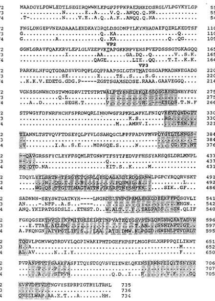
FIG. 1. Sequence of the AAV4 genome. The genomes of AAV2, AAV3, and AAV4 were aligned with the Align program (Scientific and Educational Software). The sequences of the ITRs are presented in lowercase type, and the rest of the genome is shown in capitals. TRSs are indicated by vertical arrows. Proposed transcription factor binding sites are indicated by underlining and overlining, with the name of the site above. E-box elements are indicated by highlighting, and the unique region of AAV4 in the p5 promoter is shown in boldface type. Promoters are indicated by horizontal arrows, with their corresponding map positions indicated above. Initiation and termination codons are boxed, as is the single polyadenylation site. Splice donor and acceptor sites are indicated by solid and open triangles, respectively.
on November 9, 2019 by guest
http://jvi.asm.org/
VOL. 71, 1997 CLONING OF AAV4 6825
on November 9, 2019 by guest
http://jvi.asm.org/
FIG. 1—Continued.
on November 9, 2019 by guest
http://jvi.asm.org/
FIG. 1—Continued.
VOL. 71, 1997 CLONING OF AAV4 6827
on November 9, 2019 by guest
http://jvi.asm.org/
277. Based on its position in the viral genome and efficient use of limited sequence, it is reasonable to expect that this se-quence may be involved in regulation of virus gene expression. Computer analysis of this region indicated the presence of an additional E-box element within this region. Comparison to
other E-box elements suggests that it may bind Myc with a higher affinity than it binds USF (27). In contrast to AAV3, the TATAA box present in AAV2 at nt 1823 in the P40 promoter is conserved in AAV4.
The ITRs were PCR amplified with a primer derived from the complementary sequence of the TRS contained within the
BssHII fragment and a primer in therepORF. The resulting fragments were cloned and found to contain a number of sequence changes compared to AAV2. However, all the changes were accompanied by complementary changes and thus would not affect the ability of this region to fold into a hairpin structure (Fig. 2). While the TRS motif was conserved between AAV2 and AAV4, the Rep binding site contained two alterations which expand the binding site from three (GAGC) repeats to four (16). Each of the first two repeats in AAV4 contain a T instead of a C in the fourth position. This type of repeat is present in the p5 promoter as well as in the consensus sequence which has been proposed for Rep binding (18). Gel shift experiments with random synthetic oligonucleotide probes and AAV2 Rep78 or Rep68 showed enrichment for probes containing multiple repeats of the GAGC motif. Thus, expansion of this element in the ITR of AAV4 should affect its affinity for Rep. Methylation interference data have suggested the importance of the CTTTG motif found at the tip of one palindrome in Rep binding, with the underlined T residues affecting Rep binding to both the flip and flop forms (53). While most of this motif is conserved in AAV4, the middle T residue is changed to a C and may indicate the importance of the structure of the palindrome and not the particular DNA sequence (8).
[image:6.612.63.294.65.238.2]In contrast to the high degree of homology between the Rep ORF of AAV2, AAV3, and AAV4, the capsid ORF of AAV4, which encodes a protein of 734 amino acids, is only 62 and 63% identical to the corrected AAV2 and AAV3 sequences, respec-tively (45, 51) (Fig. 3). Comparison to Moscovy duck and goose parvoviruses showed 53% homology, and little homology to other autonomous parvoviruses was observed. While the N terminus of VP1 (amino acids 44 to 133) and the start site of VP2 are conserved among AAV2, AAV3, and AAV4, the start site for VP3 is in the middle of a divergent region. By using the three-dimensional structural analysis of the canine parvovirus and computer-aided sequence comparisons (12), the regions of the AAV2 capsid that are predicted to be exposed on the exterior surface of the particle could be inferred. Comparison of the AAV2 and AAV4 sequences indicates that these regions are not well conserved between the viruses and suggests that
FIG. 2. AAV4 ITR. The sequence of the ITR is shown in the hairpin con-formation. The putative Rep binding site is boxed. The cleavage site in the TRS is indicated by an arrow. Bases which differ from the ITR of AAV2 are outlined.
[image:6.612.318.554.70.211.2]FIG. 3. Comparison of the capsid ORFs. The capsid ORFs of AAV2, AAV3, and AAV4 were compared by using the Palign program of Pcgene. Identical amino acids are indicated by a single dot. Dashes indicate gaps in the sequence added by the alignment program. The theoretical initiator codons of VP2 and VP3 are underlined and indicated above the sequence. Regions which have been proposed to be on the surface of AAV (12) are highlighted.
FIG. 4. HA. Human type O RBCs were incubated with twofold serial dilu-tions of viruses. Row 1, RBCs only; row 2, AAV3; row 3, wtAAV4; row 4, wtAAV4; row 5, rAAV4; row 6, rAAV2; row 7, adenovirus.
on November 9, 2019 by guest
http://jvi.asm.org/
[image:6.612.73.293.366.673.2]they may exhibit altered tissue tropism. Very little is known about the tissue tropism of any dependovirus. While AAV4 had been shown to hemagglutinate human, guinea pig, and sheep erythrocytes (33), it is thought to be exclusively a simian virus, since humans but not captive African green monkeys lack neutralizing antibodies (7). Additional work has involved the isolation of virus from the monkey kidney and intestine, with AAV4 antigen being detected in the kidneys, tonsils, lungs, liver, and blood following experimental infection (20). Therefore, to test AAV4 tissue tropism, we constructed
recom-binant AAV4 particles which contained the gene for nucleus-localizedb-galactosidase. Because of the structural similarities of the AAV2 and AAV4 ITRs, a genome containing AAV2 ITRs which had been previously described was used for pack-aging into AAV4 capsids.
Virus was produced as previously described for AAV2, using ab-galactosidase vector plasmid and a helper plasmid contain-ing the AAV4repandcapgenes (17). The helper plasmid was constructed to eliminate homologous sequence between the helper and vector plasmids. This step was taken to minimize the potential for wt particle formation by homologous recom-bination.
Virus was isolated from Cos cell lysates by CsCl banding, and the distribution oflacZsequences across the gradient was determined by DNA dot blot analysis of gradient fractions. The majority of packaged genomes were found in fractions with a density of 1.43 g/cm3, which is similar to that obtained for wt AAV4. Typical yields of rAAV4 particles per producer cell were three- to fivefold greater than those of rAAV2 par-ticles.
[image:7.612.65.556.68.372.2]To determine if recombinant AAV4 particles were capable of hemagglutination, purified recombinant AAV4 particles were incubated with human RBC (type O) for 2 to 3 h at 8°C. As shown in Fig. 4, both wt AAV4 and rAAV4 can hemagglu-tinate human RBCs with HA titers of approximately 1,024 and 512 HAU/ml, respectively. No HA activity was detected with AAV3, recombinant AAV2, or the helper adenovirus (Fig. 4). If the temperature was raised to 22°C, HA activity decreased 32-fold (data not shown). Comparison of the viral particle number per RBC at the end point dilution indicated that
FIG. 5. Transduction of Cos and SW480 cells. Cos and SW480 cells were transduced with either rAAV2 or rAAV4 expressinglacZat a multiplicity of infection of
500 and stained 60 h posttransduction. (A) Cos cells transduced with rAAV2. (B) Cos cells transduced with rAAV4. (C) SW480 cells transduced with rAAV2. (D) SW480 cells transduced with rAAV4.
TABLE 1. Titers of rAAV2LacZ and rAAV4LacZa
Cell type Titer of:
AAV2 AAV4
Primary rat brain 1 4.360.7
Cos 4.2310760.463107 2.2310760.253107
SW 480 7.75310661.73106 1.3310560.683105
HeLa 2.1310760.13107 1.3310660.13106
SW620 1.2310560.393105 43104
KLEB 1.2310560.353105 9310461.43104
HB 5.63105623105 3.8310461.83104
SW1116 5.23104 83103
SW1463 8.83104 83103
NIH 3T3 33103 23103
aThe different cell lines were transduced with an equal number of particles in
serial dilutions of either rAAV2 or rAAV4 expressinglacZas described in
Materials and Methods. The results are expressed as transducing units per milliliter. Because of the heterogeneous nature of the cell population in the rat brain cultures, only relative transduction efficiencies are reported.
VOL. 71, 1997 CLONING OF AAV4 6829
on November 9, 2019 by guest
http://jvi.asm.org/
[image:7.612.58.299.558.684.2]approximately 1 to 10 particles per RBC were required for HA. This value is similar to that previously reported (33).
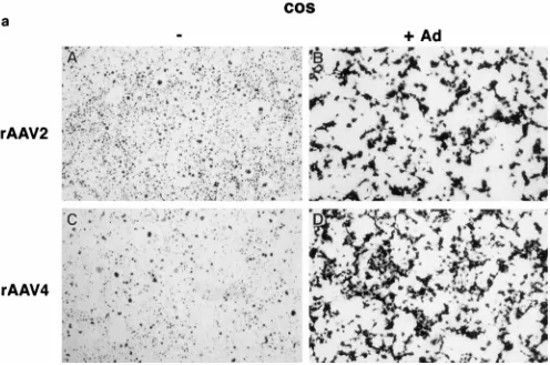
Tissue tropism was assessed by transduction of a variety of cell lines with serially diluted rAAV4 or rAAV2. As shown in Fig. 5 and Table 1, Cos cells transduced with equivalent amounts of rAAV2 and rAAV4 particles resulted in similar transduction levels (Fig. 5A and B). However, other cell lines exhibited differential transducibility. Transduction of the hu-man colon adenocarcinoma cell line SW480 with rAAV2 was 60 times higher than that obtained with rAAV4 (Fig. 5C and D). HeLa and SW620 cells were also transduced more effi-ciently with rAAV2 than with rAAV4. In contrast, tion of primary rat brain cultures exhibited a greater transduc-tion of glial and neuronal cells with rAAV4 than with rAAV2. Because of the heterogeneous nature of the cell population in the rat brain cultures, only relative transduction efficiencies are reported (Table 1). Previous work has shown that adenovirus can increase the transduction efficiency of AAV2 (21, 22). As a control for adenovirus contamination of the viral prepara-tions, Cos and HeLa cells were coinfected with rAAV and adenovirus and stained after 24 h. While the titer of rAAV2 increased in the presence of adenovirus in both Cos and HeLa cells (Fig. 6a and b, panels B), adenovirus increased the titer only in the Cos cells transduced with rAAV4 and not in the HeLa cells (Fig. 6a and b, panels D), suggesting that the dif-ference in transduction efficiencies is not the result of adeno-virus contamination. Furthermore, both vectors transduced SW1116, SW1463, NIH 3T3, and monkey fibroblast FL2 cells
very poorly. These data suggest that AAV4 may use a cellular receptor distinct from that used by AAV2.
This hypothesis was tested by cotransduction experiments with rAAV2 and rAAV4. Cos cells were transduced with an equal number of rAAV2 or rAAV4 particles containing the
lacZgene and increasing amounts of rAAV2 particles contain-ing the human coagulation factor IX gene (rAAV2FIX). At a 72:1 ratio of rAAV2FIX to rAAV4LacZ, only a twofold effect on the level of rAAV4LacZ transduction was obtained (Fig. 7). However, the same ratio of rAAV2FIX to rAAV2LacZ re-duced the transduction efficiency of rAAV2LacZ approxi-mately 10-fold. Comparison of the 50% inhibition points for the two viruses indicated a sevenfold difference in sensitivity. Previous research had shown that binding and infection of AAV2 are inhibited by trypsin treatment of cells (44). Trans-duction of Cos cells with the rAAV2LacZ gene was also in-hibited by trypsin treatment prior to transduction (Fig. 8). In contrast, trypsin treatment had a minimal effect on rAAV4LacZ transduction. This result and the result of the previous competition experiment are both consistent with the utilization of distinct cellular receptors for AAV2 and AAV4.
DISCUSSION
[image:8.612.60.556.69.398.2]AAV4 is a distinct virus based on sequence analysis, physical properties of the virion, HA activity, and tissue tropism. Sim-ilar to AAV2 and AAV3, AAV4 contains two ORFs which code for Rep proteins or capsid proteins. The genome of
FIG. 6. Adenovirus coinfection. (a) Cos cells were transduced with 43107
particles of either rAAV2 (A and B) or rAAV4 (C and D) in the presence (B and D)
or absence (A and C) of 23108
PFU of wt adenovirus. (b) HeLa cells were transduced with 43107
particles of either rAAV2 (A and B) or rAAV4 (C and D) in
the presence (B and D) or absence (A and C) of 23108
PFU of wt adenovirus.
on November 9, 2019 by guest
http://jvi.asm.org/
AAV4 is 4,767 nt in length and contains additional sequence upstream of the p7 promoter which may affect promoter ac-tivity. An additional E-box element is contained within this region; thus, AAV4 has two of these activator elements com-pared to the one of AAV2 and AAV3. Furthermore, this additional element differs in its flanking sequence from the
conserved E box and therefore is more similar to a Myc bind-ing site than to a USF site. It is interestbind-ing that the position of the new E box is still in the same place relative to the one in AAV2 and AAV3, suggesting the importance of this element in relation to the other promoter elements.
AAV4 contains an expanded Rep binding site within the
FIG. 7. Cotransduction of rAAV2 and rAAV4. Cos cells were transduced
with a constant amount of rAAV2 or rAAV4 expressingb-galactosidase and
[image:9.612.65.555.72.403.2]increasing amounts of rAAV2 expressing human factor IX (rAAV2FIX). For the competition, the number of positive cells detected in the cotransduced wells was divided by the number of positive cells in the control wells (cells transduced with only rAAV2LacZ or rAAV4LacZ) and expressed as a percentage of the control. This value was plotted against the number of particles of rAAV2FIX.
FIG. 8. Effect of trypsin treatment on Cos cell transduction. Cos cell mono-layers were trypsinized and diluted in complete medium. The cells were incu-bated with virus at a multiplicity of infection of 260, and the virus was removed following cell attachment. As a control, an equal number of Cos cells were plated and allowed to attach overnight before transduction with virus for the same time. The number of positive cells was determined by staining 50 h posttransduction. The data are presented as a ratio of the number of positive cells seen with the trypsinized group to the number seen with the control group.
FIG. 6—Continued.
VOL. 71, 1997 CLONING OF AAV4 6831
on November 9, 2019 by guest
http://jvi.asm.org/
[image:9.612.319.552.528.657.2]ITR. The Rep binding site consensus sequence suggests that multiple repeats of the GAGC motif create a higher-affinity binding site (18). This alteration could affect the ability of the ITR to function as an origin of replication or as a promoter or its ability to participate in Rep-mediated locus-specific integra-tion. However, the strong conservation of this element, as well as the TRS and Rep binding site in the p5 promoter, under-scores the importance of these elements in AAV biology.
The majority of the differences in the capsid proteins lie in regions which have been proposed to be on the exterior surface of the virus. These changes are most probably responsible for the lack of cross-reacting antibodies, the HA activity, and the altered tissue tropism compared to AAV2. However, not all of the changes are confined to the proposed exterior regions, and they may also be important for the other unique properties of AAV4 versus AAV2. The conservation of sequence in the N terminus of VP1 is in agreement with the results of mutational studies, which demonstrated that alteration of this region de-creases the yield of particles. Work by Prasad and Trempe has suggested that Rep may interact with this region and facilitate packaging (49).
The high degree of sequence conservation in the Rep ORF stresses the importance of this group of proteins in the life cycle of the virus. The data suggest that while the target cells of the two viruses may have diverged over time, the basic func-tions required for rescue, replication, and packaging carried out by the Rep proteins have been conserved. The high degree of homology among AAV4, AAV2, and AAV3 clearly places AAV4 in the family of dependoviruses, but it also possesses a high degree of homology to the goose and duck parvoviruses. The primate AAV Rep proteins are more than 90% homolo-gous but are only 40% homolohomolo-gous to the Rep proteins of the goose and duck parvoviruses. While AAV2, AAV3, and AAV5 have been isolated from humans, no clinical isolates of AAV4 have been reported. Our studies, using a recombinant version of the virus, suggest that it can transduce cells of human origin as well as those of monkey and rat origin. Furthermore, com-parison of transduction efficiencies in a number of cell lines suggests that the cellular receptor for AAV4 is distinct from that of AAV2. Cotransduction of Cos cells with either rAAV2 or rAAV4 expressinglacZand with rAAV2 expressing factor IX showed strong competition between the two rAAV2 vectors compared to that of rAAV4 and rAAV2 cotransduction. Fur-thermore, AAV2 transduction was shown to be more trypsin sensitive than that of AAV4. These results again suggest a distinct cellular receptor for AAV2 and AAV4. Unique recep-tors have also been suggested for AAV1 and AAV3 (44). While most of the autonomous parvoviruses have been shown to be hemagglutinating, AAV4 is the only dependovirus with this property. HA, which has been extensively studied with canine parvovirus, has been found to be independent of recep-tor binding and uptake (5, 63). However, in parvovirus B19, the P antigen which mediates HA is also thought to be the receptor for infectivity (9). By sequence comparisons, it is unclear to which of these two groups AAV4 belongs, and further exper-imental determination is required. The use of the ORFs of AAV4 to package a recombinant genome suggests that the cloned genome is functional. While the difference in transduc-tion efficiencies obtained with the cell lines emphasizes the individuality of this virus, the interchangeability of the ITRs and the conservation of the Rep ORF highlight the importance of these elements in viral replication and packaging.
ACKNOWLEDGMENTS
We thank Marion Taylor, Stephanie Harkin, Marzia Scortegagna, and Mike Erdos for providing different cell lines. We also thank Scott
Schors for help with the hemagglutination assay and Richard Smith for reviewing the manuscript and for helpful discussion.
J.A.C. is a Pharmacology Research Associate funded by the NIGMS.
REFERENCES
1.Arella, M., S. Garzon, J. Bergeron, and P. Tijssen.1990. Physicochemical
properties, production and purification of parvoviruses, p. 11–30.InP.
Ti-jssen (ed.), Handbook of parvoviruses. vol. 1. CRC Press, Inc., Boca Raton, Fla.
2.Bachmann, P. A., M. D. Hoggan, E. Kurstak, J. L. Melnick, H. G. Pereira, P. Tattersall, and C. Vago.1979. Parvoviridae: second report. Intervirology
11:248–254.
3.Bantel-Schaal, U., and M. Stohr.1992. Influence of adeno-associated virus
on adherence and growth properties of normal cells. J. Virol.66:773–779.
4.Bantel-Schall, U., and H. zur Hausen.1984. Characterization of the DNA of
a defective human parvovirus isolated from a genital site. Virology134:52–
63.
5.Basak, S., H. Turner, and S. Parr.1994. Identification of a 40- to 42-kDa
attachment polypeptide for canine parvovirus in A72 cells. Virology205:7–
16.
6.Berns, K. I.1995. Parvoviridae: the viruses and their replication, p. 1017–
1041.InB. N. Fields, D. M. Knipe, and P. M. Howley (ed.), Fundamental
virology, 3rd ed. Lippincott-Raven Publishers, Philadelphia, Pa.
7.Blacklow, N. R., M. D. Hoggan, and W. P. Rowe.1968. Serologic evidence for
human infection with adeno-associated viruses. J. Natl. Cancer Inst.40:319–
327.
8.Bohenzky, R. A., R. B. LeFebvre, and K. I. Berns.1988. Sequence and symmetry requirements within the internal palindromic sequences of the
adeno-associated virus terminal repeat. Virology166:316–327.
9.Brown, K. E., S. M. Anderson, and N. S. Young.1993. Erthrocyte P antigen:
cellular receptor for B19 parvovirus. Science262:114–117.
10. Buller, R. M. L., and J. A. Rose.1978. Characterization of
adenovirus-associated virus-induced polypeptides in KB cells. J. Virol.25:331–338.
11. Chang, L. S., Y. Shi, and T. Shenk.1989. Adeno-associated virus P5 pro-moter contains an adenovirus E1A-inducible element and a binding site for
the major late transcription factor. J. Virol.63:3479–3488.
12. Chapman, M. S., and M. G. Rossmann.1993. Structure, sequence and
function correlations among parvoviruses. Virology194:491–508.
13. Chejanovsky, N., and B. J. Carter.1989. Mutagenesis of an AUG codon in the adeno-associated virus rep gene: effects on viral DNA replication.
Vi-rology173:120–128.
14. Chejanovsky, N., and B. J. Carter.1989. Replication of a human parvovirus nonsense mutant in mammalian cells containing an inducible amber
sup-pressor. Virology171:239–247.
15. Chiorini, J. A., S. M. Wiener, R. M. Kotin, R. A. Owens, S. R. M. Kyo¨stio¨, and B. Safer.1994. Sequence requirements for stable binding and function of Rep68 on the adeno-associated virus type 2 inverted terminal repeats.
J. Virol.68:7448–7457.
16. Chiorini, J. A., M. D. Weitzman, R. A. Owens, E. Urcelay, B. Safer, and R. M. Kotin.1994. Biologically active Rep proteins of adeno-associated virus type
2 produced as fusion proteins inEscherichia coli. J. Virol.68:797–804.
17. Chiorini, J. A., C. M. Wendtner, E. Urcelay, B. Safer, M. Hallek, and R. M. Kotin.1995. High-efficiency transfer of the T cell co-stimulatory molecule B7-2 to lymphoid cells using high-titer recombinant adeno-associated virus
vectors. Hum. Gene Ther.6:1531–1541.
18. Chiorini, J. A., L. Yang, B. Safer, and R. M. Kotin.1995. Determination of adeno-associated virus Rep68 and Rep78 binding sites by random sequence
oligonucleotide selection. J. Virol.69:7334–7338.
19. Dixit, M., M. S. Webb, W. C. Smart, and S. Ohi.1991. Construction and expression of a recombinant adeno-associated virus that harbors a human
beta-globin-encoding cDNA. Gene104:253–257.
20. Dreizin, S., T. F. Zhuravel, A. B. Tarasova, S. G. Sobolev, G. Kozlov, and V. P. Grachev.1981. Experimental infection of green monkeys by
adeno-associated virus. Vorosy Virusol.1:82–89.
21. Ferrari, F. K., T. Samulski, T. Shenk, and R. J. Samulski.1996. Second-strand synthesis is a rate-limiting step for efficient transduction by
recombi-nant adeno-associated virus vectors. J. Virol.70:3227–3234.
22. Fisher, K. J., G.-P. Gao, M. D. Weitzman, R. DeMatteo, J. F. Burda, and J. M. Wilson.1996. Transduction with recombinant adeno-associated virus
for gene therapy is limited by leading-strand synthesis. J. Virol.70:520–532.
23. Fisher, R. E., and H. D. Mayor.1991. The evolution of defective and
au-tonomous parvoviruses. J. Theor. Biol.149:429–439.
24. Flotte, T. R., S. A. Afione, C. Conrad, S. A. McGrath, R. Solow, H. Oka, P. L. Zeitlin, W. B. Guggino, and B. J. Carter.1993. Stable in vivo expression of the cystic fibrosis transmembrane regulator with an adeno-associated virus
vector. Proc. Natl. Acad. Sci. USA90:10613–10617.
25. Flotte, T. R., S. A. Afione, R. Solow, M. L. Drumm, D. Markakis, W. B. Guggino, P. L. Zeitlin, and B. J. Carter.1993. Expression of the cystic fibrosis transmembrane conductance regulator from a novel
adeno-associ-ated virus promoter. J. Biol. Chem.268:3781–3790.
on November 9, 2019 by guest
http://jvi.asm.org/
26. Giraud, C., E. Winocour, and K. I. Berns.1994. Site-specific integration by adeno-associated virus is directed by a cellular DNA sequence. Proc. Natl.
Acad. Sci. USA91:10039–10043.
27. Grandori, C., J. Mac, F. Sie¨belt, D. E. Ayer, and R. N. Eisenman.1996. Myc-Max heterodimers activate a DEAD box gene and interact with
multi-ple E box-related sites in vivo. EMBO J.15:4344–4357.
28. Hermonat, P. L., M. A. Labow, R. Wright, K. I. Berns, and N. Muzyczka.
1984. Genetics of adeno-associated virus: isolation and preliminary
charac-terization of adeno-associated virus type 2 mutants. J. Virol.51:329–339.
29. Hermonat, P. L., and N. Muzyczka.1984. Use of adeno-associated virus as a mammalian DNA cloning vector: transduction of neomycin resistance into
mammalian tissue culture cells. Proc. Natl. Acad. Sci. USA81:6466–6470.
30. Hunter, L. A., and R. J. Samulski.1992. Colocalization of adeno-associated
virus Rep and capsid proteins in the nuclei of infected cells. J. Virol.66:
317–324.
31. Im, D.-S., and N. Muzyczka.1990. The AAV origin binding protein Rep68 is an ATP-dependent site-specific endonuclease with DNA helicase activity.
Cell61:447–457.
32. Im, D. S., and N. Muzyczka.1992. Partial purification of adeno-associated virus Rep78, Rep52, and Rep40 and their biochemical characterization.
J. Virol.66:1119–1128.
33. Ito, M., and H. D. Mayor.1968. Hemagglutinin of type 4 adeno-associated
satellite virus. J. Immunol.100:61–68.
34. Janik, J. E., M. M. Huston, K. Cho, and J. A. Rose.1989. Efficient synthesis of adeno-associated virus structural proteins requires both adenovirus DNA
binding protein and VA I RNA. Virology168:320–329.
35. Kaplitt, M. G., P. Leone, R. J. Samulski, X. Xiao, D. W. Pfaff, K. L. O’Malley, and J. M. During.1994. Long term gene expression and phenotypic correc-tion using adeno-associated virus vectors in the mammalian brain. Nat.
Genet.8:148–154.
36. Kleinschmidt, J. A., M. Mo¨hler, F. W. Weindler, and R. Heilbronn.1995. Sequence elements of the adeno-associated virus rep gene required for suppression of herpes-simplex-virus-induced DNA amplification. Virology
206:254–262.
37. Kotin, R. M., J. C. Menninger, D. C. Ward, and K. I. Berns.1991. Mapping and direct visualization of a region-specific viral DNA integration site on
chromosome 19q13-qter. Genomics10:831–834.
38. Kotin, R. M., M. Siniscalco, R. J. Samulski, X. Zhu, L. Hunter, C. A. Laughlin, S. McLaughlin, N. Muzyczka, M. Rocchi, and K. I. Berns.1990. Site-specific integration by adeno-associated virus. Proc. Natl. Acad. Sci.
USA87:2211–2215.
39. Laughlin, C. A., N. Jones, and B. J. Carter.1982. Effect of deletions in adenovirus early region 1 genes upon replication of adeno-associated virus.
J. Virol.41:868–876.
40. Laughlin, C. A., M. W. Myers, D. L. Risin, and B. J. Carter.1979. Defective-interfering particles of the human parvovirus adeno-associated virus.
Virol-ogy94:162–174.
41. Laughlin, C. A., J. D. Tratschin, H. Coon, and B. J. Carter.1983. Cloning of
infectious adeno-associated virus genomes in bacterial plasmids. Gene23:
65–73.
42. McCarty, D. M., J. Pereira, I. Zolotukhin, X. Zhou, J. H. Ryan, and N. Muzyczka.1994. Identification of linear DNA sequences that specifically
bind the adeno-associated virus Rep protein. J. Virol.68:4988–4997.
43. Mendelson, E., J. P. Trempe, and B. J. Carter.1986. Identification of the
trans-acting Rep proteins of adeno-associated virus by antibodies to a
syn-thetic oligopeptide. J. Virol.60:823–832.
44. Mizukami, H., N. S. Young, and K. E. Brown.1996. Adeno-associated virus
type 2 binds to a 150-kilodalton cell membrane glycoprotein. Virology217:
124–130.
45. Muramatsu, S. I., H. Mizukami, N. S. Young, and K. E. Brown.1996. Nucleotide sequence and generation of an infectious clone of
adeno-associ-ated virus 3. Virology221:208–217.
46. Muster, C. J., Y. S. Lee, J. E. Newbold, and J. Leis.1980. Physical mapping
of adeno-associated virus serotype 4 DNA. J. Virol.35:653–661.
47. Parks, W. P., J. L. Melnick, R. Rongey, and H. D. Mayor.1967. Physical assay
and growth cycle studies of a defective adeno-satellite virus. J. Virol.1:171–
180.
48. Podsakoff, G., K. K. Wong, Jr., and S. Chatterjee.1994. Efficient gene
transfer into nondividing cells by adeno-associated virus-based vectors. J.
Vi-rol.68:5656–5666.
49. Prasad, K.-M. R., and J. P. Trempe.1995. The adeno-associated virus Rep78 protein is covalently linked to viral DNA in a preformed virion. Virology
214:360–370.
50. Rose, J. A., M. D. Hoggan, F. Koczot, and A. J. Shatkin.1968. Genetic
relatedness studies with adenovirus-associated viruses. J. Virol.2:999–1005.
51. Ruffing, M., H. Heid, and J. A. Kleinschmidt.1994. Mutations in the carboxy terminus of adeno-associated virus 2 capsid proteins affect viral infectivity:lac
of an RGD integrin-binding motif. J. Gen. Virol.75:3385–3392.
52. Russell, D. W., A. D. Miller, and I. E. Alexander.1994. Adeno-associated virus vectors preferentially transduce cells in S phase. Proc. Natl. Acad. Sci.
USA91:8915–8919.
53. Ryan, J. H., S. Zolotukhin, and N. Muzyczka.1996. Sequence requirements for binding of Rep68 to the adeno-associated virus terminal repeats. J. Virol.
70:1542–1553.
54. Samulski, R. J., K. I. Berns, M. Tan, and N. Muzyczka.1982. Cloning of adeno-associated virus into pBR322: rescue of intact virus from the
recom-binant plasmid in human cells. Proc. Natl. Acad. Sci. USA79:2077–2081.
55. Samulski, R. J., L. S. Chang, and T. Shenk.1989. Helper-free stocks of recombinant adeno-associated viruses: normal integration does not require
viral gene expression. J. Virol.63:3822–3828.
56. Samulski, R. J., X. Zhu, X. Xiao, J. D. Brook, D. E. Housman, N. Epstein, and L. A. Hunter.1991. Targeted integration of adeno-associated virus
(AAV) into human chromosome 19 EMBO J.10:3941–3950. (Erratum,
11:1228, 1992.)
57. Senapathy, P., J. D. Tratschin, and B. J. Carter.1984. Replication of
adeno-associated virus DNA. Complementation of naturally occurring rep2
mu-tants by a wild-type genome or an ori2mutant and correction of terminal
palindrome deletions. J. Mol. Biol.179:1–20.
58. Shi, Y., E. Seto, L.-S. Chang, and T. Shenk.1991. Transcription repression
by YY1, a human GLI-Kru¨ppel-related protein, and relief of repression by
adenovirus E1a protein. Cell67:377–388.
59. Srivastava, A., E. W. Lusby, and K. I. Berns.1983. Nucleotide sequence and
organization of the adeno-associated virus 2 genome. J. Virol.45:555–564.
60. Tratschin, J.-D., I. L. Miller, and B. J. Carter.1984. Genetic analysis of adeno-associated virus: properties of deletion mutants constructed in vitro and evidence for an adeno-associated virus replication function. J. Virol.
51:611–619.
61. Trempe, J. P., and B. J. Carter.1988. Regulation of adeno-associated virus gene expression in 293 cells: control of mRNA abundance and translation.
J. Virol.62:68–74.
62. Trempe, J. P., E. Mendelson, and B. J. Carter.1987. Characterization of adeno-associated virus rep proteins in human cells by antibodies raised
against rep expressed in Escherichia coli. Virology161:18–28.
63. Tresnan, D. B., L. Southard, W. Weichert, J.-Y. Sgro, and C. R. Parrish.
1995. Analysis of the cell and erythrocyte binding activities of the dimple and
canyon regions of the canine parvovirus capsid. Virology211:123–132.
64. Urcelay, E., P. Ward, S. M. Wiener, B. Safer, and R. M. Kotin.1995. Asymmetric replication in vitro from a human sequence element is
depen-dent on adeno-associated virus Rep protein. J. Virol.69:2038–2046.
65. Walsh, C. E., J. M. Liu, X. Xiao, N. S. Young, A. W. Nienhuis, and R. J. Samulski.1992. Regulated high level expression of a human gamma-globin gene introduced into erythroid cells by an adeno-associated virus vector.
Proc. Natl. Acad. Sci. USA89:7257–7261.
66. Weitzman, M. D., S. R. M. Kyo¨stio¨, R. M. Kotin, and R. A. Owens.1994. Adeno-associated virus (AAV) Rep proteins mediate complex formation between AAV DNA and its integration site in human DNA. Proc. Natl.
Acad. Sci. USA91:5808–5812.
67. Winocour, E., M. F. Callaham, and E. Huberman.1988. Perturbation of the
cell cycle by adeno-associated virus. Virology167:393–399.
68. Wonderling, R. S., S. R. M. Kyo¨stio¨, and R. A. Owens.1995. A maltose-binding protein/adeno-associated virus rep68 fusion protein has DNA-RNA
helicase and ATPase activities. J. Virol.69:3542–3548.
69. Yakobson, B., T. Koch, and E. Winocour.1987. Replication of adeno-asso-ciated virus in synchronized cells without the addition of a helper virus.
J. Virol.61:972–981.
70. Yang, Q., A. Kadam, and J. P. Trempe.1992. Mutational analysis of the
adeno-associated virusrepgene. J. Virol.66:6058–6069.
VOL. 71, 1997 CLONING OF AAV4 6833