0022-538X/94/$04.00+0
Copyright C) 1994, American Society forMicrobiology
Requirement of
Human
Immunodeficiency
Virus
Type
1
nef
for
In
Vivo
Replication and
Pathogenicity
BETH D.JAMIESON,1 GRACEM. ALDROVANDI,1 VICENTE
PLANELLES,'
JEREMY B. M.JOWETT,'
LIANYING GAO,1 LILLIAN M.
BLOCH,'
IRVINS. Y.CHEN,2 ANDJEROMEA.ZACK`*
Division of Hematology-Oncology, Departmentof Medicine,1 and Department of Microbiology&Immunology,2 UCLA Schoolof Medicine andJonsson Comprehensive CancerCenter, LosAngeles, Califomia90024-1678
Received 17 December1993/Accepted 17 February 1994
The role of human immunodeficiency virus type 1 (HIV-1) accessorygenes inpathogenesis has remained unclear because ofthe lack of a suitable in vivo model. The most controversial of these genes is nef.We investigated the requirement for Nef for in vivo replication and pathogenicity of two isolates of HIV-1
(HIV-lJR-CSF
andHIV-1NtA-3)
in human fetalthymusand liverimplants in severe combinedimmunodeficient mice.HIV-IJR-CSF
andHIV-1NLA
3 differ in their in vitro phenotypes in thatHIV-lJR-CSF
does not induce syncytia andisrelatively noncytopathic,whileHIV-1NL4N3
ishighlycytopathicandreadilyinducessyncytia.The nefmutants of both isolates grew with kinetics similar to those of parental virus strains in stimulated peripheral blood lymphocytes but demonstrated attenuated growth properties in vivo.HIV-1NL4N3
induced severe depletion ofhumanthymocyteswithin 6 weeks ofinfection,whereas itsnefmutantdid not.Thus,HIV-1 Nef is required forefficient invivo viral replication andpathogenicity.The nef gene is conserved among primate lentiviruses and is amongthefirst viral genes transcribedfollowing infection (29). This would suggest a critical role for Nef in the virus life cycle, and therefore in the pathogenesis of lentiviral infections, yet the function of Nef has not been determined. While Nefwas originally defined as a negative factor and thought to down-regulate virus replication (4, 20, 26), recent data demonstrate that Nef is dispensable for both viral replication and cytopathic effects in vitro (11, 14, 17). The nef gene product causes down-regulation of CD4 in vitro (12, 22, 31) and in transgenic mice (32), although neither this phenomenon nor its relevance has been demonstrated in in vivo infection. In addition, Nef has been shown to be required for simian immunodeficiency virus (SIV) pathogenicity in vivo (16). The recent success utilizing nef deletion mutants of SIV as a live attenuated vaccine(9) emphasizes the importance of investigating the role of human immunodeficiency virus type 1 (HIV-1) Nef in in vivo replication and pathogenesis and determiningifmutations innef result inan attenuated phenotype in vivo.
Weand others haverecently shown that the SCID-hu mouse canbe used as anin vivo modeltostudyHIV-1 infection(15, 24) and pathogenicity (5, 6, 33). This model employs severe combinedimmunodeficient (SCID) mice(7) that are incapable ofrejecting xenografts. When human fetal liver and thymus piecesareimplanted under the murine kidney capsule (23), a
conjoint organ forms that supports the long-term
differentia-tion and maturation of human thymocytes (25) and, upon histological examination, resembles a normal human fetal thymus (5, 6, 23, 25, 33). Infection of the thymus and liver implants with HIV results in pathological changes in this organ, making the SCID-hu mouse the first animal model to display HIV-induced pathology in a lymphoid organ. The observed changes include severe depletion of CD4-bearing thymocytes (5, 6), loss of cortical-medullary junctions, and hypocellularity (5, 6, 33). There is some evidence of apoptosis
*Corresponding author. Mailing address: Division of
Hematology-Oncology, 11-934 Factor Building, Department of Medicine, UCLA Schoolof Medicine and Jonsson Comprehensive Cancer Center, Los Angeles,CA 90024-1678. Phone: (310) 794-7765. Fax:(310)825-6192.
as amechanismforthymocyte depletion inthissystem(6).In addition, Stanleyet al. have reported degeneration ofthymic epithelial cells (33).While the kinetics andseverityof pathol-ogydiffer with the virus strain used(5, 6, 33),thesechangesare consistent with pathology observed in the thymuses of HIV-infected individuals and fetuses (13, 28). Thus, the SCID-hu mouseprovidesasuitable in vivo model with whichtoexamine HIV-1 pathogenesis in lymphoid tissue and may provide conditionsnot available in tissue culture systems using mito-gen-stimulated peripheral blood lymphocytes.
Although HIV-1 Nef is dispensable for replication and cytopathicity in vitro, its importance inHIV-1infectioninvivo remains unclear. Therefore, we have used the SCID-hu model to investigate the requirement for HIV-1 Nef for in vivo replicationandpathogenicity. Ourresults demonstrate that in contrast to in vitroexperiments,replication andpathogenicity
ofHIV-1 invivo isdependentonthe presence of Nef.
MATERIALS AND METHODS
Virus and cells.Thenefmutant,
HIV-lJR-CSF-X,
was gener-atedbyaframeshiftattheXhoI siteatnucleotideposition 8914 ofHIV-lJR-CsF
(18). The proviral contruct, pNL-Anef, was constructedbydeletingthe first219nucleotides of thenefgenein plasmidpNL4-3 (1) asfollows. By PCRmutagenesis,XhoI
andMluI endonuclease restriction sites were introduced one nucleotide 3' tothe envelope gene stop codon anddirectly5' of theKpnI site within nef, respectively. The 5' coding regionof
nefwassubsequently deleted by cleaving with XhoI and MluI and inserting a 22-bp synthetic oligonucleotide linker com-posed oftwooligonucleotides with the sequences 5'-TCGAC
TGAATTCTACGCGTTAT-3' and 5'-CGCGATAACGCG
TAGAATTCAG-3'.
All virus stocks were obtained by electroporation (8) of molecular clones (50
pg)
into 107 COScells. One-, two-, and three-day virusstocks werecollected,filtered, and assayed for p249ag content by enzyme-linked immunosorbent assay (ELISA) (Coulter, Hialeah, Fla.). Virus stockswerestored at -70°C. Tostandardize infections, infectious units weredeter-3478
on November 9, 2019 by guest
http://jvi.asm.org/
A
-As
B
2 4 6 8
Days postinfection
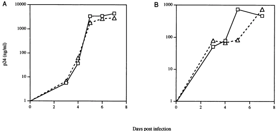
FIG. 1. In vitro replicationkineticsof HIV-1 nef mutants. (A)
HIV-1JR-CSF
andHIV-1JR
CSFx; (B)HIV-1NL4-3
and HIV-lNL,Xf.
Squares representwild-type virus, andtriangles represent the nef mutants. Viral replication in phytohemagglutinin-stimulated PBMC was quantitated by ELISAfor HIV-1 p24 antigen at the indicated days postinfection. In panel A, 5 x 10 PBMC were infected with approximately103IU of virus; 2.5 x 10³IU was added to 5 x 10⁶PBMC in panel B. Data in each were obtained from different PBMC donors.mined by limiting dilution on phytohemagglutinin-stimulated humanperipheral blood mononuclear cells (PBMC).
HumanPBMCwereobtainedfrom normal donors by veni-puncture or from leukopacks purchased from the American Red Cross. Peripheral blood lymphocytes were isolated by centrifugation over Ficoll-Hypaque and depleted of macro-phages by plastic adherence for 72 h.
Construction and infection ofSCID-hu mice. C.B-17 mice
homozygous for the SCID genetic defect (7) were originally
obtained from K. Dorshkin and subsequently bred at the
UniversityofCalifornia, LosAngeles. The micewere housed
inabiosafety level 3 facilityattheUniversity of California, Los Angeles, in accordance with institutionalguidelines. For
inva-siveprocedures, micewere anesthetized with methoxyflurene
as an inhalent and a combination of ketamine HCl with
xylazine injected intramuscularly (1 mg/10g ofbodyweight).
Humanfetal thymus andliverwere implanted under the left kidney capsule of SCID mice 6to8weeks of age as described previously(5, 6, 23, 25). Fetal tissuewasobtained (Advanced Bioscience Resources, Alameda, Calif.) from individual do-nors ranging from 16to24weeks ofgestation.InFig. 2A and 4 and Table 2, the animals are identified by numbers. The numberprecedingthehyphen indicates the fetal tissuedonor, and the number after the hyphen represents the particular
mouse. Four toeightmonthspostimplantation, implantswere injected with 200 infectious units(IU)of virus in50-,ul volumes (5). Mock implants were infected with 50
[Ii
of supernatantfrommock-electroporated COS cells.
QuantitativePCRamplification.Single-cell suspensions ob-tained frombiopsied implantswerewashedoncein
phosphate-buffered saline (PBS), lysed in urealysis buffer (4.7 Murea,
1.3% [wt/vol] sodium dodecyl sulfate,0.23 MNaCl, 0.67 mM
EDTA[pH 8.0],6.7 mMTris-HCl), andsubjectedto
phenol-chloroform extraction and ethanolprecipitation. Total nucleic acids obtained from this procedure were then subjected to quantitative PCRamplificationaspreviously described(5, 34,
35). Briefly, 25 cyclesofamplification were performed, using
the 32P-end-labeled M667-AA55 primer pair specific for the
R/U5 region of theviral long terminal repeat. To quantitatively detect cellular DNA, primers specific for human ,B-globin
(nucleotides 14to 33 and 123 to 104) (19, 34) were used with
21 cycles of amplification. To generate standard curves for HIV-1 DNA, four- orfivefold dilutions were made of cloned
HIV-lJRCSF
DNA (8) digested withEcoRI, which does not cleave viral sequences. These dilutionsweremade intoDNA from PBMC(10,ug/ml).Human,B-globin standardcurves were usually derived from 3-fold dilutions of PBMC DNA, except for theexperiments inFig.2A, in which 10-fold dilutionswere used. Standard curves for both HIV-1 and ,-globin were amplified in parallel. For PCR analysis, nucleic acids were addedto15 ,ulof low-salt PCR buffer(25mMTris[pH 8.0],2mMMgCl2,30mMNaCl,0.1mgof bovine serum albumin per
ml, 0.25 mM deoxynucleoside triphosphate). The reaction volume was brought up to 25 ,ul with S/P high-purity water (Baxter Healthcare Corp., McGaw Park, Ill.).
Following amplification, radiolabeled products were re-solvedon a6% polyacrylamide gel. Valueswere obtained by interpolation from the standard curve,using an Ambis radio-analytic imager(Ambis, San Diego, Calif.).
Flow cytometry. Thymocyte subset distribution was deter-mined by flow cytometry. Single-cell suspensions obtained frombiopsy samples werewashed once in PBS, and then 106
cellswerecostained with monoclonal antibodies(MAbs)
(Bec-tonDickinson,MountainView, Calif.)toCD4 and CD8T-cell
markers. Anti-CD4wasfluoresceinisothiocyanate conjugated.
Staining with the biotinylated anti-CD8 was followed with
avidin-allophycocyanin. As a positive control, normal human
peripheral blood was stained with anti-CD4 and anti-CD8
MAbs.An anti-mouse immunoglobulin Gl MAb was usedat
alltimepointsto stainperipheralbloodas an isotypecontrol.
After staining,erythrocytes were lysed by incubation in fluo-rescence-activated cell sorting lysingsolution (Becton
Dickin-son). Cells were then fixed in 2%
paraformaldehyde.
Datawereaccumulated on aFACStarPlus flow cytometer and
ana-lyzed with theLysis II program (Becton
Dickinson).
Forward-versus-sidescatteranalysisof mock-infectedimplantswasusedon November 9, 2019 by guest
http://jvi.asm.org/
[image:2.612.92.539.77.289.2]HIV-1 DNA STDS
A JR-CSF MOCK JR-CSF-X (COPY#)
1m
U o o o
- D N O0t
boo 0 cb m co (n aw al _ n ri L0
M
HlIV-1 _
R/U5-140bp
HUMAN DNASTDS
(CELL EQUIVALENTS)
I N m at1
9-GIobin
110bp
JR-CSF JR-CSF-X
100000
10.000l
1.000
1or,
A CD
0
a.)
C
z
8>
CL
0
0 a.
QL
z
U)
10.000
1.000
,00
*-* S
I-JR-CSF JR-CSF-X
S S
FIG. 2. In vivo replication of
HIV-lJR-CsF-x.
The number of HIV-1 genomesin 105 human thymocytes was determined in implants 3 to 4 (A and B) and 6 (C) weeks postinfection. (A) PCR data for fourHIV-lJRCSF-infected animals, four HIV-1JRCSFX-infected animals, andone mock-infected animal. Replicate samples were analyzed for human 13-globin sequences (bottom panel), allowing quantitation of viral genomes per human cell equivalent. Signals from quantitative HIV-1 DNA andcellular DNA standards assayed in parallel are shown
attheright of each panel. (BandC)Summary of PCR data for 3 to 4
to gateonthe livethymocytepopulation.Atotal of 5 x 10³to 10 X 103events were acquired, except from implantsseverely depleted ofCD4-bearing thymocytes.
RESULTS
Replication of HIV-1 wild-type and nef mutants.To investi-gate the in vivo requirement for HIV-1 Nef, SCID-hu mice wereconstructed byimplanting human fetal thymus and liver under thekidney capsule(5,6,23,25)of SCID mice(7). Four to six monthsfollowing implantation, the human thymus and liver implantswereinoculated with 200 IU ofoneof either of twodifferentwild-typeisolates of HIV-1 ortheirnefmutants:
HIV-lJR-CsF
(18) or anefframeshift mutant ofHIV-lJR-CSF
(HIV-1JR-csF-x)
orHIV-lNLA-3
(1)
or anef
deletionmutantof this isolate(HIV-lNLA,ef).
Thesetwoparental viruses differ in their in vitro phenotypes in thatHIV-lJRCSF
is relativelynoncytopathic and does not induce syncytia, whereas
HIV-1NL4-3 readily
inducessyncytia.
Asreported
fornef
mutantsof otherHIV-1molecular clones(14, 17),thenefmutantsused in these studies show in vitro growth kinetics inmitogen-stimu-lated PBMC cultures similar to those of the parental strains (Fig. 1). The in vitro syncytium-inducing phenotype of
HIV-1NLA-3
was also not affectedby
thenef
deletion(data
not shown).Implants infected with either
HIV-lJR-CSF
orHIV-IJR-CSF-Xweresubjectedtosequential biopsiesofapproximately 25%of the human organ at 3 to 4 weeks and again at 6 weeks
postinfectionandanalyzedfor viral DNAby quantitative PCR
(5, 34, 35).Anexample of this analysisatthe 3-to4-week time
point is illustrated in Fig. 2A. Implants infected with
HIV-lJRCSF
contained an average of -1,200copies of viral DNA per 105 human cells, while the implants infected withHIV-lJRCsF-x
contained an average of '30 copies per 105 cells. Thus, therewas an approximately 40-fold difference in theability of these viruses toreplicateatthistime point(Fig.2B). By the 6-week time point, average wild-type
HIV-lJR-CSF
virus load increasedapproximately twofoldto -2,800copies of viralDNAperi05humancells, while implants infected with its
nefmutant demonstrated anapproximately threefoldincrease
in viral load. Thus, at 6 weeks postinfection, an average of | 100copies of HIV-1DNAper
i05
cellswererecovered from implants infected with the mutant virus, resulting in an approx-imately 30-fold difference in genome number between thesetwoviruses
(Fig. 2C).
HIV-lNL-3
replicatesmorerapidly thanHIV-lJRCSF
inthis system, such that infected implants contained an average of approximately 1,800copies ofHIV-1 DNAper 105cellsat 3 weeks postinfection. However, at this time point, viral DNA wasundetectable inimplantsinfected withHIV-lNL-,ef
(Fig. 3A). The inability to detect viral DNA in these implants wasmostlikelyduetosensitivityof the assay, since estimated initial
multiplicities of infection were approximately 1 IU/5 x 105
cells. Because of severe thymocyte depletion at 6 weeks
postinfection (Tables 1 and 2), only four of eight implants
infected with
HIV-lNL43
yieldedsufficientnumbers of humanthymocytes to analyze by PCR. The average copy number of
HIV-1DNAhad increasedtoapproximately 13,000 copies per
105cellsatthe6-week timepoint (Fig. 3B). Low copy numbers ofHIV-1 DNA (average ofeight copies per 105 cells) were
(B) and 6 (C) weeks postinfection. The differences in virus load betweenmutantand wildtypewerestatistically significant (Wilcoxon ranksumtest;3weeks,P= 0.0001; 6 weeks, P=0.028).
1
L
on November 9, 2019 by guest
http://jvi.asm.org/
[image:3.612.53.294.77.644.2]100,000
10,000
1,000
100
10
1
100o,000o IInI
'i
i'-.Avi
10,000
1,000
100
10
1
FIG. 3. In vivo replication ofHIV-lNL,ef. Shown are copies of
HIV-1 genomesper105 thymocytes 3 (A) and 6 (B) weeks
postinfec-tion. HIV-1NLN43orHIV-dNL?,,ef(200 IU) wasinjected into thymus
and liver implants derived from fetal donors 17 to 24 weeks of gestational age. All implantswere sequentially biopsiedat the indi-cated timesandanalyzedfor HIV-1genomesandhuman 1-globinas
described previously (5, 34, 35). Points positioned on the baseline indicate undetectable viral DNA. The differences in virus load between thesemutantandwild-type strainsweresignificantatboth timepoints (3 weeks,P= 0.011; 6 weeks,P= 0.03).
detected at thistimepointinHIV-1NL,,?efinfected implants,
confirming that mutant virus was introduced and remained
viable in vivo. A single implant (14-26) infected with HIV-4NLA,efwas subsequently analyzed at 12weeks postin-fection and contained -700copiesof HIV-1 DNAper105cells (data not shown). PCR analysis of the viral nef gene in thymocyte DNAfromthis time point demonstrated the pres-enceof thedeletion, establishingthatthis viral DNAwasdue to replication of the mutant virus and not to selective
out-growth ofalow-level wild-type contaminant (datanotshown). These results demonstrate that while the nefmutantsofbothof these parental viruses are replication competent, they are
growth attenuated in vivo.
Pathogenicity
of viruses invivo. Todetermine ifthymocytesweredepletedas aresult ofHIV-1infection,allimplantswere
analyzed by two-color flow cytometry for human CD4 and CD8.Nodepletion of CD4+ human thymocyteswasobserved inimplants infected with either
HIV-lJRCSF
orits nefmutant at3or6 weekspostinfection compared with controlsconsistingof mock-infected implants (data not shown). At 9 weeks postinfection, one of five
HIV-1JR-CSF-infected
implants wasdepleted of bothmature and immature CD4-bearing
thymo-cytes. No depletionwas seeninthefour implantsinfectedwith
HIV-lJR-CSF-X
(data not shown). We and others previously demonstrated that theHIV-lJR-CsF
isolate depletesthymo-cytesin vivolessefficientlythansomeother viralisolates(5, 6, 33). Consequently,we analyzed eight implants infected with
the
HIV-1NL43
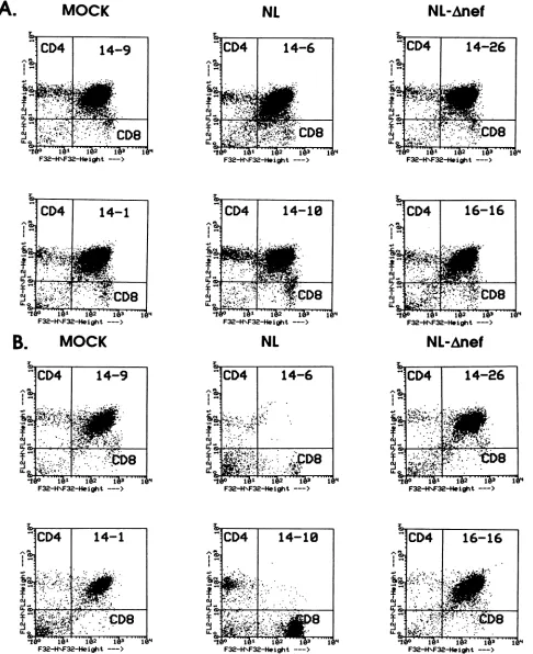
isolate, which is more cytopathic than theHIV-lJRcsF isolateboth invitroandinvivo. These implants appeared normal at 3 weeks postinfection, demonstrating approximately 75to85% CD4+ CD8+ cells,with themajority of the remaining cells being positive for either CD4 or CD8
(Fig. 4). However, by 6 weeks postinfection, all eight
HIV-1NL4-3-infected implants demonstrated severe depletion of
CD4-bearing thymocytes (Fig. 4; Tables1and2). Both
imma-ture CD4 CD8 double-positive and mature CD4+ CD8-thymocytes were depleted, resulting in a relative increase of CD4- CD8+ andCD4- CD8-thymocytes.Somedepletion of the CD4- CD8+ T-cell subset was observed in one implant
(16-9) (Table 2). By histological analysis, all eight of these implants demonstrated depletion of cortical thymocytes, simi-lar to that previously described (references 5 and 6 and data
notshown).When analyzedat 9weekspostinfection,three of four
HIV-1NL4-3-infected
implants clearly remained depleted of CD4-bearing thymocytes. In addition, implant 11-8, al-though depleted ofthymocytes (by histological analysis), dis-played reemergence of CD4+ thymocytes when assessed byflow cytometry. We and others have previously observed a
decrease in virus loadfollowingthedepletionofCD4-bearing thymocytes in thymus andliver implants (5, 6). It is possible that thedepleted thymus,inthepresenceoflesseramountsof virus, retains the ability to support regeneration of new
thy-mocytes. Therefore, the observed reemergence ofCD4+
thy-mocytes in implant 11-8 may reflect a dynamic interaction
betweeninfection anddepletionoftargetcells and the
regen-erationofnewthymocytes.
In contrast to
HIV-1NL4-3-infected
implants, depletion of CD4-bearing thymocytes was not observed in anyof the five HIV-1NLAiefinfected
implants by6 weekspostinfection(Fig. 4; Tables 1 and 2). Histological analysis revealed that these implants all hadnormal cortical and medullary regions (dataTABLE 1. Summaryofthymocytesubset distribution inmock-,HIV-1NL43-,andHIV-lNL-,efinfectedimplantsa
Infection 3wkpostinfection 6wkpostinfection
CD4+ CD4+CD8+ CD4-CD8- CD8+ CD4+ CD4+CD8+ CD4-CD8- CD8+
Mock 15.3± 10.9 76.1 ±13.9 3.1±2.0 5.3±2.9 14.7± 7.1 67.7±12.4 8.3±5.7 9.2±6.9
HIV-lNL4-3 8.6± 3.0 81.5+4.5 3.4±0.7 6.3±2.0 16.2± 11.1 3.0 +5.6 43.4±30.8 37.3±23.7
HIV1NLJ,ef 8.2±3.0 84.1 ±4.4 3.0±0.8 4.4± 1.2 7.0±3.8 71.3 ± 17.6 16.2± 12.9 5.3±3.2
a Thymocytes from implants infectedasindicatedwereanalyzedforCD4 andCD8 surface markers.QuadrantswereselectedasshowninFig.4. Mock-infected
implantswereassayedinparallelwith theexperimental implants.Valuesareaverages±standard deviations of the individual values shownin Table 2.
NL
NL-Anef
A
-mo
Q
2
._
Q
0 0
B
on
=
U)CD
Q
I.-en
._
0
0
W
-_
NL NI -Ano-f
on November 9, 2019 by guest
http://jvi.asm.org/
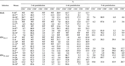
[image:4.612.62.564.633.707.2]TABLE 2. Thymocyte subset distribution in individualimplants'
Infection Mouse 3 wk postinfection 6 wkpostinfection 9wkpostinfection
CD4+ CD4+ CD8+ CD4- CD8- CD8+ CD4+ CD4+ CD8+ CD4- CD8- CD8+ CD4+ CD4+ CD8+ CD4- CD8- CD8+
Mock 9-16" NT NT NT NT 20.9 63.7 2.5 12.7
10-1"C 31.7 48.3 7.7 12.3 13.3 71.3 8.1 7.3
10-10C 28.7 65.7 1.7 3.9 15.1 63.9 17.5 3.5 7.6 80.9 6.8 4.6
11-17"c 24.4 69.8 1.5 4.3 17.4 71.7 2.1 8.8
12-16 NT NT NT NT 7.0 66.4 12.8 13.7 6.2 87.6 2.5 3.6
14-1b"C 6.3 86.3 2.8 4.4 8.2 72.7 15.2 3.7
14-3 NT NT NT NT 15.6 71.8 7.0 5.4
144b,c 9.7 80.9 3.9 5.4 28.3 40.5 6.8 24.3
14-9c 7.5 86.8 2.5 3.0 6.6 87.2 2.9 3.1 12.3 81.0 3.2 3.4
16-14c 7.5 86.6 2.0 3.7 NT NT NT NT 17.5 70.2 2.2 9.9
17-1 7.0 84.7 2.5 5.5 NT NT NT NT 10.1 81.3 5.3 3.1
HIVNL4-3
11-4" NT NT NT NT 29.3 1.0 4.6 65.011-8 NT NT NT NT 31.1 1.8 15.3 51.6 6.3 50.3 39.4 3.0
11-10b NT NT NT NT 3.0 0.4 55.5 40.9
11-11b NT NT NT NT 13.7 0.8 41.4 43.9
14-6c 6.7 85.2 3.6 4.4 21.0 1.1 61.8 16.6
14-10c 12.3 78.7 2.5 6.3 19.0 1.7 15.3 63.8 2.9 5.6 78.6 12.7
16-2c 5.7 85.5 3.1 5.4 11.6 16.8 55.8 15.5 11.8 2.8 39.0 46.2
16-9a," 9.9 76.7 4.2 9.1 0.8 0.4 97.5 1.1 2.7 1.0 90.1 6.0
HIVNL-Anef 14-16 8.8 83.8 2.8 4.4 4.9 86.7 5.2 3.1 9.8 75.9 13.2 0.9
14-26 6.1 87.3 2.2 4.2 5.4 80.9 9.2 4.3 17.8 69.7 6.5 5.9
14-29" 6.3 87.3 2.6 3.5 13.7 52.8 23.1 10.3
16-13 13.2 76.7 3.5 6.4 6.1 51.7 35.6 6.5 19.8 15.4 43.9 20.7
16-16" 6.5 85.6 4.1 3.7 5.1 84.6 7.7 2.4
aThymocyte subsets were determined as described for Table 1. Values shown for the 3- and 6-week timepointsarefor the individual implants summarized in Table
1.NT, not tested.
bSacrificed prior to the 9-week biopsy.
cDataobtained at the 3- and 6-week time points werepreviouslypublished (5).
not shown). One of three
HIV-1NLAf-afinfected
implants(16-13) testedat9weekspostinfection exhibited minor
deple-tion ofCD4+ thymocytes (Table 1). However,virus loadwas notassessedatthis timepoint.Theonly
HIV-lNLa,,nerinfected
implant tested at 12 weeks postinfection (implant 14-26)
exhibitednoCD4+thymocyte depletion(datanotshown).The results of the flow cytometric analysis demonstrate that the
pathogenic nature of
HIV-lNLA3
is severely attenuated byinactivation of the nefgene.
DISCUSSION
Our resultsdemonstrate that thenefmutantsoftwo
pheno-typically distinct HIV-1 strains are replicationcompetent yet
attenuatedforgrowth and cytopathicityinvivo. Thus, thenef
geneis required for efficient invivoreplication and
pathoge-nicity
ofHIV-1. The invivosystempresentedheremayprovide a means to determine the function of Nef. In the rhesus macaque,introductionofSIVnefmutantsresults ininefficient virusreplication,lack of diseaseprogression, and generation ofa protective immune response (9, 16). One interpretation of
these data is that SIV nef mutants are intrinsically more
immunogenic or less able to escape the immune response.
However, the attenuation of the HIV-1 nef mutants in
SCID-hu miceoccursmostlikelyinthe absence ofanimmune response. SCID mice lack functional T and B cells, as they cannotrearrangeimmunoglobulinorT-cellreceptor genes(21,
30).
In addition, it is unlikely that the differentiating humanthymocytes in these animals generate an effective anti-HIV
responsein thehumanimplant.Therefore, attenuation of the
nef
mutantsismost likelydueto an alteration of theinterac-tion between HIVand itstargetcell.
Oneinteraction of thenefgenewith CD4+ Tcells that has been documented in vitro is Nef-induced down-regulation of surface CD4 (12, 22, 31). No down-regulation of CD4 was observed in our in vivo system in thymocytes infected with either of thetwo HIVstrainsortheirnef mutants,asmeasured by intensity of CD4 staining using flow cytometry (Fig. 4 and data notshown).Itispossible that CD4 down-regulation may nothave been detectableatthe3- or6-week timepoint, as, at most, only 15% of thymocytes were infected. However, down-regulation of CD4 is not responsible for the observed loss of CD4+ thymocytes in implants infected with HIV, as histological and flowcytometric analyses of infected implants
showatrophyandseverehypocellularity(5,6,33), reflectinga
significant loss of cell numbers.
Thelower viral burden inimplants infected withnefmutants demonstrates that Nef isrequired for the efficient replication
of virus in thymocytes. The relationship between high virus load and celldepletion in implants infected with
HIV-1NL4-3
suggeststhatthe decreaseinpathogenicity of the nefmutants is relatedtoviral replication. Thedichotomy between in vitro systems, in which Nef appears to be dispensable for virus replication, and this in vivo model,inwhichnefmutantsdisplay attenuated replication, suggests that insight into the mecha-nismof Nefaction maybefound in the differencesbetween the two models themselves. One difference between the two systems is the target cell type. Most in vitro systems utilizeterminally differentiated, actively replicating T lymphocytes.
Thymocytes, however,areundergoingdifferentiation,andonly
arelativelylow percentage(10to 20%) (datanot shown) are
traversingthe cellcycleatanygiventime. The function of Nef
may therefore be related to the difference in the activation/
differentiation state of these two target cell types. Early
on November 9, 2019 by guest
http://jvi.asm.org/
[image:5.612.60.554.88.351.2]NL-Anef
B.
MOCK
F32-H\F32-Height --->
NL
_CD4
14-9
_
-I".W.S
.Oi- ij2 i73 '
F32-H\F32-Height --->
CD4
14-1
_
N >:0 .
I
CD4
14-6
.k\ ..
I
Sd , ,.';
cI
Ii.I
'''i ' '''' 2 " i
F32-H\F32-Height --->
CD4
14-10
L-. I
CD4
14-26
-jL.
CD8
F32-H\F3a Height >
QS
'jCD4
16-16
A~IFt.
C ,r;.,
S .=w .,...
s
I
(U 4
F -H\FT32 .igh j*34.6o'w
F32-H\F32-Height ---> F32-H\F32-HeightF32-HNF3a-HFight---> --->
FIG. 4. Flowcytometric analysisofHIV-1NLA-3-andHIV-lNL-Anefinfectedimplants. Implantswereinfected with 200 IU of the indicatedvirus
orwith 50 ,ul of supernatant frommock-electroporated cells.Thymocyteswereanalyzedfor CD4 and CD8 surface antigens bytwo-color flow
cytometryat3(A)and 6(B)weekspostinfection.Dotplotsfrom tworepresentative implantsareshown for eachgroup:mock-,HIV-lNL4N3-,and
HIV-lNL ,Xefinfectedimplants.
A.
MOCK
NL
NL-Anef
I
4
...8
:-S...
I
A.~lJ8
'. .1
--,..CD8
0 'Na,
bR
on November 9, 2019 by guest
http://jvi.asm.org/
[image:6.612.78.564.82.678.2]studies, although controversial, suggested that Nef negatively influencesthe transcription of HIV by interacting with cellular factors (4, 20). In contrast to those studies, our results dem-onstrate that nef mutants do notreplicate as well as wild-type HIV-1 in differentiating thymocytes. It is conceivable that Nef positively influences the expression of HIV-1 in this cell type. Studies using the SCID-hu model are currently under way in this laboratory to determine themechanism(s) responsible for the attenuated replication bynefmutants as well as to define the regions of Nef critical for its function in vivo.
Our resultsindicate that Nef is required for optimal growth and cytopathic properties of HIV-1 in the human thymus in vivo. These results are in agreementwith previously published work in theSIV system (9, 16) andsuggest that the attenuated properties of SIV nef mutants are likely due to inefficient replication in lymphoid tissues. This inefficient replication could allowfor appropriate antigen presentation and immune activation before significant depletion oflymphocytes occurs, resulting in host control of the virus and protective immunity. Poor replication in lymphoid tissues and an effective immune response could also prevent virus from becoming sequestered inlymphoid tissues which, inHIV-infected humans, have been shown to act as areservoir of viral particles and viral antigens during the clinical latency period (10, 27).
Administration of nef deletion mutants of SIV to rhesus macaques appears to be the most effective method of antilen-tivirus vaccination to date (9). However, since SIV and HIV-1 are not genetically identical, absolute parallels between these two viruses cannot be drawn. The SCID-hu model is the only animal model currently available in which HIV-1-induced pathology can be demonstrated (5, 6, 33). Our results suggest that the SCID-hu mouse may be a useful modelfor quantita-tively assessing the pathogenic properties of candidates for live attenuatedHIV-1 vaccine strains. By using this system, it may be possible to identify mutations in other accessory genes, individually or in combination with nef mutations, which may result in amore attenuated phenotype. This type of assessment is notpossible inimmunocompetent primate models, in which the host immune response may affect viral replication. How-ever, because thebest test of vaccine potential is the ability of the immune response to control both replication and pathoge-nicity, studies in SCID-hu mice would be complemented by studiesinvolving infection ofimmunocompetent animals such aschimpanzees (3) or pigtailed macaques (2) to determine if the alteredviruses would be able to evoke immune responses. This type of coordinated research effort may provide an approach for preclinical assessment of live attenuated HIV-1 vaccinestrains.
ACKNOWLEDGMENTS
Wethank Gerold Feuer and Rafi Ahmed fordiscussion and critical review of the manuscript.
Thisworkwas supported by the UCUniversitywide AIDS Research Program, the UCLA CFAR (NIH), the McCarthy Family Foundation, andagift from Jim Bridger for the initiation of the SCID mouse P3 facility. G.M.A. is a Pediatric AIDS Foundation Scholar.
REFERENCES
1. Adachi, A., H. E. Gendelman, S. Koenig, T. Folks, R. Willey, A. Rabson, and M. A. Martin. 1986. Production of acquired immu-nodeficiency syndrome-associated retrovirus in human and non-human cells transfected with an infectious molecular clone. J. Virol. 59:284-291.
2. Agy, M. B., L. R. Frumkin, L. Corey, R. W. Coombs, S. M. Wolinsky, J. Koehler, W. R. Morton, and M. G. Katze. 1992. Infection of Macaca nemestrina by humanimmunodeficiency virus type-1.Science257:103-106.
3. Alter, H. J., J. W. Eichberg, H.Masur, W. C. Saxinger, R.Gallo,
A. M. Macher, H. C. Lane, and A. S. Fauci. 1984.Transmission of HTLV-III infection from human plasma to chimpanzees: an animal model for AIDS. Science 226:549-552.
4. Ahmad, N., and S. Venkatesan. 1988. Nef protein of HIV-1 is a
transcriptional repressor of HIV-1 LTR.Science241:1481-1485. 5. Aldrovandi, G. M., G. Feuer, L. Gao, B. Jamieson, M. Kristeva,
I. S. Y. Chen, and J. A. Zack. 1993. The SCID-hu mouse as a
model for HIV-1 infection. Nature (London) 363:732-736. 6. Bonyhadi,M. L., L. Rabin, S. Salimi, D. A. Brown, J. Kosek,J. M.
McCune, and H.Kaneshima.1993. HIV induces thymusdepletion invivo. Nature (London) 363:728-736.
7. Bosma, G. C., R. P. Custer, and M. J. Bosma. 1983. A severe
combined immunodeficiency mutation in the mouse. Nature (Lon-don)301:527-530.
8. Cann, A. J., Y. Koyanagi,andI.S.Y.Chen. 1988.Highefficiency transfection of primary human lymphocytes and studies of gene expression. Oncogene 3:123-128.
9. Daniel, M. D., F. Kirchoff, S. C. Czajak, P. K. Sehgal, and R. C.
Desrosiers. 1992. Protective effects of a live attenuated SIV vaccine with a deletion in the nef gene. Science 258:1938-1941. 10. Embretson, J., M. Zupancic, J. L. Ribas, A. Burke, P. Racz, K.
Tenner-Racz, and A. T. Haase. 1993. Massive covert infection of
helper T lymphocytes and macrophages by HIV during the incubation period of AIDS. Nature (London) 362:359-362. 11. Fisher, A. G., L. Ratner, H. Mitsuya, L. M. Marselle, M. E.
Harper, S. Broder, R. C. Gallo, and F. Wong-Staal. 1986.
Infec-tious mutants of HTLV-III with changes in the 3' region and markedly reduced cytopathic effects. Science 233:655-659. 12. Garcia, J. V., and A. D. Miller. 1991. Serine
phosphorylation-independent downregulation of cell-surface CD4 by nef. Nature (London) 350:508-511.
13. Grody, W. W., S. Fligiel, and F. Naeim. 1985. Thymus involution in
the acquired immunodeficiency syndrome. Am. J. Clin. Pathol. 84:85-95.
14. Hammes, S. R., E. P. Dixon, M. H. Malim, B. R. Cullen,and W. C.
Greene. 1989. Nef protein of humanimmunodeficiency virus type 1: evidence against its role as a transcriptional inhibitor. Proc. Natl. Acad. Sci. USA86:9549-9553.
15. Kaneshima, H., C.-C. Shih, R. Namikawa, L. Rabin,H. Outzen,
S. G.Machado, and J. M. McCune. 1991. Human
immunodefi-ciency virus infection of human lymph nodes in the SCID-hu mouse. Proc. Natl. Acad. Sci. USA88:4523-4527.
16. Kestler, H. W.,III,D. J. Ringler, K.Mori, D. L. Panicali, P. K. Sehgal, M. D. Daniel, and R. C. Desrosiers. 1991. Importance of thenef gene for maintenance of high virus loads and for develop-ment of AIDS. Cell65:651-662.
17. Kim, S., K. Ikeuchi, R. Byrn, J. Groopman, and D. Baltimore. 1989. Lack of a negative influence on viral growth by the nef gene of human immunodeficiency virus type 1. Proc. Natl. Acad. Sci. USA 86:9544-9548.
18. Koyanagi, Y., S. Miles, R. T. Mitsuyasu, J. E. Merrill, H. V.
Vinters, and I. S. Y. Chen. 1987. Dual infection of the central nervous system by AIDS viruses with distinct cellular tropisms. Science236:819-822.
19. Lawn, R. M., A. Efstratindus, C. O'Connell, and T. Maniatis. 1980. The nucleotide sequence of the human l-globingene. Cell 21:647-651.
20. Luciw, P. A., C. Cheng-Mayer, and J. A. Levy. 1987. Mutational analysis of the human immunodeficiency virus: the orf-Bregion down-regulates virus replication. Proc. Natl. Acad. Sci. USA 84:1434-1438.
21. Malynn, B. A., T. K. Blackwell, G. M. Fulop, G. A. Rathbun, A. J. W.Furley, P. Ferrier, L. B. Heinke, R. A. Phillips, G. D. Yancopoulos, and F. W. Alt. 1988. The scid defect affects the final step of the immunoglobulin VDJ recombinase mechanism. Cell 54:453-460.
22. Mariani, R., and J. Skowronski. 1993. CD4downregulation by nef allelesisolated from HIV-1 infectedindividuals. Proc. Natl. Acad. Sci. USA 90:5549-5553.
23. McCune, J. M., R. Namikawa, H.Kaneshima, L. D. Shultz, M. Lieberman, and I. L. Weissman. 1988. The SCID-hu mouse: murine model for theanalysis of human hematolymphoid
on November 9, 2019 by guest
http://jvi.asm.org/
entiation andfunction. Science 241:1632-1639.
24. Namikawa, R., H. Kaneshima, M. Lieberman, I. L. Weissman, and J. M.McCune. 1988.Infection of the SCID-hu mouse by HIV-1. Science 242:1684-1686.
25. Namikawa, R., K. N.Weilbaecher, H. Kaneshima, E. J. Yee, and J. M. McCune. 1990. Long-term human hematopoiesis in the SCID-hu mouse. J. Exp. Med. 172:1055-1063.
26. Niederman, T. M. J., B. J.Thielan, and L. Ratner. 1989. Human immunodeficiencyvirus type 1 negative factor is a transcriptional silencer. Proc. Natl. Acad. Sci. USA 86:1128-1132.
27. Pantaleo, G., C. Graziosi, J.F.Demarest, L. Butini, M. Montroni, C.H.Fox,J. M.Orenstein, D.P.Kotler, andA.S. Fauci.1993.HIV
infection is active and progressive in lymphoid tissue during the clinicallylatent stage of disease. Nature(London)362:355-358. 28. Papiernik, M.,Y.Brossard,N.Mulliez, J. Roume, C. Brechot,F.
Barin, A.Goudeau, J.-F. Bach, C. Griscelli, R. Henrion, andR. Vazeux. 1992. Thymic abnormalities in fetuses aborted from human immunodeficiency virus type 1 seropositivewomen. Pedi-atrics 89:297-301.
29. Robert-Guroff, M., M. Popovic, S. Gartner, P. Markham, R. C. Gallo,andM. S.Reitz.1990. Structure andexpression of tat-, rev-, andnef-specific transcriptsof humanimmunodeficiencyvirus type
1 in infected lymphocytes and macrophages. J. Virol. 64:3391-3398.
30. Schuler, W.,I.J.Weiler,A.Schuler,R. A.Phillips, N. Rosenberg,
T. W. Mak, J. F. Kearney, R. P.Perry, and M. J. Bosma. 1986. Rearrangementof antigen receptor genes is defective in mice with severecombinedimmunodeficiency. Cell 46:963-972.
31. Schwartz, O., Y. Riviere, J.-M. Heard, and 0. Danos. 1993. Reduced cellsurface expression of processed human immunode-ficiency virus type Ienvelopeglycoproteininthe presence of Nef. J.Virol. 67:3274-3280.
32. Skowronski, J., D. Parks, and R.Mariani. 1993. Altered Tcell activation and development in transgenic mice expressing the
HIV-1nef gene. EMBO J. 12:703-713.
33. Stanley, S. K., J.M.McCune, H. Kaneshima, J. S. Justement,M.
Sullivan,E. Boone, M. Baseler, J.Adelsberger, M. Bonyhadi, J. Orenstein, C.H.Fox, and A.S. Fauci. 1993. Human immunode-ficiency virus infection of the human thymus and disruption of the thymic microenvironment in the SCID-hu mouse. J. Exp. Med. 178:1151-1163.
34. Zack, J. A., S. J. Arrigo, S.R.Weitsman,A.S.Go,A.Haislip,and
I. S. Y. Chen. 1990. HIV-1 entry into quiescent primary lympho-cytes:molecular analysis revealsalabile, latent viralstructure.Cell 61:213-222.
35. Zack, J. A.,A. M. Haislip,P.Krogstad,andI.S. Y. Chen. 1992. Incompletely reverse transcribed human immunodeficiency virus type1genomesinquiescent cellscanfunctionasintermediates in the retrovirus life cycle. J. Virol. 66:1717-1725.