JOURNAL OFVIROLOGY, Aug. 1994, p. 5074-5083 0022-538X/94/$04.00+0
Copyright C) 1994, American Society for Microbiology
Evaluation
of Human
Immunodeficiency
Virus Type 1
(HIV-1)-Specific Cytotoxic
T-Lymphocyte
Responses
Utilizing
B-Lymphoblastoid Cell Lines Transduced with the
CD4
Gene and Infected with
HIV-1
M.JULIANAMcELRATH,l* MIKE RABIN,1 MARKHOFFMAN,' SARAKLUCKING,1 J. VICTOR GARCIA,2ANDPHILIP D. GREENBERG1'3
Department of Medicine1 andDepartment of Immunology,3 University of Washington School of Medicine, Seattle, Washington, and Department of Virology and Molecular Biology, St.JudeChildren'sResearchHospital,
Memphis, Tennessee2
Received 17December1993/Accepted6May 1994
Analysis ofmajorhistocompatibility complex-restricted cytotoxic T lymphocytes (CTL) capableofkilling humanimmunodeficiency virustype 1(HIV-1)-infectedtargetsis essential forelucidating thebasis for HIV-1 disease progression and the potential efficacy of candidate vaccines. Theuse ofprimary CD4+ Tcells with variableinfectivityastargetsfor suchstudieshas significantlimitations, and immortalautologouscells with high levels ofCD4 expression thatcanbeconsistently infectedwith HIV-1 would be of much greaterutility.
Therefore,wetransducedEpstein-Barr-virus-transformed B-lymphoblastoidcell lines (LCL)witharetroviral vector, LT4SN, containing the human CD4 gene. Stable LCL in which more than 95% of cells expressed
membraneCD4wereobtained.AliquotswereinfectedwithHIV-1,and,after 4to7days, nearlyall ofthe cells contained cytoplasmicgag and produced high levels ofp24 antigen. Theabilityofmajorhistocompatibility
complex-restricted CD8+ CTL to lyse such HIV-1-infected CD4-transduced LCL (LCL-CD4H ) was
evaluated.Theseautologous targets werelysed byCTLgenerated from anHIV-1-uninfected vaccineeover a
broadrangeof effector-to-target ratios. Similarly,the
LCL-CD4HWV
1wereefficiently lysed byfreshcirculating CTLfrom HIV-1-infectedindividuals,aswellasbyCTL activatedbyin vitrostimulation. BothHIV-1env-andgag-specific CTLeffectorslysed
LCL-CD4HIV1,
consistent with the cellularexpressionof both HIV-1genes.TheLCL-CD4HWv
also functioned as stimulator cells, and thus are capableofamplifying CTLagainst multiple HIV-1 gene products in HIV-1-infected individuals. The ability to produce HIV-1-susceptible autologous immortalized cell lines that can be employed as target cells should enable a more detailed evaluation ofvaccine-induced CTL against both homologous and disparate HIV-1 strains. Furthermore, the use of
LCL-CD4HIV-1
should facilitate the analysis of the range ofHIV-1 gene products recognized by CTL in seropositivepersons.CytotoxicTlymphocytes (CTL) are essentialforhost
resis-tance to and control ofmost viruses, including HIV-1. Thus, evaluation ofCTLresponsestoHIV-1 antigensisimportantin understanding the pathogenesis of HIV-1 infection and in characterizing the potentially protective immune responses
elicited by candidate HIV-1 vaccines (10, 22, 35). Assays to detect HIV-1-specific major histocompatibility complex (MHC)-restricted CTLactivityhave reliedprimarilyuponthe
useofrecombinant vaccinia virusestoexpressHIV-1 genesin
autologous stimulator and/or targetcells (5, 34, 37, 41). This approach has been very informative, but has several limita-tions. First, in evaluating the HIV-1-specific responses of
recipientsofvaccines employingrecombinant vacciniaviruses, the CTLresponse to thevaccinia virus vector may interfere
with detection of theresponse tothe product ofthe inserted
gene.Second, although CTL thatcanrecognize and lysetarget cells expressing abundant amounts of a particular HIV-1
protein can be identified with recombinant vaccinia virus
reagents, onlyinferential assumptionscanbe made about the efficiencywithwhich such CTL willlyse HIV-1-infected target
*Correspondingauthor.Mailingaddress:Department ofMedicine,
University ofWashington, ZB-30, c/o Pacific Medical Center, 1200
12th Ave. South, Room 9301, Seattle, WA 98144. Phone: (206)
326-4177. Fax: (206) 326-4178.
cells expressing the full range of HIV-1 proteins. Finally,
analysisofthe relativeimmunogenicityof distinct viral isolates cannotbeeasily performedwith vacciniaviruses,asitrequires cloning the gene from each new isolate of interest and constructingnewrecombinant vacciniaviruses.
To directly test MHC-restricted CTLresponses to HIV-1-infected cells, autologous target cells expressing the CD4 molecule, anaccessorymoleculethat contributes to T-helper-cellactivation but alsoserves asthemajorcellularreceptorfor HIV-1 (21, 26), areusuallyused. CD4+Tcells derivedfrom peripheralblood mononuclear cells(PBMC)canpotentially be
used astargetsfor CTL analysisbut also have several limita-tions. First, the CD4+ T-cell population must be activated priortoinfectionwithHIV-1,whichcanbeproblematicwith
thedysfunctional CD4+ populationoftenobtained from HIV-1-infected individuals.Secondly, CD4+Tcellsarenot immor-tal andthususuallymustbeobtained fresh andpreparedanew
for eachassay;thismayalsobeproblematicinHIV-1-infected individuals with lowCD4+counts.Finally,the infectionrateis variable andunpredictable, and the level of HIV-1expression
maybe low, with manycells in thepopulation notexpressing HIV-1genes.
The use of an immortalized, highly infectable, autologous
cell would be very useful for these studies. B lymphocytes
possess many of the essentialqualitiesfor such acell. Bcells
5074
Vol.68,No.8
on November 9, 2019 by guest
http://jvi.asm.org/
are readily immortalized with Epstein-Barr virus (EBV) and thus can be expanded and maintained in culture (39). Such EBV-immortalized B-lymphoblastoid cell lines (LCL) have been demonstrated to be efficient in vitro stimulators and targets for CTL responses (33). Additionally, LCL, in which a subset of cells express varying amounts of endogenous CD4, have been shown to be susceptible to HIV-1 infection (6, 7, 9, 25, 31, 38, 40). The ability to infect LCL with HIV-1 has been
variable (25, 38) but appears to correlate with the proportion
andlevel of CD4 expression (40). Therefore, we have explored methods to increase CD4 expression in immortalized B cells andhave evaluated whether such cells are more susceptible to HIV-1 infection and thus are more suitable stimulator and target cells to examine HIV-1-specific CTL responses.
Ahighly efficient means for stable introduction of exogenous genes into replicating cells is infection with retroviral particles
containing recombinant retroviral vectors (30). LXSN is a
retroviralvector which contains the neo gene under the control
ofthe Moloney simian virus 40
(SV40)
promoter and contains asitefor introduction of a gene with expression driven off the Moloney leukemia virus long terminal repeat (29). Thecom-plementaryDNA encoding human CD4 has been inserted into
this site, and infectious viral particles have been produced. In this report we describe the transduction of LCL with the CD4 gene in this retroviral vector and the efficient infection of these cells with HIV-1. CD4-transduced LCL infected with HIV-1 have been tested as stimulator and target cells in CTLassays from both naturally infected individuals and in
HIV-1-uninfected persons who have been immunized with candidate
HIV-1 vaccines. Our findings indicate that immortalized B cells with stable expression of CD4 can be generated by
transduction with the CD4 gene, that such cells are highly
susceptible to HIV-1 infection, and that HIV-1-infected LCL
may have broad utility in the analysis ofCTL responses.
MATERIALS AND METHODS
Study population. Heparinized blood was obtained either from healthy unimmunized HIV-1-seronegative individuals as
controls; from initially seronegative or consenting subjects
participating in National Institutes of Health AIDS Vaccine
Evaluation Group (AVEG) Protocol 002, which includes one
to two immunizations with a recombinant vaccinia virus
ex-pressing the
HIV-1LAl
envelope glycoproteingpl60
(Bristol-Myers Squibb), followed by one to two booster injections with a recombinant HIV-1LAI
gpl60
subunit protein vaccine(Mi-croGeneSys)
(13); or from asymptomatic HIV-1-seropositiveparticipants ina blinded-controlled trial (AVEG Protocol 103)
examining augmentation of immunity with a recombinant
envelopesubunit vaccine, ENV 2-3 (Biocine).
CD4 transduction and HIV-1 infection of LCL. The
Molo-ney virus-based retroviral vector containing the human CD4
gene, pLT4SN, was constructed by insertion into pLXSN of an
EcoRI-BamHI
fragment from plasmid T4-pMV7 (26), kindlyprovidedby D. Litman, containing the full open reading frame
for CD4 (29). The resultant vector contains the CD4 gene under the control of the 5' long terminal repeat and the
neomycinphosphotransferase
Neor
gene, for positive selectionof transduced cells, under the control of the internal
SV40
promoter. The vector was transfected into the amphotropic
packaging line PA317, and high-titer virus stocks were
ob-tained by growing the packaging line with the inserted plasmid to 80 to 90% confluence and replacing the drug selection medium with a minimal amount of growth medium for 24 h (28, 29). The absence of helper virus in the producer line was
confirmed bythe S+L- assay (3, 28). Viral supernatants from
clones 1C3 and 1B4 which exhibited titers
>106
on NIH 3T3cells and resulted in
expression
of CD4 on the transducedfibroblasts were used in the
experiments
described below. Theculture
supernatant
washarvested, passed
through a0.45-mm-pore-size
filter,
and stored at-70°C
until needed.Autologous
EBV-immortalized B-LCL were prepared aspreviously
described(39).
One milliliter ofhigh-titer retroviralsupernatant
was added to 1 to 10 millionactively dividing LCLin the
presence
of,ug
4 ofPolybrene
at37°C,
5%CO2.
After48
h,
the cells were washedby
centrifugation,
resuspended ingrowth
medium(RPMI
1640supplemented
with 10%heat-inactivated fetal calf
serum,
50 U ofpenicillin
perml, 50 mg ofstreptomycin per
ml,
and 2.0 mMglutamine),
and selected by culture with 1.5mg
ofG418per
ml. Cellsexpressing CD4 werefurther enriched from the
drug-resistant
LCL population bypositive
selection on CD4 MicroCELLector flasks (AppliedImmune
Sciences, Inc.,
MenloPark,
Calif.).
CD4expression bythe transduced cells was evaluated by flow cytometry after
labeling
the LCL with fluorescein isothiocyanate(FITC)-conjugated
anti-Leu-3A monoclonal antibody(Becton-Dickin-son,
SanJose,
Calif.)
or with the irrelevant FITC-conjugatedMAb MOPC 21
(goat
anti-mouseimmunoglobulin
Gl[IgG1],
kindly
provided by
E.Clark,
University
of Washington,Seat-tle).
Thepurified
LCL transduced with CD4 were maintainedin
culture at
37°C
ingrowth
mediumsupplemented
with 0.5 mgofG418
per
ml.Onaverage,
more than 50% of thetransducedcells remained
CD4+
after 3 months of culture. Positiveselection on the anti-CD4-coated flasks was repeated at
monthly
intervals to
prevent
the
potential
outgrowth of a subpopulation ofCD4-
cells.One million LCL-CD4 in 1 ml of
growth
medium wereinfected with 200
50% tissue culture infective
doses ofHIV-1LAI,
HIV-1SF-2
orHIV-lMN
(the
latter
twokindly provided
by
K.Steimer,
ChironCorporation)
in 24-well plates (Costar,Cambridge,
Mass.)
and washed within 48
h. HIV-1 infectionwas
analyzed by p24
antigen
capture
(Abbott
Laboratories,Abbott
Park,
Ill.)
of culture
supernatant.
To
evaluate thepercentage
of infected
cells,
fluorescence-activated
cell sorter(FACS) analysis
for
p55
core
antigen
expression
in thecyto-plasm
of
permeabilized
cells was
performed
by utilizing
murineMAb KC57-PE
(Coulter
Corp,
Hialeah,
Fla.)
accordingto theproduct
instructions.
HIV-1envelope
expression
wasdeter-mined
by
FACS
analysis
of membrane staining
with mouseMAb
110-4,
an
IgGl-recognizing HIV-1LAI
gpl20
(kindlyprovided by
S.-L.
Hu,
Bristol-Myers
Squibb
Research
Institute,Seattle,
Wash.),
and
FITC-conjugated
goat
anti-mouse Ig(TAGO, Burlingame,
Calif.).
Cell-surface expression
of theHIV-1
envelope
was further verified
by
measuring
underphasemicroscopy
the
ability
of
LCL-CD4 infected
with HIV-1(LCL-CD4HIV-l
denotes infection withHIV-1LAI
strainunlessotherwise
indicated)
to form
syncytia
with SupTl
cellsaccord-ing
to a modified
procedure
previously
described (32).
LCL-CD4HIVl
were incubated with
SupTl
cells
ina
1:10 ratio inmicrotiter
plates,
and
syncytia
were
easily
visualized
byin-verted
phase
microscopy
within 6 h. No
syncytia
werevisual-ized in wells
containing
uninfected LCL-CD4 and
SupTlcells.The
production
of infectious virions
by
LCL-CD4HIVl
wasdetermined
by
adding
filtered culture
supernatants
to CEMcells,
washing
the cells twice
one hlater,
and testingthe culturesupernatants
of the CEM cells for HIV-1
infection by p24antigen production.
Based
on these infectivity studies,autolo-gous
LCL-CD4HIV-l
were used
astarget
or stimulator cells after 4 or more days of infection.Effector
cells. PBMC were isolated from heparinized
bloodby Ficoll-Paque
density
centrifugation
as
previously
described(4),
and effector cells for CTL
assays
were fresh
PBMCeitheron November 9, 2019 by guest
http://jvi.asm.org/
5076 McELRATH ET AL.
tested in a direct assay or following in vitro stimulation to amplify HIV-1-specific
CD8+
effectors. PBMC werestimu-lated in vitro with one of threetypesof autologousstimulator cells:
LCL-CD4HIV-l,
PBMC infected with a recombinantvaccinia virus(multiplicity of infection of10) expressing either the
HIV-1LAj
env(v-env)orHIV-lLAIgag(v-gag)geneproduct(18, 19) (kindly provided byS.-L.Hu)ormacrophagesinfected
with the monocytotropic strainHIV-lBa-L(11).Atotalof 20x 106 PBMC were stimulated with 2 x 10⁶ LCL-CD4HIVl,
PBMCv
env,orPBMCv-gag
for 9 daysat37°C,5% CO2 inaT25flask, without supplemental exogenous recombinant
interleu-kin 2. Alternatively, PBMC (2 x 10⁶perwell)werestimulated for two 7-day intervals without supplemental recombinant interleukin 2 at
37°C,
5% CO2 in 24-well plates withautolo-gous adherent macrophages (2 x 10⁵ per well), previously
infected for1week with
HIV-lBa-L,
aspreviously described(5,27).
Target cells. Target cells for assessing cytolytic activity included LCL-CD4,
LCL-CD4HIV-1
(infected with LAI, SF-2,orMNstrains), and LCLorLCL-CD4infected for 90min with
eitherv-env, v-gag, orvacciniavirus (NYstrain), aspreviously
described (5). No interference with expression of HIV-1 gene
products has been detected in the CD4-transduced LCL infected with vaccinia virus recombinants. A total of 2 x 10⁶
target cells werelabeledovernight with 100 mCi of5NaCrO4
and werewashed prior to use in the cytotoxicityassay.
Cytotoxicity assays. Standard 4-h chromium release assays
were performed in triplicate by using mixtures of 104 target
cells with variednumbersof effector cells inatotal volumeof
200ml.Themaximumspontaneous5'Crreleasein the absence of effector cells was less than 25% of maximal release by
detergentlysis.
RESULTS
Characteristics of LCL transduced with the CD4gene and infected with HIV-1. LCL from 13 donors were transduced
with the human CD4 gene by culture with viralsupernatants
produced with the LT4SN retroviralvectorandwerepositively
and negatively selectedasdescribedinMaterials andMethods.
CD4 expression of transduced cellswas evaluated by
fluores-cence after exposure to FITC-conjugated anti-Leu-3A. The CD4gene wastransduced successfullyfor all 13donorstested,
and the analysis of CD4 expression from cells from two
representative donors is shown in Fig. 1. LCL from the two
donors demonstratednosignificantexpressionof CD4priorto
transduction (Fig. 1A and B). Incontrast, more than 98% of
the CD4-transduced LCL from bothdonorsexpressed CD4,as
evidenced by a shift in the fluorescence histogram following
stainingwith antibodyto CD4 (Fig.1Cand D). Similarlevels of CD4 expressionwere observedin theCD4-transduced LCL
from the other 11 donors. Repeated analyses over time
re-vealedthat CD4 expression remained stable inG418-selected cells.
The susceptibility of the CD4-transduced LCL to HIV-1
infectionwas examinedby measuring p24 antigen production
in the culturesupernatant ofLCL-CD4cells infectedwith200 50% tissue culture infective doses of HIV-1. HIV-1 p24
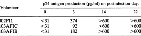
antigen was detectable within 3 days of infection, with
HIV-1LAI
in LCL-CD4 from three donors tested(Table 1).
Theproduction of p24 peaked within 2 weeks and remained elevated. Syncytium formationwas not observed by inverted
phase microscopy in cells containing infected LCL-CD4.
HIV-1 infection in the CD4-transduced LCLfrom 8ofthe 13
donorswas examined, and thekinetics of p24 productionwas
similar to that shown in Table 1 for the three donors. In
6
z
A) 002F11
350
ri
I I
11
I
0 1 0
0 1600....200
C) 002F11
400-A
0 100 200
B) 002FI6
350-0 100 200
D) 002FI6
400j
0 100 200
Fluorescence Intensity
FIG. 1. Cell-surface expression of CD4 in LCLand LCL
trans-duced with CD4 from twodonors.Single-cellsuspensions of105 cells werestained withFITC-conjugatedanti-Leu-3A(anti-CD4)or FITC-conjugated MOPC21 (goat anti-mouseIgGl) monoclonalantibodies,
andfluorescence intensity wasmeasuredby flowcytometry. The gates were set to exclude probable nonviable cells and debris. The histo-gramsrepresentfluorescenceintensityonthexaxis and cell numberon
the y axis. Stainingwith anti-Leu-3A isrepresented by the solid line ( ),and stainingwith MOPC21bythe dashedline(--), and the lines in panels A and B are superimposable. (A) LCLfrom subject 002F11. (B) LCL from subject 002F16. (C) LCL-CD4 from subject 002F11. (D)LCL-CD4 fromsubject 002FI6.
comparison experiments, measurable levels of p24
antigen
production detected in the nontransduced LCL were either absent or substantially delayed following invitroinfection.
The high levels of HIV-1 p24 antigen production in the culture supernatants following incubation of LCL-CD4 with HIV-1 confirmed that infection had occurred, but it was
unclear if this resulted from a few cells
producing
large
quantities of virus or from more uniform infection of the majority of cells. Although the presence ofasmall number of infected cells might permit the use of this population as
stimulator cells for in vitro responses, the use of
LCL-CD4HIV-1
as targets for cytolytic assays requires that the majority of cells expresstheprotein and besusceptibletolysis.Therefore, to determine the percentage of cells
infected,
LCL-CD4HIV-1
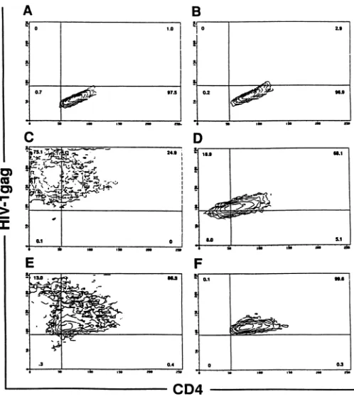
were examined by flow cytometry for the presence of Gag proteins in the cytoplasm. Minimal nonspe-cific staining for Gag was detected in permeabilized HIV-1-uninfected LCL-CD4 (Fig. 2A and B). Within 4days
ofTABLE 1. Measurement ofHIV-lLAI infectionby p24antigen
production in LCL transducedwith CD4'
p24antigenproduction(pg/ml)onpostinfectionday:
Volunteer
0 3 14 22
002F1I <31 374 >600 >600
103AFIC <31 92 >600 >600
103AFIB <31 182 >600 >600
a One million LCL-CD4were infected with 200 50% tissue cultureinfective doses ofHIV-l,AI, and culture supernatants were analyzed by p24 antigen
capture. p24 antigen control values were <31 pg/ml, and 600pg/ml was the
maximum p24 antigendetectablebytheassay.
J.VIROL.
0
on November 9, 2019 by guest
http://jvi.asm.org/
[image:3.612.326.562.628.688.2]USE OF LCL-CD4HIV-I TO DETECT HIV-1-SPECIFIC CTL RESPONSES 5077
B
0 ~~~~~~~~~~~~~~1.0
C__
U, 24.9
> _
4 r
- J
.30.4
0 L
i
.
u
" inaw.~~~~~~~~~~~~~.
D
'I~~~~~~~~~~~~.
s
~~~~~~~~~~~i
I ,
F
0.1 39.6'
i
b
I~~~~~~~~~~~~~~~~~~~~~~
_ 0 03
_ In see M no a"
[image:4.612.57.555.94.652.2]fnA
-fFIG. 2. Analysisbyflowcytometryof HIV-1infection and CD4 expression inCD4-transduced LCL (105 cells) fromtwodonors, depicted by
contourplots. LCL-CD4 before and after 4daysofinfection with eitherHIV-1LA1 orHIV-lSF-2were permeabilized, fixed, and stainedwith
PE-conjugatedKC57(y axis),amurine monoclonalantibodytothe HIV-1coreproteinp55,andFITC-conjugated anti-Leu-3A (x axis). (A) Subject 002F11, uninfected LCL-CD4. (B) Subject002FI6, uninfected LCL-CD4. (C) Subject 002F11, LCL-CD4 infectedwithHIV-1LA,. (D) Subject 002F16,LCL-CD4infected withHIV-1LAI. (E) Subject 002F11, LCL-CD4 infected withHIV-1SF-2. (F) Subject 002FI6, LCL-CD4 infectedwith
HIV-lsF-2-VOL. 68, 1994
A
C
aU
on November 9, 2019 by guest
http://jvi.asm.org/
5078 McELRATH ET AL.
infection with either HIV-1LAI or
HIV-lsF-2,
the majority of LCL-CD4 demonstrated Gag cytoplasmic staining (Fig. 2C to F). Only background or low levels of Gagcytoplasmic staining were detected in nontransduced LCL infected with HIV-1. Expression of envelope inLCL-CD4HIV-l
was demonstrated by both immunofluorescence and the formation of syncytia withSupTl cells, as described in Materials and Methods(data not shown). To investigate whether LCL-CD4 maintain their susceptibility to HIV-1 infection following cryopreservation, aliquots of LCL-CD4 were frozen in liquid nitrogen for more than 1 month, thawed, and then infected with HIV-1 as described above. The pattern of infection and HIV-1 gene expression was similar to that observed with fresh cells (data not shown). Thus, CD4 geneexpression is stable in fresh and frozen LCL-CD4, and such cells are uniformly susceptible to infection with HIV-1.The effect of HIV-1 infection on CD4 expression of the transduced LCL wasdetermined, since CD4+ cellsproducing
gpl20 have been reported to down regulate surface CD4 expression (7, 21). LCL-CD4 prior to and following infection were double-labeled with anti-CD4 and anti-Gag MAb and wereexamined by flow cytometry. CD4 was presentin double-labeled, uninfected LCL-CD4 (Fig. 2A and B). However, following in vitro infection with HIV-1, particularly with the LAI strain, CD4expression was reduced in a large portion of the transduced LCL (Fig. 2C toF).
Theability of LCL-CD4 to supportproductiveinfectionwas evaluated by assessing the supernatants of HIV-1-infected LCL-CD4 for the presence of infectious virus. Supernatants derived from cultured
LCL-CD4HIV-1
4 days after infection were filtered and transferred to uninfected LCL-CD4 and CEM cells, and the production of p24antigenwasmeasured. On day 4 after infection with the culture supernatants, LCL-CD4 and CEM cells produced 332 and.600
pg/ml ofp24, respectively, with control values of.60 pg/ml and maximum values of .600 pg/ml. Thus LCL-CD4 are susceptible to infection with vaccine prototype strains of HIV-1, express HIV-1 genes,and produce infectious virions.Use of
LCL-CD4HIV-l
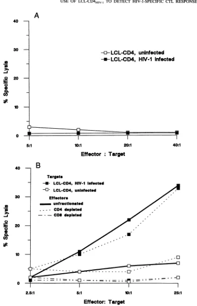
astarget cellsforCTL generatedfrom HIV-1 vaccines. CTLrecognizing envelope-expressing targets have been previously detected in an HIV-1-seronegative vac-cine recipient, designated002F11,
who had been immunized with a recombinant vaccinia virusexpressing HIV-1gp160and had been boosted with recombinant gp160 (5, 27). To deter-mine if HIV-1-infected LCL-CD4 could berecognizedby such envelope-specific effectors, PBMC were obtained from the vaccinee and stimulated in vitro for two 1-week cycles with HIV-1-infected autologous macrophages. The cytolytic effec-torcells generated were tested either unfractionatedor follow-ing enrichment for CD8+ T cells for lysis of autologousLCL-CD4HIV-1
targets. To determine that these target cells werelysable only by appropriateeffector cells, PBMC fromtwo seronegative individuals who had never received an HIV-1 vaccine and from another seronegative vaccine recipient who had notpreviously demonstrated any CTL activity to envelope-expressing targets were stimulated under similar conditions andtestedforlyticactivity. No CTL activity was detected from these control individuals against autologous LCL-CD4 in-fected with HIV-1LAI. Results from a representative CTL assay for one of theunvaccinated individuals are shown in Fig. 3A. In contrast, effectors from vaccine recipient002F11,
in whom we had previously detected envelope-specific CTL, demonstrated specific lysis of autologousHIV-1-infected LCL-CD4 targets at an effector-to-target ratio (E/T) aslow as 5:1, with minimal lysis of the uninfected autologous LCL-CD4 target (Fig. 3B). CD8+ rather than CD4+ T cells wereprimarily responsible for the lytic activity (Fig. 3B), as demon-strated after enrichment using antibody-coated plates. Thus,
LCL-CD4HIV-l
appear useful as autologous target cells to determine if the CTL activity induced by candidate HIV-1 vaccinescan lyseHIV-1-infected cells.Theanalysis of CTL responses toindividual HIV-1proteins in vaccine recipients has been complicatedby the presence of high levels of vaccinia-virus-specific lytic activity which inter-feres with the use of target cells expressing HIV-1 genes following infection with recombinant vaccinia viruses. This problem with identification of CTL responses has been exag-gerated when recombinant vaccinia viruses have been usedto elicit the invivo and in vitro responses. Therefore, we exam-ined whether the env-specific CTL precursors present in PBMC from thepreviously described vaccinee 002F11 could be amplified in vitro by stimulation withv-env-infected autologous PBMC and detected by testing for lytic activity with HIV-1-infected LCL-CD4. To ensure thatthe recognition andlysis of
CD4-LCLHIV-1
reflected specific activation ofprimed precur-sors and not an in vitro primary response, PBMC were similarly stimulated withv-gag-infected PBMC. Minimal lysis was detected against theLCL-CD4HIV-1
targets by effectors that were generated following stimulation withv-gag-infectedcells, suggesting that lytic activity reflected neither T-cell effectors generatedbyprimary in vitro stimulationnor nonspe-cific effectors activated by vaccinia virus (Fig. 4A). However, v-env-stimulated effectors lysed
LCL-CD4HIV-1
targets at anE/Taslow as 5:1, and didnotlyse uninfected LCL-CD4 targets (Fig. 4A). The MHC restriction of these CTL responseswas evaluated by testing the effector population against both autologous andallogeneic(mismatched atall classIandclass II loci)
LCL-CD4HIV-1
andLCL-CD4 targets.Althoughsome lytic activity was detected against the allogeneicLCL-CD4HIV-j
athigherE/Ts (16% at anE/Tof40:1), presumably reflecting the presence of small numbers ofnonspecific cyto-lytic effectors such as NK cells or ADCC activity, this was significantly lower than the lytic activity observed for the autologousLCL-CD4HIV-
target(37%at anE/Tof40:1) (Fig. 4B). These results indicate that use ofLCL-CD4HIV-1
may provide a means to more efficiently analyze HIV-1-specific CTL responses in recipients of recombinant vaccinia virus vaccines.Use of
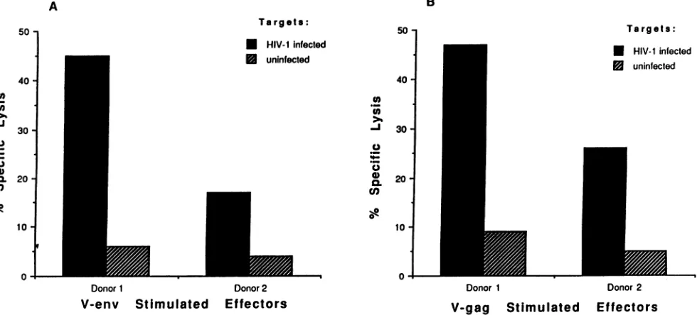
LCL-CD4HIV-l
as targetcells to assess CTLactivity in seropositive individuals. Our studies demonstrated that LCL-CD4HIVl produceinfectious virions and express multiple HIV-1 genes and thus should be usefulastargets to assessthe broad range of responses to many HIV-1 gene productsexpressed in infected cells in HIV-1-infected individuals. Therefore, PBMC from two HIV-1-seropositive individuals (103AFIC and 103AFIB)werestimulated with eitherv-env- or v-gag-infected autologous PBMC and were tested for lytic
activity against autologous LCL-CD4 and
LCL-CD4HIV-1.
Lysis ofthe uninfected LCL-CD4 was minimal at an E/T of 20:1, whereas CTL reactive to env and gag gene productsexpressed by the autologous
LCL-CD4HIV-1
target cells from both donors were demonstrable following amplification with v-env- and v-gag-infected stimulators, respectively (Fig. 5).Direct assays of CTL responses in fresh PBMC have been useful formonitoringimmunity in HIV-1-infected individuals. Toevaluate whether
LCL-CD4HIV-1
could be usedastargets in direct CTL assays in HIV-1 seropositive individuals, freshlyisolated PBMC were tested for lytic activity against
LCL-CD4HIV-l
and against LCL-CD4 expressing HIV-1 genes in recombinant vaccinia viruses. A representative experiment from anHIV-1-infected individual (103AFIB) isshown inFig. 6. No lytic activity was detected against the uninfectedLCL-J.VIROL.
on November 9, 2019 by guest
http://jvi.asm.org/
A
-E-
LCL-CD4,
uninfected
-*- LCL-CD4, HIV-1 Infected
T
10c1 20c1
Effector
; Target
B
Targets
-U- LCL-CD4, HIV-1 Infected -0- LCL-CD4, uninfected
Effectors unfractionated
- - - - CD4 depleted
- - - CDI depleted
--U.
0
2.5c1 5c1 lOci 25cl
Effector:
Target
FIG. 3. Evaluation of HIV-1-specific CTL in two HIV-1-uninfected individuals. CTL precursors were stimulated for 2 weeks with
HIV-1-infectedmacrophages andweretestedatawideE/Tinastandard4-h chromium releaseassay.(A)HIV-1-specific CTL inanindividual
who had neverreceived an HIV-1 candidate vaccine. Effectorswere tested against uninfected or HIV-1-infected (strain SF-2orstrain MN)
LCL-CD4. (B) HIV-1-specific CTL froman individual (002F1l) receiving recombinant vaccinia virus containingHIV-1,A,gpl60 followed by
rgpl6O boosting. Effectors, including unfractionated, CD4-depleted, and CD8-depleted populations, were tested against autologous CD4-transducedLCLtargets,either uninfectedorHIV-1LAI-infected.
40
30
a
I.-0
0
a~
20
10
-0
-scl
-41 40t1
40
30
a a
0%1 0 cn
at
00.
CO) 20
10
0
-I
on November 9, 2019 by guest
http://jvi.asm.org/
[image:6.612.117.514.53.669.2]5080 McELRATH ET AL.
A B
-* LCL-CD4, HIV-1 infected
_-- LCL-CD4, uninfected Effectors
30 - v-env stimulated - v-gag stimulated
10:1
40
-'In
._1
0
=~
20-0.
Co
10-0
20:1
Targets LCL-CD4
- autologous, HIV-1 infected
* autologous,uninlected -.--.-- allogeneic,HIV-1infected
.---- allogeneic,uninfected
A'
5....~~~~~~~~~~~~~
5:1 10:1 20:1
IT S-_4&-. Tafmrwft
Effector : Tarqet
rttector
: *argeiFIG. 4. Analysis of CTL againstHIV-1-infected targets in a seronegative vaccinee(002F11)by in vitrostimulation of effectors for 9 days with autologous PBMC infected with v-env or v-gag. (A) Autologous LCL-CD4 targets were either uninfected or infected with HIV-1LAI. (B) MHC-restrictedCTLactivity was assessed by the ability of the v-env- or v-gag-stimulated effectors to lyse autologous versus allogeneic LCL-CD4 targets(mismatched at all class I andIIloci) that were either uninfected or infected withHIV-1LAI.
CD4, affirming that expression of HIV-1 genes in the target cellwasessential for detection oflyticactivity.Specific lysis was demonstrated in this direct assay against the HIV-1-infected LCL-CD4 at an E/Taslow as 25:1. Moreover, nodirect lytic activity against v-env- or v-gag-infected LCL was detected in this assay, despite the fact that such targets are efficiently recognized by in vitro activated CTL (27). Similar results in which
LCL-CD4HIV-l
were moresensitive to lysis byunstimu-A
lated effectors obtained directly from peripheral blood lym-phocytes in two other HIV-1-infected individuals were ob-served. These data suggest that HIV-1-infected LCL-CD4 may be useful astargetscells in direct assays of activated cytolytic effectors fromtheperipheralblood ofseropositive individuals and may improve sensitivity, since lysis of this target reflects the collective activities of effectors recognizing all HIV-1 gene products.
B
Targets: * HIV-1 infected
0 uninfected
507
40 -0
>1
-J
0._0 n
_o
oL 30
-
20-10
-0
-t---Targets: * HIV-1 infected
* uninfecled
Donor1
V-gag Stimulated
Donor 2
Effectors FIG. 5. CTLresponsesagainst LCL-CD4targets,uninfectedorinfected with HIV-lLAI, intwoHIV-1-infected subjects: donor1,103AFIC,and donor2, 103AFIB. Bulk PBMCwerestimulated with autologous PBMC infected with eitherv-envor v-gagfor 9days. Percentspecific lysis is presentedatanE/T ratio of 20:1 and shown for v-env-stimulated effectors (A) and v-gag-stimulated effectors (B).
40
-i
0
0.
cn
20
10
-0
40:1
50
40
-._a
0n
30
-J
0 0. 20-Co
Donor1 Donor 2
V-env Stimulated
Effectors
J. VIROL.
on November 9, 2019 by guest
http://jvi.asm.org/
[image:7.612.69.556.71.300.2] [image:7.612.74.564.468.690.2]30
-._a
-J 0 ._
U) 0. o,
20
10
-0
Targets LCL-CD4
.
-U--U
50
HIV-infected
uninfected
40 v-envinfected
v-gaginfected
I
a
12.5:1 25:1 50:1
(a
.n -i
._
0
U)
0.
q)
100:1
Effector : Target
FIG. 6. Direct CTL analysis in an HIV-1 infected individual,
103AFIB. Fresh PBMCweretestedatmultiple E/Ts inastandard 4-h chromium release assayagainst autologous LCL-CD4 uninfected or
infected withv-env, v-gag,orHIV-1LAI.
LCL-CD4HWVl1
asstimulatorcellstoinduce CTLresponses. The ability ofLCL-CD4HIV-l
tofunctionasstimulator cells forthe amplification of HIV-specific CTL precursors was also
examined. PBMC obtained from both seropositive and
sero-negative vaccinees were stimulated with
LCL-CD4HIV-l
andtested forlysis againstLCL and fibroblastsinfected with either
v-env, v-gag, or vac, and uninfected or HIV-1-infected
LCL-CD4. PBMC from theseropositive individual 103AFIC, stim-ulated with LCL-CD4HIV-l and enriched for CD8+ effector cells, lysed autologous LCL and fibroblasts expressing HIV-1
gag (Fig. 7). (Lysis of target cells expressing HIV-1 gene
productswasnotdetected in adirect assay offresh
unstimu-lated PBMC.) Substantially lower levels oflytic activity were
observed with target cells expressing HIV-1 env rather than
HIV-1gag,consistent with several studiessuggesting thatgagis
the major target of the CD8+ CTL response in seropositive individuals (12). The vaccinia-virus-infected LCLwere more
readily lysed as targets than were fibroblasts. In addition,
HIV-1-infected LCL-CD4, butnot the uninfectedLCL-CD4,
werelysed bythe induced effectorpopulation, suggesting that an HIV-1-specific response could be distinguished from a response toEBVexpressed inthe LCL.
Instudies with PBMC derived fromseronegative vaccinees,
theLCL-CD4HIV-l provedtobe unsuitableasstimulator cells
because of the presence of strong EBV-specific responses.
Induction of high levels of EBV-reactive lytic activity was
detected in these cultures, resulting in high background lysis againstthe vac-infected LCL and uninfectedLCL-CD4targets
(datanotshown). Thus,theuseofLCL-CD4HIV-las stimula-torcellsmaybe restrictedtoassaysdesignedtoamplifyeffector cells from individuals who lack sufficient immunocompetence togenerate strong EBV-specific responses.
DISCUSSION
Thisreport describes a methodto produce human
autolo-gouscell lines thatcanbereproduciblyinfected with HIV-1.By transducing EBV-transformed LCL with theLT4SN retroviral
30-20
10
-O v-envinfected
2
v-saginfected M vac-infected * HIV-1 infected15
uninfectedFibroblasts
Targets
FIG. 7. Use of
LCL-CD4HIV-1
asstimulator cells to amplify HIV-specific CD8+ CTL precursors from an HIV-1-infected subject, 103AFIC. The effector population was stimulated with autologousLCL-CD4HIV-l for 9 days and was enriched for CD8+ cells by
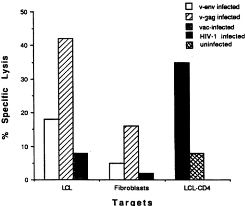
adherence to anti-CD4-coated flasks prior to testing. Autologous targetstested include LCL andfibroblasts infected with v-env, v-gag, andvac andLCL-CD4, either uninfected or infected withHIV-1,A1.
Percentspecificlysis is presented at anERTof 20:1.
vector,it was possible to generate stable B-celllines expressing CD4, and such LCL-CD4 becamesusceptibletoinfection with severalHIV-1 strains.Followinginfection, the
LCL-CD4HIVI1
remained viable for more than 2 weeks, expressed multiple HIV-1 genes, and produced infectious virions. The ability to produce such autologous infectable permanent cell lines pro-vides many opportunities for the study of HIV-1-specific immunity.The analysis of CTL responses inuninfected, immunocom-petentHIV-1vaccinerecipients participatinginseveral clinical trials (5, 15) has been obstructed by the paucity of shuttle vectorsavailable toefficiently express HIV-1 genes in autolo-gous cells. The most common approach for detecting HIV-1
envelope-specific CTL in the recipients of envelope vaccines
has been to use autologous cells infected with vaccinia virus recombinants expressing HIV-1 envelope gene products as stimulator and target cells. Frequently, the envelope-specific
CTL responses have been difficult to distinguish from the high-levelvaccinia-virus-specific CTL responses in the immu-nocompetentindividuals withpreviousvaccinia virus exposure or who have been recently immunized with recombinant vaccinia virus vaccines. The
LCL-CD4HIV-1
described in thisstudyexpress no vaccinia virusantigens and thuscan
poten-tiallycircumvent thisproblem. Our results demonstratethat,
following amplificationof PBMCwith v-env-infected
stimula-tors,even in individuals recently immunized with a recombi-nant vaccinia virus,
LCL-CD4HIV-l
proved to be sensitive targets to detect MHC-restricted HIV-1-specific CD8+ CTL without interferencebyvaccinia virus responses.LCL-CD4 may have increased utilitywith thenext genera-tion of candidate HIV-1 vaccines, which will likely contain multiple HIV-1 gene products in the context of either a
recombinant virus, a particle containing several HIV-1
pro-teins, orpossiblyan attenuatedvirus (8, 16). Thus, it maybe
necessary to test recipients for CTL responses to
multiple
on November 9, 2019 by guest
http://jvi.asm.org/
[image:8.612.316.555.73.274.2] [image:8.612.57.293.74.283.2]5082 McELRATH ET AL.
HIV-1 gene products. Our results demonstrate that
LCL-CD4HIV-1
can be employed as a single target to detect CTL effector responses inHIV-1-seropositive individualstoseveral HIV-1 gene products. Therefore, for analysis ofresponses tocomplex vaccines,HIV-1-infected LCL-CD4 may prove useful
as a screening target, and, if lytic activity is demonstrated,
subsequent detailedanalysesofactivity against specificHIV-1
proteins could beperformed.
LCL-CD4 should behelpful formonitoring CTLactivityin HIV-1-infected individuals. A decline in CTL responses to
HIV-1-specific
proteins in seropositive individuals as HIV-1 disease progresses (14, 17) has been demonstrated, but little information is available concerning the extent to which lyticactivity
is lostagainstHIV-1-infected cells.Althoughthesearepresumably related, the analysis of the response to a single
protein will vary in individuals possessing different MHC
haplotypes
andinfected with different strains.Thus,longitudi-nal monitoring of changes in CTL activity against
HIV-1-infected cells such as
LCL-CD4HIV-1
may provide a more accuratemeans ofassessing the relationship of thedecline in CTL responses and diseaseprogression.
In addition tousingLCL-CD4HIV-1
in suchstudies of the naturalhistoryofHIV-1infection,
it may be informative to include it in the panel of target cells used to evaluateaugmentation
of immunere-sponsesfollowing
immunotherapy
ofseropositiveindividuals,
particularly
if, as in previousstudies,
theimmunogen
is arecombinant
protein
expected topredominantly
augmentCD4+ helper
responses. Inthissetting,
theaugmentation
of thehelper
response may beexpected
toamplify
all CTL responses, and thus it will be necessary to evaluate the full spectrum ofCD8+ Tcellsspecific forHIV-1proteins
(23).It has been well documented that many HIV-1 mutations result from the frequent errors in reverse transcription, and that some ofthese mutants may
subsequently
be selected in treated hosts because of resistance to antiretroviral therapy(24, 36)
orthrough
loss of theepitope
recognizedby
autolo-gousneutralizing
antibodies(1,
2).Similarly,
viralmutantsmay evade eliminationby
CTL in HIV-1-infected individualsthrough
an alteration in the epitopes that interact with thepresenting
class I MHC molecule or the T-cell receptorrecognizingtheMHC-peptide complex(20, 35). However,the
importance
of viral escape from recognition byhost CTL in diseaseprogression
has been difficult to assess, since it hasrequired
molecularmappingof theepitopefromaninitial viral isolaterecognized byhostCTL, followedbyisolation of virusduring
periodsofdiseaseprogressionforcloningandsequenc-ing
of the relevant gene(35).
Longitudinalmonitoring
of CTL responsesin HIV-1-infected individualsmightbemoresimplyachieved
by studying
CTLreactivitywithautologous
LCL-CD4 infected with serialautologous
HIV-1 isolates.Inthissituation,
thefailure ofaviralisolatetosensitizeatargetfor
lysis
bythe CTL wouldimply
thatepitope
selection had occurred as onepossibility,
and inonly
selected instanceswould thisneedtobe confirmed by cloning and sequencing. In this paper we have demonstrated that LCL-CD4 can be infected with severallaboratory
strains ofHIV-1,andfuture studies will needtobeperformed
to determine theefficiency
with which clinical isolatescan infect these target cells.LCL-CD4HIV-1
may also have certain advantages as stimu-lator cells in the amplification of HIV-1-specific CTL in HIV-1-infected individuals. CTL precursor responses tomul-tiple
HIV-1 geneproductscanbeamplifiedwith the use of asingle
stimulatorcell,and itshould thus bepossibletoanalyzeand
quantify
HIV-1-specificresponsesby usingLCL-CD4HIV-1
as stimulator cells in limiting dilution precursor frequency assays and forcloning.
After such clones are generated, thefinespecificity for individualHIV-1 proteinscanbe identified and confirmed by testing with targets expressing individual HIV-1 genes.
Inconclusion,ourstudies describeamethodtoestablish cell
lines from any individual that can serve as an autologous sourceofhighlysusceptible HIV-1-infected cells. We believe
thatsuchLCL-CD4HIv
1will
have broadutilityin theevalua-tion ofCTLresponsesinducedby candidate vaccinesdesigned
to prevent HIV-1 infection and in the study of the natural
history of HIV-1 infection and immunological intervention
withseropositive individuals.
ACKNOWLEDGMENTS
Theauthors express appreciationtothe vaccinestudyparticipants and toDavidBerger,Jacqueline Burke, Nancy Coomer,Pam Easter-ling,andLawrenceCoreyfor support and assistance with this investi-gation.
This work wassupported by National Institutes of Health grants AI-26503, AI-27757,AI-30238,andAI-05065.
REFERENCES
1. Albert, J.,B.Abrahamsson, K. Nagy, E.Aurelius, H.Gaines, G. Nystrom, and E. M.Fenyo. 1990. Rapid development of isolate-specific neutralizing antibodies after primary HIV-1 infection and consequentemergence of virus variants which resist neutralization by autologoussera.AIDS 4:107-112.
2. Arendrup, M., C. Nielsen, J. E. S. Hansen, C. Pederson, L. Mathiesen, and J. 0. Nielsen. 1992.Autologous HIV-1 neutraliz-ing antibodies: emergence of neutralization-resistant escape virus andsubsequent development of escape virus neutralizing antibod-ies. J.Acquired Immune Defic. Syndr. 5:303-307.
3. Bassin, R. H., N. Tuttle, and P. J. Fischinger. 1971. Rapid cell culture assay technique for murine leukaemia viruses. Nature (London)229:564-566.
4. Cooney, E. L., A. C. Collier, P. D. Greenberg, R. W. Coombs,J. Zarling, D. E. Arditti, M. C.Hoffman, S. L. Hu, and L. Corey. 1991. Safety of and immunological response to a recombinant vaccinia virus vaccine expressing HIV envelope glycoprotein. Lancet337:567-572.
5. Cooney,E.L.,M.J.McElrath, L.Corey,S. L.Hu, A.Collier,D. Arditti, M. Hoffman, R. W. Coombs, G. E. Smith, and P. D. Greenberg. 1993. Enhanced immunity to HIV envelope elicitedby a combined vaccineregimenconsisting of priming with a vaccinia recombinant expressing HIV envelope and boosting with gpl60 protein. Proc. Natl. Acad. Sci. USA 90:1882-1886.
6. Dahl, K., K. Martin, and G. Miller. 1987. Differences among humanimmunodeficiencyvirus strains intheircapacities toinduce cytolysis of persistent infection of a lymphoblastoid cell line immortalizedby Epstein-Barr virus. J. Virol. 61:1602-1608. 7. Dalgleish, A. G., P. C. L. Beverley, P. R.Clapham, D. H. Crawford,
M. F.Graves, and R. A. Weiss. 1984. The CD4(T4) antigenis an essential component of the receptor for the AIDS retrovirus. Nature (London) 312:763-767.
8. Daniel, M. D., F. Kirchlof, S. C.Czajak,P. K.Sehgal, and R. C. Desrosiers. 1992. Protective effects of a live attenuated SIV vaccine withadeletion inthenef gene. Science 258:1938-1941. 9. DeRossi, A., S. Roncella, M. L. Calabro, E. D'Andrea, M. Pasti,
M.Panozzo, F.Mammano,M.Ferrarini, andL.Chieco-Bianchi. 1990.Infection ofEpstein-Barrvirus-transformedlymphoblastoid B cells by the human immunodeficiency virus: evidence for a persistent and productiveinfection leadingto B cellphenotypic changes.Eur. J.Immunol. 20:2041-2049.
10. Fauci,A. S.,R. C.Gallo, S. Koenig, J. Salk, and R. H. Purcell. 1989.Developmentandevaluation ofavaccine for human immu-nodeficiency virus (HIV infection). Ann. Intern. Med. 110:373-385.
11. Gartner, S., P.Markovits,D. M.Markovitz, M. H. Kaplan,R.C. Gallo,and M.Popovic. 1986. The role of mononuclearphagocytes inHTLV-III/LAVinfection. Science 233:215-219.
12. Gotch, F.M., D.F. Nixon, N. Alp, A. J. McMichael,and L.K. Borysiewicz. 1990. High frequency of memory and effector gag J. VIROL.
on November 9, 2019 by guest
http://jvi.asm.org/
specificcytotoxic lymphocytes in HIV seropositive individuals. Int. Immunol. 2:707-712.
13. Graham, B. D., T. J. Matthews, R. B. Beishe, M. L. Clements, R. Dolin, R. F. Wright, G. J. Gorse, D. H. Schwartz, M. C. Keefer, D. P.Bolognesi, L. Corey, D. M. Stablein, J. R. Esterlitz, S. L. Hu, G. E. Smith, P. E. Fast, W. C.Koff, and the NLIAD AIDS Vaccine Clinical Trials Network.1993. Augmentation of HIV-1 neutraliz-ing antibody by priming with gpl60 recombinant vaccinia and boosting withrgpl6O.J.Infect. Dis.167:533-537.
14. Guillon, J.-M., B. Autran, M.Denis,M.P.Fouret, F. Plata, C. M. Mayaud, and G. M. Akown. 1988. Human immunodeficiency virus-related lymphocytic alveolitis. Chest 94:1264-1270. 15. Hammond, S. A., R. C. Bollinger, P. E. Stanhope, T. C. Quinn, D.
Schwartz, M. L. Clements, and R. F.Siliciano.1992.Comparative clonalanalysis ofhuman immunodeficiency virus type 1 (HIV-1)-specificCD4+ and CD8+ cytolytic T lymphocytes isolated from seronegativehumans immunized with candidate HIV-1 vaccines. J. Exp. Med.176:1531-1542.
16. Haynes, B. F. 1993. Scientific andsocial issues of human immu-nodeficiency virusvaccine development. Science 260:1279-1286. 17. Hoffenbach, A., P.Langlade-Demoyen, G. Dadaglio, E. Vilmer, F.
Michel, C. Mayaud, B.Autran, and F. Plata. 1989.Unusually high frequencies of HIV-specific cytotoxicTlymphocytes in humans. J. Immunol. 142:452-462.
18. Hu, S. L.,S. G.Kosowski, and J. M. Dalyrmple. 1986.Expression of AIDS virusenvelope in recombinant vacciniaviruses. Nature (London) 320:537-539.
19. Hu, S.-L., B. M.Travis, J. Garrigues, J. M. Zarling, P. Sridhar, T. Dykers, J. W.Eichberg, and C. Alpers.1990. Processing,assembly, andimmunogenicity of human immunodeficiencyvirus core anti-gensexpressed by recombinant vacciniavirus.Virology 179:321-329.
20. Kalams, S. A., R. P.Johnson,A.K. Trocha, M. J. Dynan, H. S. Ngo, R. T. Aquila, J. T. Kurnick, and B. D. Walker. 1994. Longitudinal analysis of T cell receptor (TCR) gene usage by human immunodeficiency virus 1 envelope-specific cytotoxic T lymphocyteclones revealsalimited TCRrepertoire. J. Exp. Med. 179:1261-1271.
21. Klatzmann,D., F.Barre-Sinoussi, M. T. Nugeyre, C. Danguet, E. Vilmer, C.Griscelli, F.Brun-Vazinet, C. Rouzioux, J. C. Gluck-man,J.C. Chermann, and L. Montagnier.1984. Selectivetropism oflymphadenopathyassociatedvirus(LAV) for helper-inducerT lymphocytes.Science 225:59-63.
22. Koff, C. W., and F. D. Hoth. 1988. Development and testing of AIDSvaccines. Science241:426-432.
23. Kundu, S. K., D. Katzenstein, L. E. Moses, and T. C. Merigan. 1992. Enhancement of human immuniodeficiency virus (HIV)-specific CD4+ and CD8+ cytotoxic T-lymphocyte activities in HIV-infected asymptomatic patients given recombinant gpl60 vaccine. Proc. Natl. Acad.Sci.USA89;11204-11208.
24. Larder, B. A., B. Darby, and D. D. Richman. 1989. HIV with reduced sensitivitytozidovudine(AZT)isolated duringprolonged therapy. Science 243:1731-1734.
25. Levy, J. A., J. Shimabukuro, T. McHugh, C. Casavant, D. Stites, and L. Oshiro. 1985. AIDS-associated retrovirus (ARV) can productively infect other cells besides human T helper cells. Virology147:441-448.
26. Maddon, P.J., A. G.Dalgleish,J. S. McDougal, P. R.Clapham,
R.A.Weiss, andR.Axel. 1986. The T4 gene encodes the AIDS
virus receptor and is expressed in the immune system and the brain. Cell 47:333-348.
27. McElrath, M. J., M.Hoffman, S. Klucking, L. Corey, and P. D. Greenberg.HIV-infected macrophages as efficient stimulator cells for detection ofcytotoxic T cell responses to HIV in seronegative and seropositive vaccine recipients. AIDS Res. Hum. Retrovi-ruses, inpress.
28. Miller, A. D., and C. Buttimore. 1986. Redesign of retrovirus packagingcell lines toavoid recombinationleading to helper virus production.Mol. Cell. Biol.6:2895-2902.
29. Miller, A. D., and G.Rosman. 1989. Improved retroviralvectors forgenetransferandexpression.BioTechniques 7:980-990. 30. Miller, D. 1992. Human gene therapy comes of age. Nature
(London) 357:455-460.
31. Montagnier, L., J. Gruest, S. Chamaret, C. Dauguet, C.Axler,D. Guetard, M. T. Nugeyre, F. Barre-Sinoussi, J. C. Chermann, J.B.
Brunet, D.Klatzmann,and J. C.Gluckman. 1984.Adaptation of lymphadenopathy associated virus(LAV)toreplication in EBV-transformed Blymphoblastoidcell lines.Science225:63-66. 32. Nara, P. L. 1990. Quantitative infectivity syncytium-forming
mi-croassay, p. 77-86. In A. Aldovini and B. D. Walker (ed.), Techniques in HIV research. Stockton Press, New York. 33. Nilsson, K., and G. Klein. 1982. Phenotypic and cytogenetic
characteristicsof human Blymphoidcell lines andtheirrelevance for theetiologyofBurkitt'slymphoma.Adv. Cancer. Res. 37:319-380.
34. Orentas, R. J., J. K. E. Hildreth,E. Obah, M.Polydefkis, G. E. Smith, M. L. Clements, and R. F. Siliciano. 1990. An HIV envelopeprotein vaccine induces CD4+ human cytolytic Tcells activeagainst HIV-infected cells.Science 248:1234-1237. 35. Philips,R.E., S.Rowland-Jones, D. F. Nixon, F. M. Gotch, J. P.
Edwards, A.0.Ogunlesi,J. G.Elvin, J.A.Rothbard, C. R. M.
Bangham, C. R. Rizza, and A. J. McMichael. 1991. Human immunodeficiency virusvariants that escapecytotoxicT-cell rec-ognition.Nature(London)354:453-459.
36. Richman, D., C. K. Shih, I. Lowy, J. Rose, P. Prodanovich, S. Goff, and J. Griffin. 1991. Human immunodeficiency virus type 1 mutants resistant to nonnucleoside inhibitors of reverse tran-scriptaseintissue culture. Proc. Natl. Acad. Sci. USA 88:11241-11245.
37. Riviere, Y., F. Tanneau-Salvadori, A. Regnault, 0. Lopez, P. Sansonetti, B.Guy, M. P. Kieny, J. J. Foumel, and L.Montagnier. 1989. Humanimmunodeficiencyvirus-specificcytotoxic responses ofseropositiveindividuals: distinct typesofeffector cells mediate killing oftargets expressing gag and env proteins. Virology 63:
2270-2277.
38. Salahuddin, S. Z., D. V.Ablashi,E. A.Hunter,M. A.Gonda, S. Sturzenegger, P.D.Markham, andR.C.Gallo. 1987. HTLV III infection ofEBV-genome-positive B-lymphoidcells withor
with-outdetectable T4antigens.Int.J.Cancer39:198-202.
39. Sugden, B., and W. Mark. 1977. Clonal transformation ofadult human leukocytes byEpstein-Barrvirus. J. Virol. 23:503-508. 40. Tozzi, V., S. Britton, A. Ehrnst,R. Lenkei, and0. Strannegard.
1989. PersistentproductiveHIVinfection inEBV-transformedB lymphocytes.J.Med. Virol. 27:19-24.
41. Walker,B.D., S.Chakrabarti, B. Moss, T. J. Paradis,T.Flynn, A. Durno, R. S.Blumberg, J. C. Kaplan, M. S. Hirsch,and R.T.
Schooley. 1987. HIV-specific cytotoxic T-lymphocytesin seropos-itive individuals. Nature(London)328:345-348.