A DESCRIPTIVE STUDY ABOUT ABDOMINAL
TUBERCULOSIS IN GOVERNMENT RAJAJI
HOSPITAL MADURAI
Dissertation Submitted for
MS DEGREE (BRANCH I) GENERAL SURGERY
APRIL 2012
The Tamilnadu Dr.M.G.R Medical University
Chennai – 600 032.
CERTIFICATE
This is to certify that this dissertation titled “A DESCRIPTIVE STUDY
ABOUT ABDOMINAL TUBERCULOSIS IN GOVERNMENT RAJAJI
HOSPITAL” submitted by Dr. JAYAPRAKASH .N to the faculty of General
Surgery, The TamilNadu Dr. M.G.R. Medical University, Chennai in partial
fulfillment of the requirement for the award of MS degree Branch I General
Surgery, is a bonafide research work carried out by him under our direct
supervision and guidance from June 2009 to May 2011.
Prof. Dr. M.Gobinath, M.S., Prof. Dr. Dr.G.V.Rajambigai M.S.,FACRSI., Professor and Head of the Department, Professor & Unit Chief,
Department of General Surgery, Department of General Surgery, Madurai Medical College, Madurai Medical College,
Madurai. Madurai.
DECLARATION
I, Dr. JAYAPRAKASH . N solemnly declare that the dissertation titled “A
DESCRIPTIVE STUDY ABOUT ABDOMINAL TUBERCULOSIS IN
GOVERNMENT RAJAJI HOSPITAL” has been prepared by me. This is
submitted to The TamilNadu Dr. M.G.R. Medical University, Chennai, in
partial fulfillment of the regulations for the award of MS degree (Branch I) General
Surgery.
Place: Madurai Dr. JAYAPRAKASH . N
ACKNOWLEDGEMENT
At the outset, I wish to express my sincere gratitude to our unit chief Prof.
Dr.G.V.RAJAMBIGAI M.S, FACRSI., for her expert supervision and valuable
suggestions. I wish to express my whole hearted thanks to our assistant Professors
Dr.P.Sundareswari M.S.,D.G.O., Dr. J.Ravishankar M.S., Dr.Celine Foustina
Mary M.S, D.G.O, for their constant encouragement and excellent guidance.
My sincere gratitude to my former unit chief Prof. Dr.R.VIJAYAN M.S.,
for his valuable motivation and guidance to initiate this study.
I wish to thank Prof.Dr. M.GOBINATH, M.S., Professor and Head of the
Department of Surgery for his valuable guidance and advices. I am greatly
indebted to Prof.Dr. A.EDWIN JOE, M.D (FM), B.L., Dean, Madurai Medical
College & Government Rajaji Hospital, Madurai for their kind permission to
allow me to utilize the clinical material from the hospital.
I whole heartedly thank all the patients who willingly co-operated and
rendered themselves for the study without which the study couldn’t have been a
CONTENTS
No. TITLE
PAGE No.
1.
INTRODUCTION
1
2.
REVIEW
OF
LITERATURE
3
3.
AIMS & OBJECTIVES
34
4.
MATERIALS
AND
METHODS
35
5.
RESULTS
37
6.
TABLES
AND
FIGURES 40
7.
DISCUSSION
50
8.
CONCLUSION
59
ANNEXURES
BIBLIOGRAPHY
PROFORMA
MASTER CHART
INTRODUCTION
Tuberculosis is a disease caused by the bacterium Mycobacterium species
which is one of the commonest disease known to mankind. It is a well known fact
that it has its reputation of being one of the greatest killer diseases. There has been
a trend of increased incidence of tuberculosis in human race in the last three
decades which can be partly attributed to increase in population, social deprivation
and HIV infection.
The morbidity and mortality due to tuberculosis leads to a discovery of
newer drugs and more emphasis on the disease. With improved socioeconomic
conditions and disease diagnosis in the western countries, Tuberculosis in the
western countries impose little clinical problem. But in most developing countries
like India, it remains to be a major health hazard and major health related
mortality, morbidity both physically and psychologically. As we know,
Tuberculosis is one of the important gastroenterological problems in Tropics.
Gastrointestinal Tuberculosis is one of the earliest known diseases which
still remain the disease with diagnostic enigma due to its perplexing protean
clinical manifestations. Importance of the condition lies in the diversity of the
presentations and its wide spread effects on the body. It may present as acute,
subacute or chronic forms and may give rise to malabsorption, more so when
The symptoms and signs often quite vague and laboratory investigations
and radiological findings are sometimes non conclusive. There is no single feature
which is diagnostic for abdominal Tuberculosis. In case of any localized
involvement of the structures of the abdomen the presenting clinical picture will
mimic the disease of the organ only. In various studies about abdominal
tuberculosis worldwide, the results show wide range in its disparity in the nature
course of disease, in its diagnosis and management. We planned a study on
abdominal tuberculosis to understand the nature of disease process and to evaluate
the various laboratory and radiological investigations and to study the management
of the disease.
REVIEW OF LITERATURE
The diagnosis of abdominal tuberculosis is obscure. Joseph Walsh in
1909 remarked that “It is impossible to diagnose abdominal tuberculosis with
any degree of certainty” 23.Unfortunately this remains even today in situations
where it is relatively common. The signs and symptoms are often vague and
laboratory investigation and radiological findings are non-specific. Many cases go
unnoticed until a surgically removed specimen is examined histopathologically. A
majority of stenotic lesions of the bowel are of tuberculous in nature and has been
found on the operating table has one of the common findings in cases of acute
intestinal obstruction.1
The term abdominal tuberculosis refers to tuberculous infection of the
gastrointestinal tract. mesenteric lymph nodes, peritoneum and omentum, and of
solid organs related to the gastrointestinal tract such as the liver and spleen.1
Various pathological forms of abdominal tuberculosis are
I. Peritoneal tuberculosis—acute or chronic
A. Tuberculosis of the peritoneum
(i) Wet or ascitic type: Generalized, Localized (loculated)
(ii) Dry or fibrous type:Adhesive type, Plastic type, Miliary nodular
B. Tuberculosis of peritoneal folds and contents viz. Mesenteric adenitis,
Mesenteric cysts, Mesenteric abscesses, Bowel adhesions and rolled-up omentum.
II. Gastrointestinal
1. Ulcerative
2. Hypertrophic or hyperplastic
3. Sclerotic or fibrotic
III.Tuberculosis of solid viscera (e.g. liver and spleen)
Epidemiology of abdominal tuberculosis
Starting from the mid-1980s a resurgence of tuberculosis occurred; due to a
large extent to the epidemic of acquired immunodeficiency disease. Intestinal
tuberculosis is seen more frequently in people of poor socioeconomic
circumstances. Its incidence is higher in patients with caseo-pneumonic and
advanced lung disease than in those with fibrotic lesions and early disease. The
frequency of extrapulmonary tuberculosis has increased, largely an influence of
human immunodeficiency virus (HIV) infection; over 50 per cent of individuals
with HIV and tuberculosis develop extrapulmonary disease compared to 10 to 15
per cent in those without HIV2. Finally, there is a marked increase in the
emergence of multidrug-resistant M. tuberculosis organisms, a cause of much
Microbiology
Mycobacterium tuberculosis is responsible for nearly all cases of abdominal
tuberculosis. Other pathogenic organisms such as M. bovis have been largely
eliminated by public-health measures and are rarely encountered today. Several
other atypical or anonymous mycobacteria whose pathogenicity is not yet
established have been identified. 3,4
Routes of infection and pathogenesis
Mycobacterium tuberculosis spreads to the abdomen by several routes.
Ingestion of contaminated food may cause primary intestinal tuberculosis.
Secondary intestinal disease arises from swallowed sputum containing tuberculous
bacilli; it is influenced by the virulence and quantity of the bacilli, and by host
resistance to the infection. The peritoneum, mesenteric nodes, and the intestine
may become infected during the bacteraesmic phase that may follow primary
pulmonary tuberculosis. Mycobacteria may also spread from diseased adjacent
organs, such as the fallopian tubes. When the intestines become infected by
lymphatic spread from the mesenteric lymph nodes, the nodal disease is considered
as the primary site and intestinal involvement is secondary. This conclusion is
supported by the observation that the earliest intestinal lesions are found in the
advanced abnormalities such as caseation necrosis are found in the mesenteric
nodes rather than in the intestine. The bacteria may also be disseminated in the
bile, since they are sequestrated and excreted from granulomas in the liver.
Sites of intestinal involvement
The terminal ileum and ileocaecal junction are involved most frequently5. The
other regions affected in order of decreasing frequency are; colon, jejunum, rectum
and anal canal, duodenum, stomach, and oesophagus. The site of predilection is
dictated by factors such as the abundance of lymphoid tissue, the rate of absorption
of the intestinal contents, prolonged stasis, which provides longer time of contact
with the mucosa, and the digestive activity of the contents.
PATHOLOGY
Intestinal tuberculosis
Ulcerative lesions
Tuberculous intestinal ulcers are usually deep and are transversely placed in the
direction of the lymphatics. Multiple ulcers may be seen, most often in the terminal
ileum6. Disease progression is associated with the appearance of an inflammatory
mass around the bowel. The diseased part of the gut becomes thickened and the
mesenteric fat with fat wrapping around the bowel loops. The regional nodes
become enlarged and may caseate leading to mesenteric abscess formation. Bowel
perforation is rare, and is usually confined by the perilesional inflammatory mass.
Hyperplastic lesions
In the hyperplastic form of intestinal tuberculosis, a fibroblastic reaction occurs
in the submucosa and subserosa resulting in marked thickening of the bowel wall;
this, together with involvement of the adjacent mesentery, lymph nodes, and the
omentum, results in the formation of a mass lesion7. Hyperplastic lesions are
believed to be the result of reduced bacterial virulence and increased host
resistance.
Sclerotic lesions
The sclerotic variety is associated with strictures of the intestine, typically
described as 'napkin-ring strictures', which may be single or multiple. When
multiple, the strictures may occur in a short segment of the bowel or over the entire
length of the intestine. Enteroliths can form proximal to the stricture. In some
Peritoneal tuberculosis
Acute peritonitis
Acute tuberculous peritonitis is extremely rare and is encountered under the
following circumstances: in the miliary phase of the disease, on perforation of
intestinal disease, and with local dissemination from a ruptured, caseating
mesenteric lymph node.
Chronic peritonitis
The chronic form of tuberculous peritonitis is much more common and typically
presents as ascites. The fluid is usually clear and straw colored, but may be
sanguineous. Peritoneal adhesions that range from thin and flimsy to dense and
thick may occur. In the presence of adhesions the ascitic fluid may become
loculated presenting as a localized cyst. The characteristic lesions are miliary
nodules. When the nodules increase in size and coalesce, plastic adhesions
develop, which may completely obliterate the peritoneal cavity, forming an
abdominal cocoon that may encase the intestines. The omentum thickens to form a
CLINICAL FEATURES
Abdominal tuberculosis is seen most frequently in patients between the ages
of 30 and 50 years. Females outnumber males by 2:18. The onset of illness is
usually insidious, and the initial symptoms are often vague and non-specific; this is
particularly true of peritoneal tuberculosis. As the disease progresses the individual
may develop fever, which is present in two-thirds of the patients, night sweats,
malaise, weakness, anorexia, and weight loss9. The appearance of specific
symptoms depends upon the predominant site of involvement
Peritoneal tuberculosis: Tuberculous peritonitis usually presents with ascites.
Less frequently, fluid collection is mild but the fibrotic component is more
prominent, resulting in thickening of the peritoneum with adhesions, fluid
loculation, and the classic doughy feel of the abdomen. Patients with peritoneal
tuberculosis, especially women, often have coexisting tuberculosis of the pelvic
organs, since the genital tract is frequently the portal of entry of the tubercle
bacillus. This point is worth remembering, since a good pelvic examination and
endometrial biopsy may provide the simplest method of confirming the diagnosis.
Gastrointestinal tuberculosis: The most common presentation in patients
with disease of the gastro-intestinal tract is abdominal pain. The pain may be dull
pain is often exacerbated by eating and relieved by vomiting, but only transiently
as it soon recurs. Diarrhea is another common symptom in patients with intestinal
involvement. The stools may be watery, small in amount and mixed with blood
when the disease affects predominantly the colon. In primary small-bowel disease
the stools are large in amount, foul smelling, and resemble those seen in patients
with malabsorption. Other symptoms include flatulence, nausea, altered bowel
habit, and borborygmi. Abdominal distension suggests the presence of ascites or
persistent subacute intestinal obstruction.
Physical examination
Patients with chronic abdominal tuberculosis are often malnourished and
anaemic. In some the abdomen may be completely normal on examination, but
most demonstrate some abnormal findings. There may be visible peristalsis and
the distended bowel loops can be palpated. The abdomen may show diffuse
distension and tenderness, or the signs may be more localized, usually in the right
lower quadrant. An ileocaecal mass may be felt in the righl iliac fossa or higher up
in the right lumbar region. A doughy' abdomen suggesting peritoneal disease has
become less common in recent years. A rolled-up omentum, when present. is felt
as a transversely place mass in the epigastric region. Loculated ascites, mesenteric
Patients with ascites may have shifting dullness and a fluid thrill. In patients
with large-bowel disease a diffusely thickened and tender colon may be felt. Other
findings include hepatosplenomegaly, pelvic abnormalities mimicking
gynaecological tumours, and features of gastric-outlet obstruction due to direct
involvement of the stomach or extrinsic compression of the duodenum by enlarged
mesenteric lymph nodes. Rectal examination may reveal anal fistulas, fissures, or
stricture.
Complications:
Intestinal obstruction and malabsorption are the most frequent
complications10,11. Much less common are bowel perforation and massive
gastrointestinal haemorrhage. Perforation is usually confined, owing to the
presence of surrounding adhesions; as a result, signs of free perforation of the
bowel are rarely seen. Acute bleeding from the rectum or haematemesis are rare,
but can occasionally be severe and life threatening. Fistulas, both internal between
adjacent bowel loops or with other hollow organs (uterus, vagina), and external to
DIAGNOSIS
Since the discovery of the tubercle bacillus it has been possible to make a
precise diagnosis of tuberculosis. However, in patients with abdominal tuberculosis
the causative organism is often difficult to identify and the diagnosis is generally
made by indirect methods.
Laboratory tests
The most common abnormality on routine blood tests is an elevated
erythrocyte sedimentation rate, found in over 90 per cent of patients. Other
abnormalities include varying degrees of anaemia and leucopenia with relative
lymphocytosis. A positive tuberculin skin test (Mantoux test) is an excellent
screening test in non-endemic countries, but is of little use in endemic countries
because of high rates of positivity in healthy individuals and in those who have
received the bacillus Calmette-Guerin (BCG) inoculation.
Analysis of ascitic fluid
The ascitic fluid has a high white blood-cell count, with a predominantly
lymphocytic response. A total white-cell count of 500/mm or more has a
sensitivity of 81 per cent, a specificity of 48 per cent, and a diagnostic accuracy of
46 per cent for tuberculosis. If a high count is associated with a predominantly
cent and 78 per cent, respectively. The diagnostic usefulness of the white-cell
count is reduced in the presence of coexisting conditions such as cirrhosis and HIV
infection, both of which are associated with an increased incidence of abdominal
tuberculosis. In patients with cirrhosis and AIDS the response of white cells is poor
and the total white blood-cell count is often within the normal range. Another
characteristic of tuberculous ascites is high total protein, usually 2.5 g/dl or more,
and the serum ascitic fluid albumin gradient (SAAG) is less than 1.1. However,
high total protein is seen in several conditions, and the sensitivity, specificity, and
diagnostic accuracy of this test for tuberculosis are only 65, 78, and 74 per cent,
respectively. As with the white blood-cell count, the protein content of ascitic fluid
in cirrhotic patients with peritoneal tuberculosis is significantly lower; the
concentration is less than 2.5 g/dl in 30 to 50 per cent of patients, and the SAAG is
highly variable. Other biochemical tests, such as lactate dehydrogenase (elevated
to over 90 units/I), low pH, and an ascitic fluid:blood glucose ratio of less than
0.96, are usually positive in tuberculous ascites, but these tests are non-specific and
of little diagnostic value12.
Adenosine deaminase:
Lately, adenosine deaminase (ADA) in ascitic fluid has received much
lymphocytes, and erythrocytes. Its concentration in body fluids correlates with the
number and degree of stimulation of lymphocytes, and is a marker of host
immune response. Several studies have shown that ADA activity is an extremely
useful diagnostic test for tuberculous ascites, with specificity and sensitivity of
over 95 per cent. However, in the presence of cirrhosis, seen in more than 50 per
cent of patients with peritoneal tuberculosis in the West, the sensitivity of ADA
drops to 30 per cent; this is related to the abnormal T-lymphocyte activation and
proliferation in those with cirrhosis. For similar reasons, ADA activity is likely to
be less useful in HIV-positive patients, limiting the effectiveness of this test in
subgroups of patients who are highly susceptible to tuberculosis. However, in
those without these predisposing conditions, ADA is extremely useful and should
be a first-line investigation, obviating the need for more invasive and expensive
diagnostic tests13,14,15. Another test that may prove useful is assay of interferon
(produced by T-lymphocytes) in the ascitic fluid16.
Bacterial isolation and culture:
Positive identification of M. tuberculosis provides the definitive diagnosis of
tuberculosis. Acid-fast smears prepared from ascitic fluid have an extremely low
yield, with positive results in fewer than 5 per cent of patients. Culture of ascitic
patients. An important disadvantage of culture is that the results take 4 to 6
weeks17, which severely limits the clinical usefulness of the test. While awaiting
the results of culture, antituberculosis treatment should be started, which can be
modified later based on drug-sensitivity findings.
Laparoscopy and peritoneal biopsy:
Blind needle biopsy of the peritoneum can be performed in the presence of
ascites using special needles (Abrams' or Cope's). Most workers note a low
complication rate, but fatality has been reported. The main risk of blind biopsy is
bowel perforation, particularly in patients with adhesions between bowel loops
and the anterior abdominal wall. Open biopsy of parietal peritoneum under local
anaesthesia is much safer. A less traumatic and more useful approach is to obtain
targeted biopsy specimens from diseased areas such as enlarged lymph nodes
under ultrasonographic or computed tomographic (CT) guidance. The single most
sensitive diagnostic test is laparoscopic examination of the peritoneum18. The
peritoneal lining loses its smooth, glistening appearance and becomes rough,
irregular and dull. The characteristic finding, as described above, is the presence
of miliary tubercles. In addition, fibrous adhesions ranging from tiny filaments to
thick bands may be seen. Ascitic fluid, if present, should be sent for biochemical
LAPAROSCOPY – PERITONEAL TUBERCLES & ASCITES
from the miliary tubercles. Peritoneal biopsy should be performed even if
peritoneal nodules are not seen; histological examination shows caseating
granulomas in nearly 90 per cent of patients. The combination of the distinctive
visual and histological appearances accurately identifies nearly all patients.
However, positive identification of M. tuberculosis histologically or by culture is
made in only one-half of the patients. The complication rate of laparoscopy is low
(less than 5 percent) but care should be taken in the presence of adhesions. This
procedure is especially rewarding in patients with AIDS and cirrhosis, because of
a much broader differential diagnosis of ascites and the poor sensitivity of the
other tests.
Imaging studies
Plain radiographs:
Plain X-ray may show calcification of the mesenteric lymph nodes and
calcified granulomas in the spleen, liver or pancreas, but these findings do not
imply active disease. Patients in intestinal obstruction may show dilated bowel
loops and air-fluid levels. An abnormal chest radiograph indicating pulmonary
tuberculosis helps in the differential diagnosis but is seen in fewer than 50 percent
of patients with gastrointestinal disease19,20.
Ultrasonography:
assumes a thickened, irregular, echo-poor, sheet-like or nodular appearance.
Ultrasound is very sensitive in detecting small quantities of fluid Because of their
small size, peritoneal nodules are rarely detected, but are better visualized in the
presence of ascites and appear as tiny, echo-poor deposits21. A characteristic
finding is the presence of alternating echogenic and echo-free layers produced by
the bowel wall, the serosa, and the adjacent bowel loop with interloop fluid
collections; this is termed the 'club sandwich' appearance. In addition, isolated or
matted enlarged lymph nodes, with hypoechoic or anechoic centres caused by
caseation necrosis, may be seen.
Computed tomography:
Similar findings as noted on ultrasound, but with better definition and
resolution, are seen with the CT scan17. The ascitic fluid has high-density
appearances because of its elevated protein content. The thickening and nodularity
of the peritoneum and mesentery can be more easily identified with CT than
ultrasonography. In patients with intestinal disease there is thickening of the bowel
wall and of the ileocaecal valve. The ultrasound and CT findings, although fairly
characteristic, are not pathognomonic of abdominal tuberculosis. Similar
appearances can be seen in other diseases such as lymphoma, metastatic
CT ABDOMEN – ILEOCAECAL TUBERCULOSIS
Barium studies:
Barium studies provide useful information on the extent and severity of the
intestinal disease. The earliest abnormalities include altered bowel motility, and
irregularity and thickening of the mucosal folds. Barium swallow may show
compression of the oesophagus by a mediastinal node, with or without mucosal
ulceration. In patients with more advanced disease, mucosal ulcers, deformity of
the bowel lumen, and stricture formation are noted. Rarely, sinus tracts and
fistulas may be seen. Thickening of the ileocaecal valve, a wide-open valve
accompanied by narrowing of the terminal ileum (Fleischner sign)19, and a
fibrotic terminal ileum opening into a contracted caecum (Sterlin sign) are
characteristic of intestinal tuberculosis. Enteroclysis (small-bowel enema) is
generally considered the ideal method of assessing the small bowel. This
technique involves passing a tube into the distal duodenum, which greatly
reduces enthusiasm for the test, both by the radiologist and the patient. Most
radiologists therefore prefer the use of small-bowel series. The regular 20 per
cent wt/vol barium mixture has a high density, which often prevents proper
examination of overlapping bowel loops. Low-density barium preparations (13
per cent wt/vol) containing methylcellulose have been introduced in the belief
that they permit better dispersion of a barium that is less prone to flocculation
BARIUM MEAL STUDY - ILEAL STRICTURE
visualization of overlying loops. Whether these preparations are indeed
better remains to be confirmed. For the large bowel the study of choice is
air-contrast barium enema.
Overall, barium studies have a low sensitivity and are abnormal in only
about 60 per cent of patients with endoscopically proven disease. A rapid transit
and lack of barium retention may occur in inflamed segments of the small bowel,
resulting in false-negative results. Moreover, the barium abnormalities are
non-specific and can be mimicked by other conditions such as Crohn's disease and
lymphoma.
Endoscopy with biopsy
Fibreoptic endoscopes have made it possible to visualize directly the
gastrointestinal tract. The ileocaecal region can be accessed easily by
colonoscopy, and the use of enteroscopes allows examination of the proximal
small bowel. The usual endoscopic findings are mucosal ulcerations, nodularity,
deformity, narrowing, and stricture of the bowel. Rarely, there is diffuse disease
of the colon with hyperaemia and friability of the mucosa, mimicking ulcerative
colitis. The endoscopic abnormalities in tuberculosis can be mistaken for Crohn's
disease. Ulcers seen in tuberculosis are usually transversely located and have
LAPAROSCOPY – PERITONEAL TUBERCLES
Crohn's disease the ulcers are serpiginous, often longitudinal, with a
relatively normal surrounding mucosa22-24.
Biopsy specimens obtained at endoscopy show granulomas and epithelioid
cells in 40 to 74 per cent of patients with intestinal tuberculosis. However,
confirmatory findings of caseation necrosis are present in only 8 to 21 per cent,
acid-fast bacilli are observed infrequently, and positive cultures are obtained in
only 6 to 40 per cent of patients. Although endoscopy provides a definitive
diagnosis in only about one-third of the patients, it is very useful in excluding other
conditions such as lymphoma, carcinoma, and caecal amoeboma.
Serological studies
Because of the difficulty in identifying M. tuberculosis in tissue material, a
sensitive and specific serological test should be very helpful. Infection with M.
tuberculosis stimulates a cell-mediated as well as a humoral immune response.
The tuberculin skin test is a manifestation of the cell-mediated immune response.
Serological tests based on the detection of specific antibodies to M. tuberculosis
have been under investigation for several years. Mycobacterium tuberculosis has a
complex structure and contains numerous antigens that cross-react with each other as
immunosorbent assays based on the use of one of several bacterial antigenic
preparations are commercially available, but they provide sensitivity, specificity,
and diagnostic accuracy of only 80 per cent, and moreover are unable to identify
clearly25 (a) active disease from dormant infection, (b) M. tuberculosis infection
from other mycobacterial infections, and (c) individuals with previous BCG
inoculation.
Polymerase chain reaction
The low positive identification rate for M. tuberculosis in intestinal
tuberculosis is related to the presence of small numbers of organisms in the tissues.
By amplifying tiny quantities of DNA and RNA, the polymerase chain reaction is
thus ideally suited for this condition. The technique has been used in a variety of
clinical specimens including sputum, cerebrospinal fluid, pleural and peritoneal
fluids, and biopsy tissues, with sensitivity, specificity and positive predictivity of
85, 99, and 95 per cent, respectively. Based on studies with pulmonary secretions,
the polymerase chain reaction can detect as few as 50 organisms per reaction,
which is at least fivefold below the lower limit of detection by culture. The
technique has also been successfully used on endoscopically obtained biopsy
specimens but much more work is required before it can be recommended for
TREATMENT
Medical treatment
The treatment of abdominal tuberculosis is primarily medical. Since acid-fast
bacilli and caseation necrosis are seen in a minority of patients, and culture
results take several weeks, chemotherapy plays very important role in the
treatment of abdominal tuberculosis.28
Short-term therapy:
The current guidelines put forward by the World Health Organization and
the United States Centers for Disease Control recommends a much shorter course
of therapy. The initial treatment consists of a three-drug regimen of isoniazid (300
mg), rifampicin (450 mg), and pyrazinamide (1.5 g) in countries where the
resistance rate to isoniazid is below 4 per cent. In countries with higher
drug-resistance rates, a fourth drug is added, ethambutol (25 mg/kg) or streptomycin (1
g). All drugs are administered daily for 2 months, after which the patient is
switched to two drugs; isoniazid 300mg/day and rifampicin 450 mg/day for 4
months. Facilities for directly observed therapy are available, initial treatment is
mg/day) for 4 months Thus, the total length of treatment for both short-term and
directly observed therapies is 6 months.
Although these guidelines are based on studies in pulmonary tuberculosis,
there is no evidence that patients with abdominal tuberculosis require longer
therapy. Patients should be monitored every 4 to 6 weeks for drug-related
side-effects. Isoniazid should be discontinued if the liver enzymes (alanine and
aspartate amino-transferases) increase threefold over baseline. It is advisable to
administer pyridoxine hydrochloride (5-10 mg/day) to all patients to prevent
isoniazid-induced peripheral neuropathy. If a coexisting open pulmonary lesion
is present, the patient should be isolated for 2 weeks. If not, the patient may be
treated at home.
The clinical response to treatment is excellent. Systemic symptoms such as
fever, malaise, and weight loss subside in a few weeks. Mucosal abnormalities
take longer, but follow-up barium studies and endoscopy demonstrate regression
of the lesions in most individuals. The majority of patients (70%) with
symptoms of subacute bowel obstruction and evidence of intestinal stricture
show complete resolution of the radiological abnormality.28
Antituberculosis-drug resistance
1990s when several outbreaks were reported in patients with HIV. In 1994. the
World Health Organization initiated global surveillance for antituberculosis-drug
resistance in 35 countries and results of the first 4 years (1994-1997) of this
project were recently published. Among patients with no prior treatment, a
median of 9.9 per cent (range 2-41 per cent) were resistant to at least one drug and
multidrug resistance was seen in 1.4 per cent (0-14.4 per cent). The resistance
rates to individual drugs were: isoniazid 7.3 per cent, streptomycin 6.5 per cent,
rifampicin 1.8 per cent, and ethambutol 1 per cent. Much higher rates of drug
resistance were observed in patients with history of previous antituberculous
treatment.
Drug resistance is a major threat to tuberculosis control programmes
worldwide. Patients with resistant strains are less likely to be cured, and the
treatment is more expensive and more toxic than in those with susceptible
organisms. Multidrug resistance occurs more frequently in non-compliant patients,
in the presence of HIV infection, and in malnourished individuals. The current
emphasis is to create more facilities for directly observed therapy. Studies show that
unsupervised treatment has a drug-completion rate of only 25 to 50 per cent, while
directly observed therapy results in 85 to 90 per cent cure, with relapse rates of
ROLE OF SURGERY
Indications:
Surgery is reserved for mechanical complications of tuberculosis or when
medical therapy fails. Emergency surgery is indicated in the presence of acute
complications such as free perforation of the bowel and severe intestinal
haemorrhage. The most common indication for surgery is intestinal obstruction
secondary to stricture formation. In these patients, unless there is complete
obstruction, conservative management is advocated. If symptoms persist. elective
surgery is performed at a later stage, with significantly less morbidity. Predictors
for surgical intervention are long strictures (12 cm or more in length) and multiple
areas of involvement. Other indications for surgery are bowel adhesions,
intra-abdominal abscess due to a confined perforation, mesenteric abscess, and internal
or external fistulas. Surgery is also appropriate if the diagnosis is in doubt and
when malignancy cannot be ruled out with reasonable accuracy.15, 29,30
Surgical procedures:
Patients presenting with acute bowel perforation should be treated by
resection of the involved segment and primary anastomosis. Simple closure of
perforation is followed by a high incidence of reperforation and fistula formation.
MESENTRIC ADENITIS
TUBERCULOUS ILEAL STRICTURE
because of increased risk of abdominocutaneous fistulas if they are left in
place for more than 7 days.
Surgical techniques for patients with bowel obstruction have evolved over
time. At one time, bypass procedures such as ileotransverse colostomy and
enteroenterostomy were practiced commonly, but have now been abandoned
because of complications such as blind-loop syndrome, malabsorption, and
perforation. Similarly, radical bowel resection and extensive dissection of
mesenteric lymph nodes have fallen out of favour because of complications.
including the development of short-bowel syndrome. The order of the day is
minimal surgery. The procedure of choice for an ileocaecal mass is limited
resection with 5-cm margins from visibly abnormal tissue in place of the standard
right hemicolectomy. Segmental bowel resection has become rare and is only
performed if multiple strictures are located in close proximity. Most surgeons
prefer stricturoplasty for lesions involving the small bowel. Other procedures
advocated are ileocaecoplasty and coloplasty for ileocaecal and colonic strictures.
respectively.31-33
Approach to intestinal adhesions:
adhesive bowel obstruction. Patients with florid sepsis may be treated by
laparostomy, followed by secondary abdominal closure once the sepsis is
controlled; this prevents recurrent adhesions and reduces postoperative morbidity
and mortality. Patients with enterocutaneous fistulas are treated by resection of the
fistulous tract, drainage of any intra-abdominal abscess. and intestinal anastomosis.
A multicentre, prospective, randomized trial has shown that the interposition of a
barrier film for a sufficient length of time between diseased areas of intestine
contributes to adhesion-free healing. Seprafilm and Interceed are a few of the
commercially available barrier films proved to be effective. Some of these patients
may need stenting in addition to barrier-film placement.
SPECIFIC FORMS OF TUBERCULOSIS
Appendiceal:
Tuberculosis of the appendix is reported in 0.1 to 3 per cent of patients
with tuberculosis. Isolated tuberculosis of the appendix is rare.
Appendectomy followed by antituberculosis chemotherapy is the treatment of
choice.
Anal canal:
fissures, or perianal abscesses. Treatment consists of drainage of the abscess
combined with antituberculous chemotherapy.34-36
Gastric:
Involvement of the stomach is rare, occurring in 0.3 to 2.3 per cent of
patients with pulmonary tuberculosis. The rarity of this location is related to
several factors including the resistance of the gastric mucosa to infection, the lack
of lymphoid tissue in the stomach, and the constant flow of contents through the
stomach. Tuberculosis of the stomach presents as an ulcerative, granulomatous,
or fibrosing lesion; the last two types may result in gastric-outlet obstruction. At
endoscopy, the lesion may be mistaken for peptic ulcer disease, gastric
carcinoma. or gastric sarcoidosis. Similar appearances are also seen in syphilis of
the stomach. The diagnosis may be confirmed on endoscopic biopsy specimens.
Antituberculous chemotherapy should be initiated in all patients; this is
curative in most, especially those with ulcerative lesions, which can be confirmed
on repeat endoscopy and biopsy. Surgical intervention is required if gastric-outlet
obstruction persists despite treatment. The usual surgical approach is a partial
gastric resection such as a Billroth gastrectomy or a sleeve resection. Gastric
cancer presents in a similar fashion, and both tuberculosis and cancer may coexist
to proceed with frozen-section biopsy followed by surgical resection if
indicated.37-43
Liver and spleen:
Hepatic tuberculosis has become exceedingly rare these days. The diagnosis
is usually made accidentally during exploratory laparotomy or at autopsy in
immunocompromised patients. The lesions typically are granulomas, with or
without central caseating necrosis, calcified masses, and biliary strictures.
Tuberculous periportal lymph nodes may cause obstructive jaundice by
compressing the bile duct. Patients with hepatic tuberculosis usually have
hepatomegaly, with or without jaundice. Symptoms related to abdominal
tuberculosis often overshadow those due to liver disease. Liver enzymes, in
particular serum alkaline phosphatase, are usually elevated. Tuberculosis should
be differentiated from other conditions associated with hepatic granulomas such as
leprosy, sarcoidosis. Hodgkin disease, brucellosis, infectious mononucleosis,
inflammatory bowel disease, drug-induced liver damage, and syphilis. Chronic
active hepatitis may also mimic tuberculosis of the liver. The treatment of hepatic
tuberculosis is chemotherapy. It should be remembered that most antituberculous
drugs (except ethambutol) are hepatotoxic, and may aggravate the liver damage
observation during antituberculous chemotherapy.
Splenic tuberculosis is also rare and may present as a splenic abscess or
with hypersplenism. The presence of multiple hypoechoic lesions on
ultrasonography of the spleen in a HIV-positive patient is highly suggestive of
disseminated tuberculosis. The diagnosis is usually made following surgical
resection of the diseased spleen.
'Silent' tuberculosis:
Subclinical or 'silent abdominal tuberculosis is seen mainly in patients
belonging to upper socioeconomic groupings in the Third World countries. The
onset of illness is often precipitated by stressful conditions. The patients present
with atypical symptoms, resulting in frequent misdiagnosis. The response to
short-term antituberculous drug therapy is excellent.
Abdominal Tuberculosis in childhood: Abdominal tuberculosis in children is
seen most frequently in Immunosuppressed individuals and in those who have not
been vaccinated with BCG. Clinically. weight loss and malaise occur in 95 per
cent of these children, followed by abdominal distension (83 per cent), abdominal
pain (79 per cent), and anaemia (76 per cent). Laboratory tests show leucocytosis
and gastrointestinal contrast studies, are helpful in localizing the defect The
diagnosis is confirmed on tissue obtained by endoscopic biopsy or with echo- or
CT-guided fine-needle aspiration; samples may be stained by the Ziehl-Nielsen
method for acid-fast bacilli, subjected to microbiological culture and
drug-sensitivity testing, and examined histopathologically. A short course of
chemotherapy is an effective and economical treatment, and is associated with
minimal side-effects in children.
HIV and tuberculosis:
Abdominal tuberculosis in HIV-infected patients is invariably a
manifestation of disseminated disease and results in significant mortality.
Extrapulmonary tuberculosis is seen in over 50 per cent of patients. Fever, weight
loss, lymphadenopathy, and splenic abscesses are seen more commonly in
HIV-infected patients than in those without HIV infection. The diagnostic techniques
are the same as in uninfected individuals and include bacteriological testing of
body fluids, abdominal ultrasound and CT scanning combined with guided-needle
aspiration biopsies, barium examination, fibreoptic endoscopy, and laparoscopy.
Serological tests such as enzyme-linked immunosorbent assay lack sensitivity due
to a poor humoral immune response. Most patients respond well to conventional
course of therapy (9 months). Despite treatment, a few patients experience a
downhill course, which may be due to drug resistance or the presence of
overwhelming infection.
PROGNOSIS
The prognosis of uncomplicated abdominal tuberculosis is good. Most
patients respond well to medical therapy and the outcome of surgical management
of complications is generally good. Bowel obstruction or perforation associated
with plastic adhesions and the development of enterocutaneous fistulas with
intra-abdominal abscesses are associated with increased morbidity and poor prognosis.
GENERAL RECOMMENDATIONS
The main advance in the diagnostic approach to patients with suspected
abdominal tuberculosis is the use of endoscopy. Most patients with peritoneal
tuberculosis can be diagnosed by laparoscopy, since the peritoneal appearances are
fairly distinctive and the histological yield is high. If this facility is not available,
adenosine deaminase in ascitic fluid should be assessed because of its high positive
predictive value for tuberculosis, especially in patients without coexisting cirrhosis
or HIV infection. In contrast to peritoneal tuberculosis, the diagnosis of
gastrointestinal tuberculosis is more difficult All endoscopically accessible lesions
bacilli, and culture. Even if a positive diagnosis of tuberculosis is not made,
diseases such as carcinoma and lymphoma can be excluded. The next step in
endemic countries is a therapeutic trial with antituberculous drugs. The majority of
patients, even those with strictures, show a good response. In the West, where
tuberculosis has made a major comeback, the main differential diagnosis is with
Crohn's disease, and the clinical, radiological, and histological features of the two
are often indistinguishable. Here, a specific diagnostic test for M. tuberculosis such
AIMS & OBJECTIVES OF THE STUDY
1. To find out demographic profile of abdominal tuberculosis
2. To study the clinical presentations of abdominal tuberculosis.
3. To evaluate the diagnostic modalities of abdominal tuberculosis.
4. To discuss the medical and surgical modalities of treatment of abdominal
MATERIALS AND METHODS
STUDY AREA:
Department of general surgery in Madurai medical college, government Rajaji
hospital , Madurai.
SAMPLE SIZE:
72 cases of abdominal tuberculosis
STUDY DESIGN:
Descriptive study
STUDY PERIOD:
June 2009 to May 2011
METHOD OF SAMPLING:
Convenient sampling
INCLUSION CRITERIA:
All patients who were diagnosed abdominal tuberculosis based on
Radiological and/or Laparoscopic evidence with or without histopathological
evidence.
EXCLUSION CRITERIA:
METHODOLOGY:
This descriptive study was done for a period of 2 years from June 2009 to
May 2011 in Government Rajaji Hospital, Madurai, India. The proforma
(Annexure) contains the following details demography , presentation, lab reports,
radiological reports and management of these patients ,which were recorded after
RESULTS
The total number of cases who were diagnosed to be of abdominal
tuberculosis in our study period was 72 patients. The male and females in our study
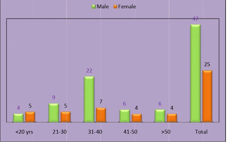
group include 47 and 25 respectively. The sex ratio was 1.9:1 between males and
females. (Table 1, Fig 1). There was a higher incidence of abdominal tuberculosis
in the age group between 31-40 years of age in both the genders followed by the
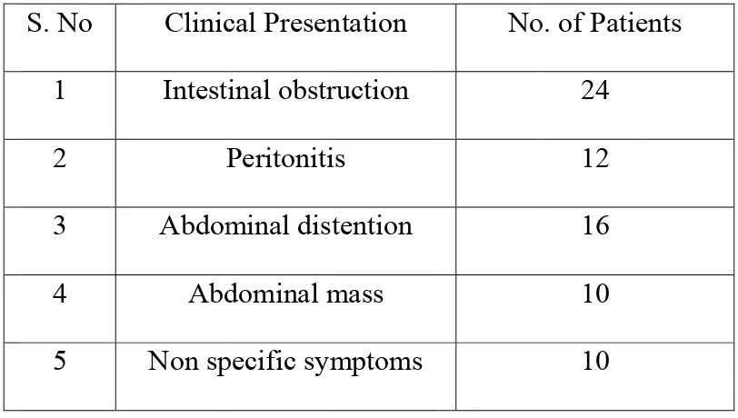
patients between 21-30 years of age. (Table 2, Fig 2). The clinical manifestations
varying from Acute intestinal obstruction to non specific illness, with acute
intestinal obstruction was a initial clinical presentation in one third of the patients,
abdominal distension in 16 patients, features of peritonitis in one sixth of patients,
mass in the abdomen in 10 patients, and non specific symptoms in 10 patients.
(Table 3, Fig 3).
All the patients underwent routine laboratory investigations like total blood
count and Erythrocyte sedimentation rate (ESR). Lymphocytosis was found in
59.71% of patients and raised ESR was found in 90.2 % of the patients in the study
group. The ascitic fluid analysis which was done in 22 patients who presented with
ascites showed the biochemical and microbiological parameters of tuberculosis in
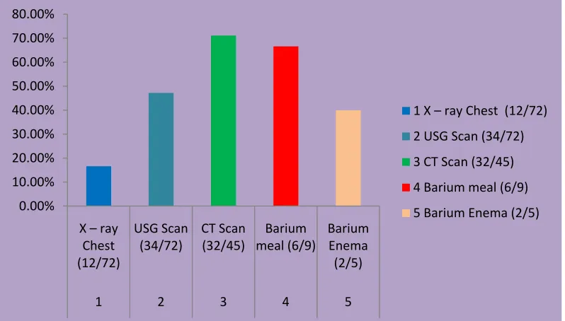
All patients underwent Roentgenogram of chest which showed features of
active and healed pulmonary lesions, pleural effusion in one sixth of the patients
with sensitivity of 16.6% for diagnosis of tuberculosis. X-ray of abdomen done in
all patients who showed features of intestinal obstruction, perforative peritonitis,
and calcified lymph nodes. Ultrasonographic evaluation of abdomen which was
done in all patients revealed features suggestive of tuberculosis in 34 patients
(47.2%). The computed tomography (CT) of abdomen was done in 7 patients
suspected to be having features of abdominal tuberculosis clinically and
ultrasonography did not provide a conclusive diagnosis. Also CT of abdomen was
done in 38 other patients who underwent ultrasonographic examination which did
not provide a conclusive diagnosis or other diagnosis for further diagnostic
evaluation. CT done in total of 45 patients concluded to be of abdominal
tuberculosis in 32 patients. Barium meal and Barium enema done in 9 and 6
patients respectively for small bowel and large bowel lesions when indicated
showed a sensitivity of 66.6% and 40% respectively. (Table 5, Fig 5).
Colonoscopic examination done in 8 patients when large bowel lesions were
suspected revealed tuberculous lesions in 2 patients. Diagnostic laparoscopy was
done in 28 patients of our study group which included the patients who did not
have conclusive diagnosis after radiological diagnosis and also patients with
confirmatory evidence and tissue diagnosis. It showed a sensitivity of 92.8 % in
our study group. (Table 6, Fig 6)
Of the 72 patients in the study group, only 35 patients needed surgical
interventions whereas 37 patients were managed with chemotherapeutic drugs
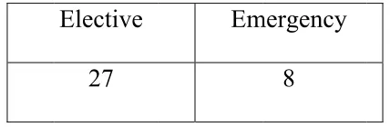
(Table 7, Fig 7). Emergency surgical management was done in total of 8 patients
which include Right hemicolectomy in 2 patients, stricturoplasty in 2 patients,
limited resection in 2 patients, and perforation closure and covering ileostomy in
the other 2 patients. 27 patients had undergone management electively after
complete investigatory evaluation, the surgical procedures include right
hemicolectomy in 7 patients, adhesiolysis in 4 patients, stricturoplasty in 6
patients, limited resection in 3 patients, abscess drainage in 2 patients, adhesiolysis
and abscess drainage through laparoscopy in 3 patients, sealed perforation closure
which presented with interloop abscess in a case. (Table 8,9 and Fig 8,9). Of the 72
Fi
TABLE
TU
ig 1: Sex D
E 1: SEX
UBERCU
M Distributio M Female 35%X DISTR
ULOSIS I
Male 47on of abdo
Male : fem
RIBUTIO
IN OUR
Female 25 ominal tub male RatioON OF AB
STUDY
Total 72 berculosis – 1.9:1 M 6BDOMIN
GROUP
in the stud
Male 65%
NAL
P
Table
Fig 2:
2: Age D
<20 y 21-3 31-4 41-5 >50 Tota
:
AGE D
4 5 <20 yrs
Distributi
yrs 0 0 0 0 alDISTRIB
9 5 s 21‐30ion Amon
Male 4 9 22 6 6 47UTION A
STUD
22 5 7 31‐40 Mng The G
Fe
AMONG
DY GRO
6 7 0 41‐50 Male FemaleGenders I
emale 5 5 7 4 4 25G THE G
UP
6 4
0 >50
e
In Our S
Tota 9 14 29 10 10 72
GENDER
47 4 0 TotStudy Gro
alRS IN OU
25
al
oup
[image:52.612.122.500.158.394.2] [image:52.612.117.499.480.718.2]Tab
Fig 3:
ble: 3 Cli
S. No
1
2
3
4
5
Clinical
inical pre
Clinic
Intesti
P
Abdom
Abd
Non sp
presenta
22 14% 1 Inte 3 Abd 5 Nonesentatio
cal Presen
inal obstr
Peritoniti
minal dist
dominal m
pecific sym
ation of a
2% % 14% estinal obstruct dominal distent n specific symp
n of abdo
study
ntation
ruction
is
tention
mass
mptoms
abdomina
% tion 2 Perit tion 4 Abdo ptomsominal tu
No
al tuberc
33 17% tonitis ominal massuberculo
o. of Patie
24
12
16
10
10
culosis in
3%osis in our
ents
our stud
r
TAB
S. No
1
2
3
BLE 4: LA
Laborator Lymphoc ESR Ascitic Fl
Fig
0.00% 10.00% 20.00% 30.00% 40.00% 50.00% 60.00% 70.00% 80.00% 90.00% 100.00%ABORAT
PATIEN
ry I
nvestig
cytosis luid
4: Sensit
Lymphocytosi 1TORY IN
NTS AND
gations
N i 77
2
tivity of L
is (43/72)
NVESTI
D ITS SE
No. of Pati investigate 72 72 22
Laborato
ESR (65/72) 2GATION
ENSITIV
ents d Pos 43 65 10ory inves
Ascitic FNS DONE
VITY
sitivestigations
luid (10/22) 3E IN OU
[image:54.612.100.517.396.676.2]TABLE 5: RADIOLOGICAL INVESTIGATIONS DONE IN OUR
PATIENTS.
S.No Radiological
Investigations
No .of Patients
Performed
Positive Sensitivity
1
X – ray Chest
72
12
16.6%
2 USG
Scan
72 34
47.2%
3 CT
Scan
45 32
71.1%
4 Barium
meal
9 6
66.6%
5 Barium
Enema
5
2 40%
Fig 5: Sensitivity of various radiological investigations in our study
group.
0.00% 10.00% 20.00% 30.00% 40.00% 50.00% 60.00% 70.00% 80.00% X – ray Chest (12/72) USG Scan (34/72) CT Scan (32/45) Barium meal (6/9) Barium Enema (2/5)1 2 3 4 5
1 X – ray Chest (12/72)
2 USG Scan (34/72)
3 CT Scan (32/45)
4 Barium meal (6/9)
[image:55.612.109.508.479.706.2]TABLE 6: Sensitivity of colonoscopy and diagnostic laparoscopy
S. No. Investigations Total No. of
patients underwent
Positive Sensitivity
1. Colonoscopy 8 2 25%
2 Laparoscopy 28 26 92.8%
Fig 6: Sensitivity of colonoscopy and diagnostic laparoscopy
0% 10% 20% 30% 40% 50% 60% 70% 80% 90% 100%
Colonoscopy (2/8) Laparoscopy (26/28)
Colonoscopy (2/8)
TABL
Fig
LE: 7 - M
g 7: Mana
51%
MANAGE
agement
EMENT O
IN OUR
Surgical 35of abdom
SurgOF ABDO
R PATIE
Mminal tub
gical MedicOMINAL
NTS.
Medical 37berculosi
calL TUBE
is in our
RCULO
patients.
49%
OSIS
[image:58.612.199.413.174.244.2]
T
[image:58.612.172.390.388.567.2]Fig.8
TABLE 8
Time of
23%
: Time O
Elective
27
surgical
Of Surgic
Em
managem
cal Mana
mergency
8
ment in o
77%
gement
our patie
Electiv
Emerg
ents
ve
TABLE 9: DETAILS OF SURGICAL PROCEDURE DONE FOR
ABDOMINAL TUBERCULOSIS IN OUR PATIENTS
S.No Procedure Elective Emergency Total
1 Right Hemicolectomy 7 2 9
2 Laparotomy and adhesiolysis 4 - 4
3 Laparotomy and stricturoplasty 6 2 8
4 Laparotomy & limited Resection 3 2 5
5 Laparotomy, Primary closure &
Diverting ileostomy
1 2 3
6 Laparotomy & drainage 2 - 2
7 Laparoscopy & Intervention
(Adhesiolysis, Abscess drainage)
3 - 3
8 Anal canal dilatation 1 - 1
Fig
g 9: Detai
0 1 2 3 4 5 6 7 8
ils of surg
tub
gical pro
berculosi
ocedures
is in our
perform
patients.
med for ab
.
bdominal
Elective
Emergency
[image:60.612.90.525.145.505.2]DISCUSSION
In our study commonest age group affected was 2nd to 4th decade of life.
Age incidence of present series is similar to reported by other workers [Adams and
Miller,1946, Dutta Guptha1950,Sharma et al ,1972 B.K Bhansali,1968]. Pritam
Das and Sukula .et.al 1978 , also reported same age group. The male and females
in our study group include 47 and 25 respectively. The sex ratio was 1.9:1 between
males and females Addison et al 1981 reported high incidence in males.
The clinical manifestations varying from Acute intestinal obstruction to non
specific illness with acute intestinal obstruction was a initial clinical presentation in
one third of the patients, abdominal distension in 16 patients, features of peritonitis
in one sixth of patients, mass in the abdomen in 10 patients, and non specific
symptoms in 10 patients. Badaoui E ,BerneyT , Kaiser L et al 2000,also reported
similar presentation.
M P Sharma and Vikram Bhatia stated in their review article about the
clinical presentation dominated by constitutional symptoms. 40-70% of patients
present with fever, 80-95% with pain, 11-20% with diarrhea, 40 -90% with weight
loss. N.Rangabashyam.et.al cited vague symptoms to be the predominant
presenting symptoms. The abdominal pain accounts for in 77-94% of patients,
followed by vomiting and abdominal distension.
of 72 patients had presented with an acute abdomen and underwent emergency
surgery for the same. J.M.Findly 1981 found that acute presentation was present in
13.5% patients. Commonest symptom is vague abdominal pain .Other common
symptoms were vomiting, fever, loss of appetite and loss of weight. Pritam Das
and Sukula had reported abdominal pain in 94% of cases Bockus et al 1964
emphasised abdominal pain , anorexia ,loss of weight as common presenting
symptoms. Shukula 1970 and L.E Hugas also reported these symptoms as common
presentation
Any patient present with vague abdominal pain, vomiting , loss of appetite ,
loss of weight and not fitting with any other clinical diagnosis , diagnosis of
abdominal tuberculosis should be considered.
Routine laboratory tests have limited value in the diagnosis of abdominal
TB. Although the mantoux test is freely available, its value in the diagnosis active
TB remains uncertain. Lymphocytosis was found in 59.71% of patients and raised
ESR was found in 90.2 % of the patients in the study group.
Radiological studies
Chest X-ray:
Evidence of tuberculosis in a chest Roentgenogram supports the diagnosis of
abdominal tuberculosis but a normal chest X-ray does not rule it out. Sharma et al
lesions on chest X-ray in 22 (46%). X-rays were more likely to be positive in
patients with acute complications (80%). In Prakash’s series of 300 patients, none
had active pulmonary tuberculosis but 39 per cent had evidence of healed
tuberculosis. Tandon et al found chest X-ray to be positive in only 25 per cent of
their patients. Hence, about 75 per cent cases do not have evidence of concomitant
pulmonary disease. Plain X-ray abdomen may show enteroliths, features of
obstruction i.e., dilated bowel loops with multiple air fluid levels, evidence of
ascites, perforation or intussusception. In addition, there may be calcified lymph
nodes, calcified granulomas and hepatosplenomegaly.
In our study group, out of all 72 patients, of chest x ray in 12 patients
(16.67%) ,9 patients showed features of healed pulmonary tuberculosis whereas
active pulmonary lesions found in 3 patients in contrast to the above literatures.
83.4% of patients in our study group did not have any evidence of concomitant
pulmonary tuberculosis. Chest X- ray gives evidence of old or active pulmonary
tuberculosis but rather they provide the indirect suspicion of abdominal
tuberculosis.
30 patients in our study group out of 72, had evidence of features of
ray gave diagnostic evidence of abdominal tuberculosis but rather they provide the
features of various presentations of abdominal tuberculosis. X ray abdomen though
plays an important role in evaluation, but do not provide the diagnostic conclusion
of abdominal tuberculosis.
Small bowel barium meal and large bowel barium enema: The features which
may be seen: Accelerated intestinal transit; hyper segmentation of the barium
column (“chicken intestine”), precipitation, flocculation and dilution of the barium;
stiffened and thickened folds; luminal stenosis with smooth but stiff contours
(“hour glass stenosis”), multiple strictures with segmental dilatation of bowel
loops, may also be found; and fixity and matting of bowel loops. Barium meal was
done in 9 of our patients which revealed the features of abdominal tuberculosis in 6
patients. Barium meal studies do have limitations in its diagnosis and application
for the patients and cannot be used in all cases but has its significance in cases of
strictures with proximal dilation. Barium enema with radiological diagnostic
features as explained earlier was found in 2 of our patients but the features were
nonspecific. Barium enema gives supportive evidence to the diagnosis. Barium
studies though accurate for intrinsic bowel abnormalities, do not detect lesions in
the peritoneum. Enteroclysis followed by a barium enema may be the best protocol
Ultrasonography
Ultrasonography is a cheap and valuable investigation in the evaluation of
abdominal tuberculosis when the clinical features have suspicion of abdominal
tuberculosis. They are particularly useful in cases of abdominal mass (Coacoon),
lymph nodes, ascites and dilated bowel loops. USG was found to be 47.2% of
patients in our study group whereas other study groups from Uvqur et al showed
sensitivity of 57% and Dobok G et al viewed a sensitivity of 54.2%.
Ultrasonography is a non expensive diagnostic tool which can be used as initial
radiological diagnostic modality.
Computed tomography:
CT of abdomen has high sensitivity than USG for diagnosis of cases with
diffuse lymphadenopathy, low density lymph node with multilocular appearance
following intravenous contrast, High density ascites, and mottled low density
masses in the omentum thickening of the Bowel wall adjacent to mesentery and
also with hepatic pseudo tumors. CT abdomen has a sensitivity of 71.1% in 45
patients in which CT was done which was marginally comparable to the
Colonoscopy
Colonoscopy is an excellent tool to diagnose colonic and terminal ileal
involment but is still often underutilized in many of the institutions. Mucosal
nodules of variable sizes (2 to 6 mm) were found in 2 of the 8 patients in which the
colonoscopy was performed. Colonoscopy has its limitations in its indications and
observer variant.
Immunological tests
Chawla et al reported a high sensitivity of ELISA for soluble antigen and
Bhargava et al used competitive ELISA with monoclonal antibody against 38 Kd
protein and found a sensitivity of 81 per cent, specificity of 88 per cent and
diagnostic accuracy of 84 per cent. These above tests were not evaluated in our
study. The values of immunological tests are not clearly understood in our clinical
evaluation.
Ascitic fluid examination:
The ascitic fluid in tuberculosis is straw coloured with protein >3g/dl, and
total cell count of 150-4000/ μl, consisting predominantly of lymphocytes (>70%).
gradient is less than 1.1 g/dl. In our study group laboratory and microbiological
evidence done in 22 patients revealed features of tuberculosis in 10 patients.
Ascitic fluid microbiological evidence needs 6-8 weeks of period for organism
growth and it has its diagnostic limitation.
ADA levels were determined in the ascitic fluid of 49 patients by Dwivedi et
al. The levels in tuberculous ascites were significantly higher than those in
cirrhotic or malignant ascitis. In coinfection with HIV the ADA values can be
normal or low. Falsely high values can occur in malignant ascitis. High
interferon-γ level in tubercular ascites have been reported to be useful diagnostically.
Combining both ADA and interferon estimations may further increase sensitivity
and specificity.
Laparoscopic findings:
Bhargava et al studied 87 patients with high protein ascites, of which 38
were diagnosed as having tuberculosis. They found visual appearances to be more
helpful (95% accurate) than histology, culture or guinea pig innoculation (82% and
37.5% sensitivity respectively). Caseating granulomas may be found in 85-90 per
cent of the biopsies.
In our study group where 28 patients underwent diagnostic laparoscopy, 26
patients had features diagnosis of tuberculosis based on visual appearances. Tissue
Laparoscopy plays a very important role in the diagnosis of abdominal tuberculosis
and it needs to be evaluated as initial diagnostic tool.
Management:
On discussing about the surgical management of abdominal tuberculosis,
Surgery is reserved for mechanical complications of tuberculosis or when medical
therapy fails. Emergency surgery is indicated in the presence of acute
complications such as free perforation of the bowel and severe intestinal
haemorrhage. The most common indication for surgery is intestinal obstruction
secondary to stricture formation. Predictors for surgical intervention are long
strictures (12 cm or more in length) and multiple areas of involvement Other
indications for surgery are bowel adhesions, intra-abdominal abscess due to a
confined perforation, mesenteric abscess, and internal or external fistulas. Surgery
is also appropriate if the diagnosis is in doubt and when malignancy cannot be
ruled out with reasonable accuracy.15, 29,30
Surgical procedures:
In our study group, only 35 patients needed surgical interventions whereas
37 patients were managed with chemotherapeutic drugs . Most frequent
interventions were right hemicolectomy (9patients), stricturoplasty (8patients),
limited resection (5patients), adhesiolysis (4patients), laparoscopy
were made by Balasubramaniam et al.
Surgical techniques for patients with bowel obstruction have evolved over
time. At one time, bypass procedures such as ileotransverse colostomy and
enteroenterostomy were practiced commonly, but have now been abandoned
because of complications such as blind-loop syndrome, malabsorption, and
perforation. Similarly, radical bowel resection and extensive dissection of
mesenteric lymph nodes have fallen out of favour because of
complications,including the development of short-bowel syndrome. The procedure
preferred for an ileocaecal mass is limited resection in place of the standard right
hemicolectomy. Other procedures advocated are ileocaecoplasty and coloplasty for
ileocaecal and colonic strictures. respectively.31-33 Drainage tubes are not
recommended and, if used, should be removed early because of increased risk of
abdominocutaneous fistulas if they