TECHNIQUES FOR DIAGNOSIS OF MALARIA
Dissertation submitted to
THE TAMILNADU DR.M.G.R. MEDICAL UNIVERSITY
in partial fulfillment of the regulations for the award of the degree
of
M.D.(MICROBIOLOGY)
BRANCH – IV
GOVERNMENT STANLEY MEDICAL COLLEGE & HOSPITAL
THE TAMILNADU DR.M.G.R. MEDICAL UNIVERSITY
CHENNAI, INDIA.
This is to certify that the dissertation entitled “A STUDY ON RECENT
ADVANCES IN LABORATORY TECHNIQUES FOR DIAGNOSIS OF MALARIA” is
the bonafide original work of DR.B.SANTHY, in partial fulfillment of the requirements for M.D(BRANCH-IV) MICROBIOLOGY examination of Tamilnadu Dr.MGR Medical University, to be held in September, 2006.
DEAN DR.P.R.THENMOZHI VALLI, M.D., Govt. Stanley Medical College Professor & Head of the Department, and Hospital, Department of Microbiology,
I, DR. B. SANTHY, solemnly declare that the dissertation titled “A STUDY ON RECENT ADVANCES IN LABORATORY TECHNIQUES FOR DIAGNOSIS OF MALARIA” is a bonafide work done by me at Government Stanley Medical College & Hospital during 2003 – 2006 under the expert guidance and supervision of Dr.P.R.Thenmozhi Valli, M.D., Professor and Head of the Department, Department of Microbiology.
The dissertation is submitted to the Tamilnadu Dr. M.G.R. Medical University towards partial fulfillment of requirement for the award of M.D.DEGREE (BRANCH - IV) in Microbiology.
Place : Chennai. Date : 10 - 04 - 2006
I express my thanks to honourable Dean Dr. M. VASANTHA, M.D., Government Stanley Medical College, and Hospital, Chennai for permitting me to carryout this study.
I express my heartfelt thanks to my esteemed Professor
Dr. P.R. THENMOZHI VALLI, M.D., Professor and Head, Department of Microbiology,
Stanley Medical College, Chennai for allotting me this topic to work and her constant encouragement to carry out this study successfully.
I express my immense thanks to Dr. S. SHANTHA, M.D., Ph.D., Professor of Immunology, Stanley Medical College for her valuable suggestions to carry out this study.
I am extremely grateful to Dr. THYAGARAJAN RAVINDER, M.D., Additional professor, Department of Microbiology, Stanley Medical College, Chennai for his valuable advice, excellent guidance and encouragement given to me throughout the study.
My profound thanks to Dr. ROSY VENNILLA, M.D.,
Additional professor, Department of Microbiology, Stanley Medical College, Chennai for her valuable suggestions and constant support throughout the study.
Professor, Department of Microbiology, Stanley Medical College for her valuable suggestions.
My sincere thanks to THE DIRECTOR, JAWARHARLAL NEHRU INSTITUTE OF SOCIAL PAEDIATRICS, and Professor and Head Department of General Medicine, Stanley Medical College Hospital for permitting me to collect sample to carryout this study.
My special thanks to Dr. A. SUNDARAM, M.D., Professor and Head, Department of Pathology, Stanley Medical College for his suggestions.
I am extremely grateful to Dr. S. KRISHNA, Health Officer and
Mr. B. DHANRAJ, Chief Vector Control Officer, Corporation of Chennai for permitting me
to do the Q.B.C. test in the malaria clinic.
I am immensely grateful to THE DIRECTOR and Mr. D.T.SELVAM, Scientist, Microbiology Division, Defence Research and Development Establishment, Gwalior, Ministry of Defence for permitting me to do the DOT ELISA and PCR techniques in their Institute.
Medicine, Stanley Medical College for his suggestion.
I also thank all my colleagues and all the technical staff, and other staff members of the Department of Microbiology and Immunology for their kind co-operation to carryout this work successfully.
S.NO
TITLE
PAGE NO
1. INTRODUCTION 1
2. REVIEW OF LITERATURE 4
3. AIMS AND OBJECTIVES 43
4. MATERIALS AND METHODS 44
5. RESULTS 55
6. DISCUSSION 61
7. SUMMARY 72
8. CONCLUSION 74
9. BIBILIOGRAPHY
10. ABBREVIATION
11. APPENDIX
INTRODUCTION
“Humanity has but three great enemies fever, famine and war; Of
these by far the greatest, by far the most terrible is fever”. William Osler30.
Human malaria is basically a febrile illness caused by species belonging to the genus Plasmodium. Malaria has always been a major public health concern, probably representing the most important parasitic disease in human. Malaria is one of the most common cause for school absentism.
It has been infecting humankind for millennia. Earliest recordings of this disease are available from description of the Chinese and Egyptian civilization between 1700 & 1500 B.C. Recordings can also be found in the Vedic writings of 1600 BC in India. Charaka and Sushrutha gave vivid descriptions of malaria and even associated it with the bite of mosquitoes56. Malaria was also a scourge
of the western civilization even at the beginning of this century.
Malaria is a protozoan (Coccidiean-Apicomplexa) disease transmitted by the bite of infected female Anopheles mosquitoes, which breed in fresh water66. The species that infect humans are four in number. P.falciparum,
A heavy burden on tropical communities poses a threat to non-endemic countries and a danger to travellers30. Malaria results in loss of economy and
productivity. The annual direct and indirect cost of malaria in India has been estimated to be an extent of 0.5 to 1 billion US dollars. Malaria remains uncontrolled to date due to various reasons viz. emergence of drug resistant parasite, pesticide resistant mosquito vector and non availability of suitable and effective malarial vaccine67.
Presently, the disease malaria seems to be a diagnostic challenge to both the physician and to the laboratories in most of the countries. Hence we need to design and review the current methodology and approach to the diagnosis of malaria in a practical and cost effective way helpful for the patients, the laboratory and for the physician caring for the patient. Concurrently the World Health Organization has begun a dialogue with scientists, clinicians and manufacturers of malaria diagnostic test devices regarding the realistic possibilities for developing accurate and cost effective rapid diagnostic tests for malaria2.
Malaria rapid diagnostic devices (MRDD) have been developed with the hope that they would offer accurate, reliable, rapid, cheap and easily available alternative to traditional method of malaria diagnosis15. Assay for rapid
instances in which skilled microscopy is not readily available2. Conventional
peripheral blood smear examination for demonstration of malarial parasite remains the “gold standard” for diagnosis of malaria52. The Quantitative Buffy
Coat (Q.B.C), assay for detection of antigen Histidine rich protein (HRP –2) and Pan Malarial Antigen (PMA) by Immuno chromatographic test (ICT), only HRP –2 antigen by Dot ELISA for P.falciparum and detection of specific nucleic acid sequences (P.f 918 bp, P.v 523 bp) by Polymerase chain reaction (PCR) have been studied here particularly to overcome the disadvantages of conventional peripheral blood smear study.
Rapid, accurate diagnosis is fundamental to effective management and control of malaria. Modern methods of malaria diagnosis include fluorescent microscopy, flow cytometry, automated blood cell analysers, serology-antibody detection, molecular methods and laser desorption mass spectrometry (Hanscheid 1999,Demirev et al 2002)15, Immunochromatographic assay detect
HISTORY OF THE MALARIAL PARASITE AND ITS DIAGNOSIS
Malaria is caused by the sporozoan parasite belonging to the Genus Plasmodium. Malaria, meaning ‘bad air’ with its well known symptom of intermittent fever has been recorded in papyri, from ancient Egypt.
5TH CENTURY B.C. : Hippocrates in Greece gave a detailed account of the
clinical picture and observed the prevalence of the disease in certain places and seasons37.
6TH CENTURY A.D. : The relation between the disease and stagnant
waters, swamps and marshy lands was recognized and measures to control the disease by effective drainage were practiced in Rome and Greece by 6th century A.D37.
18TH CENTURY : The name malaria ‘mal-bad, aria – air’ was given in Italy,
as it was believed to be caused by foul emanations from the marshy soil. Disease named “Malaria” by Sir Horace Walpole. ‘Paludism, another name for malaria also has a similar origin from palus, Latin for ‘marsh’. The recent demonstration of a specific parasitic antigen in Egyptian mummies indicates that malaria was present thousands of year’s ago37.
In 1820 : Quinine isolated from cinchona bark in Indonesia61.
specific causative agent of malaria in the red blood cells of a patient54,61.
In 1883: Marchiafava used Methylene blue for staining malarial parasites54, 61.
In 1886 : Golgi in Italy described the asexual development of the parasite in
red blood cells (erythrocytic schizogony), which therefore came to be called
Golgi cycle61.
1886-1890 : Three different species of malarial parasites infecting man,
P.vivax, P. malariae, P. falciparum were described in Italy37.
In 1897 : The mode of transmission of the disease was established when
Ronald Ross in secunderabad, India identified the developing stages of
malarial parasites in mosquitoes. This led to various measures for the control
and possible eradication of malaria by mosquito control. It is tragic that one
century down we were still grappling with the problem61.
In 1900 : Observation of mosquito transmission by Ross was supported by
Patric manson54, 61.
Both Ronald Ross (1902) and Alphonse Laveran (1907) won the Nobel Prize
for their discoveries in Malaria37.
In 1922 : The fourth species P.ovale was identified37.
In 1948 : Shortt and Garnhalm demonstrated the site of the exo-erythrocytic
pre-erythrocytic schizogony of P.falciparum54, 61.
In 1955 : The World Health organization launched a Worldwide Malaria
Eradication Programme. This continued until 1970, when it was official
declared a failure45, 54.
With the spread of malaria, mosquito control measures were tried. The first large-scale demonstration of this was in Cuba in 1899 and in India in 1935-193922. The World Health Organization between 1953-1958 embarked on the Malaria Eradication Programme22,45. But 1972-1978 saw the resurgence of malaria in endemic tropical countries including India.
1965 – 1976 as part of the reorientation of the Malaria Control strategy, extensive scientific studies were done on the development of a malarial vaccine (between 1965-1976), which culminated in the production of continuous in-vitro culture technique for P. falciparum by Trager and Jensen in 1976.
Operation under the NMEP came into force from 1st April 1977. In 1999, the Govt. of India decided to drop the term “National Malaria Eradication Programme” and renamed it “National Anti-malaria Programme”56.
Serological tests - Indirect Fluorescent antibody test (IFAT) & Indirect Haemagglutination Assay (IHA) in study of malaria were developed in early 1960s and there is a sustained interest in this aspect to develop an ideal test which would be effective in the field trials for use in sero-epidemiological surveillance26.
A test that could be easily interpreted within 5 to 15 minutes of specimen would be a substantial aid to clinicians. Of the numerous new techniques developed for the diagnosis of malaria, Quantitative Buffy Coat (Q.B.C.) technique appears to be a promising test46.
Other enzymes within the glycolytic pathway of the malarial parasite have been recognized and considered as target for rapid malaria diagnostic tests. Aldolase is a key enzyme in this pathway. Malarial antigens currently targeted by rapid diagnostic test are HRP-2, pLDH and Plasmodium aldolase2.
malaria2, 17.
EPIDEMIOLOGY
Malaria is one of the important parasitic disease of human being affecting 103 countries in the world involving a population of over 2.5billion people and causing 1 to 3 million deaths each year. Around 40% of the World’s population is deemed under malaria risk53. It is ranked with acute respiratory diseases and diarrhoeal diseases as one of the leading causes of the mortality world wide18.
GEOGRAPHICAL DISTRIBUTION :
Malaria has been recorded in places as far north as Archangel, Russia and as far south as Cordoba, Argentina and in places as low as the Dead sea (400 meters below sea level) and as high as Cochabamba, Bolivia (2800 meters above sea level). However, malaria is essentially a focal disease and its distribution is patchy in most parts28.
The relative prevalence of four species of malarial parasites varies in different geographical regions. P. vivax is the most widely distributed, being common in Asia, North Africa and Central and South America. P. falciparum,
WORLD WIDE DISTRIBUTION : At the end of 2004, 107 countries and
territories had areas at the risk of malaria transmission. Some 3.2 billion people lived in areas at risk of malaria transmission. An estimated 350 to 500 million clinical malaria episodes occur annually; most of these are caused by infection with P.falciparum and P.vivax75.
MALARIA IN INDIA : 65% of malaria in India is caused by P.vivax and
35% by P.falciparum. Nearly 0.98 million cases of malaria were reported during the year 2001 of which nearly 0.46 million were falciparum cases. Highly endemic areas for malaria include the north – eastern region and tribal forested and hilly areas of several states including Maharashtra, and selected non-tribal districts. Nationwide, the reported incidence of laboratory confirmed cases has declined from 3.0 million in 1996, 2.1 million in 2001 and 1.78 million in 200375.
IN TAMILNADU : 43,604 cases were reported in 200375, Chennai in the year
2004, 28,224 cases were reported. (Courtesy of Chennai Corporation).
TRANSMISSION : Man is the only source of malarial infection. Infected man
transmission. But unnatural methods of transmission as a result of blood transfusion when the donor harbours malarial parasite (Bruce Chwatt 1974, 1980) drug addiction and congenital infection of the new born from an infected mother (Bruce chwatt 1980) have also been reported in the first half of this century28.
AGENT:
PLASMODIA : Malarial parasites belong to Phylum Apicomplexa; Class
Telosporea, Order Eucoccidia, Suborder Haemosporina and Genus Plasmodium. Of the 120 existing species of Plasmodia, only four are the primary agents of human malaria- P.falciparum, P.vivax, P. malariae & P.ovale (Phillips 1983)23.
The malarial parasite passes its life cycle in two different hosts. Man being the intermediate host allows the asexual cycle to continue. This cycle consists of primary-erythrocytic schizogony, erythrocytic schizogony, and gametogony and in P. vivax alone the secondary exoerythrocytic schizogony. The duration of each phase varies with the species.
In Mosquitoes : The female Anopheles mosquito forms the definitive host.
sporozoites, which are infective to man23.
Vector : only female Anopheles mosquitoes can transmit human malaria. The
male Anopheles feeds exclusively on nectar and fruit juices while the female feeds on warm blooded animals. There are about 422 species of Anopheles mosquitoes through out the world, but only some 70 species are vectors of malaria under natural conditions28.
In India the predominant vector is A. culifaciens though A. fluviatilis, A. stephensi, A. sundaicus, A. minimus and A. philippinensis also exist (Bruce Chwatt 1980). The vectors of major importance are A. culifaciens in rural areas A. stephensi in urban areas; Breed in fresh water; Anopheles mosquitoes have nocturnal feeding habit, between dusk and dawn28.
Environment : Variations in climatic conditions has a profound effect on the
mosquito and the malarial parasite. A temperature of 18oC – 30oC and humidity of 60%-70% are optimal for the vector and the parasite. Rainfall is also important by increasing breeding sites and humidity (WHO 1979, Bruce Chwatt 1980)28.
disadvantages that in areas where antimalarial measures have been instituted. The spleen rate classification of endemicity is not applicable (Bruce Chwatt 1980) and the splenomegaly especially in tropical regions is a non-specific sign, which may be due to causes other than malaria (Kagan 1972).
o During the past few years, there has been an increased awareness of the importance of trained and qualified personnel to perform diagnostic procedure.
o These results obtained by intensive basic research are currently leading to improved diagnostic procedure.
o The rapid development of new methods and techniques in the area of molecular biology has gained new insights into the genetic and structural features of malarial parasite.
PATHOGENESIS AND PATHOLOGY OF MALARIA
schizonts. P.vivax invades the youngest red cells where as P. falciparum invades red cells of any age16.
The pre-erythrocytic stage of infection produces minimal histopathological changes and absolutely no detectable symptoms or functional disturbances in the host. Pathological process in malaria is the result of the erythrocytic cycle. In case of P. vivax and P. ovale infections, arrested development of some sporozoites in liver cells results in dormant forms, hypnozoites from which the infection may later relapse. P. falciparum and P. malariae have no persistent exoerythrocytic phase but recrudescences of fever may result from multiplication of parasites in the red cells which have not been eliminated by treatment and immune processes.
The fever, febrile paroxysms, headache, a variety of aches and pains and prostration, the most familiar and consistent symptoms of an acute malaria attacks, are probably the result of cytokines released from macrophages at the time of schizont rupture. The malarial toxin released at schizont rupture is the lipid, glycosyl phosphatidyl inositol (GPI) anchor of a parasite membrane protein (MSP1). Cytokines such as tumor necrosis factor (TNF), and interleukin-1 (IL-1) have been implicated as the cause of malarial fever when the mature schizont ruptures releasing red cell fragments, merozoites, malarial pigment and other parasitic debris. Macrophages and polymorphs phagocytose these and release large quantities of endogenous pyrogens leading to elevation of temperature21.
Pathology : The pathology in malaria is due to haemolysis of infected red cells and adherence of infected red blood cells to capillaries. Malaria is always accompanied by haemolysis and in a severe or prolonged attack anaemia may be profound. Typical pathological changes are seen primarily in the spleen, liver, bone marrow, lungs, kidney and brain.
Chloroquine resistant P. falciparum was first reported in Columbia and Thailand in the late 1950s55. Several mechanisms can account for changes in drug sensitivity in the malarial parasite.
of previously existing drug resistant cells from a mixed population under drug pressure.
- Spontaneous mutation.
- Mutation of extranuclear genes
IMMUNITY
Host immunity in malaria is of two types--Natural or innate immunity
-Acquired immunity
Innate Immunity: Innate resistance is seen in certain population e.g. person
with red cells negative for Duffy blood group determinants are resistant to P. vivax infections (Phillips 1983). Young babies have a reduced susceptibility especially to P. falciparum, probably due to maternal antimalarial antibodies and also the presence of haemoglobin F which is less suitable for the parasite’s existence and multiplication (Phillips 1983). Glucose -6- phosphate dehydrogenase deficient individuals are protected against malaria. This enzyme is necessary for the respiratory process of the parasite52.
P. falciparum does not multiply in sickle red cells containing abnormal haemoglobin S38. Severe malnutrition and iron deficiency may confer some protection against malaria. During severe famine in North Africa, malaria was rare but on providing food and iron supplements the patients began to develop malaria. Spleen plays an important role in immunity against malaria. Splenectomy enhances susceptibility to malaria62.
associated with protection from malaria38.
Acquired Immunity : Both humoral and cellular immunity play a protective
role in malaria.
Humoral Component : Where malaria is hyperendemic, the acquisition of
clinically evident protective immunity in young children is associated with significant increase in serum gamma - globulin and a greater increase is seen in adults (McGregor 1981). Of the increased gamma globulins, only 5-10% are associated with specific action against the malarial parasite (curtain et al 1964)49.
CLINICAL MANIFESTATIONS
The main clinical features are fever peaks with rigor followed by anaemia and splenomegaly. The clinical features of malaria vary from mild to severe and complicated according to the species of the parasite present, the patient’s state of immunity, the intensity of infection and also the presence of concomitant condition such as malnutrition.
Prodromal period : Malarial paroxysm is preceded by a prodromal period,
which varies from a few to several days. Non-specific symptoms such as malaise, myalgia, headache and fatigue are usually seen during the prodromal period3.11.60.
Malarial paroxysm : Occur with definite intermittent periodicity depending
upon the species of the parasite involved. Comprises of three successive stages cold, hot and sweating stage4,13,71.
Cold Stage : Lasting for 15-60 minutes; the patient experience intense cold and shivering4,13,71.
Hot Stage : Lasting for 2-6 hrs, when the patient feels, intense heat. Patient develops high fever (40-40.6oC), severe headache, nausea and vomiting4,13,71.
Sweating stage : Patient is drenched in profuse sweat. The temperature drops
The paroxysm usually begins in the early afternoon and lasts for 8-12 hrs. The periodicity of the attack varies with the species of the infecting parasite (P.v. 48 hours, P.f. 48 hours., P.o. 48-50 hours, P.m. 72 hours)4,13,71.
All clinical manifestations in malaria are due to products of erythrocytic schizogony and host reactions to them. The exoerythrocytic liver cycle and gametogony do not appear to contribute to clinical illness4.
Anaemia : Anaemia is normochromic and normocytic. It is severe in falciparum malaria. The pathogenesis of anaemia appears to be multifactorial. These include.
- The lysis of parasitized and non-parasitized red blood cells.
- The suppression of erythropoiesis in the bone marrow.
- Increased clearance of non-parasitized RBC’s by the spleen.
Hepatosplenomegaly : Physical examination in uncomplicated malaria shows
moderate splenomegaly and tender hepatomegaly or may even reveal no abnormalities. Splenomegaly due to massive proliferation of macrophages, phagocytize both parasitised and non-parasitised red blood cells. Lymphadenopathy does not occur in malaria4.
Factors contributing to virulence of P. falciparum :
High level of parasitaemia : In P. falciparum malaria, parasite density exceeds more than 250,000-300,000 / ml of blood. Nearly 30% - 40% of total body red cells are parasitized. This is caused by invasion of erythrocytes of all ages (young and old) by P. falciparum. A level of 25% parasitaemia is usually fatal.
Sequestration of parasite: Sequestration means a condition of holding back of
mature parasites in vital organs. This phenomenon is shown exclusively by P. falciparum and is due to its ability to cytoadhere4,13. Once inside the erythrocytes, P. falciparum merozoites produce a protein within erythrocyte surface membrane in form of a deformation called knobs. These knobs produce high molecular weight, strain specific adhesive protein. These proteins in turn mediate attachment of parasites to receptors on the endothelium of capillaries and venules. This causes sequestration of parasitized erythrocytes in the small post capillary venules of the internal vital organs specifically, the CNS, kidneys, spleen and lungs4,13.
Gametocyte – infected erythrocytes do not have any knobs, so do not stick to the venular endothelium, hence are not sequestrated, and therefore are demonstrated in the peripheral blood smears. This phenomenon of sequestration does not occur in any other malarial parasites.
TNF and IFN
γ
. These cytokines act specifically on a number of receptorspresent on the surface of endothelial cells in the small capillaries and post capillary venules49.
P. vivax malaria may not initially present with the classical symptoms, due to the release of non-synchronous broods of parasites. Herpes labialis is common in vivax malaria. Cerebral irritation, polyuria and in children fulminant infections and severe anaemia have been reported (Bruce Chwatt 1980). Vivax malaria is important not due to its fatality but due to the debility it can produce by its relapses (Bruce Chwatt 1980).
When more than 3% - 5% of erythrocytes are parasitized, the condition is known as severe falciparum malaria Complications of severe falciparum malaria are4,13.
- Pernicious malaria
- Black water fever
- Tropical splenomegaly
- Pulmonary oedema
- Renal failure
DIAGNOSIS OF MALARIA
The diagnosis of malaria can be subdivided into clinical and laboratory which include parasitological, serological and molecular biological detection70.
Importance of diagnostic methods in malaria:
- Diagnosis of individual cases.
- Prognosis of cases.
- Monitoring drug resistance.
- Epidemiological studies.
Clinical Diagnosis : The condition is considered in any person who has a
febrile illness and who has come from the area endemic for malaria, received blood transfusion or used intravenous drugs55.
Laboratory diagnosis :
Specimen : Peripheral blood : Under strict aseptic precautions blood
obtained by pricking a finger or earlobe is the ideal sample because the density of developed trophozoites or schizonts is greater in blood from this capillary rich area2.
malarial parasite. Blood should be taken at any time over the course of infection before starting antimalarial treatment, but the best time is midway between paroxysm of chills and fever when the greatest number of intracellular organisms are present55.
Parasitic diagnosis : Parasitic diagnosis depends on the demonstration of parasite in the blood.
Methods of examination :
1. Light Microscopy – Thick & Thin peripheral smear 2. Fluorescence microscopy :
- Quantitative Buffy Coat (Q.B.C.)
- Acridine Orange Staining (A.O.)
- Benzo thio Carboxy Purine Staining (B.C.P.) 3. Micro Concentration technique
Light Microscopy : Conventional light microscopy of stained blood smear is
the “gold standard” for confirmation of malaria. Both thick and thin smears are prepared from the capillary blood. They are stained with one of the Romanowsky’s stains such as Leishman’s’, Fields’, Wrights’, Giemsa’s or Jaswantsingh Bhattacharjee stain55.
on a small surface by a factor of 20 to 30 and is stained as an unfixed preparation. The thick blood film provides enhanced sensitivity of the blood film technique and is much better than the thin film for detection of low levels of parasitaemia and reappearance of circulating parasites during infection recrudescence or relapse. Atleast 100-200 fields, each containing 20 WBC, should be examined before a thick smear is reported as negative for malaria2. It is not used for species diagnosis.
Thin Smear : Because of the fixed monolayer of RBC available in this procedure, the morphological identification of the parasite to the species level is much easier and provides greater specificity than the thick-film examination. The thin film is often preferred for routine estimation of parasitaemia because the organisms are easier to see and count. The ability to count parasites in sequential blood films enables the response to therapy to be monitored, particularly for P. falciparum infection2, 31.
The diagnosis of malaria is ruled out by obtaining negative thick blood smears on atleast three different occasions. (12-24 hrs; 3 subsequent days) Light Microscopy is a sensitive method. In the hand of a skilled microscopist
5-10 parasites / µl of blood can be detected by microscopy. Microscopy is the most inexpensive test to confirm malaria55.
take up to 60 minutes of preparation time to produce a stained thin or thick film and is labor-intensive. Interpretation requires considerable expertise, particularly at low levels of parasitaemia. In addition patients with P. falciparum malaria may have parasites sequestrated in the deep capillaries (Spleen, liver, bone marrow), and organisms may not be seen in equivalent numbers in circulating peripheral blood obtained by venepuncture or fingerstick. Thus a P. falciparum infection may easily be missed because there are insufficient numbers of parasites for detection in the blood films. Post treatment examination of blood film can result in the observation of a number of circulating parasites; however, the organisms may be the non-viable parasites yet to be cleared by host, thus prolonging the assumed positivity of the patient and extending treatment2.
Chinese workers recommend that intradermal smear to be taken from multiple punctures on the upper forearm using a 25-gauge needle. The puncture should not bleed, but a serosanguinous fluid can be expressed on to a slide by squeezing. This method is said to have a better sensitivity for diagnosing as this can pick up cases in which the blood smear may be negative due to peripheral sequestration39.
Q.B.C. Assay : In 1976, Stephen Wardlaw and Robert Levine invented the
haemoparasitic diseases. This is a sensitive method for detection of malarial parasites. It is based on ability of acridine orange to stain nucleic acid containing parasites and leucocytes take up the dye, which is fluorescent when examined under a fluorescent microscope. Acridine-orange stained malarial parasites appear brilliant green. This elegant method is easy to perform, easily interpreted within 5-15 minutes. Q.B.C. is more sensitive method than the thick
blood smear. It can detect as low as 3-4 parasites /µl, of blood 7,46,55,59,70 Q.B.C. assay has advantages beyond any doubt in terms of speed, ease, reliability and sensitivity in diagnosis of malaria. In addition it has been of immense help in identifying the infective carriers with a very low parasite burden. Macroscopic examination of QBC tube often enables to predict a positivity of malaria. It has also been noted that colour of plasma in buffy coat also indicates about anaemia59.
QBC assay has also been helpful in detection of microfilaria after Hetrazan provocative test and / or night blood examination ; Other uses are the diagnosis of relapsing fever and to study the haematological parameters (e.g.) hamatocrit platelet count, WBC count etc.,59
Disadvantage : Cost
Species identification is not possible
A.O. Staining method : In this method blood smears are prepared on a
red, and aids in the identification of the malarial parasite. The stained slide is examined either with a fluorescence microscope or a microscope equipped with an interference filter system (Kawamoto1991), allows quicker screening of films, because parasites are more readily recognized and a lower power lens may be used.42,44,46,51,55,70.
Benzothio carboxy purine (BCP) staining : BCP can be applied directly to a lysed blood suspension or to an unfixed but dry thick blood film and intensely stains the nucleic acid of parasites after penetrating RBCs. The dye does not stain nuclei of WBCs. This is useful in field laboratory for mass screening because of rapid staining and evaluation but it requires a fluorescent microscope2,10.
With experience, workers using methods involving fluorochrome compounds are able to detect parasites rapidly and accurately. However, an important limitation of method, involving both A.O. and BCP is their inability to easily differentiate among Plasmodium species2.
Microconcentration technique: Blood is collected in microhaematocrit
tube and centrifuged at high speed. The sediment is mixed with normal serum and smear prepared. Although, the positivity rate is increased by this technique, but there is change in morphology of the parasite10.
Immunological methods provide the means for detecting either the parasite antigens or the host antibodies directed against the parasite. The detection of antigens may be an acceptable alternative to parasite detection, particularly if the assay is robust, inexpensive, easy to use in field conditions and does not require a microscope, but the detection of antibodies merely provides information on past malaria experience and is of limited use for individual diagnosis70.
Serological tests commonly used for detection and measurement of
malarial antibodies26 :
Test Current application
Immuno precipitation Epidemiological studies and research Indirect fluorescent antibody test (IFAT) Epidemiological studies, research and aids to diagnosis Indirect Haemagglutination (IHA) Epidemiological surveys
Enzyme Linked Immunosorbent Assay Epidemiological studies and aid to diagnosis
Radio immuno assay Research
Merozoite inhibition in culture Research
epidemiological purpose when parasitaemia is low. Other applications of serological techniques are the diagnosis of hyperactive malaria splenomegaly and the screening of blood collected for blood banks. It should be remembered however that these tests are of limited value in diagnosis of acute phase of diseases in high prevalence areas26.
ANTIGEN DETECTION :
In contrast to antibody detection, where blood as well as plasma and serum can be used, a positive antigen detection assay is most useful in detection of current infection. The ideal target antigen
- It should not persist after parasitaemia disappears.
- Should be abundant in the blood (or other bodily fluids such as urine) to maximize sensitivity.
- Should be highly specific to malaria without any cross reactions70.
Malarial antigens currently targeted by rapid diagnostic test (RDT) are HRP-2, pLDH and Plasmodium aldolase.
Histidine rich protein : (HRP)
P. falciparum infected RBC’s synthesize three histidine-rich proteins, HRP-1, HRP-2 & HRP3. HRP-1 was identified in all knob-positive P. falciparum parasites. HRP-2 was identified in all P. falciparum parasites regardless of the knob phenotype and was recovered from culture supernatants as a secreted water-soluble protein. HRP-3 was present at the lowest abundance compared to HRP-1 & HRP-2. Neither HRP-1 nor HRP-2 was found in a range of other knob-positive and negative strains of non-P.falciparum malaria. HRP-2 is a water soluble protein produced by asexual stages and young gametocytes of P. falciparum. It is expressed on the RBC membrane surface and because of its abundance in P. falciparum it was the first antigen to be used to develop on RDT for its detection2.
pLDH and aldolase
aldolase have been suggested as target antigen for RDT for species other than P. falciparum2.
Enzyme parasite lactate dehydrogenase (pLDH) detection had been used to monitor in vitro drug susceptibility assay (Makler and Hinrichs 1993). Now it has also become useful for detection of Plasmodium parasitaemia (Knobloch and Henk 1995). The principle of this assay is that pLDH has different biochemical characteristics from human LDH and may therefore be differentially measured using a simple colorimetric assay. This assay can be used for both quantitative and qualitative estimation of parasitaemia; Can detect as low as 0.1% parasitaemia; not species specific. Now the sensitivity of the assay has been currently improved, by combining it with an antigen capture method using a monoclonal antibody to pLDH; this improves sensitivity 100 times (Piper et al 1995)70.
Assay for detection of HRP-2
Three commercial dipstick formats for HRP-2 detection are
- Parasight-F
- ICT Pf
- PATH FALCIPARUM, Malaria IC.
Parasight F (Shiff, Premij and Minjas 1993) is a commercial detection test in dipstick format in which a monoclonal antibody captures, a specific antigen of P. falciparum (HRP2, a molecule present in the parasite throughout the erythrocytic cycle) if antigen is present a positive result is indicated by a visible line on the dipstick12,32,70.
Advantage :
1. Rapid and Simple 2. Takes only 10 minutes.
This simple, robust assay requires no additional equipments or trained personal; Can be used in field surveillance.
Disadvantage : 1. Cost
2. Quantitation is not possible12,70.
malaria negative. HRP-2 antigen has been shown to persist and is detectable after the clinical symptoms of malaria have disappeared and parasites have apparently been cleared from the host2.
Assay for detection of pLDH
The commercial dipstick format for pLDH detection is OptiMAL-IT assay. This assay is based on the detection of intact and functional pLDH specific for P. falciparum and pan-pLDH (PMA) an antigen that is common to all four species of malaria in whole blood. The test uses two specific monoclonal antibodies that have been immobilized across the test strip2.
The OptiMAL rapid malaria test is a patient point of care immunochromatographic test that can be performed with a drop of finger stick blood. The entire process takes approximately 15 minutes and results are visually interpreted. The OptiMAL dipstick compared well with blood film microscopy for monitoring antimalarial treatment and could be a useful replacement for microscopy to monitor treatment in places where facilities for microscopy are either lacking or inadequate. In developed countries it could be a useful adjunct to blood film microscopy and it permits a reduction in duration of hospitalization and give an early warning of treatment failure31, 68.
The ICT Malaria P.f/P.v.: This is a rapid in vitro immunodiagnostic
common to all four species of malaria, pan malarial antigen (PMA) in whole blood. The test uses two specific monoclonal antibodies that have been immobilized across the test strip. The results are interpreted within 10 minutes19, 31, 35, 74. A limitation of this test is that it cannot speciate P. falciparum mixed infections.
DOT ELISA FOR P. falciparum : Polyclonal antibodies to HRP-2 antigen
are coated to the Nitrocellulose membrane. Antigen present in plasma / serum binds to this antibody. A revealing antibody with high sensitivity and specificity is added. The resulting complex is detected using enzyme substrate reaction. Appearance of brown coloured dots on 2 NC tips indicates positive result.
- This kit detects the P. falciparum specific HRP-2 antigen.
- No specialized training required for use of the kit.
Urinary antigen in Malaria :
- Rodriguez del et al (1991) first demonstrated that diagnosis of falciparum malaria can be made by detection of specific antigen in the urine.
- The main advantage of testing the urine is that unlike blood and serum, which need to be collected by an invasive procedure requiring technical expertise and disposable syringes, the collection of urine is a non-invasive procedure.
- The urine specimens can be collected easily and frequently without causing any inconvenience to the patient.
- This would make the urine as a specimen of choice, particularly in children, elderly and those unwilling to give their blood for investigation47.
Molecular Technique :
Molecular metholds based on DNA amplification have been applied to malaria diagnosis since the late 1980s. Their value lies in their high sensitivity,
Real time PCR methods are a recent development of established PCR methodologies. They use fluorescent labels for continuous monitoring of amplicon formation throughout the reaction. The advantages of these techniques are quantification of pathogen, reduced risk of contamination and availability of results within 3 hours (including sample preparation)50,58.
PCR
- Highly sensitive technique
- Highly species specific
- Can detect parasites in dried blood specimens
- Detect drug resistant in P. falciparum
- A single P. falciparum can be detected in 20 µl of blood using PBRK-1 primer, 100 times more sensitive than of thick smear55.
The application of DNA or RNA hybridization to malaria diagnosis has several advantages over traditional methods. Although the methodology is never likely to be useful at the peripheral level of health care in its present form, it may have a place as a research tool to monitor malaria control programmes, to perform quality control checks on microscopic diagnoses or to determine the distribution of important genes (e.g. genes associated with drug resistance).
synthesized and labeled either with radioactive 32p or a non-radioactive
colorimetric reagent and this ‘probe’ is used to detect parasite nucleic acid, taking advantage of the fact that complementary sequences will hybridize.
The simplest version of this technique is the use of DNA probes to detect parasites directly in a drop of patient’s blood immobilized on a filter paper (Franzen et al 1984); in this test format, the sensitivity and specificity of the technique expends largely upon the choice of nucleic acid sequence. Species-specific small subunit ribosomal RNA sequence help to differentiate between P. falciparum, P. vivax, P. ovale, or P. malariae (snoop et al 1993)70
DNA probe : - Highly sensitive
- Detect low parasitaemia
- Useful to detect drug resistance.
Recent improvement in molecular technologies like nucleic acid hybridization and PCR has come to the diagnostic field with immense promises. Nucleic acid probes to either DNA or RNA have shown to be highly specific for target sequences of all the four species of human plasmodium. PCR method because of its high sensitivity has been utilized for screening of blood donors to exclude transfusion malaria. PCR assay has also been utilized for epidemiological study of malaria7.
Culture is not used for routine diagnosis of malaria. Bass and Johns (1912) reported for the first time the in vitro cultivation of malaria parasites (P. falciparum and P. vivax). In 1976, Trager and Jensen successfully cultivated and maintained P. falciparum in vitro in human red blood cells. In this procedure, culture medium consisted of RPMI 1640 medium, a thin layer of stationary human blood cells and an overlay of medium containing human serum, maintained under an atmosphere with 7% Co2 and 1% - 5% O2. Recently various modifications of the culture media have been described. Rabbit serum or bovine serum supplemented with hypoxanthine has been found to support parasite growth, though at a reduced rate. P. falciparum now remains the only parasite widely cultivated in its asexual intra-erythrocytic stage.
Invitro culture of malarial parasites at different development stages is essential for
- Studies on the mechanism of invasion of erythrocytes by merozoites.
- Analysis of parasite antigen and the isolation of protective antigens for the development of vaccines, and as source of antigen for sero-epidemiological studies.
- Bio-chemical and metabolic studies including stage specific changes and
Treatment :
Treatment of malaria is primarily based on
- Specific antimalarial chemotherapy
- Supportive therapy.
Antimalarials inhibit growth of malarial parasites by increasing internal pH of parasites by concentrating within acid vesicles of parasite. They also inhibit utilisation of haemoglobin and metabolism of parasite. Chloroquine, Quinine, Quinidine, Mefloquine, halofantrine and artemisinin compounds are the rapidly acting drugs for treatment of acute malaria55.
Chloroquine is the drug of choice for drug sensitive P. falciparum. It is used for treatment of cases with acute uncomplicated P. falciparum malaria and for prophylaxis. A combination of sulphadoxine and pyrimethamine (fansidar) was used for treatment of chloroquine resistant P. falciparum. Fansidar resistant P. falciparum has also been reported in the early 1980s. Mefloquine is a blood schizonticide. It is used for prophylaxis or treatment of multidrug resistant P. falciparum malaria. Recently mefloquine resistant P. falciparum have also been reported from several endemic areas of the world. Artemisinin and its derivatives are the newer antimalarials being used for mefloquine - resistant and multidrug resistant cases of P. falciparum malaria.
of the parasites and primaquine to treat the persistent hypnozoite stage of parasites54.
Prevention :
Chemoprophylaxis :
Chemoprophylaxis for travelers visiting endemic areas provides effective protection. The prophylactics of choice include chloroquine primaquine, proguanil, amodiaquine and Fansidar in weekly doses or doxycycline daily. Long-term use of antimalarial prophylactics may produce serious side effects10,55.
Immunoprophylaxis :
A number of vaccines have been tested during last three decades. SSP-2 is currently the most likely vaccine of promise for use in falciparum malaria. The most current approach is cocktail vaccine, which makes use of combination of multiple epitopes from different malarial antigens and from different stages of parasite in its life cycle55.
Control Measure :
preventive measures such as the use of repellents, protective clothing, bed nets (impregnated with delta methrin) and mosquito coils56a.
AIMS AND OBJECTIVES
i. To meet the need for a reliable diagnostic adjunct to microscopy for malaria in the clinical settings.
ii. To overcome the problem of time constraints and low sensitivity in diagnosing malaria with a low level of parasitemia by peripheral blood smear microscopy.
MATERIALS AND METHODS
The present study on “Recent advances in laboratory techniques for diagnosis of malaria” was conducted in the Department of Microbiology, Stanley Medical College and Hospital during the period from May 2004 to April 2005 in collaboration with the Department of Medicine and Jawaharlal Nehru Institute of Social Paediatrics.
The study group includes :
I. 190 patients clinically diagnosed as malaria.
II. 20 patients diagnosed as Enteric fever - Disease Control III. 20 apparently healthy individuals - Healthy Control.
Total of 230 blood samples were collected, of these 190 blood samples were collected from clinically diagnosed as malaria patients with symptoms of fever and chills irrespective of age and sex. 20 blood samples each were collected from patients diagnosed with enteric fever (disease control) and from healthy individuals (healthy control). The criteria for the selection of healthy control were the history of absence of fever for a period of one month prior to the study.
simple random method and tested by recent diagnostic tests such as Immunochromatographic test for antigen detection (HRP-2 and pLDH), DOT ELISA for P. falciparum, DRDE (HRP-2 antigen) and P.C.R. assay.
Control groups (20 disease control and 20 healthy control) were subjected to Immuno chromatographic test (Now ICT), DOT ELISA for Plasmodium falciparum (DRDE) and P.C.R. assay.
Collection of Blood Samples :
Under strict aseptic precautions, thick and thin peripheral blood smears were prepared by finger prick, and 3-5 ml of blood samples were collected in a pre-sterilized aliquot with anticoagulant (EDTA) by venepuncture under strict aseptic precautions from the patients seeking treatment in in-patient and outpatient department of Government Stanley Hospital before administration of anti malarial drug.
Transport and storage of samples :
The blood samples collected from patient were transported to the laboratory immediately. Thick and Thin smears were prepared and stained by JSB technique. Q.B.C. assay was also carried out simultaneously. Remaining portion of the samples was aliquoted and stored at 4-8oC for subsequent ICT, Dot ELISA for Plasmodium falciparum and PCR assay.
I. Peripheral blood smear : Thick and thin peripheral blood smears were prepared on a clean grease free glass slide.
a. Thin smear : Smears were air dried, fixed with methanol, dipped
in JSBII stain for 2-3 sec, washed with buffer water to remove excess stain, dipped in JSB I stain for 40-50 sec and air dried.
b. Thick smear : The smears were dehaemoglobinized using buffer
water for 5-10 minutes and continued the staining procedure as for thin smear. Both thick and thin smears were examined under oil immersion.
II. Q.B.C. Assay (Quantitative Buffy Coat)
The Q.B.C. blood parasite detection system consists of capillary tubes coated with acridine orange, floats, closures, a capillary tube holder, microcentrifuge and an ordinary light microscope with customized fluorescence – paralens attachment. The QBC capillary tube was filled with
concentrated in defined areas within the tube, which result in great reduction of examination times to 1-3 minutes.
The principle of Q.B.C. is based on the fact that on centrifugation at a high speed, the whole blood separates into plasma, buffy coat and packed red cell layer. The float gets buoyed by the packed blood cells and is automatically positioned within the buffy coat layer. Blood cells in the buffy coat layer separate according to their densities; platelets remaining at the top, lymphocytes and monocytes within the middle layer and granulocytes at the bottom. Due to acridine orange the malarial parasite stains green (DNA, nucleus) and orange (RNA, cytoplasm). The tube is examined in the region between the RBC and granulocytes and within the granulocytes and mononuclear cell layer where parasites are most abundant.
Immunochromatographic test (detection of HRP-2 and PMA)
Test Principle : This is a rapid in vitro immunodiagnostic test for the detection of circulating P. falciparum HRP-2 and an antigen common to all four species of malaria, pan malarial antigen (PMA) in whole blood. The test uses two specific monoclonal antibodies that have been immobilized across the test strip. One antibody is specific for the histidine-rich protein-2 antigen of P.f., (HRP-2). The other antibody is specific for an antigen that is common to all four plasmodium species.
will always appear in area C of the test window, if the test has been performed
correctly. 15µL of whole blood is applied to a sample pad impregnated with colloidal gold-labeled antibodies, which are directed against the malarial antigens. When a positive sample is applied, malarial antigens bind to the gold-conjugate antibodies in the sample pad. Reagent is then added which allows the immune complexes formed to migrate along the test strip where they are captured by the immobilized antibodies. When capture occurs, one or two pink lines will form in the test window. When a negative sample is applied only the control line will appear.
Test Procedure :
- All test components were brought to room temperature prior to use.
- With EDTA capillary tube blood sample was applied to cover the entire purple sample pad.
- Two drops of Reagent A was applied to the white pad immediately below where the blood was applied.
- Four drops of Reagent A was applied to the pad located at the top of the left hand side of the test card.
- Adhesive on the right hand side of the test card was removed and discarded.
- The blood sample was allowed to run up the full length of the test strip.
- The card was closed immediately.
- The result was read through the viewing window after 10 minutes.
Plasmodium falciparum infection : A positive test result is indicated by any visible line in the test window next to T1 together with a line in area C.
Plasmodium falciparum infection or a mixed infection (Plasmodium
falciparum,Plasmodium ovale, Plasmodium malariae, Plasmodium vivax) :
A positive test result is indicated by any visible line in the test window next to T1 and T2, together with a line in area C.
Plasmodium ovale, Plasmodium malariae, Plasmodium vivax or a mixed
infection of all three : A positive test result is indicated by any visible line in the test window next to T2, together with a line in area C.
Negative Test Result : The test is negative if only the C line appears.
DOT ELISA FOR P. falciparum (DRDE) :
Test Principle : Rabbit polyclonal antibodies to HRP-2 antigen are coated to
the Nitrocellulose membrane. Antigen present in plasma/serum binds to this antibody. In the next step a revealing antibody with high sensitivity and specificity is added. The resulting complex is detected using enzyme substrate reaction. Appearance of brown colored dots on 2 NC tips indicates positive result and a brown dot on 1 NC tip indicates negative result.
Test Procedure :
All the kit contents were brought to room temperature.
The cap of 2-tip NC vial (Green cap vial) was labeled.
1 drop of blood sample was added in the containing dilution buffer and incubated at room temperature for 20 minutes; discarded the contents after incubation.
Few drops of wash buffer were added in the vial cap with the help of syringe and rings. Then 1 ml of wash buffer was added in the vial; this vial was closed with cap containing NC tips and incubated with the diluted sample; shake vigorously for 20 to 30 seconds for washing the NC tips; discarded the contents.
1 ml of fresh wash buffer was added and the washing was repeated.
Again the used wash buffer was discarded and the cap was dried.
4 drops of monoclonal antibody was added and incubated for 20 minutes at room temperature; washing was repeated as before.
4 drops of conjugate was added over the NC tips in the cap and incubate at room temperature for 20 minutes; washing was repeated for 3 times
Half spoon of substrate powder was added in the vial. The vial was half filled with substrate buffer and closed with cap.
The vial with substrate solution was shaked vigorously for 2 to 3 minutes and the appearance of colored dots on the NC tips was observed.
Interpretation :
PCR Amplification of target DNA sequence :
Chromosomal DNA preparation :
Chromosomal DNA was extracted from about 300 µl of whole blood with a slight modification of the standard procedure of Maniatis et al (1982). The red blood cells were harvested by centrifugation of the whole blood at 3000X G for 2 minutes. The cells were washed three times in cold Phosphate Buffer Solution. The pellet was resuspended in 1 ml of Phosphate Buffer
Solution. About 10 µl of 5% saponin was added and gently mixed for a final concentration of 0.05%. After lysis was observed, centrifugation was done
immediately at 6000X G for 5 min. To the pellet, 25 µl of lysis buffer and
75µl of distilled water was added and was incubated at 370C for approximately
3hours with intermittent mixing by hand. After the incubation, about 100 µl of
distilled water and 200 µl of tris saturated phenol was added, mixed well and centrifuged at 12,000 rpm for 8 minutes. The supernatant was collected and
Polymerase chain reaction
[image:59.595.81.535.213.384.2]Primers specific for P. falciparum and P.vivax are presented in Table 1.
Table 1. Primers synthesized for detection of P. falciparum and P.vivax
PRIMER PRIMER SEQUENCE Product size Pf 1 5’ AGA AAT AGA GTA AAA AAC AAT TTA 3’ 918 bp Pf 2 5’ GTA ACT ATT CTA GGG GAA CTA 3’
P.v.1 5’ CCG AAT TCA GTC CCA CGT 3’ 523 bp P.v.2 5’ GCT TCG GCT TGG AAG TCC 3’
Polymerase chain reaction was performed as per the method of Mullis and Falona (1987) and Saiki and co-workers (1988) using Qiagen Taq PCR core kit. The kit reagents of 10X PCR buffer, dNTP mix, Q buffer and primer solutions were thawed and master mixture was prepared as described in Table 2. To the appropriate volume of master mix, template DNA was added. The PCR programme was run in Biorad Gene cycler. The amplification involved initial denaturation at 94oC for 5 minutes and 30 cycles of 1 minute
denaturation at 94 oC, 1 minute of annealing at 55 oC, 1 minute of primer
extension at 72 oC and a final extension at 72 oC for 10 minutes. To standardize
the annealing temperature of the primers, a temperature gradient was run with the range from 500C to 580C. In all, 8 reactions were run at annealing
Table 2. Standard PCR protocol
COMPONENT VOLUME (L) CONCENTRATION 10X PCR buffer 2.5 1X
Q buffer 5.0
-dNTP mix (10mM each) 0.5 200mM of each dNTP Primer - Pfcsp1f 0.5 0.1 – 0.5 ΜM
Primer - Pfcsp1r 0.5 0.1 – 0.5 ΜM Taq DNA polymerase 0.25 1.25 units Distilled water 10.75
-Template DNA 5.0 50 ng/reaction Total Volume 25
-Table 3. PCR conditions
TIME TEMPERATURE Initial denaturation 5 min 940C
3 step cycling
Denaturation 1 min 940C
Annealing 1 min 550C
Extension 1 min 720C
Repeat cycle 30 times
Final extension 10 min 720C
Agarose gel electrophoresis
The purity of the DNA was checked in submarine gel electrophoresis. Agarose gel (Sigma Chemicals, USA) 0.8 percent was prepared in TBE
[image:60.595.77.515.435.618.2]after solidification and run in the electrophoresis tank with TBE buffer in 100V along with molecular size marker. The gel was then visualized under UV irradiation.
STATISTICAL ANALYSIS
Qualitative data were given in frequencies with their percentages. QBC, ICT and PCR occurrence were compared with Gold Standard (Peripheral blood smear) and diagnostic statistics like Sensitivity, Specificity NPV and PPV were given with their estimate and 95% Confidence interval.
RESULTS
A total of 190 patients clinically diagnosed as malaria were taken up for this study and 40 cases of control (disease control diagnosed as enteric fever n=20 and healthy control n=20) were included in the study from May 2004 to April 2005. Blood samples were collected from patients who attended the in-patient and outpatient departments of Medicine and Jawaharlal Nehru Institute of Social Paediatrics, Government Stanley Hospital.
[image:62.595.85.514.663.759.2]190 cases were tested with peripheral blood smear and QBC. Out of this 61 samples were selected by simple random method and tested with recent techniques such as ICT, Dot ELISA for P. falciparum and PCR. Control groups (Disease control n=20 and Healthy control n=20) were also tested with Peripheral blood smear, QBC and ICT Dot ELISA for P. falciparum and PCR assay. All these new technologies were evaluated by comparing with the acceptated “gold standard” method. The results were analyzed statistically.
Table-1
Results of JSB Stained Peripheral blood smear Examination n=230
S. No. Study Group
JSB Stained Peripheral blood smear Positive Thick
smear
Thin smear
P.v P.f
Positive Percentage
%
1. 190 cases 56 55 1 29.4
In the present study out of the 190 cases 55 were positive for P. vivax and 1 case positive for P. falciparum by the thin smear examination. In thick smear 56 cases (29.4%) were positive for malarial parasite and its 95% confidence interval was 23% to 36%. Disease control & Healthy control (n=40) were negative for malarial parasite by JSB staining method.
Table-2 (a)
Results of QBC Assay n=230
S. No. Study Group PositiveQBC PercentagePositive %
1. 190 cases 109 57.3
2. 20 Disease control 0 0
3. 20 Healthy control 0 0
Among the total of 190 clinically diagnosed cases, QBC assay detected the presence of malarial parasite in 109 (57.3%) cases and its 95% confidence interval was 50% to 64%. Control groups were found to be QBC negative.
Table-2 (b)
Comparison between JSB stained Peripheral blood smear and QBC n=230
QBC assay
JSB Stained Peripheral blood smear Positive Negative
Total
Positive 56 53 109
Negative 0 121 121
Total 56 174 230
[image:64.595.120.477.251.396.2] [image:64.595.97.495.560.702.2]51.3% and NPV of 100% of QBC assay. By comparing the QBC assay and smear method, QBC assay shows higher positivity.
Table-3 (a)
Results of Immunochromatographic test n=101
S. No. Study group ICT PercentagePositive %
1. 61 cases 32
(P.v 29, Pf 3) 52.4
2. 20 Disease control 0 0
3. 20 Healthy control 0 0
32 of the 61 cases were positive for malarial parasite (P.v. 29, P.f. 3) and percentage positivity 52.4% by ICT method and its 95% confidence interval was 40% to 65%. Control groups were found to be negative by this technique.
Table-3 (b)
Comparison between JSB stained Peripheral blood smear and Immuno chromatographic test (ICT) n=101
ICT
JSB Stained Peripheral blood smear Positive Negative
Total
Positive 20 12 32
Negative 1 68 69
Total 21 80 101
[image:65.595.113.490.162.313.2] [image:65.595.109.487.490.665.2]Speciation of malarial parasites also obtained by ICT.
Table-4 (a)
Results of Dot ELISA for P. falciparum n=101
S. No. Study group
DRDE DOT ELISA FOR P. falciparum
1. 61 cases 3
2. 20 Disease control 0
3. 20 Healthy control 0
Among the total 61 cases only 3 cases were positive by Dot ELISA for P. falciparum (DRDE). Control groups were negative by this technique. Dot ELISA for P. vivax was not done. So this method was not included in the comparative study with peripheral blood smear.
Table-5 (a)
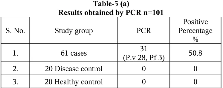
Results obtained by PCR n=101
S. No. Study group PCR
Positive Percentage
%
1. 61 cases (P.v 28, Pf 3)31 50.8
2. 20 Disease control 0 0
3. 20 Healthy control 0 0
31 out of 61 cases were positive for malarial parasite (P.v.28, P.f.3) by PCR technique with a percentage positivity of 50.8% and its 95% confidence interval was 38% to 63%. Control groups were negative by PCR method.
[image:66.595.109.490.134.281.2] [image:66.595.112.485.463.611.2]Comparison between PCR technique and JSB stained Peripheral blood smear n=101
PCR
JSB Stained Peripheral blood smear
Positive Negative Total
Positive 19 12 31
Negative 2 68 70
Total 21 80 101
[image:67.595.113.484.116.240.2]The sensitivity, specificity, PPV and NPV of PCR assay were calculated against the gold standard of Peripheral blood smear. The sensitivity of 90.5%, Specificity of 85%, PPV of 61.3% and NPV of 97.1% by PCR assay when compared to Peripheral blood smear.
Table-6
Evaluation of recent techniques in diagnosis of malaria
Test Sensitivity Specificity PPV NPV
QBC 100% 69.5% 51.3% 100%
ICT 95.2% 85% 62.5% 98.5%
PCR 90.5% 85% 61.3% 97.1%
Out of 190 cases tested with different techniques in detecting malarial parasite, the predominant species identified was P. vivax.
Table-7
[image:67.595.80.515.431.530.2]Test No. Of sample Positive % VivaxP. P. falciparum PERIPHERAL
BLOOD SMEAR 190 29.4 55 1
QBC 190 57.3 -
-ICT 61 52.4 29 3
PCR 61 50.8 28 3
Dot Elisa for P.
falciparum 61 - - 3