JOURNAL OFVIROLOGY, June 1996, p. 4188–4192 Vol. 70, No. 6
0022-538X/96/$04.0010
Copyrightq1996, American Society for Microbiology
A Plasmid-Based Reverse Genetics System for
Influenza A Virus
STEPHAN PLESCHKA, S. RICHARD JASKUNAS,† OTHMAR G. ENGELHARDT, THOMAS ZU¨ RCHER,‡
PETER PALESE,ANDADOLFO GARCI´A-SASTRE§*
Department of Microbiology, Mount Sinai School of Medicine, New York, New York 10029
Received 27 December 1995/Accepted 7 March 1996
A reverse genetics system for negative-strand RNA viruses was first successfully developed for influenza viruses. This technology involved the transfection of in vitro-reconstituted ribonucleoprotein (RNP) complexes into influenza virus-infected cells. We have now developed a method that allows intracellular reconstitution of RNP complexes from plasmid-based expression vectors. Expression of a viral RNA-like transcript is achieved from a plasmid containing a truncated human polymerase I (polI) promoter and a ribozyme sequence that generates the desired 3*end by autocatalytic cleavage. The polI-driven plasmid is cotransfected into human 293 cells with polII-responsive plasmids that express the viral PB1, PB2, PA, and NP proteins. This exclusively plasmid-driven system results in the efficient transcription and replication of the viral RNA-like reporter and allows the study ofcis- andtrans-acting signals involved in the transcription and replication of influenza virus RNAs. Using this system, we have also been able to rescue a synthetic neuraminidase gene into a recombinant influenza virus. This method represents a convenient alternative to the previously established RNP transfec-tion system.
In contrast to positive-strand RNA viruses, the naked genomic RNA of a negative-strand RNA virus is not able to initiate infection when expressed or transfected into a permis-sive cell line. The minimal infectious particle of this type of virus is the transcriptionally active ribonucleoprotein (RNP) complex. This complex is composed of the genomic viral RNA (vRNA) complexed with the viral nucleoprotein and the RNA-dependent RNA polymerase proteins (P proteins). Genetic manipulation of negative-strand RNA viruses has been made possible only by the establishment of reverse genetics tech-niques (for a review, see reference 8). These techtech-niques are based on the expression and/or transfection of functional viral RNA polymerase and RNP complexes in a host cell. Reverse genetics techniques have allowed the rescue of synthetic vRNA segments into infectious influenza viruses, which are seg-mented negative-strand RNA viruses (1, 3, 6, 7, 16, 30, 33), as well as the rescue of nonsegmented negative-strand RNA vi-ruses, such as rabies virus, vesicular stomatitis virus, respiratory syncytial virus, and measles virus, from full-length cDNA clones (4, 14, 25, 26, 31).
In the case of influenza A virus, Luytjes et al. (19) first described a reverse genetics system (also known as RNP trans-fection) based on the transfection of in vitro-reconstituted RNP complexes into helper influenza virus-infected cells. RNP complexes were made by incubating synthetic RNA transcripts with purified NP and P proteins (PB1, PB2, and PA) from influenza viruses. The helper virus was used as an intracellular
source of viral NP and P proteins and of the other vRNAs. Site-directed mutagenesis of single influenza virus genes was achieved by the same RNP transfection technique in combina-tion with a seleccombina-tion method against the corresponding RNA segment of the helper virus (6). Several modifications of the method of in vitro reconstitution of the viral RNPs have been published (7, 20, 28, 32, 33).
Other systems have been described for the expression of viral-like RNAs. When expressed by vaccinia virus or simian virus 40 recombinants, NP and P proteins support the tran-scription and replication of transfected model RNAs (2, 5, 11, 21). These proteins are also functional when induced in a stably transfected murine cell line by dexamethasone (13). In addition, Neumann et al. have successfully achieved RNP for-mation of viral model RNAs in influenza virus-infected cells after expression of the RNA from a murine RNA polymerase I (polI) promoter-responsive plasmid (23). Zhang and Air have also shown that both a model vRNA and the NP and P proteins can be expressed intracellularly from plasmids containing T7 promoters when the cells are infected with a recombinant vaccinia virus expressing T7 polymerase (34). However, to date, only the RNP transfection system has been successfully used for the generation of transfectant influenza viruses con-taining specific mutations in their genomes.
In this report we describe a reverse genetics system for influenza virus in which model vRNAs are effectively tran-scribed and amplified as a result of transfecting five different plasmids. Infection with a recombinant virus is not required, eliminating the possible interference of a heterologous virus in influenza virus-based replication and transcription. Since the method is plasmid driven, it allows the study of bothcis- and trans-acting signals involved in influenza virus transcription and replication in tissue culture. Finally, the system can also be utilized for the generation of transfectant influenza viruses when an influenza helper virus is provided. Compared with the previously described RNP transfection method, this new method of generating recombinant viruses eliminates the need
* Corresponding author. Mailing address: Department of Microbi-ology, Box 1124, Mount Sinai School of Medicine, 1 Gustave L. Levy Pl., New York, NY 10029. Phone: (212) 241-5923. Fax: (212) 534-1684. Electronic mail address: [email protected].
† On sabbatical leave from Lilly Research Laboratories, Indianap-olis, IN 46285.
‡ Present address: Centro Nacional de Biotecnologı´a (CSIC), Uni-versidad Auto´noma, Cantoblanco, Madrid, Spain.
§ On leave of absence from the Department of Biochemistry and Molecular Biology, Faculty of Medicine, University of Salamanca, Salamanca, Spain.
4188
on November 9, 2019 by guest
http://jvi.asm.org/
of purifying the viral NP and P proteins for RNP reconstitution in vitro.
The plasmid-based reverse genetics system for influenza vi-rus is schematically represented in Fig. 1. Five plasmids were cotransfected into human 293 cells. Four of the plasmids (pHMG-NP, pHMG-PB1, pHMG-PB2, and pHMG-PA, which were kindly provided by J. Pavlovic, University of Zu¨rich, Zu ¨r-ich, Switzerland) were used to express the NP, PB1, PB2, and PA proteins of influenza A/PR/8/34 virus under the control of a mouse hydroxymethylglutaryl-coenzyme A reductase (HMG) promoter (10). The fifth plasmid, pPOLI-CAT-RT, contains the chloramphenicol acetyltransferase (CAT) open reading frame in negative polarity flanked by the noncoding regions of the NS gene of influenza A/WSN/33 (WSN) virus. To ensure the correct 59end of the vRNA, the sequence of a truncated human RNA polI promoter (positions2250 to21) was fused directly to the end of the viral cDNA. We selected this pro-moter on the basis of the successful use by Neumann et al. of a similarly truncated murine polI promoter to drive the expres-sion of influenza virus model RNAs in virus-infected cells (22, 23). Since the polI promoters are species specific, it was not clear whether the truncated version of the human polI pro-moter would also be functional (29). To ensure the correct 39 end of the vRNA, the sequence of the hepatitis delta virus genomic ribozyme was included (24).
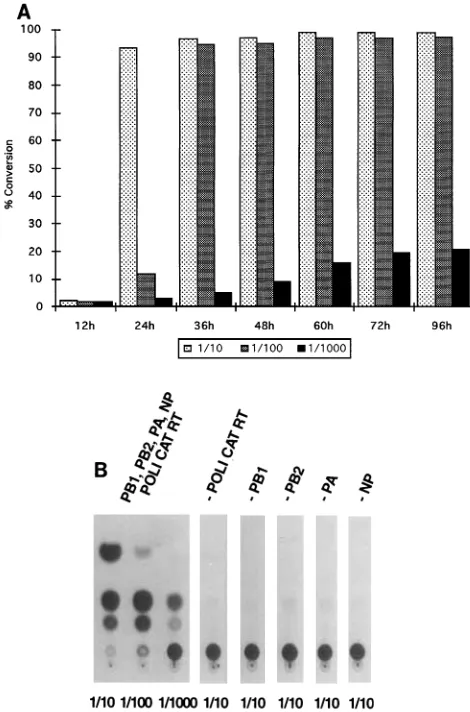
The transfection of all five plasmids into 293 cells resulted in the expression of CAT protein, as measured by its enzymatic activity (Fig. 2A). This indicates that a negative-sense RNA synthesized from the pPOLI-CAT-RT plasmid was reconsti-tuted intracellularly into functional RNPs. These RNPs were
[image:2.612.319.554.69.425.2]then transcribed by the viral RNA polymerase into mRNA, which was translated into CAT protein. As expected, CAT expression required coexpression of all four viral proteins (PB1, PB2, PA, and NP) (Fig. 2B). This result is in agreement with earlier findings with different expression systems for the NP and P proteins (11). In addition, the intracellularly recon-stituted RNPs were packaged into progeny influenza viruses when the plasmid-transfected cells were infected with influ-enza WSN virus (data not shown).
FIG. 1. Schematic representation of the plasmid-driven reverse genetics sys-tem. Five plasmids are cotransfected into 293 cells. The first plasmid, pPOLI-CAT-RT, is a pUC19-derived plasmid which contains the CAT open reading frame in minus sense, flanked by the 39and 59noncoding regions (39NCR and 59NCR, respectively) of the NS RNA segment of influenza A/WSN/33 virus (19). Expression of the influenza virus-like RNA is driven by a truncated human RNA polI promoter, which includes nt2250 to21 of the natural polI promoter (12). The correct 39end is ensured by the use of a sequence derived from the hepatitis delta virus genomic ribozyme (R). Junctions between influenza virus-specific sequences and the polI promoter and the ribozyme-specific sequences are shown. Underlined nucleotides in italics correspond to the noncoding ends of the influ-enza virus RNA. Arrows indicate the sites of polI initiation of transcription and of ribozyme cleavage. The other four plasmids, pHMG-PB1, pHMG-PB2, pHMG-PA, and pHMG-NP express the influenza A/PR/8/34 virus PB1, PB2, PA, and NP proteins from a hydroxymethylglutaryl-coenzyme A reductase promoter (HMG). These proteins are able to amplify and transcribe the influenza virus-like RNA expressed by pPOLI-CAT-RT into mRNA, resulting in the detection of CAT activity in transfected human 293 cells. The represented regions in plasmid constructs are not drawn to scale.
FIG. 2. CAT expression in 293 cells transfected with pPOLI-CAT-RT and pHMG expression vectors for influenza virus PB1, PB2, PA, and NP proteins. (A) Time course of plasmid transfection in 293 cells. Approximately 106293 cells
in 1.5 ml of Dulbecco modified Eagle medium containing 10% heat-inactivated fetal calf serum were transfected in suspension with pPOLI-CAT-RT (1mg), pHMG-PB1 (1mg), pHMG-PB2 (1mg), pHMG-PA (1mg), and pHMG-NP (2
mg) by using 30ml of DOTAP lipofection reagent (Boehringer Mannheim) according to the manufacturer’s instructions. Cells were then plated onto 35-mm-diameter dishes, incubated at 378C, and harvested at the indicated time points. After low-speed centrifugation, cells were resuspended in 100ml of 250 mM Tris-HCl buffer (pH 7.5), and cell extracts were made by freezing and thawing three times. CAT assays were done as previously described (19). The assay mixtures contained 2ml of [14
C]chloramphenicol (NEN), 1ml of 70 mg of acetyl coenzyme A (Pharmacia) per ml, and 50ml of cell extracts at the indicated dilution (1/10, 1/100, or 1/1,000). Incubation was for 2 h at 378C. The results are expressed as percentages of chloramphenicol conversion into its acetylated forms. (B) Requirements for CAT expression. 293 cells were transfected with pPOLI-CAT-RT, pHMG-PB1 (PB1), pHMG-PB2 (PB2), pHMG-PA (PA), and pHMG-NP (NP) as described for panel A or with different subsets of these five plasmids. For example, -PB1 indicates that cells were transfected with pPOLI-CAT-RT (1mg), pHMG-PB2 (1mg), pHMG-PA (1mg), and pHMG-NP (2mg) in the absence of pHMG-PB1. Cells were harvested at 48 h posttransfection, and CAT assays were performed as described for panel A. The dilution of cell extracts used in CAT assays is indicated below each lane.
on November 9, 2019 by guest
http://jvi.asm.org/
[image:2.612.65.296.72.242.2]
During influenza virus replication, vRNA is transcribed by the RNA polymerase into two different RNA species, cRNA copies and mRNA. The 39ends of the mRNAs lack the last 15 to 16 nucleotides (nt) of the cRNAs and contain instead
poly(A) tails. The cRNA is used again as a template by the RNA polymerase for the production of new vRNA. Proof that both cRNA and mRNA syntheses from the model vRNA mol-ecules were achieved upon transfection came from RNase pro-tection assay experiments. Total RNA isolated from trans-fected cells was subjected to hybridization with a minus-sense RNA probe containing 209 nt identical to the 59 end of the CAT model vRNA and 53 extra nonviral nt (18). Thus, this probe has the potential to hybridize to the terminal 209 and 193 nt at the 39ends of the CAT-specific cRNA and mRNA, respectively. After hybridization, samples were digested with RNase A and T1, as previously described (18). Two protected
RNA fragments, corresponding in size to cRNA and mRNA transcripts, were observed (Fig. 3). Identical signals were de-tected by the RNase protection assay when helper virus-in-fected cells were transvirus-in-fected by the previously described RNP transfection method (17, 18).
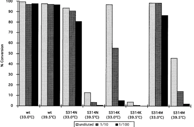
We provide an example to show that the plasmid-based reverse genetics system can be used to study thetrans-acting elements involved in influenza virus RNA replication and tran-scription. In these experiments, we investigated different tem-perature-sensitive (ts) phenotypes of the viral NP protein. For this purpose, we mutated amino acid 314 of the NP protein from serine to asparagine, lysine, or methionine (S-3143N [S314N], S314K, and S314M mutants, respectively). An S314N change in the WSN NP is known to be responsible for thets phenotype of thets56 mutant virus (15). All of the mutated NP proteins of influenza A/PR/8/34 virus exhibitedtsphenotypes when coexpressed with the viral P proteins and the CAT model RNA in 293 cells (Fig. 4). The S314K NP mutant, followed by the S314N and S314M NP mutants, was the most sensitive to high temperatures.
[image:3.612.93.259.69.295.2]The present system was also used for the rescue of transfec-tant influenza viruses carrying mutated vRNAs. For this pur-pose, we constructed pPOLI-NA-RT. This plasmid contains the NA gene of WSN virus in negative sense under the tran-scriptional control of the human polI promoter. In order to distinguish between NA genes derived from pPOLI-NA-RT
FIG. 3. Identification of CAT-specific RNA species of positive sense in plas-mid-transfected cells. 293 cells were cotransfected with pPOLI-CAT-RT alone (lane 1) or with pPOLI-CAT-RT, pHMG-PB1, pHMG-PB2, pHMG-PA, and pHMG-NP (lane 2), as described in the legend to Fig. 2. At 48 h posttransfection, cells were lysed and total RNA was extracted, as described before (18). Ten micrograms of isolated RNA was subjected to RNase protection assay using a negative-sense 262-nt-long probe, as previously described (18). Hybridization and RNA digestion yielded products of 209 and 193 nt, corresponding to the CAT-specific cRNA and mRNA, respectively, as indicated on the left. The positions of molecular size (in nucleotides) DNA markers (lane 3) are indicated on the right.
FIG. 4. Effects of mutations in the NP on CAT expression. pHMG expression vectors for several NP mutants containing amino acid substitutions at position 314 were constructed. 293 cells were cotransfected as described in the legend to Fig. 2 with the corresponding pHMG-NP plasmid, expressing wild-type (wt) or mutant (S314N, S314K, or S314M) NP, and pPOLI-CAT-RT, pHMG-PB1, pHMG-PB2, and pHMG-PA. Transfected cells were incubated for 48 h at 33.0 or 39.58C, cell extracts were made, and CAT assays were performed at the dilution indicated. The results are expressed as percentages of chloramphenicol conversion into its acetylated forms.
4190 NOTES J. VIROL.
on November 9, 2019 by guest
http://jvi.asm.org/
[image:3.612.154.465.479.683.2]and those from wild-type WSN virus, we inserted two silent mutations in the NA cDNA at nt 1358 and 1360, creating a novelSacI restriction site. pPOLI-NA-RT (1mg) was cotrans-fected into 293 cell monolayers on 35-mm-diameter dishes
together with pHMG-NP (2mg), pHMG-PB1 (1mg),
pHMG-PB2 (1mg), and pHMG-PA (1mg). At 36 h posttransfection, cells were infected with WSN-HK helper virus and incubated at 378C for an additional 12 h in 1.5 ml of Dulbecco modified Eagle medium containing 2% fetal calf serum. WSN-HK virus is a reassortant containing the NA-specific RNA segment de-rived from influenza A/Hong Kong/8/68 virus and all the other segments from WSN virus (27). Supernatants derived from 293 cells were used to infect fresh MDBK cell monolayers in 80-cm2flasks. MDBK cells were incubated at 378C for 3 additional
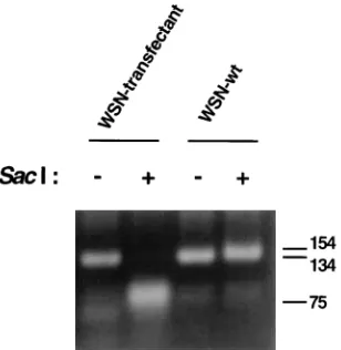
days in 10 ml of REM containing 0.2% bovine serum albumin (6). Under these conditions, transfectant viruses containing the NA RNA segment derived from plasmid pPOLI-NA-RT were rescued; only viruses containing the NA RNA segment of WSN virus are able to grow in MDBK cells in the absence of trypsin (27). The identity of the transfectant virus was further confirmed as follows. vRNA was isolated from purified viruses, and the 59end of the NA-specific RNA segment was amplified by reverse transcription-PCR using specific DNA primers, as previously described (9). Restriction analysis of the amplified product withSacI enzyme revealed that the NA segment of the transfectant virus was derived from the pPOLI-NA-RT plas-mid (Fig. 5). These results illustrate that the plasplas-mid-based reverse genetics system described above can also be used for the genetic manipulation of influenza virus genomes. By using the plasmid-driven reverse genetics system, approximately 10 transfectant viruses per transfection were obtained in three independent experiments. This is in contrast to rescue efficien-cies of between 103and 104transfectant viruses per
transfec-tion when the previously described RNP transfectransfec-tion system was used (7). The differences in the efficiency of rescue be-tween these two systems may be caused by differences in the levels of replication of the helper virus in MDBK (used for RNP transfection) and 293 cells. It should be noted that WSN
virus grows to 1-log-higher titers in MDBK cells than in 293 cells.
In summary, the plasmid-based reverse genetics system for influenza virus described in this report allows intracellular re-constitution of viral RNPs and permits us to study the repli-cation and transcription of model vRNAs. In a typical exper-iment, the levels of CAT activity were at least 10 times higher than those obtained by our previously described RNP trans-fection method. This plasmid-based reverse genetics system allows not only the study of cis-acting signals in the vRNA responsible for the replication and transcription of RNA seg-ments but also the study oftrans-acting elements and domains in the viral NP and P proteins necessary for RNA replication and transcription. The absence of a requirement for recombi-nant viruses, such as vaccinia virus, to drive the expression of influenza virus protein and/or RNA eliminates the possibility of interference between the replication and transcription of influ-enza virus-like RNAs and the replication of recombinant viruses. The use of a polI promoter to generate transcriptionally competent influenza virus model RNAs has been previously reported by Neumann et al. (22, 23). They used mouse polI promoter and terminator sequences to ensure the correct for-mation of the 59and 39ends of model RNAs (23, 35). We have reported here that similar results are achieved by using human polI promoter and hepatitis delta virus genomic ribozyme se-quences. However, the main difference between the two polI promoter-based systems is the source of the NP and P proteins required for the replication and transcription of the vRNA. The use of polII promoter-based plasmids instead of infectious influenza viruses to drive the expression of the NP and P proteins resulted in approximately 100-times-higher levels of CAT expression in 293 cells (data not shown). In addition, the trans-acting elements required for the replication and tran-scription of influenza virus RNAs can be analyzed by using the plasmid-based system.
Although several other transfection systems have been de-scribed for influenza viruses, only the RNP transfection method has been used for the rescue of transfectant influenza viruses. This method requires the purification of viral NP and P pro-teins for RNP reconstitution in vitro. In contrast, the plasmid-based transfection method eliminates the burden of the viral protein purification steps by reconstituting viral RNPs in tissue culture cells from expression plasmids. Thus, this system may represent a convenient alternative method for the rescue of transfectant influenza viruses. More importantly, since repli-cation and transcription of the vRNA is achieved in the ab-sence of helper virus, coexpression in 293 cells of the eight vRNA segments in combination with the NP and P proteins may result in the rescue of infectious influenza viruses from plasmid DNAs.
We thank Jovan Pavlovic for sharing the pHMG expression plasmids and Robert Tjian for providing the genomic clone of the human polI promoter. We also thank Guangxiang Luo and John Taylor for their contributions, leading to the pPOLI-CAT-RT construct; Hongyong Zheng and Qihan Li for their help in constructing several plasmids; and Scott Kerns and Ricardo Renvill for excellent technical assistance. This work was supported by grants from the Deutsche Forschungs-gemeinschaft (S.P.) and the NIH (P.P.).
REFERENCES
1. Barclay, W. S., and P. Palese.1995. Influenza B viruses with site-specific mutations introduced into the HA gene. J. Virol.69:1275–1279.
[image:4.612.101.255.71.229.2]2. Biswas, S. K., and D. P. Nayak.1994. Mutational analysis of the conserved motifs of influenza A virus polymerase basic protein 1. J. Virol.68:1819–1826. 3. Castrucci, M. R., and Y. Kawaoka.1995. Reverse genetics system for gen-eration of an influenza A virus mutant containing a deletion of the carboxyl-terminal residue of M2 protein. J. Virol.69:2725–2728.
FIG. 5. Restriction analysis of the NA cDNA of a transfectant virus which was rescued by using the plasmid-driven reverse genetics system. The first 130 nt at the 59end of the NA vRNA from purified transfectant virus (WSN-transfec-tant) or from wild-type influenza A/WSN/33 virus (WSN-wt) were amplified by coupled reverse transcription-PCR using oligonucleotide primers 59-TGGAC
TAGTGGGAGCATCAT-39and 59-ATGCTCTAGAAGCTTAGTAGAAACA
AGG-39. The PCR products (145 nt in length) were incubated for 2 h at 378C in the presence (1) or absence (2) of 10 U ofSacI restriction enzyme. Samples were then run on a 2% agarose gel and stained with ethidium bromide. The positions of size (in nucleotides) markers are indicated on the right.
on November 9, 2019 by guest
http://jvi.asm.org/
4.Collins, P. L., M. G. Hill, E. Camargo, H. Grosfeld, R. M. Chanock, and B. R. Murphy.1995. Production of infectious human respiratory syncytial virus from cloned cDNA confirms an essential role for the transcription elongation factor from the 59proximal open reading frame of the M2 mRNA in gene expression and provides a capability for vaccine development. Proc. Natl. Acad. Sci. USA92:11563–11567.
5.De la Luna, S., J. Martı´n, A. Portela, and J. Ortı´n.1993. Influenza virus naked RNA can be expressed upon transfection into cells co-expressing the three subunits of the polymerase and the nucleoprotein from simian virus 40 recombinant viruses. J. Gen. Virol.74:535–539.
6.Enami, M., W. Luytjes, M. Krystal, and P. Palese.1990. Introduction of site-specific mutations into the genome of influenza virus. Proc. Natl. Acad. Sci. USA87:3802–3805.
7.Enami, M., and P. Palese.1991. High-efficiency formation of influenza virus transfectants. J. Virol.65:2711–2713.
8.Garcı´a-Sastre, A., and P. Palese.1993. Genetic manipulation of negative-strand RNA virus genomes. Annu. Rev. Microbiol.47:765–790.
9.Garcı´a-Sastre, A., and P. Palese.1995. The cytoplasmic tail of the neuramin-idase protein of influenza A virus does not play an important role in the packaging of this protein into viral envelopes. Virus Res.37:37–47. 10.Gautier, C., M. Mehtali, and R. Lathe. 1989. A ubiquitous mammalian
expression vector, pHMG, based on a housekeeping gene promoter. Nucleic Acids Res.17:8389.
11.Huang, T.-S., P. Palese, and M. Krystal.1990. Determination of influenza virus proteins required for genome replication. J. Virol.64:5669–5673. 12.Jones, M. H., R. M. Learned, and R. Tjian.1988. Analysis of clustered point
mutations in the human ribosomal RNA gene promoter by transient
expres-sionin vivo. Proc. Natl. Acad. Sci. USA85:669–673.
13.Kimura, N., M. Nishida, K. Nagata, A. Ishihama, K. Oda, and S. Nakada.
1992. Transcription of a recombinant influenza virus RNA in cells that can express the influenza virus RNA polymerase and nucleoprotein genes. J. Gen. Virol.73:1321–1328.
14.Lawson, N. D., E. A. Stillman, M. A. Whitt, and J. K. Rose.1995. Recom-binant vesicular stomatitis viruses from DNA. Proc. Natl. Acad. Sci. USA
92:4477–4481.
15.Li, R., P. Palese, and M. Krystal.1989. Complementation and analysis of an NP mutant of influenza virus. Virus Res.12:97–111.
16.Li, S., M. Xu, and K. Coelingh.1995. Electroporation of influenza virus ribonucleoprotein complexes for rescue of the nucleoprotein and matrix genes. Virus Res.37:153–161.
17.Li, X., and P. Palese.1994. Characterization of the polyadenylation signal of influenza virus RNA. J. Virol.68:1245–1249.
18.Luo, G., W. Luytjes, M. Enami, and P. Palese.1991. The polyadenylation signal of influenza virus RNA involves a stretch of uridines followed by the RNA duplex of the panhandle structure. J. Virol.65:2861–2867. 19.Luytjes, W., M. Krystal, M. Enami, J. D. Parvin, and P. Palese. 1989.
Amplification, expression, and packaging of a foreign gene by influenza virus. Cell59:1107–1113.
20. Martı´n, J., C. Albo, J. Ortı´n, J. A. Melero, and A. Portela.1992.In vitro
reconstitution of active influenza virus nucleoprotein complexes using viral proteins purified from infected cells. J. Gen. Virol.73:1855–1859. 21. Mena, I., S. de la Luna, C. Albo, J. Martı´n, A. Nieto, J. Ortı´n, and A. Portela.
1994. Synthesis of biologically active influenza core proteins using a vaccin-ia-T7 RNA polymerase expression system. J. Gen. Virol.75:2109–2114. 22. Neumann, G., and G. Hobom.1995. Mutational analysis of influenza virus
promoter elementsin vivo. J. Gen. Virol.76:1709–1717.
23. Neumann, G., A. Zobel, and G. Hobom.1994. RNA polymerase I-mediated expression of influenza viral RNA molecules. Virology202:477–479. 24. Perrotta, A. T., and M. D. Been.1991. A pseudoknot-like structure required
for efficient self-cleavage of hepatitis delta virus RNA. Nature (London)
350:434–436.
25. Radecke, F., P. Spielhofer, H. Schneider, K. Kaelin, M. Huber, C. Do¨tsch, G. Christiansen, and M. A. Billeter.1995. Rescue of measles virus from cloned DNA. EMBO J.14:5773–5784.
26. Schnell, M. J., T. Mebatsion, and K.-K. Conzelmann.1994. Infectious rabies viruses from cloned cDNA. EMBO J.13:4195–4203.
27. Schulman, J. L., and P. Palese.1977. Virulence factors of influenza A viruses: WSN virus neuraminidase required for plaque production in MDBK cells. J. Virol.24:170–176.
28. Seong, B. L., and G. G. Brownlee.1992. A new method for reconstituting influenza polymerase and RNAin vitro: a study of the promoter elements for cRNA and vRNA synthesisin vitroand viral rescuein vivo. Virology186:
247–260.
29. Smale, S. T., and R. Tjian.1985. Transcription of herpes simplex virustk
sequences under the control of wild-type and mutant human RNA polymer-ase I promoters. Mol. Cell. Biol.5:352–362.
30. Subbarao, E. K., Y. Kawaoka, and B. R. Murphy. 1993. Rescue of an influenza A virus wild-type PB2 gene and a mutant derivative bearing a site-specific temperature-sensitive and attenuating mutation. J. Virol.67:
7223–7228.
31. Whelan, S. P. J., L. A. Ball, J. N. Barr, and G. T. W. Wertz.1995. Efficient recovery of infectious vesicular stomatitis virus entirely from cDNA clones. Proc. Natl. Acad. Sci. USA92:8388–8392.
32. Yamanaka, K., N. Ogasawara, H. Yoshikawa, A. Ishihama, and K. Nagata.
1991.In vivoanalysis of the promoter structure of the influenza virus RNA genome using a transfection system with an engineered RNA. Proc. Natl. Acad. Sci. USA88:5369–5373.
33. Yasuda, J., D. J. Bucher, and A. Ishihama.1994. Growth control of influenza A virus by M1 protein: analysis of transfectant viruses carrying the chimeric M gene. J. Virol.68:8141–8146.
34. Zhang, H., and G. M. Air.1994. Expression of functional influenza virus A polymerase proteins and template from cloned cDNAs in recombinant vac-cinia virus infected cells. Biochem. Biophys. Res. Commun.200:95–101. 35. Zobel, A., G. Neumann, and G. Hobom.1993. RNA polymerase I catalyzed
transcription of insert viral cDNA. Nucleic Acids Res.21:3607–3614.
4192 NOTES J. VIROL.