A Dissertation on
RECONSTRUCTION OF TUBULAR BONE
DEFECTS WITH NON VASCULARIZED
FIBULAR GRAFT
Submitted to
THE TAMILNADU DR. M.G.R. MEDICAL UNIVERSITY
CHENNAI
In partial fulfillment of the regulations For the Award of the Degree of
M.S. DEGREE BRANCH –II
ORTHOPAEDIC SURGERY
KILPAUK MEDICAL COLLEGE
CHENNAI
CERTIFICATE
This is to certify that Dr. R. VASANTHARAMAN Post
graduate student (2004- 2007) in the department of orthopaedics Govt. Kilpauk Medical College Chennai has done this dissertation on “RECONSTRUCTION OF TUBULAR BONE
DEFECTS WITH NON VASCULARIZED FIBULAR GRAFT” under
my guidance and supervision in partial fulfillment of the regulation laid down by the Tamil Nadu Dr. M.G.R Medical
University, Chennai for MS (Orthopaedics) degree examination to be held on March
2007.
Prof. Dr. THIAGAVALLI
KIRUBAKARAN, M.D., Dean,
Govt. Kilpauk Medical College and Hospital,
Chennai – 600 010.
Prof. Dr. A. SIVAKUMAR, M.S (Ortho) D. Ortho., Professor – HOD,
DECLARATION
I, Dr. R. VASANTHARAMAN, solemnly declare that
dissertation titled “RECONSTRUCTION OF TUBULAR BONE
DEFECTS WITH NON VASCULARIZED FIBULAR GRAFT” is a
bonafide work done by me at Kilpauk Medical College 2004 - 2007
under the guidance and supervision of my unit chief
Prof. A. SIVAKUMAR M.S (Ortho)., D.Ortho Professor of Orthopaedic surgery.
This dissertation is submitted to Tamilnadu Dr. M.G.R
Medical University, towards partial fulfillment of regulation for the
award of M.S. Degree (Branch – II) in Orthopaedic surgery.
Place : Chennai.
Date :
ACKNOWLEDGEMENT
First and fore most I would like to thank Prof. THIAGAVALLI KIRUBAKARAN M.D., Dean, Kilpauk Medical College for permitting me to use the resources and clinical material of this hospital.
I offer my sincere thanks to Prof. SOMASEKAR M.D., Superintendent of Govt. Royapettah Hospital Chennai -14 for having permitted me to use the hospital material for this study.
I express my profound thanks to my esteemed professor and head of the department, Prof. A. SIVAKUMAR Govt. Royapettah Hospital for his valuable suggestions and help for this study
I thank Prof. K. NAGAPPAN and
Prof. K. SANKARALINGAM for their encouragement and help for this study.
I thank Dr. R. BALACHANDRAN Assistant Professor
Government Royapettah Hospital for encouraging and extending invaluable guidance as a guide to perform and complete this dissertation.
My sincere thanks to Dr. P. ELANGOVAN, Dr. T. THOLKAPPIYAN, Dr. K. MOHAN KUMAR, Dr. V. THANIGAINATH and Dr. P. VEERANAN YADAV, for encouragement throughout the period of this study.
CONTENT
SL.
NO TITLE
PAGE NO.
1 INTRODUCTION 1
2 AIM 3
3 REVIEW OF LITERATURE 4
4 MATERIALS AND METHODS 32
5 ANALYSIS 41
6 RESULTS 44
7 COMPLICATIONS 45
8 CASE ILLUSTRATIONS 46
9 DISCUSSION 53
10 CONCLUSION
BIBLIOGRAPHY
PROFORMA
INTRODUCTION
In our speciality of orthopaedics we come across many
problems involving the bone and soft tissues like, fractures,
infections, tumours etc., among these bony defects are a great
challenge to us. These defects can be due to various causes such as
post traumatic bone loss, post infective bone loss and defect
resulting after excision of tumours. These defects have to be
reconstructed to give better form and function to the patients.
These defects can be either diaphyseal or osteoarticular. To
reconstruct these defect is a surgical challenge. Surgical challenges
may include primary articular defects, diaphyseal defects and
salvage of other failed reconstructions. Many options are available
that may be tailored to the individual's need.
Autograft, allograft, prosthetic replacement or allograft
prosthetic composite are established methods for reconstructions.
Among the autograft and allograft reconstructions it can be done
either as vascularized or nonvascularized graft
Though the use of vascularized graft for reconstruction may
provide rapid biological incorporation, growth potential in
compromised soft tissue environment. This procedure may not be
possible in some of the centers for want of technical expertise
however the long term follow up of nonvascularized fibular graft
also gives good result. Hence we made an attempt to reconstruct
the bony defects with nonvascularized fibular graft which is
AIM
To study the outcome of the nonvascularized fibular graft in
reconstructing the tumour defects and gap nonunion of
tubular bones.
To share our experience of these procedures done in our
REVIEW OF LITERATURE
HISTORICAL REVIEW
History of Bone Grafting
Bone grafting is a very old surgical procedure. The first
recorded bone implant was performed in 1668 .Over 200 years ago
John Hunter demonstrated that separated fragments of bone could
survive and grow “ Adhesion of detached splinters take place ---not only in those which are attached to the soft parts
but in those which are entirely loose”. In concluding that “these pieces must retain the living principle” he made the first
accurate though quite unconscious forecast of the possibility of bone
transplantation.
Nearly two centuries later, Hunter’s words re-echoed in a
monographic “ bone looks so dead ; as if it were no more than a
block of marble carved with delicate artistry to fit the structure
around it and give them support and yet it is so very alive and
forever busy shaping itself in adoption to constantly changing
Von Meekran (1668)
Though he reported an attempt to repair a cranial defect in a
Russian soldier with a dog’s skull bone the failure of this
heterogenous graft did not become, evident in as much as Von
meekran was forced to remove the bone graft.
Ollier (1867)
The real interest in the bone graft followed the publication in
1867 of fundamental experimental work of ollier on bone grafting in
rabbits, cats , dogs and birds .
First Successful Bone Transplantation29
In 1878 Macewen in Glasgow removed the entire diaphysis of
the humerus of a 3 years old infant for persistent osteomyelitis .
Fifteen months later he was asked to amputate the flail and useless
limb. Instead he implanted a number of bone wedges excised
during corrective osteotomies from six other patients. The
procedure was done in three stages, two wedges being inserted at
measured 6 inches (15 cms) in length, more than two – third of it
being from transplanted bone.
Lexer (1907)
The clinical application of bone allografting became prevalent
in the first decade of the 20th century, after the experimental work of Ollier and Axhausen. In 1907 Lexer was the first to perform allogeneic whole joint transplantation and he had performed 25 cases by 1925.
Axhausen (1909–1911)
In 1909 Axhausen studied the osteogenic activity of
periosteum and concluded that it was highest in autograft and
lowest in heterografts.
Albee (1911)
Albee established the cortical bone grafting.
Macewan (1912)
Macewan presented paper on transplantation and
replacement of a humerus in a patient and followed this by
extensive experimental and clinical observation which was
Phemister (1914)
In 1914 Phemister showed that osteogenesis in experimental
animal occurs from periosteum also. It was Phemister who
emphasized that the fate of transplanted bone depend upon perfect
haemostasis, perfect asepsis and perfect co-aptation of parts.
Kappis (1915)
Kappis in 1915 employed full thickness rib with periosteum
to cover the dural and cranial defects. After this many
orthopaedicians have tried cortical bone grafts using fibula, tibia or
ribs in filling the gap in the treatment after excision of the tumour
of the dispensable part of long bone.
Gallie and Robertson (1918)
They proved that apart from periosteum, the preservation of
blood supply plays main role in osteogenesis and the union of the
graft with recipient bone and so undue stripping of periosteum
Inclan (1942)
In 1942 Inclan reported the storage of autogenous and
allogenous bone and this report stimulated many similar clinical
efforts at preservation, sterilization and delayed reimplantation.
T. L. Lawson et al (1952)
They removed a giant cell tumour from the lower end
of the radius with a tumour clearance of 3 cms. The gap was filled by a free autogenous fibular graft taken from the ipsilateral side. The fibular head was made to articulate with the carpal bones. The proximal end of fibular graft and distal end of the recipient radius was fixed with two cortical screws, after making a step cut. Bony union between graft and recipient bone was present after 12 weeks and the wrist function was almost normal. The patient was a policeman.
Enneking and Hammock (1965)
They have reviewed cases and reported about the
comparative study between vascularization of autogenous and
F. F. Parrish (1966)
In 1966, F.F Parrish used autogenous fibular graft in
the treatment of bone tumours by resection.
Buchardt, Busbee and Enneking (1975)
These authors studied the repair of experimental autogenous
grafts of cortical bone and have reported that the repair begin at 8
to 12 weeks during which bony union become evident.
Taylor and Daniel (1975)
The first free vascularized fibular graft was done by them in 1975 for tibial defect. They have taken the middle third of fibula with a main nutrient artery and a vein and
anastamosed with the tibial vessels by microvascular surgery and the bony union was good in 12 weeks.
WF Enneking, JL Eady and H Burchardt (1980)
They analysed the result of using segmental cortical autogenous bone grafts to reconstruct defects created by
graft must be protected through out the prolonged reparative phase until there is evidence of union, adequate stress must be transmitted to the graft during this period to stimulate the repair, enough grafts must be used to provide functional replacement in the major tubular bones.
Russell Moore et al (1983)
In 1983 Russel Moore et al used vascularized fibular graft for
the gap more than 6 cms, producing good results.
M. Campanacci and P. Picci (1984)
They reported that fibular graft for giant cell tumour gives
very good results and the wrist joint mobility is almost normal.
A. Jhow et al (1985)
In 1985 A. Jhow et al treated 18 cases of osteoclastoma lower
end of radius by excising the tumour and the gap was filled with
autogenous fibular graft. They had maximum follow up of average
7.1 years. But local recurrence in 5 patients, one died of pulmonary
metastasis, nonunion in 5 patients and fracture of the graft in 3
SOURCES OF GRAFTS28 Autograft
The transplanted bone is derived from the same person
receiving it. Autograft both cancellous and cortical, are usually
implanted fresh and are often osteogenetic, whether by providing a
source of osteoprogenitor cells or by being osteoinductive fresh
autogenous bone is preferred since it is non antigenic and has
superior osteogenic induction properties.
Isograft
Isograft is a graft exchanged between people with genetically
identical characteristics from same zygote (e.g., identical human
twins).
Allograft
An allograft is transplantation from one person to another of
the same species, but with dissimilar genetic characteristics. When
the graft is between member of different highly inbred lines, the
allograft is termed “allogeneic”. Fresh allotransplant behave
initially as fresh autogenous transplants, but ultimately induce an
immune response which may reject the bone. Before implantation,
preserving its osteogenic induction property and retaining its
mechanical strength.
Various methods are used to process the bone before storage
and most include freeze drying (lyophilization) which is generally
believed to reduce the bone antigenicity so that the immune
response in greatly delayed and reduced, permitting the induced
osteogenesis in the recipient to proceed unhindered.
The disadvantage of lyophilization in the reduction of the
mechanical strength of the implant and fatigue fracture and non
union are frequent when allogeneic bone in used for massive
segmental and osteoarticular implants. Such grafts must be
stabilized both by internal devices and external supports for
lengthy periods until union and repair are certain.
The processed and preserved allogeneic bone is not a
substitute for autogenous bone.
Xenograft
A xenograft is transplantation from one person of one species
to a person of another species. Different types of xenografts are.
• Bovine bone xenograft
• Type I collagen xenograft
TYPES OF GRAFTS18
Multiple cancellous chips or strips
This is the most osteogenic and most widely used graft. The
best source of cancellous bone graft is the ilium. It is the principle
type of graft used for fractures, nonunions and for arthodesis of the
spine.
Single onlay cortical bone graft
This was used most commonly before the development of good
quality internal fixation and was employed for both osteogenesis
and fixation in the treatment of nonunion. The tibia and split fibula
can be used. The most common indication for this graft today is
bone grafting and stabilizing the cervical spine.
Dual onlay cortical bone graft
Boyd developed the dual – onlay cortical bone graft technique
in 1941 for the treatment of congenital pseudo-arthrosis of tibia.
A version of this technique using allograft is useful for revision
Inlay bone graft
Albee popularized the inlay bone graft for the treatment of
nonunion. Inlay grafts are created by a sliding technique, graft
reversal technique, or as a strut graft. This technique is used for
treatment of nonunion of the tibia, arthrodesis and epiphyseal
arrest.
Sliding graft
A rectangular graft is cut and removed. It is simply flipped
end for end and then impacted back into the slot. This technique is
rarely used today. The disadvantages of the sliding graft is that
graft fits loosely in the bed and it creates stress risers proximal and
distal to the nonunion sites.
H–graft
The H-graft is a corticocancellous graft usually harvested
from ilium specifically designed to achieve posterior fusion of the
cervical spine.
Peg and dowel graft
Dowel grafts were developed for the grafting of nonunions in
instances, dowel grafts have been replaced by micro vascularized
fibular grafts.
Peg grafts have also been used to bridge the tibia and fibula
to produce proximal and distal tibio fibular synostosis.
Medullary graft
Medullary grafts are not indicated for the diaphysis of major
long bones. Grafts in this location interfere with restoration of
endosteal blood supply because they are in the central axis of the
bone, they resorb rather than incorporate. The only possible use for
a medullary graft is in metatarsals and metacarpals.
Osteoperiosteal graft
In osteoperiosteal grafts, the periosteum is harvested with
chips of cortical bone. They are rarely used today.
Pedicle graft
Pedicle grafts may be local or moved from a remote site using
microvascular surgical techniques. In local muscle-pedicle bone
grafts, an attempt is made to preserve the viability of the graft by
maintaining muscle and ligament attachments carrying blood
the nutrient artery. Advantage of high percentage of cell survival,
rapid incorporation and increased active participation of the
grafted cells in the healing process.
Whole Bone Transplant27
The fibula provides the most practical graft for bridging long
defects in the diaphyseal portion of bones of the upper extremity. A
fibular graft is stronger than a fill thickness tibial graft, and when
soft tissue is scant, a wound that could not be closed over dual
grafts may be closed over a fibular graft. Disability after removing
a fibular graft is less than after removing a larger tibia graft. In
children the fibula can be used to span a long gap in the tibia,
usually by a two stage procedure.
Bone Graft Substitutes9
While autograft and allografts remain the most commonly
used osteoinductive materials in reconstructive surgery, the limited
availability and high cost of the bone graft, as well as concerns of
transmitting infectious diseases have led to the development of
several synthetic bone graft substitutes. The synthetics with
clinical applications include hydroxyapatite (HA); tricalcium
calcium phosphate polymer and composites of TCP, HA, and bovine
collagen.
The products are biocompatible; they are also available as
dense or porous implant or in granular form. Ceramics act as
osteoconductors, where the product supports or acts as a scaffold,
but does not generate new bone cells. In near future, synthetic
growth factors coupled with calcium phosphate carriers are likely
to provide osteoinductive as well as osteoconductive materials
useful for skeletal reconstruction.
The ideal graft should have the following characters
according to Red fern:
• Promotes bone healing
• Replaced by host bone
• Does not transmit disease
• Immunologically inert
• Provides mechanical support and maintains
volume
ANATOMY OF FIBULA
Fibula is the slender lateral bone of the leg. It has a proximal
head, a narrow neck, a long shaft and a distal lateral malleolus.
Head
Head is surmounted by a styloid process. It articulates with
the lateral condyle of tibia. The arcuate ligament of knee joint is
attached to it. The fibular collateral ligament and the biceps
femoris are attached in front of the styloid process. The head of
fibula resembles the distal radius and distal fibula.
Shaft
Shaft has three surfaces via anterior, lateral and posterior,
corresponding to the extensor, peroneal and flexor compartments of
the leg. Peroneus longus arises from the upper two third and
peroneus brevis arise from the lower two third of the lateral surface
of shaft. The common peroneal nerve enters peroneus longus at the
neck of the bone, the nerve can be rolled against the bone here,
where it may be damaged by a plaster cast or tight bandage. It also
divides here into superficial and deep peroneal nerve. From the
longus and peroneus tertius arises. From the posterior surface
flexor hallucis longus, soleus and tibialis posterior arises.
Lateral Malleolus
The lateral malleolus articulates with talus. It gives
attachment to posterior tibiofibular ligament, anterior tibiofibular
ligament, anterior talofibular ligament, calcaneofibular ligament
and posterior talofibular ligament. The groove on the posterior
surface of the malleolus lodges the tendons of peroneus longus and
peroneus brevis.
Vascular Supply
The blood supply is through peroneal artery. The peroneal
artery gives off the nutrient artery for the fibula which enter the
bone on it’s posterior surface.
The proximal and distal ends receive metaphyseal vessels
from the genicular and ankle arterial anastomoses respectively.
Applied Anatomy
1. The fibula does not take part in articulation of knee joint.
3. Distal fibula is important in maintaining the stability
of ankle joint
Properties of Fibular Graft13
• It is a straight cortical bone
• A graft about 26 cm long can be harvested from an
adult.
• Muscle, an articular surface proximally, and in a child
a proximal physis are available.
• The dissection is superficial and straight forward
• Complications are minimal, especially if the peroneal
nerve and tibial vessels are protected.
• Usually, overlying skin and nerve are not available.
• It is useful for long bone defects.
Indications for Fibular Graft
1. Reconstruction of large defects of long bone secondary to
tumour resection
2. Reconstruction of large long bone defect secondary to
sequestrectomy
3. Reconstruction of large bony defect secondary to trauma
5. Infection at the site of nonunion of the femur or tibia
6. Avascular necrosis of femoral head
7. Bridging of joints for arthrodesis
8. Mandibular reconstruction after tumour resection
Basic Principles10
The outcome of the graft is determined by specific properties
of cortical and cancellous bone. Primary is Osteogenesis: the synthesis of new bone either by cells of graft or host origin. New
bone of graft origin is produced by the surface cells of properly
handled fresh cortical and cancellous grafts. This early bone
formation accelerates incorporation during the first few weeks after
surgery. Cancellous bone, with its large surface area covered with
cells, has a greater potential for producing new bone of graft origin
than does cortical bone.
New bone of host origin is produced through the process of
Osteoinduction. This is a process whereby mesenchymal cells of the host are recruited to differentiate into osteoblasts. The recruitment
and differentiation of these cells is probably modulated by low
molecular weight polypeptides, such as the glycoprotein bone
morphogenic protein (BMP). BMP is a hydrophobic, non species –
diaphysis, from dentin and from various bone tumors. The activity
of BMP does not require viable graft cells, and is present not only
in fresh autografts, but also in modified allografts. Autoclaving
destroys the activity of BMP.
The three – dimensional process of growth of capillaries,
perivascular tissue, and osteoprogenitor cells of the host into the
graft is termed Osteoconduction. The graft provides the “trellis” or framework for the ingrowth of this tissue.
In addition to these biologic functions, the graft may provide
structural support until the recipient tissue can bear weight. Grafts
of cortical bone most commonly provide structural integrity during
remodeling. However, the incorporation of all grafts proceeds by
creeping substitution, the gradual resorption of the grafts and
replacement by new bone. Bone graft incorporation is characterized
by five defined stages. It begins with inflammation, proceeds to
osteogenesis and remodeling, and ends in a mechanically efficient
PATHOPHYISOLOGY28
The autogenous cortical transplant in contrast to the
cancellous transplant is revascularized at a slower rate. Blood
vessels penetrate the volkmann’s and haversian canal by the sixth
day and complete vascularization occurs by 1 to 2 months. In
addition at the surface of the cortical bone, vascular - cellular tufts
termed “cutter heads” by osteoclastic resorption progressively
burrow new tunnels into the bone.
In contrast to the cancellous bone repair, the process is
initiated by osteoclastic resorption rather than by osteoblastic bone
deposition. Resorptions begin at the outer regions of the transplant.
In experimental cortical bone transplants observed in dogs, the rate
at which the interior of the haversian canals is widened is
significantly increased until the sixth week (excavation chambers),
a phase correlated with loss of mechanical strength then the
resorptive rate gradually declines to nearly normal level by the end
of one year.
The active resorption process is at first preferentially directed
towards peripherally located haversian system (osteons) reaching
the interior by the fourth week. The interstitial lamellae remain
resorption ceases and osteoblasts appears and rebuild concentric
lamellae. The appositional phase occurs initially at 12 weeks after
transplantation. The repair is at first greater at transplant – host junctions and secondarily the repair advances towards the center of the transplant.
The proportion of viable new bone to necrotic old bone
increases from 2 weeks to 6 months after transplantation, but then
the ratio appears to remains unchanged in transplants between 6
months and 2 years. Thus it can be seen that cancellous bone is
completely repaired whereas cortical bone is only partially repaired
and remains as an admixture of necrotic and viable bone.
Cellular Behaviour
Cell reaction is required to unite a bone graft with the
recipient. Vessels can invade the osseous graft only when the
shearing between the graft and host bone is eliminated. This is
accomplished mainly by callus which glues the host tissue and graft
Bone formation requires three factors:
1. Proper specialized cell
2. Proper nutrition
3. Proper stimulus
The proper cells arises from a mesenchymal stem cell which
comes from perivascular cells, reticular cells of bone marrow, cells
of trabecular surfaces and cells of cambium layer of periosteum.
The specialization of cells in influenced by nutritional and
electromechanical factors. When cells become compacted and the
oxygen supply is adequate bone formation results.
OSTEOINDUCTION ROLE IN BONE GRAFT INCORPORATION (URIST)28
Incorporation
Defined as process of envelopment and interdigitation of
The incorporation of autogenous bone graft occurs in five stages:
Stage I
Occurs within minutes to hours after surgery. This stage
consists of inflammation, activation of migratory fibroblast
like mesenchymal cells and proliferation of preosteoblasts
and preosteoclasts in the recipient band.
Stage II and III
Begins one day to a week after inflammation and surgical
injury subsides. This stage is characterized by cell
osteoinductive interactions between protein
macromolecules of donor bone matrix and cell surface
receptor sites on the fibroblast like mesenchymal cells. In an
autograft osteoinduction is activated immediately by
secretions of osteoblasts. The induction response is
regulated by a tissue- specific system consisting of a collagen-
fiber entrapped bone morphogenetic polypeptide (BMP) and
Stage IV
This stage consist of osteoconduction occuring over periods of
months to years. It is characterized by ingrowth of sprouting
capillaries and new bone. Promoted by compression between
donor and host bone structure.
Stage V
This stage occurs over many years. The graft eventually
becomes enmeshed in the structure of recipient bone.
Biophysical Behaviour
The repair process is a continuing phenomenon. In the
process of incorporation of massive autogenous cortical bone
transplant, the major amount of matrix resorption is intraosteonal,
while the interstitial lamellar remain unchanged.
The mechanical strength of the transplant is related to the
amount of resorption as related to bone mass and structural
configuration. In man a two year period is required for completion
of internal remodeling of the transplant. Union occurs between
graft and host bone in 6 to 12 months. Restoration of mechanical
In experimental cortical bone transplants within the period
from 6 weeks to 6 months post- transplantation, as porosity of the
transplant is increased, the bone is approximately 40 % to 50 %
weaker than normal. One to two years later the porosity is near
normal and the mechanical strength and radiodensity are normal.
Consequently segmented cortical transplants must be protected
during the critical phase when resorption exceeds apposition and
the transplant is susceptible to fatigue fracture and potential
nonunion occurs.
Donor Site Morbidity18
Potential early complications include wound dehiscence,
infection, seromas, hematoma, pain, injury to knee and ankle joint,
injury to muscles, injury to peroneal nerve and vessel and
compartment syndrome. Intermediate and long term complications
include an ugly or painful scar, weakness of leg, lateral
Dropped Bone Graft18
All surgeons fear the day when a important autogenous bone
graft just harvested from the patient is accidentally dropped to the
floor of the operating room.
Chapman’s approach to the dropped graft is as follows:
1. If a second graft can be harvested with no or minimal
additional morbidity for the patient then discards the
dropped graft and harvest a second graft.
2. If the dropped graft is essential and an additional harvest
would subject the patient to additional morbidity then treat
the graft as follow and use it.
3. Irrigate the graft using a pulsatile irrigator with two or more
liters of saline depending on the size of the graft.
4. Then soak the graft in either antibiotic solution or in very
dilute solution of povidone-iodine for atleast 10 minutes.
Povidone has been shown to be cytotoxic to osteoblasts, so the
solution should be weak and the surgeon must realize that
this may result in death of any viable osteablasts in the
5. The graft can be inserted directly from the antibiotic solution
or, if povidone solution was used, it should then again be
irrigated to remove any residual iodophor.
6. Careful monitoring of the patient postoperatively and
appropriate postoperative intravenous bacteriocidal antibiotic
are appropriate.
ADVANTAGES
• Ready available and technically simple
• Upper fibula is a dispensable bone
• No instability at the donor site
• Any length of bone can be taken except the lower third
of fibula.
• Head of fibula almost resembles the lower end of radius
hence giving best fit for distal radius and distal fibular
reconstructions.
• Being a cortical bone, it gives mechanical and biological
strength to the recipient bone.
• No risk of disease transmission as it is autogenic
DISADVANTAGES
• Slow graft incorporation
• Fracture of the graft
• Necrosis of the graft and failure is slightly higher than
MATERIALS
This was a retrospective study conducted at Government
Royapettah Hospital between 2002 and 2006. In this period of four
years we analysed the hospital records to find out the cases treated
by fibular strut grafts. Among the 30 cases of various tumorous
conditions that were treated by us we identified 15 cases where the
defects were treated by fibular reconstruction.
The list of trauma cases were also analysed and we identified
5 cases with significant bone defects for which reconstruction was
done with fibular graft. So tumorous condition and trauma cases
put together we had 20 cases for this study.
List of 20 cases as follows
No of cases Diagnosis
Male Female Total
Fibrous dysplasia 2 - 2
Aneurysmal bone cyst - 2 2
Osteoclastoma 6 3 9
Chondromyxoid fibroma 1 - 1
Chondro sarcoma 1 - 1
Non union humerus 2 - 2 Non union tibia 1 - 1 Non union supra condylar
femur 1 1 2
Thus in our study upper limb was involved in 11 cases and in
rest of 9 cases lower limb was involved. Humerus and distal radius
put together constitute more than 50% of the cases.
The age of the patients ranged from 8 years to 52 years. The
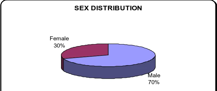
average age being 28 years. Out of 20 cases 6 cases were female
rest 14 are males.
SEX DISTRIBUTION
Male 70% Female
[image:40.612.124.503.275.434.2]30%
TABLE 1
AGE DISTRIBUTION
Age Group
(Years) Male Female Total
0 – 10 1 1 2
10 – 20 4 1 5
20 - 30 4 1 5
30 - 40 3 2 5
40 - 50 2 0 2
OUR STUDY GROUP
Benign tumors
70% Malignant
tumors 5%
Fracture nonunion
25%
As indicated by the pie chart the majority of the study group
consisted of benign tumors and the benign tumors which required
anything less than wide resection were excluded since that defect
were not significant enough to be treated by fibular strut graft. The
LIST OF BENIGN TUMORS
Male Female Total
Fibrous dysplasia 2 0 2
Aneurysmal bone cyst 0 2 2
Giant Cell Tumour 6 3 9
Chondromyxoid fibroma 1 0 1
Among the benign tumours giant cell tumour was the most
commonly encountered one comprising 64%. As described in the
literatures the distal radius is the most favoured site for giant cell
tumour in our study. These cases were either aggressive type or
late presentation necessitating wide resection. So also in case of
Aneurysmal bone cyst, we encountered presentation with extensive
involvement of the humerus not amenable for curettage or bone
grafting. In the cases of fibrous dysplasia one presented with
pathological fracture and the other case was a recurrence after an
initial treatment of curettage and bone grafting.
The one malignant tumour we had, was low grade
chondro-sarcoma of the tibial cortex which required wide resection. The
remaining cases with bone defects were due to complications of
comminuted fractures, resulting in gap non union and the cases of
The locations of the defects that were reconstructed are
shown below.
Site of reconstruction No. of patients
Humerus 6
Distal part of Radius 6
Distal part of Femur 4
Proximal part of tibia 1
Distal part of tibia 1
Shaft of tibia 1
METHODS
Preoperative evaluation done for the tumour cases were
complete haemogram, serum studies, radiography of appropriate
parts, skeletal survey. Serum studies consisted of serum calcium,
serum phosphorus and serum alkaline phosphate. CT and MRI of
the lesion and near by joint were also done. It is with these
investigations (MRI & CT) we identified the exact extent of the
lesion, cortical / articular breach etc. Based on this wide resection
was planned along with reconstructions.
However the histopathological (biopsy) study of some of the
cases helped us to plan for wide resection (aggressive giant cell
tumour). HPE was done as excisional biopsy for some of the benign
tumour of the study. The patients were selected only if
preoperative imaging had shown that a satisfactory surgical
margin could be achieved. Patients with expected defects larger
than 10cm were excluded from the study because vascularized
fibular graft in a better option in such condition27.
Fracture nonunion cases were evaluated preoperatively by
means of complete haemogram, total count, differential count,
erythrocyte sedimentation rate and radiography of the appropriate
investigation to assess the general condition of the patients
regarding fitness of the surgery were also done. All patients were
given intravenous antibiotics before surgery.
As for as approaches are concerned
1. Volar approach for wrist
2. Posterior approach for humerus
Lateral approach for tibia, femur and dorsal approach for
metatarsal were used.
Surgical Technique
In order to decrease the time of surgery and to avoid
contamination we had two operation teams one for tumour
resection and another for graft harvesting.
Tumour resection
Under anaesthesia without using tourniquet incision was
made such that it includes the biopsy scar. The tumour was
resected en-block with wide margin. The margin of clearance
ranged from 2.5 to 5 cm. At most care was taken to avoid
contamination to near by tissues. The resected segment was
Harvesting the graft27
Through posterolateral approach (Henry approach) skin
incised depending up on the requirement. If proximal third of fibula
is to be resected, the common peroneal nerve along the
posteromedial aspect of the biceps tendon in the proximal part of
the wound is identified and protected. The fascial plane between
soleus muscle and peroneus longus muscle is located and the
dissection is deepen to reach the fibula. Subperiosteal stripping was
started distally and progressed proximally in order to protect the
anterior tibial vessel that passes between the neck of fibula and the
tibia. The fibula was resected according to the length of the bony
defect. In some cases nutrient artery entering the posterior surface
of fibula required ligation. After resection was completed, the bicep
femoris tendon and the fibular collateral ligament were sutured to
the adjacent soft tissues.
As per the above technique the proximal fibula was harvested
in 6 cases (for distal radius reconstruction) and shaft of the fibula
was harvested in rest of the cases. After bony reconstruction, the
Tumour Resection
Fixation of the graft
In order to enhance union rigid immobilisation was sought
with internal fixations. The graft was fixed to the host bone either
with a plate and screws, or with lag screws if a step cut osteotomy
was performed or with Kirschner wires.
Post Operative Protocol
All our patients received five days of post operative
intravenous antibiotics. Sutures were removed 10-12 days after
surgery and sent home with plaster cast. This was maintained
usually for six to eight weeks. Then the extremities were taken out
of cast and appropriate brace or splints were given with advice for
passive movements only. In case of lower limb partial weight
bearing was allowed after 12 weeks and in case of upper limb
gentle mobilisation was started after 6 weeks.
All our patients were reviewed clinically and radiologically at
regular interval of one month upto 6 months. After 6 months they
ANALYSIS
We analysed our patients in terms of graft incorporation,
oncological evaluation and functional outcome.
Graft Incorporation5
Criteria for incorporation was absence of radiolucency at host
donor junction and presence of smooth external continuity of
cortical bone on all sides at the junction.
Non union was arbitrarily defined as an absence of union at
one year after the operation.
Oncological Evaluation20
The oncological parameters that were studied includes
survival of the patient, local recurrence and metastasis
Functional Out come
The patients were evaluated functionally as described by
Excellent
Means that the patient had no evidence of disease, was pain
free and had essentially normal function with No limitations
(excepts with regard to high performance sports activity).
Good
Means that the patient has some degree of impairment of
function that did not necessitate bracing or use of supports (such as
crutches or a cane) or interfere with the patients occupations or life
style (except with regard to sports activities).
Fair
Means that a brace or support was needed for walking or
prehension and that the patient had sufficient pain to impair
function.
Failed
Means graft removed or limb amputated. Death as a result of
The follow up period ranged from 11 months to 4 years. All
our patients were analysed in terms of graft incorporation,
oncological evaluation and functional evaluation.
Graft incorporation was assessed radiographically. In our
study the graft united in 16 out of 20 patients between 4 to 12
months period. Average time of graft incorporation was 7.2 months.
4 months in children, 6 months in case of wrist and 8 to 12 months
in case of other bones. In the remaining four cases the graft did not
incorporate due to various reasons that is discussed in later part of
this text giving poor result.
Oncological evaluation was done only for the tumours
conditions, out of 15 tumour cases 14 remained free from disease
till date and one patient was operated for recurrences of giant cell
RESULTS
Results were based on functional outcome which was
analysed according to Mankin et al criteria.
Following were our results:
Excellent - 10 cases
Good - 3 cases
Fair - 3 cases
Complicat
ions
COMPLICATIONS
The following were the complications in our study
1. Stitch abscess – one case
2. Early post operative infection – one case
3. Persistent Infection – three cases
4 Recurrence of tumour – one case
5. Donor site complications – two cases
In our study we had few post operative complications. One
case of stitch abscess and one case of early post operative infection
treated appropriately.
One case of recurrence was encountered with distal radius
giant cell tumour for which excision and centralization of the ulna
We had three cases of persistent infection which resulted in
necrosis of the graft and failure.
Regarding donor site morbidity one patient had transient
peroneal nerve palsy which recovered on physiotherapy and
splinting. Another patient had permanent peroneal nerve palsy
Graft incorporated in 4 months.
CASE -I
Pre operative X-ray Post operative (immediate)
Followup
CASE - II
14 years old kameswaran had fibrous dysplasia but presented
with pathological fracture of right humerus at first instances
planned for resection and reconstruction. Tumour measuring 3.5
cm along with 2.5 cm of clearance was resected and reconstructed
with 8.5 cm of allograft from mother fixed with plates & screws.
Graft incorporated in 4 months
CASE –II
Pre operative X- ray Post operative
(immediate) After implant exit
CASE – III
26 yrs old kottamma presented with Giant cell tumour of the
left distal radius. 10 cm autograft was fixed to the host bone with
plate and screws. This patient developed transient peroneal nerve
palsy which recovered with physiotheraphy and splint.
At six months patient developed recurrence of tumour /
infection for which removal of graft and centralization of ulna was
done.
CASE -III
Pre operative X- ray Post of X-ray
(immediate)
CASE - IV
Mrs. Aysha 52 yrs presented with giant cell tumour of right
distal radius. The histopathological specimen of the patient
revealed aggressive type of tumour, planned for resection. 10 cm of
the distal radius was resected with clearance and was
reconstructed with same size autograft. Post operatively the
patient had stitch abscess which was treated appropriately.
Graft incorporated in 6 months
CASE -IV
Pre operative X- ray Post operative (immediate)
Followup Functional Outcome
CASE – V
21 yr old Mr. Prabhu presented with recurrent
chondromyxoid fibroma of shaft of 1st metatarsal right foot following curettage and bone substitute done 1½ yrs back. This
patient had foreign body reaction with probable recurrence. 1st metatarsal along with medial cuneiform was removed and the
defect was reconstructed with 9.5 cm autograft stabilized with two
kirschner wires.
Graft incorporated in 8 months.
CASE –V
Pre operative X- ray Clinical Picture Post operative (immediate)
CASE –VI
Mr. Mani 40 year old male presented with bony defect of
right tibia following trauma who had external fixation and plastic
procedure done for grade III B compound fracture of both bone
right leg elsewhere. 6 cm bony defect was reconstructed with same
size fibular strut graft fixed with plate and screws. There was
persistent infection leading on to wound dehiscence and
sequestration of the graft. Treated with graft removal and ilizarov
fixator.
CASE –VI
Pre operative X- ray
CASE – VII
50 yr old Mr. Thangamuthu presented with giant cell tumour
of lateral femoral condyle for which curettage and fibular strut
graft was planned. The tumour was thoroughly curetted and filled
with bone cement. The lateral pillar was reconstructed with fibular
strut graft measuring about 8 cm.
Graft incorporated in 8 months.
CASE - VII
Pre operative X- ray Post operative (immediate)
Followup X-ray
DISCUSSION
The goal of treatment is to cure the patient while preserving
as much function, anatomical alignment and quality of life as
possible. Thus every effort should be made to totally eradicate the
primary lesion during the initial surgical treatment itself. Thus
enbloc resection is strongly recommended for aggressive/ recurrent
benign lesions and for some of the low grade malignant tumour.
Reconstruction is necessary after adequate resection of tumor to
preserve the function and alignment. Many reconstructive options
are available after resection.
Autograft, allograft, prosthetic replacement or allograft
prosthetic composite are established methods for reconstructions.
Although use of allograft has shown encouraging results, there are
many associated problems. Selection of suitable donors, the method
of obtaining and preserving the graft, and the technique of allograft
reconstruction deserves particular attention. The surgeon must
consider the risks of infection, graft rejection, delayed healing and
functions of the concern part. Custom-made prosthetic devices have
been used with early success, but problems with late loosening and
Among the autograft and allograft reconstructions, it can
either be with nonvascularized or vascularized graft. Vascularized
fibular autograft is technically more demanding with use of
microsurgical techniques. Nonvascularised fibular graft
incorporation as an autograft is more rapid and predictable than an
allograft4. Moreover, it is easily accessible without significant donor site morbidity3,13. It is also a biological solution and most of orthopaedic surgeons can perform this surgery in an average set
up. They are associated with relatively low rate of complication and
they survive for a longer duration, where as metal implants are
difficult to design and have shorter life span.
In our study nonvascularized fibular graft was used for
reconstructing defects in humerus, distal radius, distal femur,
metatarsal shaft and proximal tibia that araised due to resection of
tumorous conditions and complication of trauma. We had 14 cases
of benign and one case of malignant tumour which were resected
and reconstructed with non vascularized fibular strut graft.
Out of 15 tumour cases, 6 cases were giant cell tumours
involving the distal radius which was reconstructed with the
proximal fibula giving excellent results because of there structural
In another 3 cases the defects were near large joints (distal
femur and proximal tibia). In these cases the fibular graft were
augmented either with bone cement or bone grafting. Even though
we could clear the disease and achieve anatomical alignment there
were some impairment of joint movements. Thus functional
outcome was good to fair in cases of large joint involvement.
In case of distal tibia giant cell tumour after resection the
reconstruction was done by arthrodesis of tibia and calcaneum with
fibular graft augmented with Kuntscher nail. Here the functional
outcome was fair because the patient developed calcaneus
deformity.
In two cases of pediatric group the tumours were involving
the shaft of humerus for which fibula was used as intercalary graft,
taken from their mother. Inspite of it being an allograft these cases
showed early incorporation of the graft resulting in excellent
functional outcome. This was probably due to good osteogenic
potential and remodeling capacity.
We could eliminate the tumours in 14 out of 15 cases (93.3%);
one case of giant cell tumour recurred. This case was further
As for as trauma group are concerned we had five cases out of
which 3 cases resulted in failure. Out of these 3 cases, 2 cases were
compound injuries to start with and had initial treatment else
where in the form of external fixator. Both there cases presented to
us with compromised soft tissue which could not withstand our
extensive procedure of reconstruction resulting in would
dehiscence and infection. The graft sequestrated and subsequently
managed by the orthofix and ilizarov fixators.
In the third case of failure the graft did not incorporate
probably due to inadequate fixation even after 1 ½ years, which
was subsequently managed by bone grafting and replate
osteosynthesis.
In our study 65 percent (13 of 20) had stable, painless
extremity and resumed active use of the involved extremity
without protective device after 1 year. The 7 patients who did not,
were the 3 cases with fair results and 4 cases of failure. The fair
result in 3 patients were because of painful extremity and they
required assistive devices; two patients with distal femur
reconstruction had knee stiffness and flexion deformity. The other
The four patients with failure were due to infection, non union and
recurrence.
In summary considering the problems for which the
reconstruction were done 13 out of 20 patients (10 excellent and 3
CONCLUSION
¾
The bony defects arising out of wide resection of the benigntumour can be successfully reconstructed with fibular graft –
giving good functional outcome.
¾
Post traumatic bony defects with late presentation and caseswith compromised soft tissues did not give satisfactory
results with this procedure.
¾
However these bony defects can be successfully managedwith fibular reconstruction when they present early to the
surgeon.
¾
Our overall experience with nonvascularized fibular graft forreconstructing bony defects are encouraging, however we are
aware this is a short term study and would require further
4. Dhammi I.K; AK Jain, Aditya V. Maheswari; MP Singh.
Giant cell tumours of lower end of the radius problems and
solutions. Indian Journal of Orthopaedics, 39(4): 201-205, Oct. 2005.
5. Eduardo Ortiz Cruz; Mark C. Gebhardt. The results of
transplantation of intercalary allograft after resection of
tumors. Journal of Bone and Joint Surgery, 79A (1): 97-105, 1997.
6. Enneking W.F., Burcharan; Puhl J.J. et al. Physical and
biological aspects of repair in dog cortical bone transplants.
Journal of Bone and Joint Surgery, 57A: 237, 1975.
7. Enneking W.F., Eady J.L. Ana Burchardt. H. Autogenous
cortical bone graft in the reconstruction of segmental skeletal
defects. Journal of Bone and Joint Surgery. Am. 62(7): 1039-1058, Oct 1980.
8. Enneking W.F. Dunham, W. Gebhardt, M.C., Malawer. M
and Pritchard D.J. A system for functional evaluation of
reconstructive procedure after surgical treatment of tumors
9. Evelyn B., Kelly Ph.D; New frontiers in bone grafting.
Orthopaedic Technology Review, 2(9): Oct. 2000.
10. Goldberg V.M. MD and Sharon Stevenson D.V.M Ph.D
Natural History of Autografts and Allografts. Clinical Orthopaedics and Related Research, 225, Dec. 1987.
11. Goldberg V.M.; Stevenson. Shaffer J.W, Davy D. Klein. L,
Zika, Fidel. G. Biological and biomechanical property of
normal fibula. Journal of Bone and Joint Surgery, 72(6): 801-810, Jul.1990.
12. Hammack, Enneking W.F. Comparative vascularization of
autogenous and homogenous bone transplants. Journal of Bone and Joint Surgery, 42A: 811, 1960.
13. Jobe T.M: Micro surgery. Campbell’s Operative Orthopaedics 10th Edition, 4: 3335.
14. Lorenzol. Pacelli, Joel Gillard, Sean W. McLoughlin and
Mark J. Buehler. A biomechanical analysis of donor site
15. Lawson T.J: Fibular transplant for osteoclastoma of Radius.
Journal of Bone and Joint Surgery. 34 B(1): 74-5, 1952.
16. Magdy EL Sherbiny. Replacement of distal radius after
resection of primary bone tumour using nonvascularized
proximal fibular graft. Journal of the Egyptian Nat. Cancer. Inst. 15 (2): 163-168, June 2003.
17. Marco Innocenti, Luca Delcroix, Marco Manfrini, MASSIMO
Ceruso, And Rodolfo Capanna; vascularized proximal fibula.
Epiphyseal transfer for distal radial reconstruction. Journal of Bone and Joint Surgery 86A (7):1504-1511, July 2004.
18. Michael W. Chapman and Juan J. Rodrigo. Bone grafting,
Bone graft, substitutes and growth factors. Surgical
Principles and Techniques. 1: 181-215.
19. Parrish F.F. Treatment of bone tumors by total excision and
replacement with massive autogenous and homogenous graft.
Journal of Bone and Joint Surgery,48A: 968, 1966.
20. Partick, J. Getty. Complications and functional outcomes of
reconstruction with an osteoarticular allograft after intra
articular resection of the proximal aspect of the humerus.
21. Richard. G. Dias, Adesegun Abudu, Simon R. Carter. Robert.
J. Grimer and Roger. M. Tillman. Tumour transfer to bone
graft donor site a case report and review of the literature of
the mechanism of seeding. Sarcoma, 4: 51-59, 2000.
22. Shaffer J.W. MD., Greg A. Field B.A., Goldberg V.M. M.D.
Fate of Vascularized and Nonvascularized autograft.
Clinical Orthopaedics and Related Research, 197: July–Aug. 1985.
23. Robert B. Duthie. E. Musculo skeletal system transplantation
of bone. Mercer’s Orthopaedic Surgery 9th Edition. 1: 100.
24. Sarat S.K., S.C. Goel. Complication of resection and
reconstruction in giant cell tumor of distal end of radius – an
Analysis. Indian Journal of Orthopaedics, 39(4): 206-211 Oct. 2005.
25. Stein Lechner C.W.B., N.C. Mkandawire, nonvascularized
fibular transfer in the management of defects of long bones
26. Steven gitelis M.D; David heligman M.D, George quill M.D
and Patricia Piasecki, M.S. The use of large allografts for
tumour reconstruction and salvage of the failed total hip
arthoplasty. Clinical Orthopaedics and Related Research, 231, June, 1988.
27. Terry Canale S. Surgical Techniques and approaches.
Campbell’s operative orthopaedics. 10th Edition, 1: (14),
19.
28. Turek, Samuel, L. Histology and histopathology of bone,
Orthopaedics Principles and their application IV edition, 1: 50-90.
29. Wilson, J.N. Ununited fracture and the transplantation of
bone; Watson – Jones fractures and joint injuries 6th
PROFORMA
Name: Age/ Sex IP No.
Hospital: Unit: Ward:
Address:
Phone No: Date of Admission:
Date of Surgery:
Diagnosis:
Procedure:
Clinical Features:
Physical Examination:
Investigations Serum Study
Routine Blood Investigation
X-ray
Skeletal Survey
CT scan/ MRI
Treatment
Type of Defect
Type of Graft Used
Length of Graft
Mode of Fixation
Antibiotic protocol
Followup
Complications
Graft Incorporation
Oncological Evaluation
Donor site
Immediate Late
1 Karthik 8, M
Recurrent Fibrous dysplasia
(Lt)
Humerus Diaphysis 8 Screws - - - 4 20 Consolidated Excellent
-2 Kameswaran 14,M
Fibrous dysplasiawith
pathological fracture
Rt.
Humerus Diaphysis 8.5
Plates &
Screws - - - 4 29 Consolidated Excellent
Implant Exit done
3 Mohan 34, M
Low grade chondrosarco
ma
Rt. Tibia Osteo
articular 9 Screws - -
-Limitation of Joint Movement
- 8 18 Consolidated Good
-4 Kottamma 26,F Giant cell tumour
(Lt) Distal radius
Osteo articular 10
Plate, screws and 'K' wires
Transient peroneal nerve palsy
- Recurrence Failure Failure failure 17 Graft removed Failed Centralization of ulna done
5 Ayesha 52, F Giant cell tumour
(Rt) Distal radius
Osteo articular 10
Plate, Screws & K
wires
- - - 6 12 Consolidated Excellent
-6 Rajabunisha 36,F Giant cell tumor
Distal radius (Lt)
Osteo articular 8
Plate, Screws & K
wires
Peroneal
nerve palsy - - - 5 42 Consolidated Excellent
Planned for Tendon Transfer
7 Sekar 25, M Giant cell tumor
Distal radius (Lt)
Osteo articular 7.5
Plate, Screws & K
wires
- - - 7 36 Consolidated Excellent
-8 Prabhu 19, M
Recurrent chondromyxoid fibroma 1st Metatarsal (Rt) foot Osteo
articular 9.5 K wires - Infection - - - 8 11 Consolidated Excellent
-9 Thangamuthu 50, M Giant cell tumor Lateral femoral condyla (Lt) Osteo
articular 8 - - - Pain
Fixed flexion
deformity - 8 12 Consolidated Good
-10 Rajendra Prasad 24, M Giiant cell tumor
Distal radius (Rt)
Osteo articular 7
Plate, screws
& K wires - - - 6 12 Consolidated Excellent
Donor site Immediate Late Deformity or Disability Need of Brace or support Durations of followup (months) Graft incorporation Functional Outome Remarks Type of Defect Graft length (cms) Time of union (months) Mode of fixation Complications Recipient site S. No. Name Age (Yrs.) & Sex Diagnosis Site
11 Ragu 22, M Giant cell tumour
Distal femur (Rt.)
Osteo
articular 6.5 - - Infection
-Only jog of
movement Brace 11 30 Consolidated Fair
-12 Mani 40, M Fracture
non-union Tibia (Rt) Diaphysis 6
Plate &
Screws - - Infection Shortening + Brace failure 13
Failure necrosis of graft Failed
ilizarov was applied
13 Selvam 19, M Fracture nonunion
Humerus
(Rt) Diaphysis 5
Plate &
Screws -
-Implant failure, non
union
Shortening + Brace failure 42 Necrosis of graft Failed
Cortical onlay graft applied
with plate & screws
14 Shahul Hamad 32, M Fracture non union
Humerus
(Rt) Diaphysis 5
Plate &
Screws - Infection Non union Shortening + Brace failure 14 Necrosis of graft Failed
Treated with implant exit and ortho fix external fixator
15 Gobi 20, M Fracture non union
Supra condylar femur (Lt)
Osteo articular 8
Plate &
Screws - Infection
-Fixed flexion deformity and limitation of
movement
- 12 18 Consolidated Good
-16 Radika 37, F Fracture non union
Supra condylar femur (Lt)
Osteo articular 8
Plate &
Screws - -
-Shortening + knee stiffness
+
- 12 15 Consolidated Fair
-17 Pandiyan 50, M Giant cell tumour
(Rt) Distal radius
Osteo articular 8
Plate &
Screws - - - 6 14 Consolidated Excellent
-18 Kamaraj 27, M Giant cell tumour
Distal tibia (Rt)
Osteo
articular 9 k' nail - -
-Loss of ankle
movement Crutch 8 16 Consolidated Fair
-19 Kumudha 15, F Aneurysmal bone cyst
Humerus
(Lt) Diaphysis 6
Plate &
Screws - - - 5 15 Consolidated Excellent
-20 Selvi 10, F Aneurysmal bone cyst
Humerus