CORRELATION BETWEEN CYCLIN D1
EXPRESSION
AND CLINICAL BEHAVIOUR
IN SQUAMOUS CELL CARCINOMA OF THE PENIS
DISSERTATION SUBMITTED TO COLLEGE OF ONCOLOGICAL SCIENCES
CANCER INSTITUTE (WIA)
FOR M.Ch. DEGREE IN SURGICAL ONCOLOGY- BRANCH VII
THE TAMILNADU Dr.M.G.R. MEDICAL UNIVERSITY
CHENNAI
COLLEGE OF ONCOLOGICAL SCIENCES
CANCER INSTITUTE (WIA)
ADYAR, CHENNAI
CERTIFICATE
CERTIFIED THAT THIS IS THE BONAFIDE DISSERTATION DONE BY
Dr.P. RAJKUMAR
AND SUBMITTED IN PARTIAL FULFILLMENT OF THE
REQUIREMENTS FOR THE
DEGREE OF M.Ch. (SURGICAL ONCOLOGY) BRANCH-VII
THE TAMILNADU Dr M.G.R. MEDICAL UNIVERSITY, CHENNAI.
DATE PROF.AND HEAD DEPT.OF SURGICAL ONCOLOGY
DATE PRINCIPAL
THIS WORK IS DEDICATED TO CANCER PATIENTS
AROUND WHOM
OUR WORK AND LIFE REVOLVES
CORRELATION BETWEEN CYCLIN D1 EXPRESSION AND
CLINICAL BEHAVIOUR
TABLE OF CONTENTS
AIM 1
CHAPTER I
INTRODUCTION AND REVIEW OF LITERATURE
1.1 PENILE CANCER -THE PROBLEM 2
1.2 PATHOLOGY AND PROGNOSTIC FACTORS 3
1.3 CLINICAL STAGING 6
1.4 SURVIVAL 7
CHAPTER II
MOLECULAR ASPECTS OF CELL CYCLE THE ROLE OF CYCLIN D1
2.1. CELL CYCLE 8
2.2. CYCLIN DEPENDENT KINASES 8
2.3. CELL CYCLINS 10
2.4. CYCLIN D 16
2.5. CYCLIN D1 GENE 18
2.6. CELL CYCLE IN NOEPLASIA 20
2.7. CYCLIN D1 EXPRESSION PATTERNS IN HUMAN CANCERS 22 2.8. CYCLIND1 AND MOLECULAR ALTERATIONS IN PENILE CANCER 23
CHAPTER III
MATERIALS AND METHODS
3.1 PATIENT SELECTION 35
3.3 SAMPLE COLLECTION 36
3.4 IMMUNOHISTO CHEMISTRY 36
3.5 ANTIGEN RETRIEVAL 37
3.6 BLOCKING AND ANTIBODY REACTIONS 37
3.7 TREATMENT WITH CHROMOGEN DIAMINOBENZEDENE 38
CHAPTER IV
ANALYSIS 38
RESULTS 43
DISCUSSION 47
CONCLUSIONS 51
FUTURE DIRECTIONS 52
AIM
To find a molecular marker that would predict the clinical behaviour of
the local aggressiveness and their metastatic potential. CYCLIN D1 is a
molecular marker that is over expressed in most of the epithelial cancer .So for
there are only few studies reporting their significance in penile cancers. From
CHAPTER I
INTRODUCTION AND REVIEW OF LITERATURE
1.1. THE PROBLEM
Penile cancers are uncommon genitourinary malignancies in the
western countries. In Europe, the incidence is 0.1-0.9 per 100,000, and in
the USA, 0.7-0.9 per 100,000. In some areas of Asia, Africa and South
America, the incidence is significantly higher at 19 per 100.000 1 in these
countries, penile carcinoma accounts for as many as 10-20% of male
cancers. This high incidence is related to the poor personal hygiene,
socioeconomic conditions and the high prevalence of human Papilloma
viruses.
In the Madras Metropolitan Tumor registry there were 98
cases reported between 1999 and 2001 for an average total mid year male
population of 2,178,842 of which there were 1,643,901 men of 15 – 70
years age group2. The crude incidence is 1.5per 100,000 population. The
incidence remains stable in most of the western countries, however there
is a trend towards a decrease in the incidence in our country due to the
Penile cancers are generally slow growing tumors with predictable
orderly spread to the superficial and deep inguinal and to the pelvic
nodes. Distant metastases at presentation are rare. These cancers have a
spectrum of presentation with indolent slow growing lesions to highly
aggressive metastatic disease. The survival drops drastically when the
patient develops nodal metastases3. At present there are few clinical or
pathological markers which would predict the aggressiveness of these
tumors. No definite molecular marker has been identified to predict the
aggressiveness.
1.2. PATHOLOGY AND PROGNOSTIC MARKERS
Penile carcinoma essentially metastasizes via the lymphatic system
and develops mainly through the embolization mechanism instead of
lymphatic permeation. Distant metastases are very rare and are a result of
vascular dissemination 2. Spreading essentially develops in stepwise
fashion; inguinal lymphatic spread occurs first, followed by pelvic
metastases, and lastly by distant metastases. As a consequence, it is
extremely rare to observe patients with positive pelvic nodes or distant
metastasis without inguinal lymph-node involvement. The primary tumor
is localized to the glans in 48% of cases, prepuce in 21%, both glans and
prepuce in 9%, coronal sulcus in 6%, and less than 2% in the shaft 5.
(range 20-96%) 6. Of these patients, 17-45% has nodal metastases, while
the remaining patients have inflammatory disease secondary to an
infection of the primary tumor.
The histological types, grade and growth patterns of penile cancers are
TYPES OF SCC
• Classic
• Basaloid
• Verrucous and its varieties (24):
• Warty (condylomatous) carcinoma
• Verrucous carcinoma
• Papillary carcinoma
• Hybrid verrucous carcinoma
• Mixed carcinomas (warty-basaloid carcinoma, adeno-basaloid
carcinoma)
• Sarcomatoid
GROWTH PATTERNS OF SCC
• Superficial spread
• Nodular or vertical-phase growth
• Verrucous
DIFFERENTIATION GRADING SYSTEMS FOR SCC
• Broders system traditionally used as a grading system
• Maiche system score7 currently seems to be the most suitable
grading system
Of all the cancers, squamous cell carcinomas account for more than
95%. The prognostic factors are the T status, the histological grade, and
the presence of the nodal metastasis. The histological grade predicts the
development of the inguinal metastases to some extent. However
substantial number of patients with high grade N 0 disease undergoing
nodal dissection will have pathological N0 disease facing the morbidity of
1.3. CLINICAL STAGING
AJCC 2002 TNM TUMOUR STAGING
T - Primary tumour
TX Primary tumour cannot be assessed
T0 No evidence of primary tumour
Tis Carcinoma in situ
Ta Non-invasive verrucous carcinoma
T1 Tumour invades subepithelial connective tissue
T2 Tumour invades corpus spongiosum or cavernosum
T3 Tumour invades urethra or prostate
T4 Tumour invades other adjacent structures
N - Regional lymph nodes
NX Regional lymph nodes cannot be assessed
N0 No evidence of lymph node metastasis
N1 Metastasis in a single inguinal lymph node
N2 Metastasis in multiple or bilateral superficial lymph nodes
N3 Metastasis in deep inguinal or pelvic lymph nodes, unilateral or
M - Distant metastasis
MX Distant metastases cannot be assessed
M0 No evidence of distant metastases
M1 Distant metastases
1.4 SURVIVAL
An overall 5-year survival rate of 52% has been reported. This
ranges from 66% in patients with negative lymph nodes to 27% in
patients with positive nodes 8, 9 and 0-38.4% in patients with pelvic node
involvement. Most patients are elderly and the neoplasm has a slow
growth rate. Death from cancer is usually a consequence of local
complications, such as infection, haemorrhage of the ulcerated tumour or
ulcerated inguinal metastases. The crude 5-year survival rate was 95% for
patients with negative nodes, 76% when only inguinal nodes were
positive, and 0% when the pelvic nodes were positive. The adverse
prognostic factors were (1) involvement of > 3 inguinal nodes, (2)
perinodal infiltration in the inguinal area, and (3) pelvic node
CHAPTER II
MOLECULAR ASPECTS OF CELL CYCLE - THE ROLE
OF CYCLIN D1
2.1 CELL CYCLE
Cell cycle is comprised of four phases: g1, s phase, g2 and m
phase.
The g1 phase before the mitosis are the times of regulatory decisions of
the cycle.(fig 1)
2.2 CYCLIN DEPENDENT KINASES
The major transitions of the eukaryotic cell cycle is triggered by a
family of serine threonine kinases called cyclin dependent kinases (
cdk ) at least nine cdks are known so far 11. CDK activation requires the
regulatory subunits called the cyclins and phosphorylation of a conserved
threonine by the cdk activating kinase ( CAK ) which itself is a
complex of a regulatory cyclin H and a catalytic cdk7 subunit , cellular
cdk levels tend to remain in constant excess through out the normal cycle
and catalytic subunit of cdks are regulated post translationally. Cdk are
In humans, the growing list of cdks include the confounding
member, cdc2 and cdk2- cdk7. Our understanding of cdk structure and
function (fig-1) is based largely on studies of prototypical cdks of
s,pombe( cdc2 ), s. Cerevisiae ( cdc28) and vertebrates ( cdc 2 and cdk
2 ) . The typical cdk catalytic unit
Fig 1
contains 300 aminoacids. The catalytic subunit is completely in active
when it is monomeric and unphosphorylated. Cyclins are the primary
p34 CDC 2 (cdk1)
The earliest reported members of the cdk family constitute
homologous 34 kda products encoded by the yeast cell division cycle
(cdc) genes. p34 cdc2 (cdk1) is a highly conserved cyclin associated 34
kda protein kinase that becomes activated on phosphorylation.
Complexed with cyclin b it forms the maturation factor (MPF).Without
p34cdc2 the cells are unable to divide12. p34 cdc2 distribution is
cytoplasmic during the interphase, shifts into the nucleus at the beginning
of the prophase and extends through the cell in the mitiotic phase, as the
key regulators of the cycle, the cyclin dependent kinases must be tightly
regulated by extra and intra cellular signals. The activity of cyclin
dependent kinases is controlled by four highly conserved biochemical
mechanisms forming a web of regulatory pathways unmatched in this
elegance and intricacy.
2.3 CELL CYCLINS
Cyclins are a class of structurally related proteins that bind and
activate the catalytic subunit of the CDKs. To data there are eight types
of cyclins, (Cyclins A – H ) and all of them share an ∼ 150 amino acids
of the region of homology called the cyclin box which is responsible for
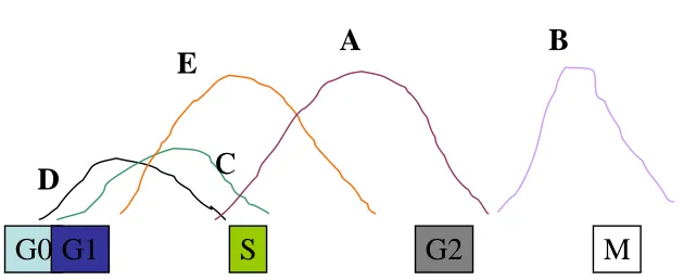
two subfamilies: the G1 cyclins and the mitotic cyclins. The G1 Cyclins
(C, D, E) are short lived and have rapid turnover throughout cell cycle(
fig 2). Their levels are determined of transcription of their m RNA. The
mitotic cyclins ( A,B) are very stable throughout the interphase but
undergo rapid proteolysis by an ubiquitin dependent pathway during
mitosis. Compared to the mitotic cyclins, the G1 cyclins have a longer C
terminal sequence after the cyclin box, and it is this part of the protein
that seems to confer instability to the G 1 cyclins 14. The function of
cyclins is primarily controlled by changes in the cyclin levels which
increase at specific stages and are often categorized by the stage at which
they are expressed. The G 1 cyclins differ from the mitotic cyclins in
their overall primary structure and this has implications for their stability
during the cell cycle. The transcription of the mitotic cyclins, Cyclin A
and B are cell cycle dependent and their levels are determined both by
transcription and proteolysis15. Protein degradation is an effective method
for promoting unidirectional cell cycle transitions because of its rapidity
and irreversibility. Three major cell cycle transitions, entry into S phase,
separation of sister chromatids and exit from mitosis, require the
degradation of specific proteins via the ubiquitination by 26S proteosome
The degradation of mitotic cyclins involves the ubiquitin
dependent proteolytic machinery and a small sequence motif called the
Destruction box located at the N terminus. This region has a small cluster
of conserved residues followed by a lysine rich stretch. The destruction
box region differs between Cyclin A and B and probably accounts for the
finding that in mitosis Cyclin A is degraded before Cyclin B. G1 cyclins
lack the mitotic destruction box but contains the PEST sequences as the
peptide motifs that are important for proteolysis. PEST sequences (rich in
proline, glutamic acid, serine, and threonine) are frequently present in
unstable proteins such as G1 cyclins and contain specific sites of
phosphorylation. Phosphorylation of PEST regions facilitates the
destructions of PEST sequence proteins. It is not the PEST sequences per
se but the specific motifs within them that actually control the PEST
Fig 2
CYCLIN WAVES
G0 G1
S
G2
M
D
C
C
E
A B
Different cyclins/CDK complexes exp at different phases
of cell cycle
[image:18.612.179.494.347.476.2]Cyclins and cyclin dep Kinases
G0
Mitogens
G1
Cyclin D/CDK4/6
S
Rest point
(pRB phosph)
Cyclin E/CDK2
G2
M
Cyclin A/CDK2
Cyclin A/CDK1
Cyclin B/CDK1
Regulatory protein subunit-CYCLINS (A-T)
Catalytic subunit(protein kinase)-CDKs (1-9)
(cdc2)
Cyclins are thought to target the CDK to specific substrate and
subcellular locations. Cyclin expression varies during cell cycle and the
periodic expression of different cyclins defines the start of each phase of
the cell cycle and also marks the transitions between the various phases.
Cyclins and their cognate CDK catalytic subunits non covalently form 1:1
complexes to produce the CDK holoenzyme. Specific CDKs operate in
distinct phases of cell cycle. At least two stages within cell cycle are
regulated in response to the DNA damage, the G1 – S and the G2 –M
transitions. These transitions are called CHECK POINTS at which the
cells delay cell cycle progress to allow repair of the DNA damage. Check
points are thought to consist of surveillance mechanisms that can detect
DNA damage, signal transduction pathways that transmit and amplify the
signal to the replication or segregation machinery and possibly repair
activities 17.The timing of all the events that transpire during the cell cycle
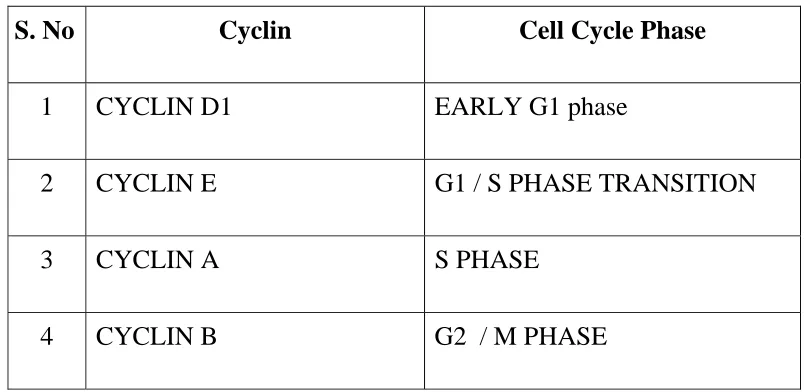
Table 1 :- Cyclins and cell cycle phases.
S. No Cyclin Cell Cycle Phase
1 CYCLIN D1 EARLY G1 phase
2 CYCLIN E G1 / S PHASE TRANSITION
3 CYCLIN A S PHASE
4 CYCLIN B G2 / M PHASE
2.4 CYCLIN D
The first groups of cyclins that are expressed after the cells are
stimulated to enter the cell cycle are the D cyclins. D type cyclins act as
growth factor sensors. Cyclin D has a relatively short half life of 20 min.
approximately and rapidly disappears with the removal of mitogenic
stimuli or the addition of antiproliferative agents.18 Cyclin D complexed
with respective partners CDK4 and CDK6, participates in the
transduction of external signals (mitogenic or proliferative) to other
components of G1 / S transition cell machinery. Cyclin D helps in
moving G0 cells into G1 And early G1 Cells into the G1 /S transition in
response to the extra cellular stimuli. The D type cyclin belongs to a
criteria. There are three genes identified under this family, Cyclin D1, D2
[image:22.612.123.506.177.369.2]and D3 19.
Table 2 Gene Locations Of Cyclins
GENE CHROMOSOME
LOCATION RNA PROTEIN
CCD1
(CYCLIND1) 11q13 4.5 KB 295AA
CCD2
(CYCLIN D2) 12p13 7KB 289AA
CCD3
(CYCLIN D3) 6p21 2.2KB 292AA
The chromosome 11q13 has been identified as a site of tumor
specific translocations .In the parathyroid adenomas, due to clonal
inversion, the cyclin D1 gene on chromosome 11q13 is juxta posed on
chromosome 11q15. This results in an increase in Cyclin D1 expression.
20,21
Frequent rearrangements of 11q13 has been noted in B cell
lymphomas and multiple myelomas. The same locus is subjected to
translocation22, retro viral insertion, gene amplification by yet poorly
defined mechanisms in lymphomas, squamous cell carcinomas and breast
carcinomas implying that Cyclin D1 acts as a Oncogene –positive
2.5 CYCLIN D1 GENE
The cyclin D1 gene (CCND1), linked closely to the bcl-1 gene on
chromosome 11q13, is sometimes referred to as the PRAD1 gene because
of an initial finding of frequent rearrangement of this gene in benign
parathyroid adenomas.49 The important role of cyclin D1 in parathyroid
neoplasia has subsequently been confirmed50. The bcl-1 locus, so called
because of the involvement of this chromosomal region in translocations
{(t11; 14) (q13, q32)} characteristic of certain B cell lymphomas (now
called mantle cell lymphomas), was originally thought to be identical to
the cyclinD1/PRAD1 gene, but it has been shown to reside 110–130 kb
upstream or centromeric to the PRAD1 gene51. However, no
transcriptional units have been identified in the immediate vicinity of the
major translocationcluster (MTC) 52 of the bcl-1 breakpoint area or any
of the breakpoints that have been found up to 63 kb telomeric to the MTC
region53. The absence of CpG islands between the original bcl-1 locus
and PRAD1’s CpG Island 51 52 54 lends further support to the notion that
no other gene lies within this interval. Thus, the probability remains that
the cyclin D1 / PRAD1 gene is the bcl-1 Oncogene. Although bcl-1 often
co-amplifies with other genes on 11q13 (such as EMS-1, FGF3, FGF4,
int-2, and hst-1), cyclin D1 and EMS-1 are the only proteins, so far, over
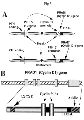
The D type cyclins contains the sequence LEU-X- Cys - Glu near
their aminoterminus (fig3). This sequence is common to DNA vital
oncoproteins SV40 T antigen, adenovirus E1A and human Papilloma
virus E7 that bind and activate pRB and pRB related proteins. The D
type Cyclins bind directly to pRB and p107 invitro and the interactions
are disrupted by point mutations of the LEU-X- Cys - Glu motif. The
Oncogene derived peptides that contain this motif compete with Cyclin d
to bind to pRB23. Cyclin D2 and D3 form more stable complexes than
cyclin d1 suggestive of differences in functional interactions not being
equivalent24. The functional interaction of Cyclin D with pRB
underscores its role as a positive growth regulator with Oncogenic
potential.
2.6 CELL CYCLE IN NEOPLASIA
Predictably, proteins involved in driving the cell cycle, such as
cyclins, are frequently over expressed in primary tumors, whereas
proteins that slow cell division, such as the CKIs, are often inactivated.
Of the many cell cycle regulators implicated in the development of
cancers, cyclin D1 is among the most prevalent. Over expression of
D-type cyclins has been shown to contract the G1 phase, decrease cell size,
and reduce the dependency of the cell on mitogens 25-27 in animal models
A link between D-type cyclins and the retinoblastoma protein
The connection between D-type cyclins and tumorigenisis is
bolstered further by compelling evidence that D-type cyclins are
important in cell cycle regulation of the retinoblastoma tumor suppressor
protein (pRB), an approximately 105 kDa nuclear phosphoprotein.
28-30
.The amount of pRB is not altered with progression of the cell cycle,
however, the phosphorylation state of pRB is cell cycle dependent31-33 .
pRB is hypophosphorylated throughout G1 phase, phosphorylated just
before S phase, and remains phosphorylated until late mitosis.
Hypophosphorylated pRB arrests cells in G1,34an effect most likely
mediated through complex formation with DNA binding proteins
(including members of theE2F family)35-37 required for transcriptional
activation of cellular genes. Phosphorylation of pRB during late G1 phase
reverses the growth suppressive effects of pRB, by untethering E2F from
its inhibitory constraint and thereby allowing the activation of genes
required for DNA replication.38Because D-type cyclins are able to bind to
pRB through an N-terminal LXCXE motif, 23,39,40they are excellent
candidates for G1 phase pRB protein kinases as part of a complex with
their specific Cdk partners. Interestingly, this LXCXE motif is common
to the SV40 T antigen, adenovirus E1A, and human Papilloma virus E7
explains the oncogenic potential of these viruses. Support for the idea that
D-type cyclins can inactivate pRB comes from reports that increased
amounts of D-type cyclins can reverse the pRB induced cell cycle arrest
and accelerate progression through G127 39 41 42.
Cells that lack functional pRB have significantly lower levels of
Cyclin D1 and Cyclin D1 – CDK 43-46 a result which has been interpreted
to mean that hypophosphorylated pRB is involved in the stimulation of
Cyclin D1 transcription. The reported ability of exogenously expressed
pRB to induce cyclin D1 is in concordance with this hypothesis .47 Thus a
negative feed back loop seems to exist in which Cyclin D1 synthesis and
activation lead to pRB phosphorylation , which in turn causes decreased
Cyclin D1 expression. 48
2.7 CYCLIN D1 EXPRESSION IN HUMAN CANCERS
In addition to parathyroid adenomas, increased cyclin D1
expression has been shown in a number of primary human tumors and
cell lines. In general, primary tumors provide more reliable information
as it is often difficult to determine when the amplification occurred
during the development of the cell line. This is because other influences,
which cause the cells to proliferate at faster rates, may up regulate cyclin
correlates in most instances with amplification of the CCND1 gene, this
is not always the case. In some tumors there is a increased cyclin D1
RNA and/or protein without apparent gene amplification, suggesting that
other cellular genes (such as the retinoblastoma gene) may impact on the
protein expression of cyclin D1,57 although all the mechanisms have not
yet been satisfactorily elucidated. DNA amplification is the most frequent
abnormality affecting the CCND1 gene. Furthermore, no major
abnormality in the coding region of the cyclin D1 gene has been detected
58,59
suggesting that it is the normal gene product that contributes to
tumorigenesis.
BREAST CANCERS
About half of all invasive breast cancers 76-79 have raised
expression of cyclin D1 compared with normal epithelium, although the
figure for gene amplification averages around 13%. 57 Some earlier
studies80-83 failed to find any significant association between 11q13
amplification and oestrogen receptor positive cancers, but a number of
materials 84–93 has now accumulated supporting a correlation between
cyclin D1 gene amplification and protein over expression with oestrogen
receptor positive tumors. Studies in mice 94 95 and man 96 link cyclin D1 to
D1 appears to be an independent activator of the oestrogen receptor. 97
Despite the correlation with oestrogen receptor status, there is lack of
agreement as to the prognostic significance of cyclin D1 in breast cancers
in general. More work need to be done to identify the subsets of patients
in whom cyclin D1 may play a more prominent role. It seems that cyclin
D1 is more important in node positive,74 well differentiated, and
particularly lobular, varieties than other types of invasive breast cancer.91-
93
The role of cyclin D1 in ductal carcinoma in situ (DCIS) 98, high grade
lesions 99 were more likely to show gene amplification but demonstrated
lower percentages of nuclei expressing cyclin D1 protein than low grade
lesions, which suggests that mechanisms other than gene amplification
may be responsible for increased cyclin D1 protein. In this situation,
assessment of cyclin D1 protein in combination with pRB may provide
more useful information.100-102 The over expression of cyclin D mRNA,
determined by in situ hybridization, was able to distinguish DCIS from
atypical ductal hyperplasia and other lesions associated with a low risk of
progression to invasive carcinoma103.
In patients with ER-positive tumors, high levels of cyclin D1
mRNA were associated with increased risk of relapse, local recurrence,
metastasis, and death. There were no clinical correlations with cyclinD1
therapy for their primary or recurrent breast cancers, there was an
apparent association between ahigh cyclin D1 mRNA level and shorter
response duration within the ER-positive subgroup. These findings
indicate thatover expression of cyclin D1 mRNA correlates with a worse
prognosiswithin the ER-positive breast cancer phenotype and may be a
contributing factor to the development of endocrine resistance in
ER-positive disease104.
CYCLIN D1 IN PANCREATIC TUMORS
Overexpression of cyclin D1 was identified in43% of cases, and no
correlation was observed with clinicalphenotype. The novel observation
of frequent over expression of cyclin D1 suggests that this established
Oncogene might be implicated in the pathogenesis of human Pancreatic
Endocrine Tumors. The absence of detectable alterations in cyclin D1
genomic structure suggests that the mechanism for its Oncogenic
activation in PETs maybe transcriptional or posttranscriptional105.
HEAD AND NECK SQUAMOUS CELL CARCINOMAS
A range of 35% to 64% of head and neck squamous carcinomas
106-111
(squamous carcinomas in the oral cavity, nasopharynx, pharynx,
hypopharynx, and larynx) show over expression of cyclin D1 and/or
specimens corresponds not only with more frequent recurrence 109 but
also with more advanced disease, lymph node involvement, and reduced
overall survival. 107 110 In one study of squamous carcinomas of the
larynx, a correlation among cyclin D1 gene amplification, mRNA over
expression, and tumor progression, was shown in a cohort of 46 patients.
107
The above and another study 110demonstrated a significant association
between molecular abnormalities of the cyclin D1 gene and pathological
measures of poor prognosis. Recently, over expression of cyclin D1
protein in resected material from head and neck squamous carcinomas
was found to be an independent prognostic factor 111 a finding that has
been confirmed. 112 In Japanese hypopharyngeal squamous cell
carcinomas, cyclin D1 gene amplification and protein over expression
correlated not only with prognosis but were also useful in identifying
optimum treatment regimens. 113 Cyclin D1 negative tumors responded
particularly well to multimodality treatment in these tumors.
ESOPHAGEAL CANCERS
In approximately 30% of esophageal cancers, amplification 113 and
over expression 114–116 of cyclin D1 have been demonstrated, with several
studies showing an association with increased mortality. 116–118 Also, the
esophageal cancer cells 119 provides strong supporting evidence for the
molecule’s role in cancers at this site.
Cyclin D1 mRNA was overexpressed in the cytoplasm of cancer
cells that showed cyclin D1 gene amplification by Southern blot
hybridization. Cyclin D1 antigen was overexpressed in the nucleus of
thesecancer cells. The distribution of cyclinD1 mRNA-positive cells was
similar to that of cyclin D1 antigen-positive cells in the cancer tissues.
The overall 5-year survival of patients with strongly stainingtumors was
significantly lower than that of patients with weakly or non staining
tumors (7 versus 59%; P < 0.01). There was no significant correlation
between cyclinD1 expression and other clinic pathologicalfactors. These
results suggest that cyclin D1 may play an important role in
carcinogenesis and that cyclin D1 over expression may be a useful
prognostic factor in esophageal cancer120.
HEPATOCELLULAR CARCINOMAS
Amplification and raised protein concentrations have been
observed in 10% of hepatocellular carcinomas. 121, 122 Among other
hypothesis, it has been suggested that hepatitis B or C viral integration
within the cyclin D1 gene or one of its adjacent regulator sequences may
hepatitis B viral genome was detected on chromosome 13 at an upstream
site close to the CCND1 gene. Another study122 showed that cyclin D1
amplification was restricted to hepatocellular carcinomas that contained
the hepatitis B or C virus. Although the evidence is scant, the possibility
of an interaction between these viruses and cyclin D1 is an intriguing one.
COLORECTAL CANCERS
Initial reports 126 using cell lines suggested that cyclin D1 was
not an important factor in colorectal adenocarcinomas, recent work has
shown not only does cyclin D1 over expression occur as an early event in
tumor progression, it may also be an independent prognostic factor.127
There is increased nuclear immunostaining in adenomatous polyps and
adenocarcinomas, but not in adjacent normal, transitional or hyperplastic
mucosa. These findings apply to both sporadic 128 and familial forms 129 of
colon cancer. Furthermore, as has been demonstrated in the oesophagus,
antisense to cyclin D1 inhibits the growth and tumorigenicity of colon
cancer cells.
GENITOURINARY CANCERS
Amplification of 11q13 has been demonstrated in between 6% and 21%
130–132
of transitional cell cancers of the urinary bladder, although nuclear
cases. Alterations in cyclin D1 appear to be an early event in
tumorigenesis of the urinary bladder, but the prognostic significance of
amplification and over expression remain to be determined. In one study
a significant relation between cyclin D1 over expression and low tumor
grade as well as T classification were observed, 133 these findings were
not found in another study. 134 Abnormalities of cyclin D1 are also
common in both vulval and cervical squamous cell carcinomas. 135 At
these sites, cyclin D1 appears to inactivate pRB in a similar manner to
oncogenic human Papilloma virus genotypes. Thus, it seems that in
vulval and cervical squamous carcinomas, human papilloma virus
proteins can circumvent cellular requirements for cyclin D1 136 or vice
versa. In endometrial carcinomas, 11q13 amplification is exceedingly
rare, but about 40% of cases show aberrant accumulation of cyclin D1. 137
Again, the effect of other genes is the likely explanation for this
phenomenon, al-though in this case the interaction of cyclin D1 with p53
138
appears to be more important than with pRB. Because cyclin D1 can
activate the oestrogen receptor independently, 98 if this molecule were
also over expressed in endometrial hyperplasia it would provide a useful
link with known pathogenetic mechanisms. In epithelial ovarian cancers,
abnormalities of cyclin D1 are early events in the progression to
transformation.139There is a strong positive correlation between cyclin D1
and c-K ras immunoexpression, 140 but no correlation with the c-erb-B2
oncogene. 141 The relation with the Ras proto-oncogene is important
because it has been shown that inactivation of Ras in cycling cells causes
a decline in cyclin D1 protein, accumulation of the hypophosphorylated,
growth suppressive form of pRB, and G1 arrest. 142 143 Strangely, the
relation between cyclin D1 amplification and oestrogen is not as clear cut
as has been demonstrated in the breast. 141
LUNG CANCERS
Studies have shown a higher frequency of bcl-1 gene
amplification in squamous cell carcinomas of the lung than in other types
of non-small cell lung cancer, 145 and an association with poor grade and
high Ki-67 labelling index. 146However, current research is contradictory
as to the prognostic usefulness of detecting cyclin D1 protein over
expression. 147–148 From studies of the resection margin epithelia of lung
cancer patients, com-pared with non-smoking controls, it seems that
genetic alteration of cyclin D1 is an early event in non-small cell lung
SKIN CANCERS
Compared to normal skin and benign lesions, cyclin D1 protein
expression is significantly greater in various malignant skin tumors,
including squamous cell carcinomas, melanomas, and malignant fibrous
histiocytomas. 149 Studies of chemically induced squamous cell
carcinomas in mice also implicate cyclin D1 (and other G1 cyclins) in the
process of carcinogenesis. 150
SARCOMAS
Amplification of the cyclin D1 gene has been detected in a small
percentage of a variety of sarcomas, 151 but the increased cyclin D1
protein expression in at least some cases may due to a mutant protein
with greater stability. 152
MANTLE CELL LYMPHOMAS
All or almost all mantle cell (centrocytic) lymphomas in
several studies51 60–67 have raised activity of cyclin D1, even in cases in
which no rearrangement at 11q13 was found. 68 Generally, however,
positive nuclear staining with monoclonal antibody to the cyclin D1
protein correlates with amplification of the CCND1 gene as well as
lymphocytes in the mantle zone impairs the capacity of these cells to exit
the cell cycle and to differentiate into mature plasma cells. 70 This
pathogenetic theory is contradicted by a recent finding of cyclin D1
protein expression in 26% of plasma cell neoplasms, however, the same
study 71 supports a relation between mantle cells, plasma cells, and their
corresponding neoplasm. An international lymphoma consensus
72
acknowledged the importance of chromosome 11q13 translocation and
increased cyclin D1 expression in mantle cell lymphomas; in the future it
may be elevated to a defining characteristic, given the high sensitivity and
relative specificity of this molecule in mantle cell lymphoma compared
with other B cell neoplasms. 61 73 Cyclin D1 protein expression and bcl-1
gene rearrangement has been identified as a key component in the
diagnosis of the blastoid variant 74 of mantle cell lymphoma as well as in
an entity closely related to mantle cell lymphoma multiple lymphomatous
polyposis.75
OTHER SITES AND MALIGNANCIES
Central nervous system malignancies such as astrocytomas 153
and glioblastomas 154 are not exempt from cyclin D1 amplification or
pancreatic adenocarcinomas (may be associated with a poor prognosis),
157
or squamous carcinomas of the gall bladder. 158
Only a few human tumors are still holding out against the storm of
cyclin D1 research. These include pituitary tumours,159 renal cell tumors,
prostate carcinoma, 160 and many haemopoietic malignancies. However,
with some of these tumors there are possible links with cyclin D1. For
example, the 11q13 region (although apparently not the CCND1 gene) is
important in pituitary neoplasms of MEN-1,and cyclins including cyclin
D1 are important in renal development.
In addition to the MEN1 tumor suppressor gene, the cyclin D1
oncogenehas demonstrated a role in the pathogenesis of parathyroid and
gastroenteropancreatic neuroendocrine tumors. Upregulationof cyclin D1
is observed early in tumor formation, implyinga possible role in tumor
initiation. Overexpression of cyclinD1 in the parathyroid glands of mice
resulted in the tandem regulation of cellular proliferation and hormonal
secretion,a feature intrinsic to neuroendocrine tumors.161
Cyclin D1 (CCND1) is a key cell cycle regulatory protein, the
over expression of which is often found in human tumors and is
associated with cell proliferation and poor prognosis.A common
results in an altered messenger RNA transcript and a longer-life protein,
which are preferentially encodedby the A allele.The CCND1 870A allele
may be associated withcolorectal cancer, and particularly with forms of
the diseasethat result in severe morbidity and mortality162.
2.8 CYCLIND1 AND MOLECULAR ALTERATIONS IN
PENILE CANCER
Alterations pointing to a disturbed p16 INK4A /cyclin D1/Rb
pathway are commonly present in penile carcinomas144. Activity of
high-risk HPV and the resulting increase in p16 INK4A expression was the
most frequently detected mechanism amongst the mechanisms that were
analyzed, followed by p16 INK4A hypermethylation and BMI-1 over
expression. In addition, there is evidence that penile carcinomas being
etiologically heterogeneous, with only a proportion of cases attributable
to HPV infection. These results are important when HPV-based
immunotherapeutic interventions are envisaged, because in contrast to
patients with cervical carcinoma, only about 26% of patients with penile
CHAPTER III
MATERIALS AND METHODS
Penile cancer patients reported to our institute between the year
1998 and 1999 who were accepted for treatment were chosen for analysis.
The data were collected from the case records of the Tumor registry
.There were 104 cases of carcinoma penis that were accepted for
treatment in our institute between the year1998 and 1999. These records
were analyzed.
3.1 PATIENT SELECTION
Of the 104 cases who were accepted for treatment at our institute,
those patients who had not completed the treatment were excluded
from the study. Those who lost to follow up and histology other than
squamous were also excluded from the study.Of those records,
37squamous cell carcinoma patientsof different grades were chosen .The
inclusion criteria are – completed treatment , regular follow up ,
squamous cell carcinoma any grade , complete pathology is available
with the paraffin blocks .
3.2 DATA COLLECTION
Case records of those patients who fulfilled the above criteria were
taken for analysis. The relevant data were taken from the case records and
3.3 SAMPLE COLLECTION
The pathology reports of these patients included for analysis were
taken and their corresponding paraffin block numbers were taken. These
blocks are then retrieved from the pathology department paraffin block
bank. Five micron sections are taken from the paraffin blocks and
incubated overnight. Suitable positive and negative controls are also
taken simultaneously from the previously Cyclin D1 positive sections
from the laryngeal cancer .After overnight Incubation the slides were
submitted foe immunocytochemistry with Cyclin D1.
3.4 IMMUNOCYTOCHEMISTRY
The paraffin embedded sections of 5 µ were placed on the APES
coated slides.The slides were treated in Xylene consecutively twice for 8
minutes each for dewaxing the sections. This was followed by treatment
in 100% alcohol consecutively twice for dehydration. The slides were
then washed in tap water for 8- 10 minutes, taking care not to disturb the
tissue sections . Followed by tap water wash , the slides were then place
in 0.3% hydrogen peroxide for 30 minutes to quench the endogenous
peroxidase activity in the tissues .
3.5 ANTIGEN RETRIEVAL
Antigen retrieval is needed in the study in order to expose the
epitopes for immune reaction .Antigen retrieval was done using10 mM Citrate buffer using , with wet autoclaving at 121° C in 10mM Citrate
buffer pH6. After antigen retrieval the slides were cooled and washed
3.6 BLOCKING AND ANTIBODY REACTIONS
The slides were arranged on a moist chamber, to prevent drying of the sections and 100 µ of 2% Bovine Serum Albumin (BSA) was added
to each section, taking care to cover the whole section and left
undisturbed for 30 min. Following gentle tipping of BSA off the slides , the primary antibody ( 75 µl ) in 1: 100 dilutions was added to each
sections .Care was taken to omit the negative control. The slides were left
overnight undisturbed to incubate the primary antibody.
The slides were washed with PBS thrice for 5 minutes each to wash off the excess of primary antibody and secondary antibody (75 µl )
was added to each slide and incubated for 35 minutes. The slides were
washed in PBS thrice for 5 minutes each followed by addition of ABC
(Avidin – Biotin Complex -75µl) onto each slide and incubated for 30
minutes.
3.7 TREATMENT WITH CHROMOGEN DI-
AMINOBENZEDENE
The slides were washed in PBS thrice for 5 minutes each to wash
excess of ABC and the slides were treated in a solution of DAB ( 150 ml
of water + 150 ml PBS+100 µl hydrogen peroxide + 150 mg of DAB) for
5 minutes . The excess DAB was washed off the slides in tap water for 5-
CHAPTER IV
ANALYSIS
The stained slides were the scored by the pathologist. Only the
nuclear staining was considered as positive. All cytoplasmic and other
nonspecific staining were not considered as positive. Since the adjoining
normal squamous epithelium did not over express Cyclin D1 and the
corresponding squamous epithelium of the cervix , were > 5 % of
expression is considered positive , the same principle was applied here .
Depending on the staining intensity, the same is scored as +, ++, +++.
The age distribution were
AGE DISTRIBUTION
0 10 20 30 40 50 60 70 80
0 10 20 30 40
SLIDE SHOWING POSITIVE CONTROL
SLIDE SHOWING PENILE CANCER WITH PREDOMINANT
CYCLIN D1 EXPRESSION
SLIDE SHOWING NEGATIVE CONTROL
RESULTS
Of the 37 cases analyzed and studied the T status distribution were :-
T status Numbers Percentage
1 6 16.2%
2 21 56.8%
3 9 24.3%
4 1 2.7%
Total 37 100%
'T' DISTRIBUTION
0 5 10 15 20 25
1 2 3 4
T STATUS
NU
M
B
E
R
The N status were
N Numbers Percentage
0 30 81.1%
1 1 2.7%
2 6 16.2%
The grade distribution were
Grade Numbers Percentage
1 16 43.2%
2 12 32.4%
3 9 24.3%
Of these 37 cases that were followed up , there were 2 local
recurrences (5.4%) that were salvaged and 10 regional recurrences(27%)
. The cyclin D1 expression pattern in all cases were analyzed . Cyclin D1
expression pattern with respect to the T status showed 14.3 % of T 2, 44.4
% of T3 tumors expressed Cyclin D1 .None of the T1 or T4 tumors
expressed .applying Chi square test did not show any statistical
Cyclin D1 expression pattern with respect to grade of the tumor
showed 41.7 % of grade II tumors and 22.2 % of grade III tumors
overexpressed Cyclin D1 which was found to statistically significant ( p
value 0.0197) Even though the recurrence pattern and the Cyclin D1
correlation showed Cyclin D1 over expression only in 18.9% which
statistically not significant ( p =0.270 ) there was trend towards increased
GRADE DISTRIBUTION
0 2 4 6 8 10 12 14 16
NUMBER
1 2 3
GRADE
GRADE AND CYCLIN D1
GRADE 1=16 GARDE 3= 9
DISCUSSION
Cyclin D1 is one of the molecular markers that is over expressed in
the actively dividing malignant epithelial cells. Most of the epithelial
cancers as described before, over express Cyclin D1 .However there are
only very few studies done to know the significance of this over
expression in penile cancers. In our study we attempted to find the
significance of this over expression with respect other clinico
pathological factors.
In our study the cases chosen are highly selected with the
criteria which was described above. Even though this may result in
selection bias , the complete details are available only for those chosen
cases .
Search of literature didn’t show any reports regarding the cut
off values for the scoring system of the expression. However there
reports using > 5% of the stained cells as positive in cervical cancers.
Using the same principle, our slides were scored .The normal squamous
cells did not show positivity in none of our cases.
Analyzing Cyclin D1 expression with the T status there was
no statistically significant difference between the expression patterns .The
increasing T status did not show corresponding over expression of
Cyclin D1 .This may be explained by the fact that the most of our
patients present late .
With regard to the nodal status again the over expression did
did show over expression .Since the number of cases analysed is small , a
large study may throw light on the correlation between Nodal disease
and Cyclin D1. All the patients who have their nodes over expressing
Cyclin D1 also had their primary over expressing Cyclin D1.
With respect to the grade, of the 37 cases analysed there were
16 cases of grade I tumors, 12 cases of grade II tumors and 9 cases of
grade III tumors. None of the cases of grade I tumor over expressed
cyclin D1 .Five cases (41 % ) of grade II tumors over expressed Cyclin
D1. However only 22% of grade III tumors over expressed Cyclin D1 .
Even though this showed statistically significant p value, the grade 3
tumors only 22 % over expressed which is paradoxical when compared to
the grade 2 tumors. Since the number of cases studied was less and the
grade of these tumors were analysed by different pathologists more
standardization and inclusion of large numbers of cases may produce
much more statistically significant values.
Even though the Cyclin D1 expression in recurrent tumors
did not reach statistically significant value (p= 0.270), there is definite
trend towards recurrence in tumors that over express Cyclin D1.a sudy
with large number of patients in the future may show a significant
correlation .
In some tumors there is a increased cyclin D1 RNA
and/or protein without apparent gene amplification, suggesting that other
cellular genes (such as the retinoblastoma gene) may impact on the
protein expression of cyclin D1,57 although all the mechanisms have not
yet been satisfactorily elucidated. .In breast cancers it seems that cyclin
particularly lobular, varieties than other types of invasive breast cancer.91-
93
The role of cyclin D1 in ductal carcinoma in situ (DCIS) 98, high grade
lesions 99 were more likely to show gene amplification but demonstrated
lower percentages of nuclei expressing cyclin D1 protein than low grade
lesions, which suggests that mechanisms other than gene amplification
may be responsible for increased cyclin D1 protein. In this situation,
assessment of cyclin D1 protein in combination with pRB may provide
more useful information.100-102 The over expression of cyclin D mRNA,
determined by in situ hybridization, was able to distinguish DCIS from
atypical ductal hyperplasia and other lesions associated with a low risk of
progression to invasive carcinoma103.
35% to 64% of head and neck squamous carcinomas 106-111
(squamous carcinomas in the oral cavity, nasopharynx, pharynx,
hypopharynx, and larynx) show over expression of cyclin D1 and/or
CCND1 amplification. Over expression of cyclin D1 in the initial surgical
specimens corresponds not only with more frequent recurrence 109 but
also with more advanced disease, lymph node involvement, and reduced
overall survival. 107 110, over expression of cyclin D1 protein in resected
material from head and neck squamous carcinomas was found to be an
independent prognostic factor 111 In esophageal cancers, results suggest
that cyclin D1 may play an important role in carcinogenesis and that
cyclin D1 over expression may be a useful prognostic factor 120
Abnormalities of cyclin D1 are also common in both vulval and cervical
squamous cell carcinomas. 135 At these sites, cyclin D1 appears to
inactivate pRB in a similar manner to oncogenic human Papilloma virus
carcinomas, human papilloma virus proteins can circumvent cellular
requirements for cyclin D1 136 or vice versa.
HPV which is the commonest etiological agent for carcinoma
penis, especially in developing countries may result in gene expression
abnormalities of cyclin d1 geneswhich may account for the over
expression .More studies with correlation to hpv genome studies Cyclin
D1 gene and other genes like Rb gene , p53 gene and their expression
patterns are required to arrive at a definite conclusion .
CONCLUSIONS
CYCLIN D1 IS OVER EXPRESSED IN PENILE CANCERS
HIGH GRADE SQUAMOUS CELL CARCINOMAS OVER
EXPRESS CYCLIN D1, WHICH IS STATISTICALLY
SIGNIFICANT
EVEN THOUGH CYCLIN D1 EXPRESSION IN RECURRENT
TUMORS IS NOT STATISTICALLY SIGNIFICANT, THERE IS A
TREND TOWARDS OVER EXPRESSION IN RECURRENT
TUMORS
CYCLIN D1 EXPRESSION DOES NOT SHOW ANY
FUTURE DIRECTIONS
1. CYCLIN D1 MAY BE USED AS A PREDICTIVE AND
PROGNOSTIC MARKER OF METASTASES , HOWEVER LARGE
STUDY IS REQUIRED TO SHOW ITS DEFINITE SIGNIFICANCE
2. MORE WORK IS NEEDED TO DETERMINE THE EXACT
ASSOCIATION BETWEEN CYCLIN D1 AND HPV VIRUS.
3. EXPRESSION OF CYCLIN D1 IN HIGH RISK NODE NEGATIVE
PATIENTS MAY BE AN INDICATION FOR ELCTIVE NODAL
DISSECTION.
4. MORE WORK NEEDS TO BE DONE ON THE ROLE OF
CYCLIN D1 IN PRECANCEROUS PENILE LESIONS.
5. EXPRESSION PATTERNS IN HIGH GRADE LESION MAY
GUIDE THE ROLE OF ADJUVANT THERAPY.
6. GENE STUDIES ON THE EXPRESSION OF CCND1 GENES.
7. CORRELATION BETWEEN CYCLIN D1 AND HPV NEEDS TO
BIBLIOGRAPHY
1. Mobilio, G, Ficarra, V.Genital treatment of penile carcinoma. Curr Opin
Urol 2001; 11: 299-304.
2. MMTR Report 2002
3. DeVita Cancer principles and practice of oncology 7 th edition Lippincott
Williams and Wilkins.
3. Burgers JK, Badalament RA, Drago JR. Penile cancer. Clinical presentation, diagnosis and staging. Urol Clin North Am 1992; 19: 247-256.
4. Sufrin G, Huben R Benign and malignant lesions of the penis. In: Adult and Pediatric Urology, 2nd edn. Gillenwater JY (ed). Chicago: Year
Book Medical Publisher, 1991, p. 1643.
6. Ornellas AA, Seixas ALC, Marota A, Wisnescky A, Campos F, de Moraes
JR. Surgical treatment of invasive squamous cell carcinoma of the penis:
retrospective analysis of 350 cases. J Urol 1994; 151: 1244-1249.
7. Maiche AG, Pyrhönen S, Karkinen M. Histological grading of squamous
cell carcinoma of the penis: a new score system. Br J Urol 1991; 67: 522-526.
8. Ornellas AA, Surgical treatment of invasive squamous cell carcinoma of
the penis: retrospective analysis of 350 cases. J Urol 1994; 151: 1244-1249.
9. Horenblas S, Squamous cell carcinoma of the penis. III. Treatment of
regional lymph nodes.J Urol 1993;149:492-497
10. Ravi R.Correlationbetween the extent of nodal involvement and
survival following groin dissection for carcinoma of the penis Br J Urol.
11 morgan , cyclin dependent kinases : engines , clocks and microprocessors ,
Ann. rev .Cell div. Biol 13:261-91;1997
12 Hamaguchi et al ,.Requireent of p34cdc12 kinaseis restricted to mitosis in
the mammalian cdc2 mutantFt210.J.Cell.Biol.117:1041-53;1992
13 Hunt et al , Cyclins and their partners , Semi. Cell. Biol. 2: 213-19;1991
14. Glotzer.M. , Murray , and krishner Cyclin is degraded by ubiquitin
pathway, Nature , 349 , 132-34;1991
15 minshall et al , The A and b type cyclins aasociated CDC2 kinases in
xenopus turn and off at different tissues in cell cycle EMBO J, 9:2865;1990
16Rechiester M and Rogers SW ,pEST sequences and regulation by proteolytic
TIBS, 21:267-271;1996
17 Hartwell and Weinert TA , Check point controls that ensure the order of the
Cell Cycle events,Science 246,:629-34;1989
18 Peter M, Nakagama,et al , In vitro disassembly of the nuclear lamin and M
phase specific phosphorylation of lamin by cdc2 kinase, Cell ,61:591-98;1990
19.Inaba t, Matsushime, Valentine et al , Genomic organization , chromosomal
localization and independent expression of human cyclin D1 genes ,
Genome,13:565-74, 1992.
20.Arnold A Kunitta , Gas RD et al , Molecular cloning and chromosomal
mapping of DNA rearranged with parathyroid adenomas , J.Clin. Invest.,83
2034-40;1989
21 Mokotura and Arnold Cyclin and oncogenes .Biochem, Biophysics
22.Resenberg et al . Rearrangements and overexpression of D11S287E a
candidate Oncogene on chromosome 11q13 in benign parathyroid tumors.
Oncogene ,6:449-453;1991
23 Dowdy SF, Hinds PW , LouieK et al , Physical interaction of the
retinoblastoma protein with Human D cyclins, Cell ,73 499-511;1993
24.Kato J,Matsushma, Hiebert et al , Direct binding of cyclin D to the Rb gene
product of pRb and pRb phosphorylation by the CyclinD dependent
kinase,Cdk4 . Genes Dev., 7: 331-42;1993
25. Ohtsubo M, Roberts JM. Cyclin-dependent regulation ofG1 in mammalian
fibroblasts. Science 1993;259:1908–12.
26. Quelle DE, Ashmun RA, Shurtleff SA. Overexpression ofmouse D-type
cyclins accelerates G1 phase in rodentfibroblasts. Genes Dev 1993;7:1559–71.
27. Resnitzky D, Gossen M, Bujard H. Acceleration of the G1/Sphase
transition by expression of cyclins D1 and E with aninducible system. Mol Cell
Biol 1994;14:1669–79.
28. Friend SH, Horowitz JM, Gerber MR. Deletions of a DNA sequence in
retinoblastomas and mesenchymal tumors:
organisation of the sequence and its encoded protein. Proc Natl Acad Sci USA
1987;84:9059–63.
29 .Lee W-H, Shew J, Hong FD. The retinoblastoma suscepti-bility gene
encodes a nuclear phosphoprotein associated with DNA binding activity.
Nature1987;329:642–5.
30. Fung Y-K, Murphree A, T’Ang A. Structural evidence forthe authenticity
31. De Caprio JM, Ludlow J, Lynch D. The product of the retinoblastoma
susceptibility gene has properties of a cell cycle regulatory element. Cell
1989;58:1085–95.
.32 .Buchkovich K, Duffy L, Harlow E. The retinoblastoma pro-tein is
phosphorylated during specific phases of the cell cycle. Cell 1989;58:1097–
105.
33 .Chen P, Scully P, Shew J-Y. Phosphorylation of theretinoblastoma gene
product is modulated during the cellcycle and cellular differentiation. Cell
1989;58:11938.
34 Hinds PW, Weinberg RA. Tumor suppressor genes. CurrOpin Genet Dev
1994;4:135–41.
35 Kaelin WG, Pallas DC, De Caprio JA, et al. Identification of the cellular
proteins that can react specifically with the E1A-binding region of the
retinoblastoma gene product. Cell 1991;64:521–32.
36 DeFeo-Jones D, Huang PS, Jones RE, et al. Cloning of cDNAs for cellular
proteins that bind to the retinoblastoma gene product. Nature 1991;352:251–4.
37 Chellappan SP, Hiebert S, Mudryj M, et al. The E2F tran-scription factor is
a cellular target for the RB protein. Cell 1991;65:1053–50 Oka K, Ohno T, Kita
K, et al. PRAD1 gene over-expression in mantle cell lymphoma but not in
other low-grade B-cell lymphomas including extranodal lymphoma. Br J
Haematol 1994;86:786–91.
38 Nevins JR. E2F: A link between the Rb tumor suppressor protein and viral
oncoproteins. Science 1992;258:424–9.
39 Ewen ME, Sluss HK, Sherr CJ, et al. Functional interactions of the
40 Kato J, Matsushime H, Hiebert SW, et al. Direct binding of cyclin D to the
retinoblastoma gene product (pRB) andpRB phosphorylation by the cyclin
Ddependent Cdk4.Genes Dev 1993;7:331–42.
41 Hinds PW, Mittnacht S, Dulic V, et al. Regulation of retino-blastoma
protein functions by ectopic expression of human cyclins. Cell 1992;70:993–
1006.
42 Jiang W, Khan SM, Zhou P, et al. Overexpression of cyclin D1 in rat
fibroblasts causes abnormalities in growth control, cell cycle progression and
gene expression. Oncogene 1993;8:3447–57.
43 Bates S, Parry D, Bonnetta L, et al. Absence of cyclin D/Cdk complexes in
cells lacking functional retinoblastoma protein. Oncogene 1994;9:1633–40.
44 Tam SW, Theodoras AM, Shay JW, et al.Differential expression and
regulation of cyclin D1 protein in normal and tumor cells: association with
Cdk4 is required for cydlin D1 function in G1 progression. Oncogene
1994;9:2663–74.
45 Muller H, Lukas J, Schneider A, et al. Cyclin D1 expression is regulated by
the retinoblastoma protein. Proc Natl Acad Sci USA 1994;91:2945–9.
46 Lukas J, Muller H, Bartkova J, et al. DNA tumor virus onco-proteins and
retinoblastoma gene mutations share the abil-ity to relieve the cell’s
requirement for cyclin D1 function in G1. J Cell Biol 1994;125:625–38
47 Muller H, Lukas J, Schneider A, et al. Cyclin D1 expression is regulated by
the retinoblastoma protein. Proc Natl AcadSci USA 1994;91:2945–9.
48 Lukas J, Muller H, Bartkova J, et al. DNA tumor virus oncoproteins and
retinoblastoma gene mutations share the ability to relieve the cell’s requirement
49 Motokura T, Bloom T, Kim HG, et al. A novel cyclin encoded by a bcl-1
linked candidate oncogene. Nature 1991;350:512–15.
50 Hsi ED, Zukerberg LR, Yang WI, et al. Cyclin D1/PRAD1 expression in
parathyroid adenomas: an immunohisto-chemical study. J Endocrinol Metabol
1996;81:1736–9.
51 Rosenberg CL, Wong E, Petty EM, et al. PRAD1, a candidate bcl1
oncogene: mapping and expression in centrocytic lymphoma. Proc Natl Acad
Sci USA 1991;88: 9638–42.
52 Lammie GA, Fantl V, Smith R, et al. D11S287, a putative oncogene on
chromosome 11q13, is amplified and expressed in squamous cell and
mammary carcinomas and linked to BCL-1. Oncogene 1991;6:439–44.
53 Meeker TC, Grimaldi JC, O’Rourke R, et al. An additional breakpoint in the
bcl1 locus associated with the t(11; 14)(q13;q32) translocation of a B-cell
malignancy. Blood 1989;74:1801–6.
54 Withers DA, Harvey RC, Faust JB, et al. Characterization of a candidate
bcl-1 gene. Mol Cell Biol 1991;11:4846–53.
55 Schuuring E, Verhoeven E, Mooi WJ, et al. Identification and cloning of
two overexpressed genes, U21B31/PRAD1 and EMS1, within the amplified
chromosome 11q13 region in human carcinomas. Oncogene 1992;7:355–61.
56 Schuuring E. The involvement of the chromosome 11q13 region in human
malignancies: cyclin D1 and EMS1 are two new candidate oncogenes—a
review. Gene 1995;159: 83–96.
57 Hall M, Peters G. Genetic alterations of cyclins, cyclin-dependent kinases,
58 Rosenberg CL, Motokura T, Kronenberg H, et al. Coding sequence of the
overexpressed transcript of the putative oncogene Prad1/cyclin D1 in two
primary human tumors. Oncogene 1993;8:519–21.
59 Rimokh R, Berger F, Bastard C, et al. Rearrangement of CCND1
(BCL1/PRAD1) 3' untranslated region in mantle cell lymphomas and
t11q13-associated leukaemias. Blood 1994;83:3689–6.
60 Rimokh R, Berger F, Delsol G, et al. Rearrangements and overexpression of
the bcl-1/PRAD-1 gene in intermediate lymphocytic lymphomas and in
t(11q13)-bearing leukae-mias. Blood 1993;81:3063–7.
61Bosch F, Jares P, Campo E, et al. PRAD-1/cyclin D1 gene overexpression in
chronic lymphoproliferative disorders: a highly specific marker of mantle cell
lymphoma. Blood 1994;84:2762–32.
62 Hayashi T, Ohno H, Yamabe H, et al. Clinical aspects of B-cell malignancy
involving the BCL-/PRAD-1 locus. Int JHematol 1994;59:281–96.
63 de Boer CJ, Schuuring E, Dreef E, et al. Cyclin D1 protein analysis in the
diagnosis of mantle cell lymphoma Blood 1995;86:2715–23.
64 de Boer CJ, van Krieken JH, Kluin-Nelemans HC, et al. Cyclin D1
messenger RNA overexpression as a marker for mantle cell lymphoma.
Oncogene 1995;10:1833–40.
65 Banno S, Yoshikawa K, Nakamura S, et al. Monoclonal anti-body against
PRAD1/cyclin D1 stains nuclei of tumor cells with translocation or
amplification at BCL-1 locus. Jpn JCancer Res 1994;85:918–26.
66 Oka K, Ohno T, Kita K, et al. PRAD1 gene over-expression in mantle cell
lymphoma but not in other low-grade B-cell lymphomas including extranodal
67 Nakamura S, Seto M, Banno S, et al. Immunohistochemical analysis of
cyclin D1 protein in hematopoeitic neoplasms with special reference to mantle
cell lymphomas. Jpn JCancer Res 1994;85:1270–9.
68 Swerdlow SH, Yang WI, Zukerberg LR, et al. Expression of cyclin D1
protein in centrocytic/mantle cell lymphomas with and without rearrangement
of the BCL1/cyclin D1 gene. Hum Pathol 1995;26:999–1004.
69 Kuroda H, Komatsu H, Nakamura S, et al. The positive nuclear staining
observed with monoclonal antibody against PRAD1/cyclin D1 correlates with
mRNA expres-sion in mantle cell lymphoma. Jpn J Cancer Res 1995;86:890–
8.
70 Banks PM, Chan J, Cleary ML,et al. Mantle cell lymphoma: a proposal for
unification of morphologic, immunologic and molecular data. Am J Surg
Pathol 1992;16:637–40.
71 Vasef MA, Medeiros LJ, Yospur LS, et al. Cyclin D1 protein in multiple
myeloma and plasmacytoma: an immunohisto-chemical study using fixed,
paraffin-embedded tissue sections. Mod Pathol 1997;10:927–32.
72 Harris NL, Jaffe ES, Stein H, et al. A revised European-American
classification of lymphoid neoplasms: a proposal from the International
Lymphoma Study Group. Blood 1994;84:1361–92.
73 Nakamura S, Yatabe Y, Kuroda H, et al. Immunostaining of PRAD1/cyclin
D1 protein as a marker for the diagnosis of mantle cell lymphoma. Leukemia
1997;11:536–7.
74 Ott G, Kalla J, Ott MM,et al. Blastoid variants of mantle cell lymphoma:
frequent bcl-1 rearrangements at the major translocation cluster region and
75 Kumar S, Krenacs L, Otsuki T, et al. bc1–1 rearrangement and cyclin D1
protein expression in multiple lymphoma-tous polyposis. Am J Clin Pathol
1996;105:737–43.
76 Gillet C, Fantl V, Smith R, et al. Amplification and overex-pression of
cyclin D1 in breast cancer detected by immunohistochemical staining. Cancer
Res 1994;54:1812– 17.
77 Bartkova J, Lukas J, Muller H, et al. Cyclin D1 protein expression and
function in human breast cancer. Int J Can-cer 1994;57:353–61.
78 Buckley MF, Sweeney KJ, Hamilton JA, et al. Expression and amplification
of cyclin genes in human breast cancer. Oncogene 1993;8:2127–33.
79 Zhang SY, Caamano J, Cooper F, et al. Immunohistochem-istry
Of cyclin D1 in human breast cancer. Am J Clin Pathol 1994;102:695–8.
80 Adnane J, Gaudray P, Simon MP, et al. Proto-oncogene amplification and
human breast tumor phenotype. Onco-gene1989;4:1389–95.
81 Tang RP, Kacinski B, Validire P, et al. Oncogene amplifica-tion correlates
with dense lymphocyte infiltration in human breast cancers: a role for
hematopoietic growth factor release by tumor cells? J Cell Biochem
1990;44:189–98.
82 Schuuring E, Verhoeven E, van Tinteren H, et al. Amplification of genes
within the chromosome 11q13 region is indicative of poor prognosis in patients
with operable breast cancer. Cancer Res 1992;52:5229–34.
83 Frierson HF Jr, Gaffey MJ, Zukerberg LR, et al. Immuno-histochemical
detection and gene amplification of cyclin D1 in mammary infiltrating ductal