0022-538X/93/084842-06$02.00/0
Copyright X 1993, AmericanSociety for Microbiology
The Herpes Simplex Virus UL37 Protein Is Phosphorylated
in Infected
Cells
ALLENG. ALBRIGHTANDFRANK J. JENKINS*
Departmentof Microbiology, Uniformed Services University ofthe HealthSciences, Bethesda, Maryland20814-4799
Received 11March 1993/Accepted 30 April 1993
The herpes simplex virus type 1 (HSV-1) UL37 open reading frame encodes a 120-kDa late ('yl), nonstructural protein in infected cells. Recent studies in ourlaboratory have demonstrated that the UL37
protein interacts in the cytoplasm of infected cells with ICP8, the major HSV-1 DNA-binding protein. Asa result of thisinteraction, the UL37protein is transportedtothe nucleusandcanbecoeluted with ICP8from
single-strandedDNA columns.Pulse-labeling and pulse-chase studiesof HSV-1-infected cellswith [35S]methio-nine and 32p; demonstrated that UL37was aphosphoproteinwhich didnothaveadetectablerateofturnover. The protein was phosphorylated soon after translation and remained phosphorylated throughout the viral replicative cycle.U137 protein expressed fromavacciniavirusrecombinantwasalsophosphorylated during
infection, suggestingthat the UL37proteinwas phosphorylatedbyacellular kinase and thatinteractionwith the ICP8proteinwasnotaprerequisiteforUL37phosphorylation.
Phosphorylation of proteins is an important posttransla-tional modification that has been shown to modulate a
variety of macromolecular events, including transcription, translation,andviral transformation (14).Mosttranscription factors arephosphorylated (14), and the phosphorylation of specific amino acid residues has been shown to (i)prevent nuclear localization, (ii) regulate protein binding to DNA
sequences, and (iii) regulatethetranscription factors' trans
activation andtrans repressionactivities (12, 19, 23). Phos-phorylationcan alsomodulateprotein functionby inducing allosteric conformational changes and by creating electro-staticrepulsive effectsonproteindomains(15, 32).
The herpes simplex virus type 1 (HSV-1) genome is a
linear, double-stranded DNA molecule of 160 kbp which encodes at least 75 separate proteins (16, 22). The HSV proteinscanbe divided into three kinetic classes, termeda,
,B, and y, whose expression is coordinately regulated and
sequentiallyordered in acascade fashionduringlytic infec-tion (13). Analysis of HSV-induced phosphoproteins in infected cells has detected at least 11 separatespecies (21, 26). HSVphosphoproteins that have beenidentifiedto date include transcriptional regulatory proteins, proteins in-volved in DNAreplication and nucleotide metabolism, and structuralproteins. Thephosphorylated regulatory proteins include theaoproteins ICPO, ICP4,ICP22,andICP27and the
alpha trans-inducingfactor(1, 20, 21, 26).Papavassiliouand coworkers demonstrated that the phosphorylation state of ICP4 modulates theprotein'sinteraction withdifferent viral promoters(25).Thephosphorylated HSV structural proteins include the gB and gE glycoproteins and the tegument
proteinsencodedby theopenreading frames US9 andUL41
(3, 5, 31). The UL41 protein is responsible for an HSV
virion-inducedshutoff of host cellulargeneexpression (18). HSV-encoded phosphoproteins with enzymatic functions include thelarge subunit of ribonucleotide reductase, alka-line exonuclease, and two protein kinases encoded by the
open reading frames UL13 and US3 (2, 6, 24, 27, 34). In addition, UL42, the 65,000-Mr double-stranded
DNA-bind-*Correspondingauthor.
ingprotein whichserves as an accessoryproteinfor theHSV DNA polymerase, is also heavily phosphorylated (20). Pulse-chase studies have shown that the phosphates of several of these proteins cycle on and off during viral replication (34). The functionalrole ofphosphorylation for mostofthese proteinsremainsunknown.
Workers inourlaboratoryhavepreviously reportedonthe identification and characterization of the HSV-1 UL37
pro-tein. TheUL37open reading frame,which is located in the unique longsequencesof the viral DNAgenome,encodes a
nonstructural protein with an apparent molecular mass of 120 kDa (Fig. 1). Analysis of the kinetics ofproduction of UL37placeit in the-yl class ofHSVgenes.Inaddition,the UL37 protein coelutes from single-stranded and double-stranded DNA columns withICP8,themajorHSV-1 DNA-bindingprotein (29).Werecently discovered that the UL37 and ICP8 proteins interact in HSV-1-infected cells (28). Thesestudiesweredone withavaccinia virus recombinant thatexpressestheUL37protein (V37) andan HSV-1 ICP8 mutant(d21)that encodesatruncated ICP8proteinthat does notbind single-stranded DNAand remains cytoplasmic in infected cells. Comparativestudies withHSV-1-, V37-, and d21-infected-cellproteinsdemonstrated that(i)theabilityof theUL37proteintobind DNA columns isdependentupon, at least, the presence of a DNA-binding-competent ICP8 protein and (ii) the transport of UL37 to the nucleus of infected cells is mediatedbyICP8.
Inthisarticle,wereportthattheUL37proteinwasstably
phosphorylatedinHSV-1-infectedcells. Phosphorylationof theUL37protein didnotrequireinteraction with the ICP8 protein and was most likely the result of the action of a cellular kinase. The phosphorylation of the UL37 protein occurred soon aftertranslation, and the phosphatedid not
appeartocycleon and offduringviralreplication. MATERIALS ANDMETHODS
Cells and viruses. Vero cells (American Type Culture Collection)weregrowninEagle'sminimalessential medium (EMEM) supplemented with 10% (vol/vol) Serum-Plus (JRH, Rockville, Md.)and 50 p.gofgentamicin (USB, Inc., 4842
on November 9, 2019 by guest
http://jvi.asm.org/
HSV UL37 PROTEIN PHOSPHORYLATED IN INFECTED CELLS 4843
TRL
A. _
B.
UL37
U L IRL IIRS
Us
4 -4
ICPS UL37
C PP PP PP H
II
I I III780
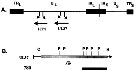
FIG. 1. (A) Arrangement of the HSV-1 DNAgenome, showing
the locations of the unique sequencesof the L and S components
(UL and Us) and of the terminal (TRL andTRs)and inverted (IRL andIRS)repeatsand the ICP8 and UL37genes.(B) Schematic of the
UL37gene. The hatched boxrepresents theUL37 coding region.
The solidbox represents the domain of the UL37 protein in the MalE-1.1-kbp UL37 fusion protein usedtogenerate the780
anti-serum described in Materials and Methods. C, ClaI; P, PstI; H,
HindIII. The location ofapotentialATP-binding domain is indicated
by the smallnarrowbar.
Cleveland, Ohio)perml. ThepropertiesofHSV-1(F)and the vaccinia virus recombinants V37 and V8 have been de-scribedpreviously (4, 28, 29). HSV-1(F) andvaccinia virus stockswerepreparedandtiteredonVero cells asdescribed previously (4, 29).
Antisera. The production of ICP8-specific and UL37-specificrabbitpolyclonal antiserahas been described previ-ously (28). The ICP8-specific antiserum is directed against wild-type ICP8 protein, and theUL37-specific antiserum 780 is directed against aMalE-UL37 fusion proteinconsistingof the Escherichia coli maltose-binding protein (product of malE)fusedtothe terminal one-third aminoacid residues of UL37(Fig. 1B).TheICP6-specificmonoclonalantibody38S wasobtained from MartinZweig, National Cancer Institute,
Frederick, Md. (30).
Preparationof32P-labeledinfected-cell proteins. Confluent monolayers of Verocellswere incubatedinphosphate-free
medium for 2 h before and after infection with 5 PFU of either HSV-1(F), V37, or V8 per cell. 32p labeling was
performed bythe addition of50,uCiof32p; (carrier free; New England Nuclear, Boston, Mass.)perml. Cellswerelabeled
from 1 to 6, 7 to 12, or 12 to 24 h postinfection (hpi) and harvestedat the end of each labeling periodby rinsing the monolayerswithphosphate-bufferedsaline andscrapingthe cells into 1 mlof RIPA buffer (150mMNaCl, 1% Nonidet P-40, 0.5% deoxycholate, 0.1% sodium dodecyl sulfate [SDS], 50 mM Tris-HCl [pH 7.5]) supplemented with 0.01 mM TPCK (tolylsulfonyl phenylalanyl chloromethyl ke-tone), 0.01 mM TLCK (Na-p-tosyl-L-lysine chloromethyl ketone), and aprotinin (1:100). Lysates were frozen at
-700C.
Pulse-chaseradiolabeling ofvirus-infected-cell proteins. (i) 35S labeling.Confluentmonolayersof Vero cellswere incu-bated in EMEMcontaining1/10the normal concentrationof methionine for 1 h prior to and following infection with 5 PFU ofHSV-1(F)percell.3S pulselabelingwasperformed
by incubating the infected cells in EMEM containing re-duced methionineplus37.5,uCiof[35S]methioninepermlfor 30min. Followingthepulse,acold chasewasperformedby
incubating the cells in EMEM containing the normal con-centration ofmethionine. Mock-infected cells were labeled in thesameway.
(ii) 32p labeling. Confluent monolayers of Vero cells were incubated in phosphate-free EMEM for 1 h prior to and following infection with 5 PFU of the appropriate virus per cell. 32plabeling was performed by incubating the cells in phosphate-free medium containing 62.5 ,uCi of32p; per ml. Cold chases were performed by rinsing the cells and incu-bating them with normal EMEM. Cells were harvested as described in the preceding section.
Immunoprecipitations and protein blot
analysis.
For immu-noprecipitation reactions, 100-,u samples of cell extracts wereincubated with 1 ,ul of the appropriateantiserum for 18 h at4°C on a rotary shaker. The resulting complexes were precipitated by the addition of 100 ,ul of 10% protein A-Sepharose (Sigma Chemicals) followed by incubation at 4°C for 6 h. The immune complexes were collected by centrifugation, washed four times in RIPA buffer, resus-pended in SDS-polyacrylamide gel electrophoresis (PAGE) sample buffer (2% SDS, 5% ,-mercaptoethanol, 50 mM Tris-HCl [pH 6.8], 5% glycerol, 0.15 mM bromophenol blue), and boiled for 2 min. Thesampleswerethen subjected to SDS-PAGE. Immunoblot analysis was performed as described before (28, 29). Bound antibodies were detected by use of an alkalinephosphatase-conjugated second anti-body. Radiolabeledproteins were detected byimage analy-sis withanImageQuantPhosphorImager(Molecular Dynam-ics).RESULTS
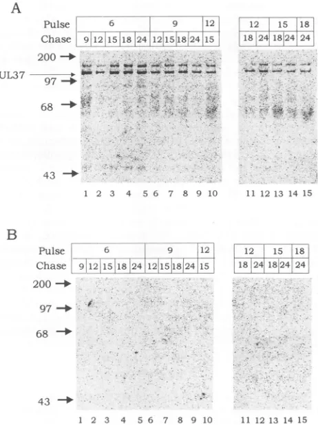
Stability
of the UL37 protein in HSV-1-infected cells. The UL37protein is first detected by immunoblot analysis and immunoprecipitations in HSV-1-infected cells atbetween 6 and 9 hpi and increases in abundance throughout the viral replication cycle (29). To determine the stability of the UL37 protein during lytic HSV replication, pulse-chase labeling experiments with [35S]methioninewere performed in HSV-1-infected cells. HSV-HSV-1-infectedormock-infectedVerocells werepulsed with[35S]methioninefor 1 h at 6, 9, 12, 15, and 18hpiand chased in mediumcontaining excesscold methi-oninefor various timeperiodsupto24hpi.Theradiolabeled infected-cell proteins were immunoprecipitated with the UL37-specificantiserum780,separated bySDS-PAGE, and transferredtonitrocellulose membranes.Phosphorimage analysis of the nitrocellulose membranes is shown in Fig. 2. Two bands of 140 and 120 kDa were
immunoprecipitated with the 780 antiserum from HSV-1-infected-cell extracts but not from mock-infected-cell ex-tracts (Fig. 2). Immunoblot analysis of the nitrocellulose membranes with the 780 antiserum showed that the
35S-labeled 120-kDa band was the UL37 protein (Fig. 3). As discussedbelow, the 140-kDa protein was identified asthe largesubunit of theHSV-1 ribonucleotide reductase thatwas
nonspecifically immunoprecipitated by rabbit antisera. Therewere nosignificantdifferences in therateofsynthesis of the UL37protein produced at the 6, 9, and 12hpi time points. Forexample, theamountofUL37protein produced
at6, 9,and 12hpiand harvested3 h later(9, 12, and15hpi, respectively)was similar (Fig. 2A). Likewise, the levels of radiolabeled UL37 presentat15hpi appearedtobeidentical for the 6-, 9-, and 12-h pulses. There was a noticeable reduction in the rate of synthesis of the UL37 protein produced at 15 and 18 hpiand harvested at 18 and 24 hpi. This pattern ofexpressionis in agreement withour
previous
dataindicatingthat theUL37proteinbelongstothe
-yl
class ofHSV genes(13, 29).UL37 protein phosphorylated in infected cells. To
deter-m
9 . . .
I I I
VOL. 67,1993
on November 9, 2019 by guest
http://jvi.asm.org/
[image:2.612.65.300.69.190.2]4844 ALBRIGHT AND JENKINS
A
Pulse 6 9 112
Chase 9!1211518 24121548124115
200_-UL37
97
-4
-68 -. ;.
43 -+
1 2 3 4 5 6 7 8 9 10
12 15 118
24l18124l 24
v5W*h_4_r_ w
*-jb#1#
*..-.''. ;' *.
? !l r-S-z-e-.:;=q. *
W
''.!
ll ls 13 14 ls
A
Pulse 6 9 12'
Ch-ase '912 i15
-1
24I1158245---I !
1
-200 _UL37
-68
433
1 2 3 4 5 6 7 8 9 10
12 T 15 *181
-21-1- -,--1
_ ___ _
18T4I81 4, 247
11 12 13 14 15
B
Pulse! 6 1 9 12i
Chase 9C12 151824
12115!18124
is200
_-97 -.4
68
-[12 15 18] '18 2411824 24!
- ,~
Pulse 6 9 ,12
|___..--- _ ---r~ '.
Chase i12!15,18 !24121151182415J 200 -_
97 -_
68
-z .l.~~~~~~~~~~~'
[image:3.612.65.296.71.380.2]1 2 3 4 5 6 7 8 9 10 11 12 13 14 15
FIG. 2. Synthesis and stability oftheUL37 protein in
HSV-1-infected cells. Immunoprecipitates of(A)HSV-1-infected- and(B)
mock-infected-cell extractswere pulse labeled with
[35S]methion-ine.HSV-1-infected Vero cellswerepulselabeled andharvested at
the times indicated(inhourspostinfection)asdescribed in Materials and Methods. Proteins fromaliquotstaken at each timepointwere
immunoprecipitated with the UL37-specific antiserum 780,
sepa-ratedby SDS-PAGE,and transferred to nitrocellulose membranes. "S-labeledproteinsweredetectedby phosphorimage analysisofthe membranes. Positions of size markersareindicatedonthe left(in kilodaltons).
mine whether the UL37protein is phosphorylated, HSV-1-infected cellswerelabeled with32Pifrom either 6 to 12or12 to 24hpi. Immunoprecipitation of the radiolabeled proteins with the 780 antiserum detected the presence ofa 120-kDa phosphoprotein thatwasnotimmunoprecipitatedwith either ICP8-specificor normal rabbitantiserum(Fig. 4).To deter-mine whether the UL37 protein was phosphorylated by either a viral or cellular kinase, 32P-labeled extracts from V37-infected cellswereimmunoprecipitatedwiththe UL37-specific, ICP8-UL37-specific, and normal rabbit antisera. V37 isa
recombinant vaccinia virus thatexpressestheUL37 protein (29). As shown in Fig. 4, a 120-kDa phosphoprotein was
immunoprecipitatedwiththe 780 antiserumbutnot withthe ICP8-specific or normal rabbit antiserum, suggesting that
phosphorylationwastheresult ofacellular kinase, although atthispointwecannotruleoutthepossibility thataprotein kinase encoded by vaccinia virus is responsible for UL37 phosphorylation in theV37-infected cells.
AsacontroltoensurethatphosphorylationofUL37in the V37-infected cells was protein specific, 32P-labeled V8-infected-cell proteinextractswerealso immunoprecipitated with the UL37-specific, ICP8-specific, and normal rabbit
43 -_4
1 2 3 4 5 6 7 8 9 10 11 12 13 14 15
FIG. 3. Identification of the UL37protein. Photographof
immu-noblotanalysis of(A) HSV-1-infected- and(B)mock-infected-cell
proteins. Proteins processed as described in thelegend to Fig. 2
weresubjectedtoimmunoblotanalysiswith the 780 antiserum and goat anti-rabbit immunoglobulin antiserum conjugated to alkaline
phosphatase.
antisera. V8 is arecombinant vaccinia virus thatexpresses
the ICP8 protein, which has been reported to have no
posttranslational modifications in HSV-1-infected cells (17, 28). As shown in Fig. 4, immunoprecipitations of the 32p_ labeled V8-infected-cell extractsfailed to detect any phos-phoprotein.
To demonstrate that the 120-kDa phosphoprotein immu-noprecipitated from HSV-1- and V37-infected-cell extracts
wasUL37,thenitrocellulose membraneswere probedwith either UL37-orICP8-specificantiserum andthen withgoat
PtAb N 8 37 N 837 N 8 37 N 8 37 N8.37 N 8 37
UL37 UL37
68-=
ILabel 6-12 12-24
HSV-1
6-12 12-24 6-12 12-24
V8 V37
FIG. 4. Identification of the UL37proteinas aphosphoprotein.
"P-labeled protein extracts from HSV-1-, V8-, and V37-infected cells were immunoprecipitated with either normal rabbit serum
(lanes N), ICP8-specificantiserum(lanes 8),orUL37-specific
anti-serum (lanes 37). Immunoprecipitated proteinswereseparated by
SDS-PAGE and transferredtonitrocellulose membranes. Radiola-beledproteinswere detectedby phosphorimage analysis.
B
12 r 15 118
-8
2411t8124
44. > Ce 4
J. VIROL.
on November 9, 2019 by guest
http://jvi.asm.org/
[image:3.612.319.548.71.376.2] [image:3.612.318.551.577.655.2]HSV UL37 PROTEIN PHOSPHORYLATED IN INFECTED CELLS 4845
PptAb N- 8 37 N- 8 37
Label 61.'.122''4 8
N 837 N 8 37
- L-3L7
A
Pulse 6
Chase 7 12 10 -5
200 _ ;
UL37 -_ ,
68
6-12 12-24
ca37
43 _B
1 2 3 4PptAb N 8 37N 8 37
ICP8 a
Label 6-12 I2-24
u8
;837X- 8 37
A In,x;:.
:.6112-:.':
B
-UL3, Pulse 6 9
Chase 7 12 10 15
200-_
UL37 --- -e
68 O
u'37
PptAb N; 8 37N 837
ICP8 -_
Label 6-12 12-24 6-.12 12-2.
Cx8 0t37
FIG. 5. Detection of UL37and ICP8proteins. D gels shown inFig. 4weresubjected toimmunoblc
eitherICP8-specificorUL37-specific antiserumanc
phatase-conjugatedgoatanti-rabbitimmunoglobulin
each panel, the blot on the left was probed wit
antiserum (a8) and the blot onthe right wasprob specificantiserum (a37). (A)HSV-1-infected-cellpi
infected-cellproteins; (C)V37-infected-cellproteins
anti-rabbitimmunoglobulinantiserumconjugal phosphatase. As shown in Fig. 5, the UL37
serumimmunoprecipitated UL37 protein fron V37-infectedcells,while theICP8-specificant
noprecipitated the ICP8 protein from HSV-fectedcells. The 120-kDaphosphorylatedpro
alignedwith theUL37protein.
Stability ofUL37phosphorylation. To detei bility of thephosphate groupsonthephosphc protein, HSV-1-infected and mock-infected V pulse labeled with
32Pi
for 1 h at 6 and 9 b harvested immediately or chased with mediu cold phosphate for an additional 5 h. Theproteinswere immunoprecipitated with the 7 separated by SDS-PAGE, and transferred to
membranes. As shown inFig. 6,theamount
latedUL37proteindidnotchangebetween the
1UL37 FIG. 6. 32P pulse-chase experiment. Immunoprecipitations of
HSV-1-infected-cell(lanes1to4)ormock-infected-cell(lanes 5to8) proteinextractspulse labeled with
32pi.
HSV-1-infected Vero cellswerepulse labeled and harvestedat the times indicated (in hours postinfection)asdescribed in Materialsand Methods. Proteins from
aliquotstakenateach timepointwereimmunoprecipitated with the UL37-specificantiserum780, separatedbySDS-PAGE, and trans-4 ferred to nitrocellulose membranes. (A) 32P-labeled proteinswere
detected by phosphorimage analysis of the nitrocellulose
mem-branes. (B)Nitrocellulose membraneswere subjectedto immuno-uplicatesof the blot analysis with UL37-specific antiserum and alkaline
phos-)t analysis with phatase-conjugated goatantiserum.
d alkaline
phos-antiserum. For
h ICP8-specific
hed
with UL37- thesubsequent
5-hcold chase.Thus,
thephosphorylation
ofroteins; (B)V8- UL37 does not appear tocycleonand off but rather appears
to occur soon aftertranslation, and thephosphates remain with the protein throughoutthe viralreplication cycle.
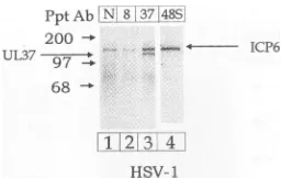
Identification of the 140-kDa protein. The UL37-specific 780 antiserum immunoprecipitated a 140-kDa protein from tedtoalkaline 35S-labeled, HSV-1-infected cells but not from mock-in-'-specific anti- fected cells (Fig. 2). The 780 antiserum failed tobind the n HSV-1- and 140-kDa bandinimmunoblotexperiments, indicating thatit iserumimmu- was not the UL37 protein (Fig. 3). Similarly, a 140-kDa 1- and V8-in- phosphoprotein was immunoprecipitated from HSV-1-in-teins inFig.4 fected cells with either UL37-specific, ICP8-specific, or normal rabbit antiserum (Fig. 4). These results suggested rmine the sta- that the 140-kDa band represented an HSV-1
phosphopro-)rylated UL37 tein thatwas immunoprecipitated byrabbit antiserum. The erocellswere mostlikelycandidate forthisproteinis the 140-kDaHSV-1 ipi and either ribonucleotide reductase (ICP6)encodedbythe UL39gene
,im containing product. ICP6 is one of themajor HSV-1 phosphoproteins radiolabeled andshows, attheaminoacidlevel,astronghomologywith '80 antiserum, ribonucleotidereductasesencodedin othersystems(33, 34). nitrocellulose ICP6 exhibits 38% relatedness atthe amino acid level with ofphosphory- the large subunit of ribonucleotide reductase from E. coli 1-hpulseand (33). The identity of the 140-kDa band as ICP6 was
con-A
ICP8
--6 9
7 12 10 l5
5 6 7 8
6 9
7 12 10 15
C
N 8 37N 837
43 *
1 2 3 4 5 6 7 8
VOL.67, 1993
on November 9, 2019 by guest
http://jvi.asm.org/
[image:4.612.62.298.71.446.2] [image:4.612.333.545.71.359.2]PptAb N 8 3748S
2 00 ICP6
UL37
68
1!2 I;4
IIsv- I
FIG. 7. Identification of the 140-kDa proteinasICP6. Vero cells wereinfected with HSV-1, labeled with32Pifrom 6to 12 hpi, and harvested at 12 hpi as described in Materials and Methods. 32p_
labeled protein extractswere immunoprecipitated with either
nor-mal rabbitserum(lane N),ICP8-specific antiserum (lane 8),
UL37-specific antiserum (lane 37), or a monoclonal antibody directed against ICP6 (lane 48S). Immunoprecipitated proteins were
sepa-ratedby SDS-PAGE and transferred tonitrocellulose membranes. Radiolabeled proteinsweredetected by phosphorimage analysis.
firmedby immunoblot analysis with the ICP6-specific mono-clonal antibody 38S (Fig. 7).
DISCUSSION
Previous studies in our laboratory demonstrated that the UL37 open reading frame of HSV-1 encoded a nonstruc-tural, 120-kDa protein belonging to the -yl class of HSV
genes(29). The UL37 proteininteracts withICP8, the major HSV-1 DNA-binding protein, in the cytoplasm of infected cells. Inarecentstudy,wedemonstrated thattworesultsof this interaction were transport of the UL37protein to the nucleus of infected cells and coelution of the UL37protein with ICP8 from both single- and double-stranded DNA columns(28).Inthisarticle,wereportthat theUL37protein exhibited littleorno apparent turnoverduring HSV replica-tion. Asaresult of thisstability, the UL37protein accumu-lated in the cellduringviralreplication.We have also shown that the UL37proteinwas phosphorylated in both HSV-1-infected and V37-HSV-1-infected cells. Thephosphategroupswere added soon after translation and remained on the protein throughoutthe viralreplication cycle and therefore didnot
appear to cycle on and off as has been reported for other HSVphosphoproteins (34). Furthermore, thestateof phos-phorylationof theUL37 proteinrelativetotherateof UL37 synthesis did not appear to increase or decrease during HSV-1replication.
The phosphorylation of a UL37 protein expressed by a recombinant vacciniavirus (V37) suggests that the protein kinase responsible for phosphorylation was a cellular
en-zyme.Theidentityof the cellular kinaseresponsibleforthis phosphorylationisnot known.
We have also demonstrated that theHSV-1 ICP8protein is not phosphorylated in either HSV-1-infected or V8-in-fected cells. This is an important point regarding
phospho-rylation specificity, sincethe UL37proteinisnotoneof the
major HSV phosphoproteins. The UL37 and ICP8 proteins have a similar number of amino acid residues (1,123 for UL37 and 1,196forICP8), andwehave previously demon-strated that the ICP8 and UL37 proteins comigrate on
SDS-PAGE,with identicalapparent molecular weights (28). Thephosphorylation of UL37butnotICP8 in both HSV-1-infected cells and cells infected with vaccinia viruses
ex-pressingeachprotein (V37 and V8) suggests that the phos-phorylation of UL37 is not due to an adventitious
phosphorylationofarelatively large protein, since ICP8, a
viral protein of the same apparent molecular weight as UL37, is notphosphorylated. While the number and loca-tions of the phosphorylation sites within the UL37 protein are not known, there are numerous potential sites. Within the 1,123 amino acids of the UL37 protein, there are 162 serine, threonine, and tyrosine residues (71, 71, and 20 residues,respectively). Analysis of the amino acid sequence for motifs recognized as potential sites of phosphorylation by known cellular kinases revealed two potential sites for cyclicAMP-dependent protein kinase, 14 potential sites for casein kinase II, and 14potential sites for protein kinase C. Studies are under way in the laboratory to determine the precise number and location(s) of phosphorylation sites on the UL37protein.
Asmentionedearlier,theUL37 and ICP8proteinsinteract inthe cytoplasm of HSV-1-infected cells(28). The phospho-rylation of the UL37 protein from V37-infected cells indi-cates that the interaction with the ICP8 proteinwas not a prerequisite for phosphorylation. In addition, preliminary resultshave indicated that the phosphorylated form of UL37 coeluted with ICP8 fromsingle-stranded DNA-agarose col-umns(data notshown). This suggests that phosphorylation ofthe UL37protein did not inhibit the interaction of UL37 with the ICP8protein. We have begun studiestodetermine whether the phosphorylation of UL37 is required for its interaction with the ICP8 protein.
The interaction of UL37 with ICP8 may result in a modification of an ICP8 function that occurs late in viral replication. The ICP8 protein has been implicated in the negative regulation of ICP4, the major immediate-early regulatory protein of HSV-1 (7-9). Mutations in the ICP8 protein have also been shown to result in increased
tran-scriptionof several earlyand lateHSV genes(10, 11). Gao andKnipe (8) recentlydescribedatransdominantmutantof theICP8proteinthatinhibited theexpressionof several late proteins during HSV replication. This inhibition occurred independentlyof any block in DNAsynthesiscausedbythe mutant ICP8protein.These results have ledto the hypoth-esis that at late times in infection, the ICP8 protein binds eitherto small single-stranded DNAregions, keeping pro-moterregionsopenfortranscription,or tospecificstructures or sequences inHSV-1late-gene promoters.
In order for ICP8torecognizetheselate-gene promoters, it may require direct interactions with other viral and/or cellularproteins. Fromthe results ofourearlierstudies,we
postulated that the UL37 protein would be a reasonable candidate for such aviralprotein (28). Previous studies in several laboratories have demonstrated that protein phos-phorylation can affect nuclear localization, DNA binding, and transactivation and repression (12, 19, 23). While the exact role ofphosphorylation in the function of the UL37 protein in viral replication has yet to be determined, it is interesting to speculate that it might be involved in the interaction with the ICP8protein. Studiesare inprogressin ourlaboratoryto determine whether the phosphorylation of UL37 is involved in theprotein'snuclear localization orits interactionwith the ICP8 protein.
ACKNOWLEDGMENTS
We thank William T. Ruyechan and Kathryn V. Holmes for a
criticalreadingof themanuscript.
Thisinvestigationwassupported byUSUHS grant R07396.
on November 9, 2019 by guest
http://jvi.asm.org/
[image:5.612.115.243.69.150.2]HSV UL37 PROTEIN PHOSPHORYLATED IN INFECTED CELLS 4847 REFERENCES
1. Ackermann, M., D. K. Braun, L. Pereira, and B. Roizman.1984. Characterizationofherpes simplexvirus 1 aproteins0, 4,and 27with monoclonal antibodies. J. Virol. 52:108-118.
2. Banks, L. M., I. W. Halliburton, D. J. M. Purifoy, R. A. Killington, and K. L. Powell. 1985. Studies on the herpes simplexvirusalkaline nuclease: detectionoftype-commonand type-specificepitopes on the enzyme. J. Gen.Virol. 66:1-14. 3. Edson, C. M., B. A.Hosler,and D. J. Waters. 1987.
Varicella-zostervirusgpIand herpes simplexvirusgE: phosphorylation andFcbinding. Virology 161:599-602.
4. Ejercito, P. M.,E. D. Kieff, and B. Roizman. 1968. Character-ization ofherpessimplexvirus strains differing in theireffects onsocialbehaviorof infected cells.J.Gen.Virol. 2:357-364. 5. Frame, M. C., D. J. McGeoch, F. J. Rixon, A. C.Orr, and H. S.
Marsden. 1986. The 10K virion phosphoprotein encoded by geneUS9fromherpessimplexvirus type 1.Virology 150:321-332.
6. Frame, M.C., F. C. Purves, D. J. McGeoch, H. S. Marsden, and D. P. Leader. 1987. Identification of herpes simplex virus protein kinaseastheproductof viral geneUS3.J. Gen. Virol. 68:2699-2704.
7. Gao, M., and D. M. Knipe. 1989.Genetic evidenceformultiple nuclear functions of the herpes simplex virus ICP8 DNA-bindingprotein.J.Virol. 63:5258-5267.
8. Gao, M., and D. M. Knipe. 1991. Potential role for herpes simplex virusICP8 DNAreplication proteinin stimulation of late geneexpression. J. Virol. 65:2666-2675.
9. Godowski,P.J., and D. M.Knipe.1983.Mutationsinthemajor DNA-bindingproteingeneofherpessimplexvirus type 1result in increased levels ofviralgene expression. J. Virol. 47:478-486.
10. Godowski, P. J., and D. M. Knipe. 1985. Identification of a
herpessimplexvirus function that represses late gene expres-sionfromparental viralgenomes. J. Virol. 55:357-365. 11. Godowski,P.J., and D. M. Knipe. 1986.Transcriptional control
of herpesvirus gene expression: gene functions required for positiveand negative regulation. Proc. Natl. Acad. Sci. USA 83:256-260.
12. Gonzalez, G. A., K. K. Yamamoto, W. H. Fischer, D. Karr, P. Menzel, W. BiggsIll,W. W.Vale, and M. R. Montminy. 1989. Aclusterofphosphorylation sitesonthecyclic AMP-regulated nuclear factor CREBpredicted byits sequence. Nature (Lon-don)337:749-752.
13. Honess, R.W.,and B. Roizman.1974.Regulation of herpesvirus macromolecularsynthesis.I.Cascaderegulation of the synthe-sis of threegroupsofviralproteins.J.Virol. 14:8-19. 14. Hunter, T., and M. Karin. 1992. Theregulation of transcription
byphosphorylation. Cell 70:375-387.
15. Hurley,J. R., A. M. Dean, J. L.Sohl, D. E.J.Koshland,and R. M. Stroud. 1990.Regulation ofanenzymeby phosphoryla-tionattheactive site.Science 249:1012-1016.
16. Kieff,E.D.,S. L.Bachenheimer,and B.Roizman. 1971. Size, composition, and structure of the deoxyribonucleic acid of herpes simplex virus subtypes1and 2. J. Virol. 8:125-129. 17. Knipe, D. M., M. P.Quinlan, and A.E.Spang. 1982.
Charac-terization of two conformational forms of the major DNA-binding protein encodedby herpes simplex virus 1. J. Virol. 44:736-741.
18. Kwong, A. D., J. A. Kruper, and N. Frenkel. 1988. Herpes simplex virusvirion hostshutoff function. J.Virol. 62:912-921. 19. Luscher,B.,E.Christenson,D.W.Kitchfield,E.G.Krebs,and
R. N.Eisenman.1990. MybDNAbinding inhibited by phospho-rylation at a site deleted during oncogenic activation. Nature (London) 344:517-522.
20. Marsden, H. S., M. E. M. Campbell, L. Harr, M. C. Frame, D.S. Parris, M.Murphy, R. G. Hope, M. T. Muller, and C. M. Preston. 1987. The 65,000-Mr DNA-binding andvirion
trans-inducing proteins of herpes simplex virus type 1. J. Virol. 61:2428-2437.
21. Marsden, H. S., N. D.Stow, V. G. Preston, M. C. Timbury, and N.M. Wilkie.1978. Physical mappingofherpes simplex virus-inducedpolypeptides.J. Virol.28:624-642.
22. McGeoch, D. J., M. A. Dalrymple, A. J. Davison, A. Dolan, M.C. Frame, D. McNab, L. J.Perry,J. E.Scott, and P. Taylor. 1988. ThecompleteDNA sequenceof thelong unique regionin the genome of herpes simplex virus type 1. J. Gen. Virol. 69:1531-1574.
23. Moll, T., G. Tebb, U. Surana, H. Robitsch, and K. Nasmyth. 1991. Theroleofphosphorylationand the CDC28proteinkinase in cell cycle-regulated nuclear import of the S. cerevisiae transcriptionfactorSWI5.Cell 66:743-758.
24. Overton, H. A., D. J. McMillan, L. S. Klavinskis, L. Hope, A. J. Ritchie, and P. Wong-Kai-In. 1992. Herpessimplexvirus type 1 geneUL13 encodes aphosphoproteinthat is a componentofthe virion. Virology 190:184-192.
25. Papavassiliou,A.G., K. W. Wilcox, and S. J. Silverstein. 1991. The interaction of ICP4 with cell/infected-cell factors and its state of phosphorylation modulate differential recognition of leader sequences in herpes simplex virus DNA. EMBO J. 10:397-406.
26. Pereira, L.,M. H.Wolff,M. Fenwick, and B. Roizman. 1977. Regulation of herpesvirusmacromolecularsynthesis.V. Prop-ertiesofpolypeptidesmade in HSV-1 and HSV-2 infectedcells. Virology 77:733-749.
27. Purves, F. C., and B. Roizman. 1992. TheUL13geneofherpes simplex virus 1 encodes the functions for posttranslational processing associatedwith phosphorylation of the regulatory proteina22. Proc. Natl.Acad. Sci.USA89:7310-7314. 28. Shelton, L. S. G., A. G. Albright, W. T. Ruyechan, and F. J.
Jenkins.Submittedforpublication.
29. Shelton, L. S. G., M. N. Pensiero, and F. J. Jenkins. 1990. Identificationand characterization of theherpes simplex virus type 1 protein encoded by the UL37 open reading frame. J. Virol. 64:6101-6109.
30. Showalter, S. D., M. Zweig, and B. Hampar. 1981. Monoclonal antibodiestoherpessimplex virustype 1proteins, includingthe immediate-early protein ICP4. Infect. Immun. 34:684-692. 31. Smibert, C. A., D. C. Johnson, and J. R. Smiley. 1992.
Identi-fication andcharacterization of thevirion-induced host shutoff product of herpes simplex virus gene UL41. J. Gen. Virol. 73:467-470.
32. Sprang,S. R., K. R. Acharya, E. J.Goldsmith, D.I.Stuart, K. Varvill, R. J.Fletterick, N. B. Madsen, and L. N. Johnson. 1988. Structuralchanges in glycogenphosphorylaseinducedby phos-phorylation. Nature(London)336:215-221.
33. Swain,M.A., and D. A.Galloway.1986.Herpessimplexvirus specifies two subunits of ribonucleotidereductase encodedby 3'-coterminal transcripts.J. Virol. 57:802-808.
34. Wilcox,K.W., A. Kohn, E.Sklyanskaya,and B. Roizman.1980. Herpessimplexvirusphosphoproteins. I. Phosphatecycleson
andoffsomeviralpolypeptides andcan alter theiraffinityfor DNA. J.Virol.33:167-182.
VOL.67, 1993