JOURNAL OFVIROLOGY, Sept. 1993,p.5623-5634 0022-538X/93/095623-12$02.00/0
Copyright © 1993,American Society forMicrobiology
V3
Loop of the Human
Immunodeficiency Virus Type
1
Env
Protein:
Interpreting Sequence Variability
LYNN MILICH,' BARRY MARGOLIN,23 ANDRONALD SWANSTROMl.3*
DepartmentofBiochemistry and Biophysics,1Departmentof Biostatistics,2andLineberger Comprehensive Cancer
Center,3 University of
North CarolinaatChapel Hill, Chapel
Hill,
North Carolina27599-7295
Received 6 January 1993/Accepted 10 June 1993
Two different states ofhumanimmunodeficiency virus type 1 areapparent in the asymptomatic and late stagesof infection.Important determinants associatedwiththesetwo stateshavebeen found within the V3loop of the viral Envprotein.Inthisstudy,twolargedatasetsofpublishedV3 sequenceswereanalyzedtoidentify patterns ofsequence variabilitythat would correspond to thesetwo states ofthe virus. Wewereespecially
interested in thepatternof basicamino acidsubstitutions, since thepresenceofbasic amino acidsinV3 has
been shown tochange virus tropismin cellculture. Fourfeatures of the sequence heterogeneity in V3were
observed: (i) approximately 70%o of all nonconservative basicsubstitutionsoccuratfourpositionsinV3,and V3sequenceswithabasicsubstitutioninatleastoneof these fourpositions containapproximately95% of all nonconservative basic substitutions; (ii) substitution patternswithin V3 areinfluencedby the identityofthe amino acidatposition25; (iii) sequencepolymorphismsaccountforasignificant fraction of uncharged amino
acid substitutions atseveral positions inV3, and sequence heterogeneity other thanthese polymorphisms is
mostsignificantat twopositions nearthe tip ofV3;and (iv) sequenceheterogeneity inV3(inadditiontothe basicaminoacidsubstitutions) isapproximately twofoldgreaterin V3sequencesthat contain basic amino acid
substitutions. By using this sequence analysis, we were able to identify distinctgroups of V3 sequences in
infected patients that appear to correspond to these two virus states. The identification of these discrete
sequencepatternsinvivodemonstrates how theV3sequencecanbe usedas ageneticmarker forstudyingthe twostatesof humanimmunodeficiencyvirus type 1.
The viralglycoproteins displayedonthe surface of
envel-opedvirusesareresponsibleforinteracting with thereceptor
onthetargetcellandarealsoexposedtohumoral selection bycirculating antibodies. Selection by antibodiescanresult
intheappearanceofsequenceheterogeneity within discrete
regionsoftheglycoproteinsequence, ashasbeenseenwith
the influenza virus HA protein, for which the biological
consequence of thisheterogeneityis antigenic drift(66, 77, 80).Genomicdiversity amongindependenthuman immuno-deficiency virus type 1 (HIV-1) isolates (reference 68 and references therein), to a lesser degree among sequential
isolates from thesamepatient (28, 30, 65),andevenwithina
single patient isolate (54) is awell-characterized feature of
HIV-1. Althoughthis sequence heterogeneity is distributed throughoutthegenome,mostof theheterogeneityis located intheenvgene (13,27, 50). Comparisonofpredicted amino acidsequences from several different isolates revealed that amino acidheterogeneityisclusteredinfive variableregions (Vl through V5)of the surfaceglycoprotein gp120 (43) (Fig. 1).Whenenvgene sequences fromsequential isolateswere
examined, amino acidchangesingp120werefoundtooccur
predominantly inthe samehighlyvariableregions, suggest-ing that sequence variability mayplaya significant role in
generatingmutantscapable ofescaping neutralization (43). During disease progression, a more virulent strain of
HIV-1emerges,suggestingthatHIV-1exists intwodifferent statesearlyand late ininfection(2, 6, 19, 59, 74, 75).These stateshavebeenmeasuredbythereplicative capacityof the isolate, defined as slow-low and rapid-high; by cytopathic abilities, usually defined as non-syncytium inducing (NSI)
andsyncytiuminducing (SI);orbyalterations inhostrange,
*Corresponding author.
including virus growthin macrophages (macrophage tropic)
versus adaptation to growth in transformed T-cell lines (T-cell-line tropic) (2, 6, 18, 19, 59, 60, 61). Althoughthese threedesignationsrefertodifferent propertiesofHIV-1,it is likelythatrapid-high, T-cell-line tropic, and SI all definethe
same state, which is distinct from a less-virulent state characterized asslow-low, macrophage tropic, and NSI.
Thebiological significance of thesequenceheterogeneity
in V3, the third variable region ofgpl20, has come under study (reviewedinreference24). Althoughthisregionisonly 35 amino acidslong,considerablesequencevariabilityexists within it when sequences from different isolates are
com-pared (37). Inspiteof thisvariability,V3 contains determi-nants that mediate virus interactions with CD4+ cells, the primary targets ofHIV-1 infection, and with the host
im-mune system. Antisera to recombinant Envproteins or to synthetic peptides that include amino acids within V3 are
able toblocksyncytium formation inducedbyavirus with thehomologousV3sequence(25, 32, 48, 53).V3 alsoserves as amajortargetforneutralizing antibody (29, 32, 33, 41, 48, 53). Antibodies tothe V3 region do notprevent binding of gpl20totheCD4 receptor but dopreventvirusuptake (67). There have been additional reports which show that V3 contains epitopes that elicit both cytotoxic T-lymphocyte
responses(10, 69,71, 72)andhelperT-cellresponses(11, 49,
70). Finally, molecular recombinants have been used to
show that determinants of virus tropism lie within the V3 region (3, 8, 9,31, 46, 62, 78). Consequently, the heteroge-neitycharacteristic of V3 mayrepresent inpartthe sumof selective pressures associated with these different
proper-ties.
Initial attempts have been made toidentify specific pat-terns of sequence variability in V3 associated with virus tropism and the cytopathic abilities of both
laboratory-5623
Vol. 67,No. 9
on November 9, 2019 by guest
http://jvi.asm.org/
su
signaVgpl2o gpl2O/gp4l
TM
%/ Vl V2 V3 V4 V5
DItH
lull|E
CTRPNNNTRKSIHIGPGRAFYTTGEIIGDIRQAHC
FIG. 1. Variable domains of the HIV-1 Envprotein. The five variable regions (Vl through V5)in thegpl20 (SU) portion of the envgene are indicated by black boxes. Shown below is the V3 consensus sequence(37). The shaded box represents the membrane-spanningdomainwithin thegp4l(TM) portion of theEnvprotein.
adapted andprimaryisolates of HIV-1. Severalstudieshave
shown that coupledchanges flanking the GPGR sequence,
which is present in most V3 sequences, are sufficient to
change virus tropism to T-cell-line tropic (9, 15). Mutant
viruses have been used to demonstrate thata minimum of three amino acidsubstitutionsin V3 canconfermacrophage
tropismand alterT-cell-linetropism(63). de Jongetal. (15)
provided evidence that HIVisolateswith an SI,
fast-repli-catingphenotype haveahighernetchargein V3 because of
the addition of basic amino acids. Comparison of V3
se-quencesfrom setsofbiological clones isolated sequentially
from 12 patients showed a correlation between a basic
chargeat twopositions (positions11and
25)
andthetransi-tionfrom an NSI toanSIphenotype(21).Theimportanceof these two positions was confirmed by mutational analysis (14).
We analyzed more than 250 published V3 sequences to
identifysequence patternsthatdistinguishbetween thetwo
states of HIV-1. Our starting point for the analysis of sequencepatternswas twolargedatasetsof V3 sequences, which included examples ofmacrophage-tropic and
T-cell-line-tropicviruses (37, 44). Inthe second part of thisstudy,
weusedthe sequencepatterns identifiedfrom thepublished
sequences to analyze the virus population in infected pa-tients.
MATERIALSAND METHODS
Source of V3 sequences.The 175 V3 sequences used in the
initialanalysiswereselected from the datasetpublished by
LaRosaetal.(37)withcorrections(38,
39)
andaredescribedin the legend to Fig. 2. An additional 83 V3 sequences obtainedpredominantlyfromthe HIV-1 data base(44)were used in the analysis of V3 sequence variability and are available upon request. These sequences were determined
forisolates from 64individuals and represent both in
vivo-derived sequences and sequences from cultured virus. In almost all cases, no more than two sequences for isolates from a single patientwere included, representing the pre-dominant macrophage-tropic-like sequence and a distinct sequence variant when present. The viruses were isolated
predominantlyfromtheblood,althoughsomefromthe brain
and the lung are included. When HIV-1 was transmitted betweenindividuals,theisolate(s) from onlyonepatient was used. All of the isolates in the data set except SF1703 (7)
representisolates from North America or Europe. A subset of this data setwhich includes 10 pairs of in vivo-derived sequencesisfurther described inFig. 3B.
Viral DNA isolated from peripheral blood mononuclear
cell (PBMC) DNA of two HIV-1-infected individuals was
usedas a source for thepatient V3 sequences described in
Fig. 6. Patients A and B were enrolled in the AIDS Clinical Trials Group at theUniversity of NorthCarolina Hospitals, Chapel Hill, N.C. Both patients hadCD4+ cellcountsof less than 100/mm3 and had received antiviral therapy.
PCR amplification and cloning.PBMCs were isolated from patient blood bycentrifugation on Ficoll-Hypaqueas previ-ously described (12).Approximately 107 PBMCswere resus-pended in 0.28 ml of 1x STE(0.15 M NaCl, 10 mM Tris [pH 8.0], 1 mMEDTA). Lysis buffer (500 ,ug of proteinase K per ml, 0.5% sodium dodecyl sulfate [SDS], 10 mM Tris [pH 7.5], 30 mM NaCl, 20 mM EDTA) was added to a final volume of 0.5 ml, and the cells were incubated for 2 h at 50°C. Samples were then phenol extracted and ethanol precipitated.
Nested polymerase chain reaction (PCR) was used to amplify the 3-kb env gene from the uncultured PBMC DNA. Thefirst 30 cycles were donewith outerprimers5411/5428 (sequence derived from HXB2R; 5'-AGC ATC CAG GAA
GTCAGC-3') and 8546/8565 (5'-GTA CCTGAGGTGTGA
CTG GA-3') (51). Either 0.5 or 1 p,g of the patient DNA
samplewasused in a
100-pl
reaction that included 0.5 ,uMeach of the primers, 800 ,uM totaldeoxynucleotide triphos-phates, 2.0 mM MgCl2, 10 mMTris-HCl [pH 8.3], 50 mM KCl, 2.5 U ofAmplitaq[PerkinElmerCetus]). All 30cycles
consisted ofa30-sdenaturingstepat94°C, a30-sannealing
stepat 60°C, anda2-min extension step at72°C exceptfor
thefirst cycle, inwhich reactions wereboth denatured and
extended for 5 min. After 30 cycles, the reactions were extended for an additional 7 min. Three successive 10-fold dilutionsweremade from theamplified DNA,andanaliquot
of8 ,ul of each dilutionwasplaced intoa new100-,ulreaction mixture for a second 30 cycles, during which a region of
DNAwasamplifiedfrom thepreviouslyamplifiedDNA.The
second round of PCRwas done with nested primers 5503/
5537, containinganadditionalXbaI site(5'-AGCTTC GAG
ATCITAGJATT
AGG CAT CTC CTA TGG CAG GAA GAA GCG GAGACA-3'),and8421/8463,containinganXhoIsite and an additional MluI site(5'-TAAGTA CTA TACGCGC-ICATGTTlTTTCC AGGTJCTCGA.GATGCT GCT CCC ACC CCA TCT GC-3'). The parameters for the secondround ofamplificationwerethesame asforcycles2through
30 exceptthat thedenaturingstepincycle31was5 minand
theMgCl2 concentrationwas 1.5 mM.
OnlyPCR reactions thatwere not
overamplified
wereused toavoidcloning envheteroduplexes that may have formedbyreannealingathigh template concentrations.Thereaction
mixtures were purified by binding to glass beads by the manufacturer's protocol (Geneclean II; Bio 101 Inc.), and
theamplifiedDNAwasdigested withthe
appropriate
restric-tion enzymes. DNAfragmentswere ligatedinto the
phage-midpIBI31(International Biotechnologies, Inc.),and
bacte-rialtransformationwas donebyelectroporation (16).
Recombinant clones containing the 3-kb insert were screenedon indicatorplates containing100 ,ug ofampicillin
perml,0.5 mM
isopropyl-3-D-thiogalactopyranoside (IPTG)
and 40p,g of
5-bromo-4-chloro-3-indolyl-P-D-galactoside
(X-Gal) per ml. Between 10 and 20% of the white colonies(lacking lacZ)weretransferred tonitrocellulose (Schleicher
& Schuell
BA85)
and screened for HIV-1 env DNA byhybridization with an oligonucleotide probe as described
before(55, 84)exceptthat thefilterswerehybridizedat80°C
in 10 ml ofprehybridizationsolutioncontaining2.3pmolof
a 32P-5'-end-labeled oligonucleotide (56) and cooledslowly
toroomtemperature overnight. Plasmid clones identified in
thehybridizationscreen werethen screened for the presence
offull-lengthenv inserts.
on November 9, 2019 by guest
http://jvi.asm.org/
[image:2.612.60.297.67.154.2]SEQUENCE VARIABILITY IN HIV-1 V3 LOOP 5625
Taq error frequency. An error rate for Taq DNA poly-merase was determined after 60 cycles of amplification.
Roughly 100 copies of pIBI20 containing the HIV-1 env gene were mixed with 1 p.g ofuninfected high-molecular-weight
human cell DNA, and the env gene was amplified for 60
cycles under the conditions described above. The amplified
fragment was cloned and sequenced. V3 sequences were
obtained for 22 clones totalling 1,980bp. One C-to-T
transi-tion was identified, for an estimated error frequency of
approximately 0.05% after 60 cycles of amplification. DNAsequencing and sequence alignments. Single-stranded DNA for sequencing was generated after rescue of the env-containing phagemid with helper phage M13K07 (76).
The sequences of the clones were determined by dideoxy sequencing with Sequenase Version 2.0 according to the manufacturer's protocol (United States Biochemical). The sequencing primer used for V3 was 5'-AAC CAT AAT AGT ACA-3' (sequence derived from HXB2; primer 6616) (51). The sequence analysis programs SeqEd, Translate, and
PileUp (23) were used to translate and align patient V3
sequences.
Statistical
analysis.
All P values were generated by using the Fisher exact test as the significance test criterion (20).Nucleotide sequence accession numbers. Patient sequences were deposited in GenBank under accession numbers L21769 to L21836, L21980, and L21981.
RESULTS
We examined two large data sets of published V3 se-quences toidentifypatterns of sequence variability thatcan
be used to distinguish between the two different states of HIV-1.Since the work of de Jong et al. and others (9, 14, 15,
21, 63) has shown that the addition of a basic charge (by
substitution)in V3is associated with the more virulentstate
of HIV-1, we were
especially
interested in determining the distribution of basic amino acid substitutions within the V3 region. In an initial analysis, we examined the pattern ofbasic amino acid substitutions within 175 distinct V3
se-quences published previously by LaRosa et al. (37). We chose the LaRosa data set as our starting point for several reasons. First, theconsensus sequence of this data set isthe samesequence as that of theHIV-1 isolatesBaLl and JR-FL
(44, 46), both of which are known to confer a
macrophage-tropic phenotype (22, 31, 46). Second, an example of this sequence is present in the list of V3 sequences, as are examples of variants that are known to grow well in T-cell lines. We assume that this extensive list contains numerous examplesofeach type of virus.
Patterns ofbasicamino acidsubstitutions within V3.
Anal-ysisof the V3 sequences from the LaRosa data set show that basic amino acids appear frequently, both within the con-sensussequence(at five positions) and as substitutions from
theconsensussequence (Fig. 2A). Some positions tolerated conservative substitutions (9, 10, and 18), while two
posi-tions witharginine as the predominant amino acid had
lysine
substitutions only rarely (positions 3 and 31). Nonconserva-tive substitutions to basic amino acids were also discrimi-nating. Basic substitutions were most frequently seen at
positions 11, 13, 19, 23, 24, 25, and 32, with substitutions at
the four most common positions, 11, 24, 25, and 32, repre-senting approximately 67% of all nonconservative basic substitutions. Basic substitutions at positions 13, 19, and 23 account for about 19% of all nonconservative basic substi-tutions. Nonconservative basic amino acid changes were also distributed asymmetrically at these seven positions.
Four positions, 19, 24, 25, and 32, tolerated either arginine or lysine, while at the other three positions (11, 13, and 23), arginine predominated. At position 32, basic amino acid
substitutions were the only substitutionstolerated.
Analysis of an additional 83 V3 sequences, including
sequences from the HIV-1 data base (see Materials and
Methods), revealed similar basic amino acid substitution
patterns (data not shown).
When
V3 sequences from the twodata sets were combined, we found that 70% of all
noncon-servative basic substitutions occurred at the four noted positions, 11, 24, 25, and 32. In addition, the subset of V3 sequences (approximately 42%) that contained a basic amino
acid at one of these four positions contained
approximately
95% of all of the nonconservative basicsubstitutions present within the total list of V3 sequences. Thus, basic substitu-tions at four posisubstitu-tions (11, 24, 25, and 32) can be used as a reliable marker to identify virtually all sequence variants with nonconservative basic substitutions. In addition, basic substitutions in two of these fourpositions (11 and 25) have been directly implicated in virus tropism (14, 21, 63, 79).
In analyzing the V3 sequences from the LaRosa data set
501 A * Amn
0a
40-o c
-E-°
3o0
0
I 020,
men)
0 10o
B
Lys
0 Lys
-Ii
CTRPNNNTRKSI H IGPGRAFYTTGEI IGDI RQAHC
5 10 15 20 25 30
1001 B
V 0
2-11
Co o
0C
D .
'a
CTRPNNNTRKS I H I G PGRAFYTTGE I I GD I RQAHC
I S G M V A QVT N Y
[image:3.612.317.562.300.729.2]14 21 53 15 14 73 25715 22 8
FIG. 2. Distributionofbasicanduncharged amino acid substi-tutions withintheLaRosaV3sequencedataset(37). Included in the data set are175 uniqueV3 sequencespublishedbyLaRosa et al. (37).Identical sequences wereusedonlyonce.Notincludedwere11 V3 sequences with either a QR or RG insert between Ile-14and Gly-15oftheconsensussequence(LaRosaV3sequences166 to 175 and 225). Also not included were nine V3 sequences with other unusual insertions or deletions(LaRosaV3sequences64to66,178 to 180, 212, 223, and 244). Seventeen sequenceswhich contain a single amino acid deletion at position18,22,24, or27wereincluded in the data set(LaRosaV3sequences39,63,101,121,122,133,135, 144, 163, 164, 208 to 211, 222, 232, and245).(A)Numberofarginine and lysine substitutions. (B) Number of uncharged amino acid substitutions and deletions notincluding polymorphisms. Specific polymorphisms are shown belowtheconsensussequenceinpanel B. Numbers indicate how many times each polymorphism was found among the 175sequences.
VOL.67,1993
.n 0
Nn
I 0 --1on November 9, 2019 by guest
http://jvi.asm.org/
that had nonconservative basic amino acidsubstitutions,we also noted thatmostof them also hadasequencechange at
position 25, awayfrom the consensus glutamicacid or its
conservative substitute, asparticacid. This correlationwas also apparent if the sequenceswere first grouped asbeing
substitutedatposition25.Of the 88 sequences in the list that containedanonacidicaminoacidatposition 25,69 of them hadabasicamino acidatposition 11, 13,19, 23, 24,or32.By contrast,
only
10 V3 sequencesonthe list hadabasic amino acidat oneofthese sixpositionsandanacidic amino acidatposition
25. Thus, therewas a significant correlation for achange
awayfromanacidic amino acidatposition25 and the appearanceof basic amino acidsatthe other sixpositions(P
< 10-4).
Patternsofacidic anduncharged amino acid substitutions within V3. Inaddition to identifyingbasic amino acid sub-stitution patterns that may be associated with virus state,we
also wanted to address the broader question of sequence
variabilitywithin V3. To dothis,weexamined the
distribu-tion of acidic and
uncharged
amino acids in the 175 V3 sequences from the LaRosa data set. We found that, in contrastto basic aminoacids,
acidic amino acidswere rare in V3 sequences both within the consensussequenceandas sequencevariants(datanotshown).Thepredominantacidic amino acid substitutions were a conservative substitution(aspartic
acid forglutamic
acid)
atposition
25 andachangefrom
glycine
to either aspartic acid or glutamic acid atposition
24.Substitutions with
uncharged
amino acidswere character-izedbytwopatterns(Fig.
2B). First,at somepositionsthere was asingle
amino acid that accounted for40%ormoreof all substitutions atthatposition.
Second,when thesepredomi-nant amino acid
polymorphisms
were subtracted, V3 se-quenceheterogeneity
was mostsignificant
at positions 13 and 20. Thepredominant
amino acid atposition
13 ishistidine,
with the substitutionsrepresenting
small aminoacids,
while atposition
20, the consensus phenylalanine isusually replaced
with ahydrophobic
amino acid. The nextmostheterogeneous positionwasposition 25,which also had
a
frequent change
toglutamine.
Theseresults, together
with ouranalysis
ofcharged substitutions,
indicate that the ap-parent sequence variability in V3 has several distinct fea-tures,including
substitution of basic amino acids atdiscretepositions,
an underrepresentation of acidic amino acids,frequent
substitutions that may represent polymorphisms,and clusteredheterogeneityofunchargedamino acid substi-tutions at positions 13 and 20.
Sequence
changes thatcorrelate with altered tropism. Im-portant determinants ofmacrophage
tropism
versus theability
togrowin T-cell lines havebeen showntolie within the V3loop
(8, 9, 31, 63, 79),
and the addition of basicaminoacids to the V3 sequence enhances
growth
andsyncytium
formation in transformed T-celllines
(14, 15).
Itislikelythat thepredominant
nonconservative basic amino acid substitu-tions at thepositions
seen in the examination of sequenceheterogeneity (Fig. 2A)
represent the naturally occurringpositions
foratleastsomeof the sequencechangesinvolved intropism (positions
11, 24, 25, and 32). We tested thepossibility
that changes at these fewpositions coulddistin-guish
between macrophage-tropic andT-cell-line-tropic vi-rusesby examiningtheV3sequencesofviruses with knownphenotypes (Fig. 3A).
All 10 of theT-cell-line-tropicviruses hadabasicamino acid inatleastoneof thesepositions,and aswith thelarger
data set, basic substitutionsat these fourpositions
accounted for approximately 75% of all noncon-servative basic substitutions. In contrast, none of the 14macrophage-tropic viruses had a basic substitutionatanyof the four positions. There was only one example ofa non-conservative basic amino acid substitution at any position among the macrophage-tropic viruses (H13R in HIV-1
SF1703), while all V3 sequences from the group of
T-cell-line-tropic viruses contained one or more nonconservative basic substitutions. The association of at least one noncon-servative basic amino acid substitution with the
T-cell-line-tropic virusescomparedwiththe macrophage-tropic viruses
washighly significant (P<
10-').
Comparison of V3 sequences from the two phenotypes
also showed that all but three of the T-cell-line-tropic viruses had a nonacidic amino acid at position 25, while an acidic amino acid or alanine predominated at this position among macrophage-tropic viruses. Substitution of a nonacidic aminoacidatposition 25 among T-cell-line-tropic viruses, in contrast to the predominance of acidic amino acidsat this position among macrophage-tropic viruses, was found to be
significant (P = 0.01). Thus, three features that appear to
distinguish T-cell-line-tropic from macrophage-tropic vi-ruses include (i) the presence of nonconservative basic amino acid substitutions, (ii) frequent addition of basic amino acids at predominantly four positions, and (iii) a
changefroman acidic amino acidatposition 25.
We next used these features toexamine 10 pairs of V3 sequencesderived from sequences present in vivo(Fig. 3B).
These pairs of sequences represent two distinct groups of viral sequences present ineachpatient.Ineach case, thereis one sequence with nonconservative basic substitutions whicharefrequently found at the positions noted among the viruseswith the known T-cell-line-tropic phenotype. With
one exception, the sequences in this group also had a
nonacidic amino acidat position25. Theoppositewastrue for the second group of the paired sequences, in which
position25waspredominantlyacidic andonlyone
noncon-servative basic amino acid change was present (Q32K in
V12840). Thus, distinct features of viruses known to be
T-cell-linetropicarealso present withinviruspopulationsin vivo and appear to distinguish two different virus states in infectedpatients.
The link between the sequences in the larger data sets, which include sequences from cultured viruses, and the sequences presentin vivo isstrengthened by the observation that the consensus sequence of these in vivo-derived se-quences is almost identical to the macrophage-tropic con-sensussequencethatwasidentified in the LaRosa data set, the only differencebeing an equal occurrence ofglutamic acid and glutamineatposition25 (datanotshown).
Discrete patterns of basic amino acid substitutions. Since theloss ofanacidicamino acidatposition25appearedtobe
an importantdeterminant of T-cell-linetropism,we
consid-ered the
possibility
that theidentityof the amino acidatthisposition affects the pattern of substitutions at other
posi-tions. We examined 46 V3 sequences with an uncharged
glutamine at position 25 and compared these with 45 V3
sequenceswithabasic amino acidatposition25(Fig. 4).The patternsofspecificsubstitutionswereconsideredas a func-tion ofincreasingbasiccharge of the V3 loop. Overall, the
patterns of sequence changes between these two groups
appeared similar. However, there were two features that
distinguished the two groups, both ofwhich were
statisti-callysignificant. First,withabasic amino acidatposition 25,
glycine replacedserineasthepredominantuncharged amino
acid at position 11 (P <
10-4).
Second, basic amino acidsubstitutions were found at position 13 onlywhen a basic amino acidwaspresentatposition25(P= 0.012).
on November 9, 2019 by guest
http://jvi.asm.org/
SEQUJENCE VARIABILITY IN HIV-1 V3 LOOP 5627
A 11 2425 32 Reference
Consensus CTRPNNNT R KSIHI . GPGRAFYTTGEIIGDIRQAHC
TAdViruses
NL43 ---R R----K-a,1
RF --- *K ---* - --Q---K--- b
168.10.---R ....---Q- *--- 15
SF2 ..---*.- ----* .----K-5
SF33.---R -*R- * .--- * **.---K -82
HAN2.-- - - -*.-- -* 57
GUN-1(WT) ..---*.---*** K * .. 73
14558 ----***.--.*R ..--- ---KD- 9
13231.---R- -- ---.* ---K--- 9
MT Viruses
BALl--- c,22
JRFL--- 46
JRCSF - - --*--- d
ADA--- - 78
SF162.---* ..---* - -D.--- 5
SF1703.---*R .---* ---*- f
JFL---**---D ----
-g---SF128A ..---*--- .. A---.. 4,0
K3 ...-- -* .-- *.. 9
K7.-- * ..- * .-- *.*.-- 9
Q13 ....-*.*.-- 9
B 11 2425 32 References
Consensus CTR PN N NTRKSIHIH I GPGR AFY TTGEIIGDIRQAHC
TAd_Like
Dent-II -- --.*----*--K---R--- h,47
AM.Al4. 3 - . ---R * ---**.**.-- --* *--8 1
DH479-6-55.---*.---R--* -RR.--- 36
V12834 - .*---* . ** G * * - -*. -K.-.*..- 64
Z82Rl6Z.---R.----*---Q* .--.*.-- 30
S4-B-10.---R-** *-.-.-.-.-.-Q* .--. K-* - 42
A-II -- -- *-- . R*- *
.-*--B-Il -* *--. -- R**.----**RQ- -* j-.
C-Il. ---* * .--- * .---K.--- k
LCO2.DA05 .---* -* -R* * ..---** -RK.--- 1
MT-Like
Dent-I AM.Al4. 4 DH479-6-5 V12840 Z82R12
S4-B-11
A-I B-I
C-i LCO2.DA06
-*.. *.*
D.~-
-*.*--m, 47 81 36 64 30 42
n 0
p q
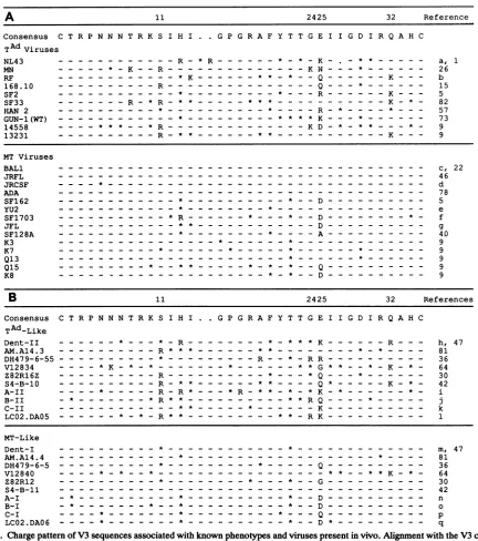
FIG. 3. Chargepattern ofV3sequences associated with knownphenotypesand virusespresentin vivo.Alignmentwiththe V3consensus
sequence of(A)sequences from viruses knowntogrow in T-cell lines(top)ormacrophages (bottom) and(B)sequencesrepresentingtwo
distinctgroups of virusespresentin each of 10HIV-1-infectedpatientsatasingletimepoint.These sequencesrepresentasubset of the 83 V3 sequences from the HIV-1 data base(44)thataredescribed in Materials and Methods. The sequenceswereobtained from uncultured PBMC DNAorplasmaRNA. Eachpairincludes thepredominantmacrophage-tropic-likesequence(bottom)andavariant that contains the sequence elements characteristic ofaT-cell-line-tropicvirus(top).Thepattern ofbasicchargesand the amino acidpresentatposition25were
features usedto sortthe sequences in eachpairinto thetwogroups of viruses. The actualphenotypeof these viruses is unknown. Amino acids shownrepresent nonconservativearginine andlysinesubstitutions. The identityof the amino acidat position25 is alsoshown. Asterisks
representother substitutions from theconsensussequence.Identitywith theconsensussequence is shown withadash. Numbersatthetop representpositionswithin theconsensussequence.TA,T-cell adapted (i.e.,T-celi-linetropic);MT,macrophage tropic.GenBank accession numbers: a, M19921; b, M12508; c,M68893; d, M38429; e, M93258; f, M66533; g, M31451; h, M90851; i, L21822;j, L21831; k, L21981;
1, M90917;m, M90848;n,L21770;o,L21778;p, L21980; q, M90918.
ison of the basic amino acid substitution patterns forthese tamine rather thanarginineorlysinewaspresentatposition two groups also showed that there was a tendency for 25.These resultssuggestthat basic amino acid substitutions
arginine to be substituted at position 11 as the first basic tendtofollowatleasttwodifferentpathways, althoughthere amino acid substitution (outside of position 25) when glu- are a few examples of sequences with elements of both
VOL.67, 1993
on November 9, 2019 by guest
http://jvi.asm.org/
[image:5.612.87.519.73.561.2]8T
11 13 19
CT R P N N N T R K S I H I G P G R A F Y T
O G(2) S (14)
+1 G(1) S(5) R,K(7) R(
A
+2 S(1) R(16) . R(2..
CT R P N
+1
+2
+3
iP G R A F Y T
R(4) G(4)R(2)
R,K(4
232425
T GE I
I I IGD
!(16)
!(13)
2)R,KQ (17) (7)
23242 T G E
R,K
(5) 5
I I G D I
t,K(14)
t,K(22)
K(3) R(4)R,KR,K(9) (3)
32
I RQA HC
0
c<C>
0)
c
o =
cJ
K(5)
R,K(9)
32 R A H C
R,K (4)
R(2)
FIG. 4. Discrete patterns of basic amino acid substitutions within V3.Specificamino acidchangesareshown for V3sequences
witheitherglutamine(top)orarginineorlysine (bottom)substituted atposition25. Substitutionpatternsarepresentedas afunction of
increasingbasicchargeinV3.Chargesfor individualV3sequences
werebasedonamino acid substitutionsatsevenpositionswithin the LaRosaconsensus sequence(11,13, 19, 23, 24, 25,and32).Atotal of 91 V3sequencesarerepresented,including43sequencesfrom the LaRosa data set(seebelow)and 48sequencesfrom othersources
(17, 21, 30,44,64):from reference30,sequencesC2, C3, D5, D7,
andE3;fromreference21,sequencesACH-15.9, ACH-320, ACH-168.7, AMS-55, AMS-16.1, ACH-479.5, ACH-182.69, ACH-320.2A.5, andAMS-175; from reference 17, sequencesW2-lalO, W2-1a6, W12-2cS,Wl-lal,andW1-1bS;from reference44,isolates HIVWMJ12, HIVNY5NEW, HIVHAN, CANOB, and FO; from reference 64, sequences91-a, 91-c, 82-c, and 87-d; and from the GenBank/EMBLdatabases,accessionnumbersM90881, M90886, M74658, M26727, M90885, M21138, M21098, M38430, M90938, M90913, M90902, M90876, M90958, M90936, K02007, M90917, M90905, M74676, M90851, and M12508. The 43sequencesfrom the LaRosadatasetinclude allsequenceswithaglutamine, arginine,or
lysine residue atposition 25 exceptforLaRosa V3sequences50, 106, 117, 126, 130, 146, 148, 149, 201, 203, 229, 235, 236, 238, and 240. Atotal of132 nonconservative basic substitutionswerefound
inthe 91 V3sequences,but in thisanalysisonly the identity of the amino acids at positions 11, 13, 19, 23, 24, 25, and 32 were
considered. Eighteen basic amino acid substitutions occurring at
otherpositions within V3arenotshown. Amino acid substitutions
are represented by a single letter. Shown in parentheses is the number ofsequenceswith each substitution. Numbers atthe top representpositions within theconsensussequence.
5
I.
4+ 3- 2- 1-0
I.
I.
LR LA IV
LowCharge (Macrophage-tropic-like)
LR LA IV
HighCharge
(T cell line-tropic-like)
FIG. 5. Extent of sequence heterogeneity associated with dis-tinct groups of V3 sequences. The mean number of amino acid differences from the V3 consensus sequence is shown for two groups of sequences in each of three separate data sets: theLaRosa dataset(LR), 63sequences from the HIV-1 data base(LA), and 10 pairs of in vivo-derived sequences(IV). Thetwogroups represented include viruses that aremacrophage-tropic-like and havealow net charge in V3 and sequence variants which haveahighnetcharge in V3 andareT-cell-line-tropic-like. TheV3sequencesin each dataset weresorted into the two groups bythepattern of basic amino acid substitutions and theidentity of the amino acidatposition 25. The number of amino aciddifferences from the V3consensussequence, not includingnonconservativebasic substitutions, and the amino acidatposition 25wasthen determined for each sequence. Shown are the means for eachgroup in the three datasets ±2 standard errors. V3 sequences from the LaRosa dataset aredescribed in the legendto Fig.2. Sequences from theHIV-1data base and the in vivo-derived sequences used in thisfigurearedescribedinMaterials and Methods and shown inFig. 3B, respectively.
pathways. This indicates that while these patterns are pre-ferred, theyare notobligatoryin the addition ofbasic amino acids toV3.
Basic substitutions and position 25 changes predict other sequence
heterogeneity.
Fromtheprevious analysis and the work of others(9, 14, 21,79),weproposethat the presence of basicamino acidspredominantly atpositions 11, 13, 19,23, 24, and 32 oftheV3loopandthe identityofthe amino
acids atposition 25 canbeused todistinguishtwodistinct sets of viral sequences. These features distinguish the se-quences of viruses with known phenotypes as well as discretesubsets of virusespresentinvivo(Fig. 3), although
the effectontropismof these individualsubstitutions in the
macrophage-tropic background has not yet been tested.
Nevertheless, we have used these features to sort V3
se-quencesfromthe three separate datasets(theLaRosadata set, the sequences from the HIV-1 data
base,
and the in vivo-derived sequences shown in Fig.3B)into twogroups. Chesebro et al. have suggested that macrophage-tropicviruses are moresimilar in sequenceas agroupthan
T-cell-line-tropicviruses (9).Weexamined theextentof sequence
heterogeneity associatedwith each group of viral sequences
ineach of the three datasets.As a measureofheterogeneity,
we determined the number ofamino acid differences from the consensus sequence for each sequence and did not includenonconservative basic substitutions in the tally.
The mean number of amino acid differences from the
consensus sequence in each of the three data sets was
approximatelytwofold greater among viruses that have basic
amino acidsubstitutions, with
approximately
2.6changesas themeanfor themacrophage-tropic-likeviral sequences and
on November 9, 2019 by guest
http://jvi.asm.org/
[image:6.612.333.541.74.218.2] [image:6.612.64.297.85.427.2]SEQUENCE VARIABILITY IN HIV-1 V3 LOOP 5629
13
-12 11
-10
9.
(n
ac
8'() 7
.o0
o 6 E
<
.5
5,-4,3
2-1
-Patient A (N5Y)30
4 2,59,
(S11R; 114V; F201; 31
tG24E;27T;H34YJ
189
(N7S)183(R31K)198/
A2(2G)68 (F20L)210
4 0
C12T; P1 3R;P16L;
G17R; A19T; D25KJ
,1X
19,205 (12T;D29N;H34Y)182187 (R9K; G17E; D29N)4
(114T;130V)165
(D25N; D29G)202,
(130M)197(N7S)13 -, (A22V) (N5T)2
P28/48
13
-12
-11.
10*
9
o
c
co
0
._c
Is
8-7.
6.
5.
4.-
3-
2-
1-0/2 3 4 5 6 7 8 9
V3charge
PatientB (R3K; GI5N; GI 7E
~10
R18K;G28K;
D29N)
(R3K; G1 5E;G28E)103
(130M)1672
R100;SI11R;112L P1 3S; T23S;G24R;F D25O
(R9G)8/ / (Gl5R)93
/11
or/(G17R)1
(D25E)l
08 (GI7R)96(R9K)110 (D25V)107
(112M)102/ (ClR)63
P7'20 U
I I I I I I 1
0/2 3 4 5 6 7 8 9
[image:7.612.64.557.78.356.2]V3 charge
FIG. 6. Identification of discrete V3sequence families in twoHIV-1-infectedpatients. Individual V3 sequences from patients A and B are plottedasV3 chargeversustotalnumber of amino acidchanges from the predominant V3 sequence in each patient. V3 charge for each clone wascalculatedfrom arginine and lysine substitutions and the amino acid residue at position 25. Represented are V3 sequences from 48 and 20clones isolated at asingle time point from patients A and B, respectively. The predominant ormacrophage-tropic-like sequence in each patient, indicated by the letterP,isasfollows:A,CIRPNNNTRKSIPIGPGRAFYATGDIIGDIRQAHC; B,CIRPNNNTRRSIPIGPGRAF YATGDIIGDIRQAHC. The subscriptrefers to the number of times that the predominant sequence appeared among the total V3 sequences examined in eachpatient. Superscriptnumbersrefer to individual clone designations. Shown in parentheses are the amino acids that differ from thepredominantsequence ineachpatient. Thenotation, e.g., A22V, includes the amino acid from the predominant V3 sequence, the position number, andthe amino acid change present in a particular clone. Doubly starredsuperscriptsrepresent an initial variant in each patientfrom which thehighly divergent viruses must have evolved. Dashed arrows represent the monoclonal outgrowth of the initialvariant fromthepredominantV3 sequence. V3 sequences at the end of solid arrows contain all of the changes found in the previous clone in addition toany newchangesshown inparentheses. Nucleotidesequences forindividualclones haveGenBank accession numbers L21769 to L21836.
approximately 5.4 changes as the mean for the sequences
that havebasic substitutions(Fig. 5).The difference between themeansfor the two typesofsequencesrepresented in the in vivo data set was not as significant as the difference betweenthemeansfor the larger data sets, possibly because of the smaller sample size. However, all three data sets followed the same trend. These results suggest that V3 sequencesderived fromputativemacrophage-tropic viruses show about twofold less sequenceheterogeneity than those sequenceswhich contain basic amino acid substitutions.
DetectionofdiscreteV3 sequencefamiliesin vivo. Weused both thebasic charge of the V3 loop and the total sequence
variability to compare individual V3 sequences present in
the virus population within aninfected individual (Fig. 6). We examined a total of 68 V3 sequences amplified from PBMC DNA obtained from two HIV-1-infected patients
(patientsAandB)at asingletime point. Themostfrequent
(predominant) V3 sequence in each patient appeared to
represent asequencefroma macrophage-tropic virus char-acterizedby the presence of an acidic amino acid at position 25 and the absence of basic nonconservative amino acid substitutions(Fig. 6);inaddition,thepredominant sequence hadaseries ofclosely related sequences clustered around it. Bothpatient samples also showed evidence of highly
diver-gentviral sequences. In thesetwopatients,thepattern of V3 sequences among the divergent sequences shared three features. First, there was evidence for a monoclonal
out-growthof the distinct variant, as shownbythe presence of
several closelyrelated sequences which werequite distinct from the predominant sequence. Second, each of the vari-antshadachangeatposition25 andatleastoneotherbasic
substitutionatposition11,13,or24.Third,inadditiontothe
basic amino acid substitutions in V3, the variant V3 se-quences had acquired numerous other sequence changes.
These changes include substitutions of hydrophobic and
small amino acidsatthetwomostvariablepositionsshown
in Fig. 2B, position 20 (patient A; F2OL) and position 13
(patient B; P13S), respectively. These results suggest that
the acquisition of basic amino acids in vivo occurs when
thereis strong selective pressure for other sequencechanges
and thatonlydiscrete sequence variantsaccumulate.
DISCUSSION
V3 is a regionwithin the HIV-1 env gene that displays
significant sequence heterogeneity (37). In
addition,
thisregion influences the ability of the virus to replicate in
macrophages and togrow in transformed T-cell lines (8, 9,
VOL.67, 1993
on November 9, 2019 by guest
http://jvi.asm.org/
31,63). V3 serves as a major target forneutralizing antibod-ies (25, 29, 32, 33, 34, 41, 45, 48, 53), suggesting that at least some of the heterogeneity may be the result of humoral
selection. In this study, we have dissected V3 sequence variability within two large data sets of V3 sequences in order toseparate sequence changes that correlate withvirus tropismfrom more diverse changes that contribute to overall sequence heterogeneity. The consensus sequence, or aver-age V3 sequence, is known to confer a macrophage-tropic
phenotype. This sequence provided a baseline fromwhich
we were able to identify distinct features of the sequence variabilityassociated with the V3region.
Charge changes in V3 associated with theabilitytoreplicate intransformed T cells.Theaddition of basic amino acidsin V3plays a rolein thephenotypicshiftbetween the NSI and the SI phenotypes (14, 15, 21). When we analyzed 175 published V3 sequences(37), we found verydistinctpatterns of basic amino acid substitutions. Nonconservative basic amino acid substitutions occurred predominantly at posi-tions 11, 13, 19, 23, 24, 25, and 32 (Fig. 2A), with approxi-mately95% of allnonconservativebasicsubstitutionseither in or linked to a basic substitutionat position 11, 24, 25,or 32. Inaddition, the presence of abasicorunchargedamino
acid at position 25 correlated closelywithbasicamino acid
substitutions at the other six positions. Thus, these two
features of basic amino acid substitutions with achange at position 25 define adistinct subset of V3 sequences.
Comparisonof 24 V3 sequences from viruseswithknown
phenotypes provided strong evidence that the pattern of basic substitutions and the change at position 25 that we identified within the LaRosa data set are distinguishing
featuresof a virus with a T-cell-line-tropic phenotype (Fig.
3A). With one exception, there were no nonconservative,
basic amino acid substitutions present in the V3 sequences of 14macrophage-tropicvirusescompared with the consen-sus sequence. Among the group of 10 T-cell-line-tropic
viruses, nonconservative basic substitutions were common and75% of themoccurredatthe fourpredominant positions
notedin ouranalysisof the 175 V3 sequences, i.e., 11, 24,
25, and32.
It was also evident from comparing the identity of the amino acid at position 25 in the sequences of these two groups of viruses that the amino acid at this positionwas usuallydifferentformacrophage-tropicandT-cell-line-tropic
viruses. Most of themacrophage-tropicviruseshad eitheran acidic amino acidoralanineatposition 25,incontrast tothe
T-cell-line-tropic viruses, which usually had a nonacidic
amino acid at this position. Together, these observations
strongly suggest that although the loss of an acidic amino acid atposition 25 is a strongpredictorof virustropism,it is the combination ofsubstitution ofamino acidswithabasic
chargeat asubset ofresiduesin V3(positions11,24,and32)
and theidentity of the amino acid at position 25 that together can be used to distinguish between macrophage-tropic and
T-cell-line-tropic viruses.
Theimportance of position 25 in predicting virus state has also been underscored previously in studies that used chi-meric molecular clones and performed sequence compari-sonsofprimary isolates. In a study by Westervelt et al. (79), amino acids in V3 associated with macrophage tropism
includedanacidicamino acid or alanine at position 25, while
isolates incapable of infecting macrophages had a basic residueatthisposition and substitutions atpositions 13 and 21. Fouchier et al. showed that among NSI viruses, the amino acidatposition 25 was either acidic or uncharged and the amino acid residue atposition 11wasuncharged(21).In
contrast, either one or both ofthese residues were basic amongSI isolates. Positions 11, 25, and 29 were shownby
mutagenesistocontributeto anSIphenotype (14).Chesebro
etal.
(9)
usedmolecular recombinantstoshow that sequence elementsoneither side of theGPGR motifcancontributetotheT-cell-line-tropicphenotype. In examiningalarger set of
sequences,wehave been abletogeneralize these patterns of sequencedifference from theconsensussequencetoallowa more consistent identification of two naturally occurring
virusstates.
Adaptivechanges may follow several pathways.
T-cell-line-tropicviruses can have V3 sequences with a single
noncon-servative basic amino acid substitution or up to three such substitutions(Fig. 3A). Whenweexamined thepositions of these substitutions as a function of the number of basic amino acid substitutions in the V3 sequence and whether the amino acid at position 25 was basic or an uncharged
glu-tamine,wefound several differences (Fig. 4). First,arginine
appeared at position 13 only when there was a basic amino acid atposition 25. Second, when there was a basic amino acid at position 25, glycine became the predominant
un-charged amino acidatposition 11 instead of serine. In this
typeofanalysis,itisnotpossibletodetermine whichchange
occurs first, although it is clear that the identities of the amino acids at positions 11 and 25 are coordinately influ-enced. Also, although these patterns represent strong
ten-dencies, they are not unique, in that some sequences with
elements of both patterns can be found. Nevertheless, the existence of these patternsmustultimately be understood in terms of the structural requirements for V3 function as reflected in either the direct or indirect interactions of these specific amino acids.
Some
heterogeneity
represents polymorphisms. When we examined uncharged amino acid substitutions within the consensus sequence, we found that a single amino acid represented a significant fraction of the substitutions away from the consensus sequence at many positions (Fig. 2B). For example, of 81 uncharged amino acid substitutions at position 22, 73 were from the consensus threonine to an alanine. Although we found no linkage between these pre-dominant uncharged amino acidsubstitutions, the apparent selection for particular types of amino acid residues at specific positions further suggests that variability in V3 is limited by certain structural constraints. In a few cases, the polymorphism was most apparent in the viral sequences with nonconservative basic substitutions (A19V andE25Q), while in the rest of the cases, the polymorphisms appeared irre-spective of the presence of nonconservative basic amino acid substitutions (Fig. 2B and datanotshown).Korber et al. found elements of sequence linkage after examining a large data set of V3 sequences (35). In the majority of cases, the most commonamino acidcombination noted represents the consensus sequence. However, there were examples among a small percentage of sequences in which the appearanceof an amino acid at one site was linked to aparticular amino acid atanotherposition. These include R13 and K25 or R25;V19 andT13; V20 and K24orR24;and Gll and H13 or R13.
Remaining sequence
heterogeneity
ispartly
clustered. When the basic amino acid substitutions, which are corre-lated with tropism, and the predominant, uncharged amino acid substitutions (i.e., polymorphisms) were subtracted from the total sequence heterogeneity in the LaRosa data set, we found that theremaining sequenceheterogeneitywas mostsignificant at two positions, 13 and 20 (Fig.2B).Both of these positions are two amino acids away from the GPGRon November 9, 2019 by guest
http://jvi.asm.org/
SEQUENCE VARIABILITY IN HIV-1 V3 LOOP 5631 sequence.This sequence wasinitially modeled as a beta turn
(37), a hypothesis that was subsequently confirmed by nuclear magnetic resonance analysis (4, 85). The initial modeling also suggested that a beta sheet conformation extends from the turn on each strand. In such a structure, the side chains at positions 13 and 20 would each be two residues away from the turn (on the N- and C-terminal sides, respectively), putting their side chains on the same face of the beta sheet and adjacent to each other across the sheet. Thus, these two variable residues may be equivalently placed to influence sequence heterogeneity, and therefore
antigenicity, in thevicinityof the tip of the V3 loop.
Charge changes and
heterogeneity
are linked. When we sorted thepublishedV3 sequences into two groups based on the parameters we have proposed to reflect the two virus states, we found that most of the sequence heterogeneity was associated with sequences that contain the nonconser-vative basic amino acid substitutions (Fig. 5). These findings wereconsistent for three separate data sets analyzed,includ-ing10pairs of V3 sequences present in vivo (Fig. 3B and 5). In all three data sets, the mean number of amino acid changes from the consensus sequence was approximately twofold greater among the group of sequences that contain basic amino acid substitutions than among the V3 sequences representing macrophage-tropic-like viruses.
Similar observations onthe extent of sequence
heteroge-neity among viruses representing the two tropism
pheno-typeshave been reported by others. Chesebro et al. noted more extensive sequence heterogeneity among
T-cell-line-tropicviruses(9). Similarly, McNearneyet al., in a study of
V3 sequences obtained sequentially from infected patients, noted that V3 sequences obtained early in infection were
more homogeneous than sequences obtained at later time
points (42). This study also showed that the consensus
sequences obtained from different patients at early time
points were similar to each other and to the
macrophage-tropic consensus sequence.
Although the significance of the additional sequence
het-erogeneity is not known, it islikelythat the total sequence
variabilityrepresents a sumof selective pressures. Early in
infection, changes in the V3 sequence may be restricted,
presumablybecauseof some functional requirement, such as
the establishment of HIV-1 infection in macrophages during transmission. As infection progresses, acquisition of basic
charges mayproduce variants that have new properties (as
evidencedbytheabilitytogrowin transformed T-cell lines) inwhich restraintsonthe level of sequence change within V3 arerelaxed.Alternatively,viruses with basic substitutions in V3 mayreplicateunder conditions in which there is greater selection for sequenceheterogeneity,aselectionpresumably
supplied bythe immune system.
Similar V3 sequence patterns are present in vivo. Basic
substitutions seem to define a distinct subset of HIV-1 V3 sequencevariants, althoughit isnotclear whether all of the variants grow in transformed T-cell lines. Thus, sequence
analysiscanbe usedtocomplementtheidentificationof this
class of HIV-1 variants. We used this type of analysis to
identifydifferent sequence variants present in vivo (Fig. 3B).
For each of 10 patients studied, two distinct groups of viruseswererepresented that could be readily distinguished from each other, in one case as a macrophage-tropic-like sequence and in the other by frequent substitution of basic amino acidsatspecific positions,andpredominantlya nona-cidic amino acid at position25. In addition, these in vivo-derived sequences alsodisplayed enhanced sequence
heter-ogeneity among the sequences with basic amino acid
substitutions (Fig. 5). Families of sequences representing these two states were alsopresent among the virus popula-tions in twopatients whom we examined (Fig. 6). Thus, the major elements of both patterns of basic amino acid substi-tutions and sequence heterogeneity appear to be similar in each of the data sets examined.
Implication of V3 sequence
heterogeneity.
The function of V3 is notknown, and at present, the V3 sequence canonly be used as a genetic marker. In our analysis of V3 se-quences, we have dissected sequence heterogeneity into three general classes: (i) patternsof basic aminoacid substi-tutions whose presence is known to be correlated with improved virus replication in T cells (9, 14, 15, 21, 63); (ii) polymorphisms representing frequent specific substitutions notcorrelated with virus state; and (iii) residual heterogene-ity that is linked to the presence of basic amino acid substitutions.The observation of reduced sequenceheterogeneity in the macrophage-tropic version of V3 isolated from diverse sources has two implications.
First,
it implies that sequences in this virus pool are separate from those in the T-cell-line-tropic viruses. One way to explain this observation is that the T-cell-line-tropic virus arises anew in each person from the macrophage-tropic virus. When we examined V3 se-quences in vivo, we found that the T-cell-line-tropic se-quences represented a discreteoutgrowth
of avariant with both charge changes and other sequence heterogeneity (Fig. 6). Thus, the increased heterogeneity associated with the T-cell-line-tropic viruses as agroup
may represent the sum of discrete viruses that evolve indifferent individuals rather than the appearance of many sequence variants in each individual.The second implication that can be drawn is that it is the macrophage-tropic viruses that are most frequently transmit-ted and that usually establish the
chrQnic
infection. Recent examination of sequences fromvirus isolates obtained from five individuals prior to seroconversion showed that the V3 sequences weresimilar to each othet and to those associated with a macrophage-tropicphenotype (83). McNearney et al. made a similar observation in a study of V3 sequences obtained earlyafterinfection fromdifferent patients (42). In each of these studies, the authors suggest that there is strong selection foralimitedpopulation of sequence variants during transmission and that only this subset of the HIV-1 quasis-pecies is capable of causing primaryHIIV
infection. Trans-mission of a single virus species or the selective outgrowth of certain genotypes during primary infection was also sug-gestedin a recent study of V3 sequences obtained at or soon afterbirth from 10HIV-1-infectedinfants and their infected mothers(58). It is interesting thatV3sequencesrepresenting macrophage-tropic-like viruses appear to have been trans-mitted in all 10 of the mother-infant pairs studied even though T-cell-line-tropic-like variants were present in the virus pool of some of the mothers around the time oftransmission. However, Roos et al. have shown that
syncy-tium-inducing isolates can be transmitted (52), and in the case of transmission from the Florida dentist, both the dentist andoneof thepatients hadtwosimilarpopulations of V3 sequenceswhich hadrecognizable sequence elements of macrophage-tropic and T-cell-line-tropic viruses (47). The persistence of both sequencevariantsinthispatientsuggests that the two virus types can be successfully transmitted,
althoughthe relativeefficiency oftransmissionofeach type
of virus and theimpact on disease course are notknown. VOL. 67,1993
on November 9, 2019 by guest
http://jvi.asm.org/
ACKNOWLEDGMENTS
We thank Charles van der Horst, Robin Anderson, and Betsy Lopezfor providing patient samples. We also thank Susan Fiscus and her staff for preparation ofPBMCs. Additional thanks go to Bruce Chesebro andBryanCullen for helpful discussions.
Thiswork was supported by Public Health Service grants U01-AI25868 and P01-CA19014.
REFERENCES
1. Adachi, A., H. E. Gendelman, S. Koenig, T. Folks, R. Willey, A. Rabson, and M. A.Martin.1986.Productionof acquired immu-nodeficiencysyndrome-associated retrovirus in human and non-human cells transfected with an infectious molecular clone. J. Virol. 59:284-291.
2. Asjo, B., M. L. Morfeldt, J. Albert, G. Biberfeld, A. Karisson, K. Lidman, and E. M. Fenyo. 1986. Replicative capacity of human immunodeficiencyvirus from patientswith varying se-verity ofHIVinfection. Lancetii:660-662.
3. Cann,A. J., M. J. Churcher, M. Boyd, W.O'Brien, J. Q. Zhao, J. Zack, and I. S. Chen. 1992. Theregionof theenvelope gene of humanimmunodeficiency virustype 1responsible for deter-mination of cell tropism. J. Virol.66:305-309.
4. Chandrasekhar,K., A. T.Profy, and H. J. Dyson.1991.Solution conformational preferences of immunogenic peptides derived fromtheprincipal neutralizing determinant of theHIV-1 enve-lope glycoproteingpl20. Biochemistry30:9187-9194.
5. Cheng,M.C.,M.Quiroga, J. W. Tung, D. Dina, and J. A. Levy. 1990.Viral determinants of humanimmunodeficiencyvirus type 1 T-cell or macrophage tropism, cytopathogenicity, and CD4 antigenmodulation. J.Virol. 64:4390-4398.
6. Cheng, M. C., D. Seto, M. Tateno, and J. A. Levy. 1988. Biologic features ofHIV-1thatcorrelatewithvirulence inthe host. Science240:80-82.
7. Cheng-Mayer, C.,J. Homsy, L. A. Evans, and J. A. Levy.1988. Identification ofhuman immunodeficiencyvirus subtypes with distinct patterns of sensitivity to serum neutralization. Proc. Natl. Acad. Sci. USA85:2815-2819.
8. Chesebro, B., J. Nishio, S. Perryman, A. Cann, W. O'Brien, I. S. Chen,and K.Wehrly.1991.Identification of human immunode-ficiency virus envelope gene sequencesinfluencing viral entry into CD4-positive HeLa cells, T-leukemia cells, and macro-phages. J. Virol.65:5782-5789.
9. Chesebro, B., K. Wehrly, J. Nishio, and S. Perryman. 1992. Macrophage-tropic human immunodeficiency virus isolates from different patients exhibit unusual V3 envelope sequence heterogeneityincomparison withT-cell-tropic isolates: defini-tion ofcritical amino acids involved incell tropism. J. Virol. 66:6547-6554.
10. Clerici, M., D. R. Lucey, R. A. Zajac, R. N. Boswell, H. M. Gebel,H.Takahashi, J. A.Berzofsky,andG.M.Shearer.1991. Detection of cytotoxic T lymphocytes specific for synthetic peptides ofgpl60inHIV-seropositive individuals.J. Immunol. 146:2214-2219.
11. Clerici, M., N. I. Stocks, R. A. Zajac, R. N. Boswell, D. C. Bernstein, D. L. Mann, G. M. Shearer, and J. A. Berzofsky. 1989. Interleukin-2 productionused to detect antigenic peptide recognition by T-helper lymphocytes from asymptomatic HIV-seropositive individuals. Nature(London)339:383-385. 12. Coombs, R. W., A. C. Collier, J.-P. Allain, B. Nikora, M.
Leuther,G. F. Gjerset, and L. Corey. 1989. Plasma viremia in human immunodeficiency virus infection. N. Engl. J. Med. 321:1626-1631.
13. Crowl, R., K. Ganguly, M. Gordon, R. Conroy, M. Schaber, R. Kramer, G. Shaw, F. Wong-Staal, and E. P. Reddy. 1985. HTLV-III env geneproducts synthesized in E. coli are recog-nizedby antibodies presentin the sera of AIDS patients. Cell 41:979-986.
14. De Jong, J.-J., A. De Ronde, W.Keulen,M.Tersmette, and J. Goudsmit. 1992. Minimalrequirementsfor thehuman immuno-deficiency virus type 1 V3domain to supportthe syncytium-inducing phenotype:analysis by single amino acid substitution. J. Virol. 66:6777-6780.
15. de Jong, J.-J., J. Goudsmit, W. Keulen,B. Klaver, W. Krone,
M. Tersmette, and A. de Ronde. 1992. Human immunodefi-ciencyvirus type 1cloneschimeric for the envelopeV3 domain differinsyncytium formationandreplication capacity. J. Virol. 66:757-765.
16. Dower, W. J., J. F. Miller, and C. W. Ragsdale. 1988. High efficiency transformation ofE.coli by high voltage electropora-tion. Nucleic AcidsRes.16:6127-6145.
17. Epstein, L. G., C. Kuiken, B. M. Blumberg, S. Hartman, L. R. Sharer,M.Clement,andJ.Goudsmit. 1991. HIV-1 V3domain variation in brain and spleen of children with AIDS: tissue-specific evolution within host-determined quasispecies. Virol-ogy180:583-590.
18. Evans, L. A., T. M. McHugh, D. P.Stites,andJ. A. Levy.1987. Differential ability of humanimmunodeficiency virus isolatesto productively infecthumancells.J.Immunol. 138:3415-3418. 19. Fenyo, E. M., M. L. Morfeldt, F. Chiodi, B. Lind, A. von
Gegerfelt, J. Albert, E.Olausson, and B.Asjo. 1988. Distinct replicative and cytopathic characteristics of human immunode-ficiency virus isolates. J. Virol.62:4414 4419.
20. Fisher, R. A.1935.Thelogic of inductive inference.J.R.Stat. Soc. 98:39-54.
21. Fouchier, R.A., M. Groenink, N. A. Kootstra, M. Tersmette, H. G. Huisman, F. Miedema, and H. Schuitemaker. 1992. Phenotype-associated sequencevariation in the third variable domain of the human immunodeficiency virus type 1 gpl20 molecule. J. Virol. 66:3183-3187.
22. Gartner, S., P. Markovits, D. M. Markovitz, M. H. Kaplan, R.C. Gallo, and M. Popovic. 1986. Therole of mononuclear phagocytesinHTLV-III/LAV infection. Science 233:215-219. 23. GeneticsComputerGroup. 1991.Program manual fortheGCG
package, version 7, April 1991. Genetics Computer Group, Madison,Wis.
24. Goudsmit, J., N. K. Back, and P. L. Nara. 1991. Genomic diversity andantigenic variation of HIV-1: links between patho-genesis, epidemiology and vaccine development. FASEB J. 5:2427-2436.
25. Goudsmit,J.,C.Debouck, R. H. Meloen, L. Smit, M. Bakker, D. M.Asher, A. V.Wolff,C.J. J.Gibbs,and D. C.GajduseL 1988. Human immunodeficiency virus type 1 neutralization epitope with conserved architecture elicits earlytype-specific antibodies inexperimentallyinfectedchimpanzees. Proc.Natl. Acad. Sci. USA85:4478-4482.
26. Gurgo,C.,H.G.Guo,G.Franchini, A.Aldovini,E.Collalti,K. Farrell,F.Wong-Staal,R.C.Gallo, and M.S.J.Reitz. 1988. Envelopesequences oftwonewUnited StatesHIV-1isolates. Virology164:531-536.
27. Hahn, B. H.,M. A. Gonda, G. M. Shaw, M. Popovic, J.A. Hoxie, R. C.Gallo,and F.Wong-Staal.1985.Genomic diversity oftheacquired immune deficiency syndrome virus HTLV-III: different viruses exhibit greatest divergence in their envelope genes. Proc.Natl. Acad. Sci. USA82:4813-4817.
28. Hahn, B.H., G. M.Shaw,M. E.Taylor, R. R Redfield, P. D. Markham,S. Z.Salahuddin,F.Wong-Staal,R.C.Gallo,E.S. Parks,and W. P. Parks. 1986.Genetic variation inHTLV-III/ LAV overtime in patients with AIDS or at risk for AIDS. Science232:1548-1553.
29. Ho,D.D.,M. G.Sarngadharan,M.S.Hirsch,R. T.Schooley, T.R.Rota,R.C.Kennedy,T.C.Chanh,and V. L.Sato.1987. Human immunodeficiency virus neutralizing antibodies recog-nize several conserveddomainsontheenvelopeglycoproteins. J. Virol. 61:2024-2028.
30. Holmes, E.C.,L.Q.Zhang, P. Simmonds,C. A.Ludlam,and A.J.Brown.1992. Convergentanddivergentsequence evolu-tionin the surfaceenvelopeglycoproteinof human immunode-ficiencyvirustype 1 withinasingleinfectedpatient.Proc.Natl. Acad.Sci.USA89:48354839.
31. Hwang, S.S.,T.J.Boyle, H. K.Lyerly,and B. R. Cullen. 1991. Identificationof theenvelope V3loopasthe primary
determi-nantof celltropismin HIV-1. Science 253:71-74.
32. Javaherian, K.,A.J. Langlois,G.J. LaRosa, A. T.Profy,D. P. Bolognesi, W. C.Herlihy, S. D. Putney, and T. J. Matthews. 1990.Broadly neutralizing antibodies elicited bythe hypervari-ableneutralizingdeterminant of HIV-1. Science250:1590-1593.
on November 9, 2019 by guest
http://jvi.asm.org/
SEQUENCE VARIABILITY IN HIV-1 V3 LOOP 5633 (Erratum,251:13, 1991.)
33. Javaherian, K., A. J. Langlois, C. McDanal, K. L. Ross, L. I. Eckler, C. L.Jells, A.T.Profy, J.R. Rusche, D. P. Bolognesi, S. D.Putney, and T. J.Matthews. 1989. Principal neutralizing domain of the humanimmunodeficiency virus type 1 envelope protein. Proc. Natl. Acad. Sci. USA86:6768-6772.
34. Kenealy,W. R., T. J.Matthews, M.C.Ganfield, A. J.Langlois, D. M.Waselefsky,and S. R. J.Petteway.1989.Antibodies from human immunodeficiency virus-infected individuals bind to a short amino acid sequence thatelicits neutralizing antibodies in animals. AIDS Res. HumanRetroviruses 5:173-182.
35. Korber, B. T. M., R. M. Farber, D. H. Wolpert, and A. S. Lapedes. Covariation ofmutations in the V3 loop of HIV-1: an information theoretic analysis. Proc. Natl. Acad. Sci. USA, in press.
36. Kuiken, C. L., J. J. de Jong, E. Baan, W. Keulen, M. Tersmette, and J.Goudsmit. 1992.Evolution of the V3envelope domain in proviral sequences and isolates of human immunodeficiency virus type 1 during transition of the viral biologicalphenotype. J.Virol. 66:4622-4627. (Erratum, 66:5704.)
37. LaRosa, G. J., J. P. Davide, K. Weinhold, J. A. Waterbury, A. T.Profy, J. A. Lewis, A. J.Langlois, G. R.Dreesman, R. N. Bosweli,P.Shadduck, L. H. Holley, M.Karplus,D. P. Bolognesi, T. J.Matthews, E. A. Emini, and S.D.Putney. 1990. Conserved sequence and structural elements in theHIV-1 principal neu-tralizingdeterminant. Science 249:932-935.
38. LaRosa, G. J.,J. P. Davide, K. Weinhold, J. A. Waterbury, A. T.Profy,J. A.Lewis, A. J.Langlois, G. R.Dreesman, R. N. Boswell, P. Shadduck, L. H.Holley,M.Karplus, D. P.Bolognesi, T. J.Matthews, E. A.Emini,and S.D.Putney. 1991. Conserved sequence and structural elements in the HIV-1 principal neu-tralizing determinant: corrections and clarifications. Science 251:811.
39. LaRosa, G. J., K. Weinhold, A. T. Profy, A. J.Langlois, G. R. Dreesman, R. N. Boswell, P. Shadduck, D. P. Bolognesi, T. J. Matthews, E. A. Emini, and S. D. Putney. 1991. Conserved sequence and structural elements in the HIV-1 principal neu-tralizingdeterminant: further clarifications. Science 253:1146. 40. LAu, Z. Q., C. Wood, J. A. Levy, and M. C. Cheng. 1990. The
viral envelope gene is involved in macrophage tropism of a human immunodeficiency virustype 1 strainisolatedfrombrain tissue. J.Virol.64:6148-6153.
41. Matsushita, S., G. M. Robert, J. Rusche, A. Koito, T. Hattori, H. Hoshino, K. Javaherian, K. Takatsuki, and S. Putney. 1988. Characterization of a humanimmunodeficiencyvirus neutraliz-ing monoclonal antibody and mapping of the neutralizing epitope. J. Virol. 62:2107-2114.
42. McNearney, T., Z.Hornickova, R. Markham, A. Birdwell,M. Arens, A. Saah, and L. Ratner. 1992. Relationship of human immunodeficiencyvirus type 1sequence heterogeneity to stage of disease.Proc. Natl. Acad. Sci. USA89:10247-10251. 43. Modrow, S., B. H. Hahn, G. M. Shaw, R. C. Gallo, F.
Wong-Staal, and H. Wolf. 1987. Computer-assistedanalysis of enve-lope proteinsequences of seven humanimmunodeficiencyvirus isolates: prediction of antigenic epitopes in conserved and variable regions. J. Virol. 61:570-578.
44. Myers, G., J. A.Berzofsky,B.Korber, R. F. Smith, and G. N. Pavlakis. 1992. Human Retroviruses and AIDS. Theoretical Biology and Biophysics Group T-10, Los Alamos, N. Mex. 45. Nara, P. L., R. R. Garrity,and J. Goudsmit. 1991.
Neutraliza-tion ofHIV-1: a paradox of humoralproportions. FASEB J. 5:2437-2455.
46. O'Brien, W. A., Y. Koyanagi, A. Namazie, J. Q. Zhao, A. Diagne, K. Idler, J. A. Zack, and I. S. Chen. 1990. HIV-1 tropism for mononuclear phagocytes can be determined by regions of gpl20 outside the CD4-binding domain. Nature (London) 348:69-73.
47. Ou,C.-Y., C. A.Ciesielski,G.Myers, C. I.Bandea, C. C. Luo, B.T.Korber, J. I.Mullins,G. Schochetman, R. L. Berkelman, A. N.Economou, J. J. Witte, L. J.Furman, G. A. Satten, K. A. Maclnnes,J. W.Curran, H. W. Jaffe,LaboratoryInvestigation Group, and Epidemiologic Investigation Group. 1992. Molecular epidemiologyof HIV transmission in a dental practice. Science
256:1165-1171.
48. Palker, T. J., M. E. Clark, A. J. Langlois, T. J. Matthews, K. J. Weinhold, R. R. Randall, D. P. Bolognesi, and B. F. Haynes. 1988. Type-specific neutralization of the human immunodefi-ciency virus with antibodies to env-encoded synthetic peptides. Proc. Natl. Acad. Sci. USA 85:1932-1936.
49. Palker, T. J., T. J.Matthews, A. Langlois, M. E. Tanner, M. E. Martin, R. M. Scearce, J. E. Kim, J. A. Berzofsky, D. P. Bolognesi,and B. F. Haynes. 1989. Polyvalent human immuno-deficiency virus synthetic immunogen comprised of envelope gpl20 T helper cell sites and B cell neutralization epitopes. J. Immunol. 142:3612-3619.
50. Rabson, A. B., and M. A. Martin. 1985. Molecular organization of the AIDS retrovirus. Cell 40:477-480.
51. Ratner, L., A. Fisher, L. L.Jagodzinski, H. Mitsuya, R. S.Liou, R. C. Gallo, and F. Wong-Staal. 1987. Complete nucleotide sequences of functional clones of the AIDS virus, HTLV-III. AIDSRes. Human Retroviruses 3:57-69.
52. Roos, M. T. L., J. M. A. Lange, R. E. Y. de Goede, R. A. Coutinho, P. T. A.Schellekens, F. Miedema, and M. Tersmette. 1992.Viralphenotype and immune response in primary human immunodeficiency virus type 1 infection. J. Infect. Dis. 165:427-432.
53. Rusche, J. R., K.Javaherian, C. McDanal, J. Petro, D. Lynn, R. Grimaila, A.Langlois, R. C. Gallo, L. 0. Arthur, P. J. Flsch-inger,D.P. Bolognesi, S. D. Putney, and T. J. Matthews. 1988. Antibodies that inhibit fusion of human immunodeficiency vi-rus-infected cells bind a 24-amino-acid sequence of the viral envelope,gpl20. Proc. Natl. Acad. Sci. USA 85:3198-3202. 54. Saag,M.S., B. H.Hahn, J. Gibbons, Y. Li, E. S. Parks, W. P.
Parks, and G. M. Shaw. 1988. Extensive variation of human immunodeficiency virus type-i in vivo. Nature (London) 334: 440-444.
55. Sambrook, J., E. F. Fritsch, and T. Maniatis. 1989. Molecular cloning: alaboratory manual, 2nd ed., p. 1.92-1.99. Cold Spring HarborLaboratoryPress, Cold Spring Harbor, N.Y.
56. Sambrook, J., E.F. Fritsch, and T. Maniatis. 1989. Molecular cloning: a laboratory manual, 2nd ed., p. 10.60-10.61. Cold Spring HarborLaboratory Press, Cold Spring Harbor, N.Y. 57. Sauermann, U., J. Schneider, J. Mous, U. Brunckhorst, I.
Schedel, K. D. Jentsch, and G. Hunsmann. 1990. Molecular cloningandcharacterization of a German HIV-1 isolate. AIDS Res. Human Retroviruses 6:813-823.
58. Scarlatti, G., T.Leitner, E. Halapi, J. Wahlberg, P. Marchisio, M. A.Clerci-Schoelier, H. Wigzell, E. M. Fenyo, J. Albert, M. Uhlen, and P. Rossi. 1993. Comparison of variable region 3 sequences of human immunodeficiency virus type 1 from in-fected children with the RNA and DNA sequences of the virus populations of their mothers. Proc. Natl. Acad. Sci. USA 90:1721-1725.
59. Schuitemaker, H., M. Koot, N. A. Kootstra, M. W. Dercksen, R. E. de Goede, R. P. van SteenwUk, J. M. Lange, J. K. Schattenkerk, F. Miedema, and M. Tersmette. 1992. Biological phenotype of human immunodeficiency virus type 1 clones at different stages of infection: progression of disease is associated with ashift from monocytotropic to T-cell-tropic virus popula-tions. J. Virol. 66:1354-1360.
60. Schuitemaker,H., N. A. Kootstra, R. E. de Goede, F. Miedema, and M. Tersmette. 1991. Monocytotropic human immunodefi-ciency virus type 1(HIV-1)variants detectable in all stages of HIV-1infection lack T-cell line tropism and syncytium-inducing ability in primary T-cell culture. J. Virol. 65:356-363.
61. Schwartz, S., B. K. Felber, E. M. Fenyo, and G. N. Pavlakis. 1989. Rapidly and slowly replicating human immunodeficiency virus type 1 isolates can be distinguished according to target-cell tropism in T-cell and monocyte cell lines. Proc. Natl. Acad. Sci. USA86:7200-7203.
62. Shioda, T., J. A. Levy, and C.Cheng-Meyer.1991. Macrophage and T cell-line tropisms of HIV-1 are determined by specific regions of the envelopegpl20gene. Nature (London) 349:167-169.
63. Shioda, T., J. A. Levy, and C.Cheng-Mayer. 1992. Small amino acidchanges in the V3hypervariable region ofgpl20can affect VOL.67, 1993