A COMPARATIVE STUDY OF ORAL MIFEPRISTONE
AND ENDOCERVICAL PGE
2GEL AS PREINDUCTION
CERVICAL RIPENING AGENT IN PARTURITION
Dissertation submitted
In partial fulfillment of the requirements for the degree of
M.D BRANCH II
OBSTETRICS AND GYNAECOLOGY
Kilpauk Medical College
The Tamilnadu Dr. M.G.R. Medical University
Chennai, Tamilnadu
CERTIFICATE
This is to certify that the dissertation entitled “A COMPARATIVE
STUDY OF ORAL MIFEPRISTONE AND ENDOCERVICAL PGE2
GEL AS PREINDUCTION CERVICAL RIPENING AGENT IN
PARTURITION” is the bonafide original work of Dr. B. Arumugaselvi
under the guidance of Prof. Dr. H.K. Fathima MD, DGO, HOD, Department
of Obstetrics and Gynecology, K.M.C.H. Chennai in partial fulfillment of the
requirements for the degree of M.D branch II Obstetrics and Gynecology
examination of the Tamilnadu Dr. M.G.R Medical University to be held in
March 2010.
Prof. Dr. H.K. Fathima MD, DGO.,
Professor and Head
Department of Obstetrics and Gynecology, Kilpauk medical College
Chennai – 600 010
Prof. Dr.V.Kanagasabai MD., The Dean
ACKNOWLEDGEMENT
I am extremely thankful to Prof. Dr.V.Kanagasabai, M.D., Dean,
Government Kilpauk Medical College and Hospital, Chennai. who has granted permission to do this study in this institution.
I take this oppurtunity to express my deepest sense of gratitude to
Prof. Dr. H. K. Fathima M.D.D.G.O, Head of the Department of obstetrics
and Gynecology Kilpauk Medical College, Chennai for encouraging me and rendering timely suggestions and guiding me through out the course of this work. I will be forever indebted to her for her constant support.
I am extremely thankful to Prof. Dr. Famida M.D.D.G.O for her
extensive support, advice and guidance in the analysis and successful completion of this study.
I sincerely thank my Prof. Dr. Meenalochini M.D.D.G.O,
Prof. Dr. Rajini M.D.D.G.O Prof. Dr. Yuvarani M.D.D.G.O and
Prof. Dr. Maheswari M.D.D.G.O. I am extremely thankful to all the Assistant
Professors of the department of obstetrics and gynecology for their guidance and support.
LIST OF ABBREVIATION
ACOG - American College of obstetrics and gynecology
RCOG - Royal College of obstetrics and gynecology
PGE1 - Prostaglandin E1
PGE2 - Prostaglandin E2
PGF2α - Prostaglandin F2 alpha
IUD - Intra uterine death
ARM - Artificial rupture of membrane
GI - SYMPTOMS - Gastrointestinal symptoms
PPH - Post partum haemorrhage.
MAS - Meconium aspiration syndrome
NICU - Neonatal intensive care unit
NN Mortality - Neonatal mortality
TABLE OF CONTENTS
S.No. Title
Page
No.
1 Introduction 1
2 Aim of the study 3
3 Review of literature 4
4 Materials and methods 42
5 Analysis of the results 48
6 Discussion 63
7 Summary 67
8 Conclusion 69
9 Bibliography
10 Annexures
Proforma
Master chart
INTRODUCTION
Human parturition has been termed ‘labour’ in recognition of the hard
work that the parturient as well as the uterine myometrium have to perform in
order to deliver the fetus. Labour refers to the onset of effective uterine
contractions leading to progressive effacement and dilatation of the cervix
resulting in the expulsion of the fetus, placenta and the membranes.1
According to Turnbull (1976)- “The spontaneous onset of labour is a
robust and effective mechanism…. And should be given to operate on its
own. We should only induce labour when we are sure that we can do
better”
The most important decision to be made when considering induction of
labour is whether or not the induction is justified. How it is to be achieved, is
a secondary decision. Whatever method is chosen to implement a justified
decision to induce labour, uterine contractility and maternal and fetal
wellbeing should be monitored closely.
In the present world, there is a spectrum of valid indications for
induction of labour. The concept of elective induction for the convenience of
but this practice is recommended or indicated when the benefits for the
mother and fetus outweigh those of continuing the pregnancy and to achieve
vaginal delivery, thus avoiding an unnecessary caesarean section2.
The present day obstetrics, calls for induction for a myriad of
obstetrical, medical and fetal indications, that include valid indications which
include emergency situations like premature rupture of membranes with
chorioamnionitis, severe preeclampsia etc., to several relative indications
which may amount to or approximate an elective induction such as a
residence at an appreciable distance from an obstetric facility or history of
rapid labour in the previous pregnancy3 .
Compromise to maternal longevity, accounts for the majority of
indications for induction of labour, while the wide diversity of fetal
indications are most often not compromising to their survival or morbidity.
Favourability of the cervix is a need for labour induction. Research in this
direction has helped in the development of various methods to ‘ripen’ the
cervix prior to uterine contractions. The discovery of prostaglandins, and
lately the antiprogesterones, have made labour induction at the disposal of the
obstetrician, enabling the delivery of the patient as and when required, thus
trauma of a prolonged or protracted and painful labour for the patient, to give
her a healthy baby without compromising her health.
AIMS OF THE STUDY
1. To compare the efficacy and safety of oral mifepristone, and
endocervical PGE2 gel for preinduction cervical ripening in term
pregnancies and prolonged pregnancies.
2. To evaluate the effect of these drugs on parturition and neonatal
outcome.
3. To critically evaluate the effect of these drugs on primigravida and
REVIEW OF LITERATURE
Induction of labour:
Induction of labour is one of the most commonly performed
interventions in modern obstetrics with upto 20% of pregnant women having
labour induced in some countries.
Induction can be defined as an intervention intended to artificially
initiate uterine contractions resulting in the progressive effacement and
dilatation of the cervix which will result in the birth of the baby by vaginal
route.
Induction rates have been influenced by several reports worldwide,
which claimed that an active induction policy, led to substantial reduction in
perinatal and maternal morbidity and mortality.
The incidence of induction of labour varies widely in different parts of
the world. It is 10-15% in developing countries and 10-25% in the developed
world. At Parkland Hospital, approximately 30% of labour were induced or
History of Induction of Labour:
History of labour induction, antedates back over the past three to four
centuries, which has been accomplished by an innumerable number of
mechanical and pharmacological methods. This exhaustive list is enumerated
below.
I. Mechanical Methods:
1. Amniotomy or artificial rupture of memberanes or the ENGLISH
METHOD was the first really effective method of induction of labour,
practised by Thomas Denman in 1756. Scheel’s method
2. Electricity for labour induction (Herder 1802, Schreiver 1843,
Renford 1842, Henning 1856, Theobald 1973)
3. Stripping or sweeping of membranes by using the forefinger
(Hamilton 1810)
4. Massage of the uterus (Uslamer and d’Outrepont 1820)
5. Massage of the breasts (Friedricgh 1939)
6. Sponge tents in the cervix (Brunninghousen 1820)
7. Instrumental dilatation of the cervix has been an age-old method.
8. Vaginal tampoons (Scholler 1842)
10. Extraamniotic injection of fluid or the COHEN’S METHOD
(1846) or Glycerin (Pelzer 1891) and Artes’ Paste.
11. Introduction of a catheter or the KRAUS’ METHOD (Moir 1855)
12. Hot vaginal douche (Kinisch 1856)
13. Hot carbolic douche (Scanzoni 1856)
14. Rubber bags in the cervix (Barnes 1861)
15. Matreurynter (Tarnier 1862), small rubber balloons made of pig’s
bladder.
16. Hygroscopic cervical dilators (Kramner 1995, Gilson 1996)
17. Laminaria tent (Wilson 1865)
18. Balloon catheter for cervical dilatation (Baners and Woodman 1863)
19. Paracentesis of amniotic fluid usually with injection of irritant
solutions
20.Extra amniotic saline infusion – EASI (Schreyer 1989)
II. Pharmacological Methods:
1. Oxytocin was the first polypeptide hormone synthesized, which was an
important milestone in labour induction. Its discovery won a Nobel prize
for Du Vigneaud in 1953 and the efforts of Turnbull and Anderson (1968)
However, it was noted that this method of induction resulted in more
postpartum hemorrhage than induction with prostaglandins (Howarts and
Botha 2001). When compared to induction with prostaglandins, evidence
suggests that oxytocin induction is associated with a lower chance of
delivery within 24 hours; there was no difference in the rate of cesarean
section. However, subgroup analysis reveals more information showing
that:
In primigravid women, there is a reduction in the number of women
satisfied with the method of induction when oxytocin is used.
In women with an unfavorable cervix, that oxytocin induction is
associated with a higher rate of cesarean section
In women with a favorable cervix, induction with oxytocin was
associated with greater satisfaction.
2. The discovery of Prostaglandins in the 1930’s from human semen and its
elucidation in the biological role in the parturition process, has
revolutionized the process of labour induction and has been the greatest
armamentorium in the induction of labour for the present day obstetrician.
Prostaglandins are autocoids detected in almost all tissues and body
nervous system, uterus, vesical gland and seminal fluid. Named by Von
Euler of Sweden in 1935, who extracted it from the seminal vesicle. Sune
Bergstrom of Sweden received a Nobel Prize for its synthesis in 1932.
Most protocols recommend the use of intracervical prostaglandin in
women with an unfavourable cervix and intact membranes; however, there
are benefits in giving prostaglandin to all women undergoing induction
regardless of cervical score. Meta-analysis by the Clinical Effectiveness
Support Group at the Royal College of Obstetricians and Gynecologists
showed improved rates of successful vaginal delivery, lower rates of cesarean
section and higher levels of maternal satisfaction in women induced with
prostaglandin compared to oxytocin. However, amniotomy and oxytocin
infusion are effective in women with a favourable cervix and in areas where
resources are limited, the cheapness of this method may outweigh the
consideration of maternal satisfaction. Vaginal PGE2 tablets appear to be as
effective as gel formulations and their use may offer financial savings. In
1992 FDA approved PGE2 (0.5mg intracervically) for cervical ripening and
labour induction.
Misoprostol is a synthetic analogue of prostaglandin E1 and is less
expensive, more stable and easier to store than PGE2. These factors have led
induction in areas of the world that have previously been unable to afford this
luxury (E1 Refaey and Jauniaux 1997). In the UK and the USA, the drug
currently has a licence for the treatment of peptic ulceration but has no
licence for the induction of labour28. Although the manufacturers have
indicated that they do not intend to pursue licensing for this purpose, the
American College of Obstetricians and Gynecologists has issued statements
that misoprostol is a safe and effective drug for the induction of labour when
appropriately used (ACOG 1999, 2000)2. In the UK the Royal College of
Obstetricians has remained more cautious, agreeing that misoprostol is a
cheap and effective agent for inducing labour but due to safety concerns, feel
that further clinical trials are required prior to recommending it’s use in
general obstetric practice (RCOG 2001a).
3. Mifepristone or RU 486, an antiprogesterone is a receptor level
antagonist, licensed in the U.K in July 1991. Mifepristone is a
19-nor-steroid with a great affinity for the progesterone receptor and thus blocks
the action of progesterone at a cellular level. As a fall in the level of
progesterone is considered one of the important events in the onset of
spontaneous labour, it therefore seems likely that this drug may be useful
in induction4. A number of studies have looked at the efficacy of
mifepristone in cervical ripening. When compared to placebo, 200mg oral
need for prostaglandins (Lelaidier et al 1993)18. There is a reduction in
the induction delivery interval when induction is performed after
mifepristone and a trend to a reduction in the rate of cesarean section
(Wing et al 2000)17.
These studies in recent literature over the last two decades, shows the
efficacy of mifepristone not only in first and second trimester induced
medical abortions, but also its use of late, as a safe, orally effective cervical
ripening and labour inducing agent.
Other Therapeutic Agents:
Purified Porcine Ovarian Relaxin (1-4mg)
Relaxin has been used both vaginally and intracervically to induce
labour but studies have failed to show any benefit compared to prostaglandin
(Kelly 2002b)1.
Hyaluronidase
Hyaluronidase given by cervical injection has been postulated to
increase cervical softening by increasing tissue water content. The problems
associated with its administration and the lack of evidence of any benefit
associated with using it, is such that its use cannot be recommended.1
Estradiol in tylose gel is not commonly used as an induction agent but
has been used previously in the belief that they may stimulate prostaglandin
release. There is no evidence to confirm or refuse their efficacy and their use
is therefore of historical interest only.
Indications for Induction of Labour
The indications for induction of labour are, where the benefits of
delivering the fetus at a specified point of time, outweighs the benefits of
allowing the pregnancy to continue.
There are two main types of induction, namely Indicated Induction and
Elective induction.
A.Indicated Induction 3
Commonly accepted indications
• Pregnancy induced hypertension
• Prelabour rupture of membranes
• Chorioamnionitis
• Severe intrauterine growth restriction
• Isoimmunization
• Maternal medical problems
Diabetes mellitus
Renal disease
• Fetal demise
• Prolonged pregnancy
• Oligohydramnios
B. Elective induction
Logistic factors such as distance from the hospital or a history of rapid
labor and delivery, may be reasonable indications. But elective induction
(without medical or obstetric indications ) is generally not recommended.
Contra indications
1. When vaginal delivery is contraindicated-
(a) Major degrees of cephalo pelvic disproportion
(b) Previous VVF repair
(c) Pelvic tumour
(d) Carcinoma cervix
(e) Previous uterine surgery disruption
(f) Active gential herpes infection.
2. Malpresentations.
3. Placental abnormalities like Vasa praevia and Type III and IV placenta
praevia.
4. Appreciable macrosomia
5. Severe hydrocephalus
Outcome of Induction
Factors influencing the outcome of induction
The process of prelabour cervical softening and dilatation is a part of a
continuum, which culminates in spontaneous labour.
The success of any method of induction depends largely on (1) Parity
and (2) The state of cervix at the beginning of induction. In most centers, the
modified Bishop score (1964) is used to assess the favourability of the cervix
both prior to and following induction.5 The partogram aids in assessing the
progess of labour.
Some definitions, useful for assessing the success or failure of
induction are enlisted below.
Successful induction
Successful induction is defined as (“Vaginal delivery of an infant in
good condition with minimum maternal discomfort and side effects,
within a specified framework of time”).
Failed induction
Defined by Duff et al (1984), (as the failure to enter the active phase
of labour, after twelve hours of regular uterine contractions). Failed
vaginally, in the absence of fetal distress, with acute events like abruption or
cord prolapse and failure of progress due to cephalopelvic disproportion or
malposition and or if the patient has not entered the active phase of labour
despite adequate management for twelve hours (Arulkumaran et al 1985).
Methods of Induction
There are only three existing broad approaches in induction of labour
practiced in the current obstetric practise. They are:
(A) Amniotomy or Artificial rupture of membranes
(B) Use of oxytocic agents
(C) Stripping of membranes or Sweeping of membranes.
(A)Amniotomy or Artificial rupture of membranes
Introduced by Thomas Denman more than 200 years ago, the
procedure represents one of the most irrevocable interventions in pregnancy,
and more than any other procedure calls for a firm commitment to delivery
within a short time scale to avoid the risk of maternal and fetal infection.
Amniotomy alone often results in vaginal delivery in most women with
good cervical score. However Patterson in 1971 found that 15% of
primigravidas and 22% of multigravidas were not in established labour more
amniotomy is usually combined with oxytocin immediately or after a variable
interval. After controlled artificial rupture of membranes without dislodging
the presenting part amniotic fluid is allowed to drain, color of liquor and any
cord prolapse is noted.
There are two types of rupture of membranes – Low rupture of
membranes (LARM) done by using a kocher’s artery forceps and high
rupture of membranes or hind water amniotomy done by using a Drew-
Smythe catheter. The low rupture of membranes is the basic procedure in
induction of labour.
Prerequisites
•Vertex presentation
•Cervix must be well applied to the presenting part
•High Bishop score
•No cephalopelvic disproportion.
Mechanism of action
1. Releases endogenous prostaglandin and may result in labour.
2. Intrauterinespace decreases progressively following amniotomy so that
Complications
Are mainly in the form of infection, chorioamnionitis, cord prolapse,
premature separation of placenta, injury to the fetus and cervix constant
drainage of liquor amni, fetal anaemia due to unrecognized vasa previa, risk
for Rh iso-immunisation.
(B)Stripping of membranes or Sweeping of membranes
Sweeping or stripping of the membranes is an old method of inducing
labour described by Hamilton in 1810. Sweeping of the membranes involve
the digital separation of the membranes from the lower segment and has been
widely used for many years in the belief that it reduces the need for formal
induction of labour. The procedure of membrane sweeping causes an
increase in the levels of prostaglandin F2alpha (McColgin et al 1993).
Several recent studies have addressed the validity of this belief and the risks
associated with this procedure.
In a randomized study of 195 women beyond 40 weeks, two-thirds of
women undergoing membrane sweeping laboured spontaneously within 72
hours compared to one-third of women in the control group (Allot and Palmer
1993). A recent meta-analysis concluded that sweeping membranes prior to
term (38-40 weeks) does reduce the frequency of prolonged pregnancy and
(Boulvain et al 2001). The same review found no evidence of serious
maternal or neonatal morbidity, such as infection associated with the
procedure.
Technically, membrane sweeping is not possible in all women
(Cammu and Haitsma 1998), usually requiring a cervical score greater than 4.
Women undergoing membrane sweeping, more frequently describe
discomfort during the vaginal examination, vaginal bleeding and contractions
not associated with the onset of labour than women not undergoing sweeping
(Boulvain et al 1999). This discomfort will not be tolerated by all women
and counselling prior to membrane sweeping is needed.
Although it is presumed to be a formal method of induction, it is still
employed by some obstetricians at term, especially when the indications for
induction are not strong enough. The forewater is stripped by a gloved index
finger passed through the cervical canal. Uterine contractions are frequently
established following the procedure resulting from the release of endogenous
prostaglandins, and labour is brought about within 3 days.
3. Oxytocin
CERVICAL RIPENING
Cervical ripening is a process by which the cervix becomes soft,
compliant and partially dilated. It is due to a combination of biochemical,
endocrine, mechanical and possibly inflammatory events.
Cervix is composed of collagen, smooth muscle and connective tissue
‘ground substance’ containing glycosaminoglycans. Cervix is predominantly
composed of types I (66%) and type III (33%). The firmness of the cervix in
the non pregnant state is mainly due to the properties of these collagen fibrils
which are bound together in the form of bundles. These bundles in turn are
embeded in ground substance consisting of proteoglycans.7
In the cervix the main glycosaminoglycan are dermatan sulphate and
chondroitin sulphate both of which are highly negatively charged and
hydrophobic. Hence they repel water and are responsible for the firmness of
the cervix. Towards term the glycosaminoglycan concentration of the cervix
alters and the dermatan and chondroitin sulphates are replaced by hyaluronic
acid. Hyaluronic acid is hydrophilic and imbibes water. Accumulation of
water within the substance of the cervix destabilizes the collagen fibrils
contributing to cervical ripening. The water content of human cervix
Collagenase is an enzyme that breaks down collagen types I, II and III
and is produced by fibroblasts and leucocytes. Leucocyte elastase is another
enzyme that that can break down elastin, collagen and proteoglycans. It is
produced by macrophages, neutrophils and eosinoplils. The levels of both
these enzymes are found to increase with advancing gestation and are
associated with progressive decline in the concentration of cervical collagen.
Cervical remodelling takes place with advancing gestation. The
mature collagen, which has many crosslinks that are responsible for its tensile
strength, is replaced by an immature collagen, which has few such crosslinks.
Functionally the newly formed immature collagen is much weaker and is
easily broken down during labour.
Cervical Ripening Methods3
Mechanical Methods:
¾ Foley Catheter
¾ Laminaria tents
¾ Hygroscopic dilators
¾ Acupuncture
Pharmacologic Methods
¾ Mifepristone (RU 486)
¾ Dinoprostone (PGE2)
¾ Misoprostol (PGE1)
¾ Nitricoxide
¾ Relaxin
Methods to assess cervical ripening
¾ Bishop score
¾ Lange score
Bishop Score
Parameters 0 1 2 3
Dilatation of cervix (cm) 0 1-2 3-4 5-6
Effacement of cervix (%) 0-30 40-50 60-70 80
Consistency of cervix Firm Medium Soft -
Position of Cervix Posterior Mid Anterior -
MIFEPRISTONE (RU 486)
Introduction:
Mifepristone, a synthetic steroid was discovered in 1980 by Dr.
Etienne – Emile Beaulieu of France. Mifepristone is an antiprogestin. There
are two types of antiprogestin
• Type I -RU486, ZK 112993
• Type II – ZK 98299.
Structure:
Mifepristone is a 19 nor steroid, chemically referred to as 11
beta-(4-dimethyl amino phenyl)-4, 9-dien-3-one. It is an antiprogestrone. It has a
molecular formula of C19H35NO26. Its molecular weight is about 429.6.
moiety, appears to be essential for the antiprogestronic activity. It also has
antiglucocorticoid and antiandrogen activity.
The structure of the gene encoding both isoforms (PRA and PRB) of the
progesterone receptor includes the location of the n-terminal initiation codon
for each isoform (AUGB and AUGA)8. The basic structure of this gene is
shared by all members of the steroid, thyroid, vitamin D, retinoic acid and
orphan receptor superfamily, with five functional domains: an n-terminal
transactivation domain (A/B), a DNA-binding domain (C), a hinge region (D)
and a hormone-binding domain (E). Regions important for heat shock
protein binding (HSP), nuclear translocation (NTS) and transcriptional
activation (TAF-I, -II) are also indicated.2
Mifepristone acts as a competitive receptor antagonist at the
progesterone receptor in the presence of progesterone and acts as partial
agonist in the absence of progesterone. Mifepristone at doses greater or equal
to 1mg/kg antagonize the endometrial and myometrial effects of
progesterone. Antiglucocorticoid effect of mifepristone is manifested at doses
greater or equal to 5.5mg/kg and antiandrogenic effect in animals is seen with
III. Receptor binding
Progesterone receptor schematic diagram.
722
(1) (2) (3)
1. Transactivation domain
2. DNA binding domain
3. Hormone binding domain
The anti progestin action of mifepristone is mediated by the PR, a
ligand activated transcription factor with domains for DNA binding, hormone
binding and transactivation. The amino acid glycine at position 722, which is
in the hormone-binding domain of the human PR, appears to be critical for
mifepristone binding and action. Substitution of glycine with cysteine in the
Mechanism of action
Progesterone and mifepristone produce a conformational change in the
form of the PR that permits it to bind to DNA.
Agonist (Progesterone)
Antagonist (mifepristone)
PR – Progesterone receptor
HSP – Heat shock protein
In the absence of ligand the progesterone receptor is associated with
heat shock proteins. Binding of progesterone or mifepristone induces
conformational changes resulting in dissociation of HSP and dimerization of
PR. The PR complex binds to specific progesterone response elements in the
complex is transcriptionally active resulting in agonistic effects whereas
mifepristone – PR complex is not transcriptionally active resulting in
antagonistic effects39.
Under certain circumstances as in the absence of progesterone,
mifepristone display progesterone agonistic activity It is related to the
existence of two isoforms of PR, PR-A and PR-B. PR-B behaves as a partial
agonist in the presence of mifepristone. When PR-A and PR-B are present
together the antagonistic effects of PR-A can override the agonistic effects of
PR-B. So agonistic or antagonistic action depends on relative expression of
PR-A and PR-B in target tissues.
Pharmaco Kinetics
Mifepristone is administered orally and is readily absorbed.
Metabolism in splanchnic circulation reduces its bioavailability to 40%.
Metabolic clearance rate is 0.55l/kg / day. It doesnot bind to cortisol binding
globulin or sex steroid binding globulin31.
Serum mifepristone levels reached a maximum in one hour after oral
administration of single dose ranging from 50 to 800mg. After single dose of
100mg or less the disappearance of mifepristone follows first order kinetics
initial redistribution phase of 6-10 hours followed by a plateau in serum
levels for 24 hours or more.
The major excretory pathway is fecal with less then 10% being
recovered in urine. Metabolism involves two step demethylation and
hydroxylation. Mifepristone metabolite cross the placental barrier during the
second trimester, the efficacy of placental transfer decreases with advancing
pregnancy.23
Clinical pharmacology:
Pregnant uterus
Mifepristone acts on receptors in decidua resulting in progesterone
withdrawal to endometrium, disruption of placental function and uterine
bleeding. Mifepristione stimulate release of PGEF2
α
.33,46,47 The increase inprostaglandin is due to marked reduction in the activity and tissue
concentration of prostaglandin dehydrogenase, the key enzyme involved in
the control of prostaglandin catabolism by mifepristone21.
Mifepristone increases the sensitivity of the myometrium to prosta
glandin due to increase in number of gap junctions so that synchronization of
uterine muscle contractility occurs. This causes enhanced electrical activity
resulting in opening of voltage dependent calcium channels, which causes
Mifepristone causes cervical ripening in women undergoing
termination of pregnancy. Mifepristone causes cervical ripening directly or
through the blockage of progesterone receptors49. Mifepristone stimulates the
release of nitric oxide and the expression of inducible nitric oxide synthase in
cervical cells of women. This is one of the mechanism by which mifepristone
initiates cervical ripening52.
Other Uses
1. Termination of early pregnancy
Medication abortion became an option for early abortion in India when
in April 2002, the Drugs Controller General approved the use of mifepristone
to terminate early pregnancies.
In December 2006, the Drugs Controller General of India granted the
permission to manufacture misoprostol and approved its use for
gynecological conditions like cervical ripening, prevention of post partum
hemorrhage and first trimester abortion with mifepristone50. While in India, a
combination of mifepristone and misoprostol is recommended for termination
of early pregnancy up to 49 days/seven weeks from the last menstrual period
(LMP); WHO recommends their use up to 63 days or nine weeks from LMP
(WHO, 2003).13
Mifepristone is an anti-progestin, which stops the pregnancy from
growing, detaches it from the lining of the uterus and softens the cervix.51,52
Recommended Drug Protocol
Day 1 200mg mifepristone orally. Anti D if Rh-ve
Day 3 400 mcg misoprostol orally/vaginally. Analgesics
Day 15 confirm completion of abortion by USG Contraceptive
2. Contraceptive
Mifepristone, a novel estrogen free contraceptive when administered in
low doses daily (2 to 10mg), it inhibits ovulation, menstruation and
significantly suppresses effects on the endometrium.30 However, due to
continuation of variable degree of follicular development, unopposed
estrogen can cause hyperplastic or malignant changes in the endometrium.
But in 2003, Baird ST et al, in their study reported that mifepristone<10mg
per day neither caused endometrial hyperplasia nor the significant effect on
the HPA-axis. Mifepristone also maintained bone density, lipids & sense of
well being. Mifepristone as a postcoital contraceptive inhibits ovulation,
blocks implantation by causing a delay in maturation of endometrium and
causes regression of the corpus luteum in the majority of women when given
in the middle or late luteal phase.32,43,48 Two randomized trial have compared
of 600mg of mifepristone given within 72 hours of unprotected intercourse
was 100 percent effective as an emergency contraceptive.34
3. Uterine myoma
For safe and effective non-surgical treatment of symptomatic fibroids,
high-dose progestin therapy and GnRh agonists have been shown to decrease
overall uterine volume by 50 percent at the end of 3 months therapy. So far
no therapy has been used on a long term basis, therefore, the effect of medical
therapy is temporary. On a long term basis, mifepristone blocks progesterone
dependent growth factors, reduces blood supply due to vascular changes and
decreases inhibition of progesterone estrogen receptor gene transcription by
the progesterone receptor - A isoform, these are some of the mechanisms
causing the antiproliferative activity of mifepristone. Mifepristone can be
used in uterine fibroids as an alternative to GnRh anlogues in the preoperative
application and if the safety of long term low dose mifepristone is
established, perimenopausal women with large, symptomatic fibroid could
avoid hysterectomies by using mifepristone till menopause.41
4. Endometriosis
Mifepristone through antioxidant property does not allow
endometriosis to proliferate. It also preserves follicular phase levels of
dose of 50mg daily. However, the use of mifepristone for the treatment of
endometriosis requires additional studies.42
5. Ovarian Cancer
Mifepristone inhibits ovarian cancer cells growth by inducing G1 cell
cycle arrest and blocking the G1-S phase transition without causing cell
death. This growth arrest is observed by a decline in cyclin – dependent
kinase 2 (cdk2) protein level and activity.44 In 2003, Xu M et al reported that
ovarian cancer cells expressed glucocorticoid receptors. Mifepristone may
drive its anticancer action by binding to glucocorticoid receptors with an
affinity similar to that for progesterone receptors and as an antioxidant to
drive G1 arrest through a p53 independent p21. In 2000, Rocereto TF et al in
their small trial conducted with 44 patients suffering from recurrent epithelial
ovarian cancer whose tumors had become resistant to standard chemotherapy,
mifepristone administration showed desirable effects against some of the
tumors. Thus, mifepristone is a single agent potent blocker of ovarian cancer
growth, however, the feasibility of using mifepristone to enhance the efficacy
of conventional chemotherapy for ovarian cancer requires further
investigations.
The sex steroid dependency of this disorder has been well established
by the absence of PMS in castrated women and women treated with GnRH
agonist analogues. Because the main symptom complex occurs in the luteal
phase when serum progesterone is at the highest level, it was proposed that an
antiprogestin, such as RU 486, may be useful in treatment of PMS.40 Dosing
schedules such as low dose daily administration to induce a acyclic pattern
may yet prove to be efficacious in the treatment of PMS.
7. Ectopic Pregnancy
The role of antiprogestin in the medical therapy of ectopic pregnancy
remains to be clearly defined. Certainly, the timing, dosing, and efficacy of
RU 486 treatment in this scenario awaits future studies.
8. Abnormal Uterine Bleeding
It has been suggested by some that antiprogestins may be useful in
treatment of dysfunctional uterine bleeding. No clinical experience in this
venue has been published. If adenomyosis is the etiology of menorrhagia, it
may be expected that treatment with an antiprogestin may be useful.
9. Breast Cancer
It has been observed that estrogen and progesterone in low doses
stimulates breast cancer growth but in high doses both inhibit breast cancer
advanced estrogen-receptor-positive tumor because of its efficacy, safety and
convenience. Antiestrogen (Tamoxifen) and antiprogestin produce tumor
regression but either agent alone only produces tumor stasis. Tamoxifen
down regulates the estrogen receptor but it favors agonists activities and
therefore up regulates the progesterone receptor. Mifepristone down
regulates both estrogen and the progesterone receptors. The finding suggests
that tamoxifen can not inhibit the progestin-mediated growth-stimulatory
effects. Thus, addition of mifepristone to tamoxifen effectively reestablishes
tamoxifen growth inhibition. It has been observed that eventually all
advanced breast cancer become hormone independent and increasingly
resistant to any subsequent therapy as a result there is limitation in potential
utility of antiprogestin and other endocrine therapies for the treatment of
advanced disease.
10. Cushing’s Syndrome
Chronic exposure to excessive corticosteroids in Cushing’s Syndrome
leads to the development of multiple metabolic abnormalities such as glucose
intolerance, dyslipidemia, hypertension, osteoporosis and weight gain. In
2001, Dwight FM et al reported that extremely ill patient with Cushing’s
syndrome, treated initially unsuccessfully by a combination of conventional
surgical, medical and radiotherapeutic approaches responded extremely well
antagonist therapy. Treatment efficacy was confirmed by the normalization
of all biochemical glucocorticoid-sensitive measurements, significant reversal
of the patient’s heart failure, the resolution of the psychotic depression and
usual return of his HPA axis to normal.25
11. Meningioma
Most meningiomas have no estrogen receptors but have substantial
concentrations of progesterone receptors. In patients with unresectable
meningiomas, objective response and subjective improvement has been
noted.29
Contraindication
Mifepristone is contraindicated in the presence of an intrauterine
device (IUD), ectopic pregnancy, adrenal failure, hemorrhagic disorders,
inherited porphyria and anticoagulant or long term corticosteroid therapy.
Side Effects
Side effects of short term use include abdominal pain, cramping,
nausea, vomiting and headache which are dose and treatment duration
dependant. Long term administration of mifepristone is associated with
adrenal insufficiency, low serum potassium levels, a slight increase in serum
creatinine levels, moderate increase in hepatic enzymes and significant
Conclusion
The combination of mifepristone plus a prostaglandin has been
approved for ending pregnancies of up to 49days. The use of mifepristone
plus an oral prostaglandin, presumably with fewer side effects,has improved
the acceptability of this method for early first-trimester abortion over
standard surgical procedure.Mifepristone has also been approved in France
for the induction of labour in the event of fetal death Adequate clinical
studies have demonstrated the safety and effectiveness of this drug and these
studies support applications to regulatory authorities in other
countries9.Cochrane review has justified further trials comparing mifepristone
PROSTAGLANDINS
Structure
Prostaglandins are biological derivatives of 20 carbon polyunsaturated
fatty acids that are released from cell membrane phospholipids. The
prostaglandins PGE2 and PGF2 alpha are widely used in obstetric practice.
There are no preformed stores of prostaglandin. They are synthesized
locally, in response to appropriate stimulus, at the rates governed by release
of arachidonic acid from cell membrane by the action of lysosomal enzyme
phospholipase A2, which is said to be the rate limiting step in prostaglandin
biosynthesis.
Free arachidonic acid enters the cyclo-oxygenase pathway and
converted to prostaglandin, by the enzyme prostaglandin synthase. In
pregnant uterus of human being, free arachidonic acid is converted to
prostaglandins in chorion leave and decidua vera, by prostaglandin synthetase
specific activity, which is greatest in the amnion.
In the amnion and chorion, PGE2 is formed. In decidua vera, both
PGE2 and F2 alpha are formed. The fetal membranes and decidua vera are
proved to be the site of synthesis of both arachidonic acid and prostaglandins
in amniotic fluid. The half-life of primary prostaglandins is about five
major site of metabolism of prostaglandins, other sites being the liver and
kidney.
Pharmacological actions
Prostaglandins act on almost every other tissue in the body. Some of
the best known actions are (a) stimulation of smooth muscle leading to either
relaxation depending upon the receptors involved (b) changes in the cervical
tissue (c) inhibition of gastric acid secretion and cytoprotection (d) inhibition
and induction of platelet aggregation (e) increase in vascular permeability (f)
thermoregulation (g) modification of steroidogenesis in the adrenals and
gonads (h) inhibition of hormone induced lipolysis (i) release of
neurotransmitters in the peripheral nervous system and the potentiation of
action of biogenic amines.
However, the most potent action of prostaglandins is their ability to
stimulate smooth muscles of the uterus, gut and vasculature. Unlike
oxytocin, which is relatively ineffective in early pregnancy prostaglandins,
are potent stimulators of uterine myometrium in all stages of pregnancy.
Uses of prostagandins in obstetrics include induction of abortion,
termination of molar pregnancy, induction of labour, cervical ripening prior
to induction of labour and abortion, acceleration of labour, management of
Muscle physiology consists of three important concepts: phasic
contraction, tonic tension and relaxation. Phasic contraction is intermittent
and may last for a short or a long period of time, whereas tonic tension is
fairly constant lasting for prolonged periods. At the myometrial cellular
level, prostaglandins have been found to induce both phasic contractions as
well as tonic tension with superimposed phasic contractions (Chamley and
Parkington 1984). In practical terms, they increase both the resting tone of
the uterine myometrium as well as the amplitude and duration of myometrial
contractions.
On a molecular level, phasic contractions are due to the influx of
sodium ions into the myometrial cell, whereas tonic tension is due the
increased availability of intracellular calcium. Both these processes are
affected by prostaglandins (Reiner and Marshall 1976).
Prostaglandins also induce the formation of gap junctions between the
myometrial cells, which help in the development of coordinated myometrial
action, giving the advantages of a functional syncytium.
There is also a differential response according to the type of
prostaglandins. PGE2 metabolites peak prior to the onset of established
directly with the duration of labour. PGE2 has a predominant effect on the
cervix, whereas PGF2alpha on the myometrium.
Contraindications
(a) Hypersensitivity to the compounds
(b) Bronchial asthma
Advantages
(a) It has got powerful oxytocic effects, irrespective of the period of
pregnancy. (b) As such it can be used independently especially in induction
of abortion with success. (c) It is useful drug not only in induction but also
for acceleration of labour. (d) it has no antidiuretic effect.
Disadvantages
(a) It is costly (b) Unpleasant side-effects caused by its stimulatory effects on
the smooth muscles, which however subside easily due to its rapid
metabolism (c) When used as an abortifacient, extensive cervical laceration
may occur (d) The hyperactivity of the uterus if occurs during induction may
continue for a variable period.
(a) Nausea, vomiting and diarrhea are common. (b) Cramping pain of uterine
origin related to the degree of uterine activity. (c) Unduly forceful uterine
contractions. (d) Anaphylaxis.
Oxytocin
The word ‘oxytocin’ means “Quick birth”. The structure of oxytocin
was determined by Du vigneaud in 1950.
Oxytocin, an octapeptide which is secreted in a pulsatile manner is a
neurohormone originating in the hypothalamus and secreted by the posterior
lobe of pituitary gland. The half life is 10-12 minutes. The metabolic
clearance rate is similar for men, pregnant women and non pregnant women.
20-27 m1/kg/minute. Recent study shows that 40 minutes are required for
any particular dose of oxytocin to reach a steady state plasma concentration.3
The sensitivity of uterus to oxytocin increases as pregnancy progresses
due to increase in oxytocin receptors in the myometrium and decidua.
Oxytocin has direct stimulatory effects on the myometrium and also
stimulates decidual prostaglandin production. The direct effect of oxytocin on
myometrium is mediated by polyphosphoinositide hydrolysis with production
mobilization of interacellular calcium ion. The principles of current clinical
usage of intravenous oxytocin, are based on the classic studies of Turnbull
and Anderson (1968).
Oxytocin is known to be a very potent uterotonic, causing uterine
contractions in a sensitized uterus. The infusion of oxytocin is relatively
ineffective in inducing labour in human pregnancies, except for dose near
term. Oxytocin is effective, only in those patients in whom preparation of the
uterus for active labour is already completed. The plasma concentration of
oxytocin in pregnant women is 2-10mcg/ml.
Advantages
It is cost effective, relatively safe, the dosage can be adjusted and
titrated according to the needs in a particular case, when combined with
amniotomy induction delivery interval is very short, labour gets established
earlier.
Disadvantages
Patient has to be confined to bed, in large doses it produces water
intoxication, there are chances of hyperbilirubinemia, when given in higher
doses, rarely it can cause rupture of uterus in multigravida and coronary
insufficiency, and the incidence of PPH in induced labour is greater.
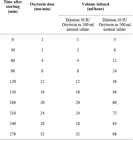
Table.2.2: RCOG guidelines for induction of labour (2001)1
Time after starting
(min)
Oxytocin dose (mu/min)
Volume infused (ml/hour)
Dilution 30 IU Oxytocin in 500 ml
normal saline
Dilution 10 IU Oxytocin in 500 ml
normal saline
0 1 1 3
30 2 2 6
60 4 4 12
90 8 8 24
120 12 12 36
150 16 16 48
180 20 20 60
210 24 24 72
240 28 28 84
MATERIALS AND METHODS
Selection of cases
This comparative study done to compare the efficacy of oral
mifepristone and endocervical PGE2 gel as preinduction cervical ripening
agents in term gestation and prolonged pregnancies was done in
uncomplicated antenatal women who had clear indication for induction of
labour, admitted in antenatal ward and labour ward at Government Kilpauk
Medical Hospital, Chennai. 100 antenatal women were selected for study 50
women received oral mifepristone 200mg and 50 women received
endocervical PGE2 gel 0.5mg.
This comparative study was done after getting clearance from ethical
committee of Government Kilpauk medical college, Chennai.
Inclusion Criteria
1. Singleton pregnancy in cephalic presentation.
2. Post dated uncomplicated pregnancy.
3. Term uncomplicated pregnancies with unfavourable cervix. (Bishop
-score < 4)
5. Congenitally anomalous babies.
6. Term or post term pregnancies with no contraindications for vaginal
delivery.
7. No contraindications for prostaglandins or mifepristone.
8. Primigravida less than 35 years and uncomplicated multigravida up to
three pregnancies.
9. Intact membranes during the time of induction.
Exclusion Criteria
1. Premature rupture of membranes.
2. Malpresentations.
3. Cephalopelvic disproportion.
4. Bad obstetric history or history of previous abortions.
5. Previous history of caesarean section or any uterine surgery.
6. Associated medical complications.
7. Multiple pregnancy.
8. Elderly primigravida (age > 35 years).
9. Oligohydramnios.
10.Rh Negative mother.
11.Placental complications like abruption or placenta praevia.
12.Abnormal fetal heart rate patterns.
14.Parity > 3
15.Active herpes infection.
16.Contra indication for prostaglandins.
17.Chorioamnionitis
18.Any febrile morbidity.
On admission, a detailed history, and complete general and obstetric
examination was carried out. Vaginal examination was done under strict
aseptic precautions and the cervical status, fetal station were assessed.
Gestational age calculated by Naegle’s rule and a routine obstetric scan for
fetal maturity and well-being was done. Once the inclusion criteria were
fulfilled and cephalopelvicdisproportion was ruled out, the patient was
prepared and transferred to the labour ward. Indication for induction was
noted after reaffirming that there was no contraindication for induction.
Informed Consent
A detailed written informed consent was obtained from the participant
and her relatives. The following were addressed in the consent form.
Indication for induction of labour, drug to be administered with its dosage
and mode of administration, side effect of the drug, risks associated with the
administration of these drugs and if complications arise, alternative mode of
Treatment Schedule
Group – I
50 pregnant women were given tablet mifepristone 200mg orally on
day1. They were observed for maternal vitals, uterine activity bleeding or
draining pv and fetal heart rate. After the wait period of 24 hours or when the
Bishop score was ≥ 6, when the cervical dilatation was > 2cm, or when the
membranes ruptured or when the patient was well in labour whichever is
earlier labour was accelerated with oxytocin drip.
Group – II
50 pregnant women pregnant were instilled endocervical PGE2 gel
0.5mg on day 1. They were observed for maternal vitals,uterine
activity,bleeding or draining pv and fetal heart rate. After the wait period of
6 hours or when the Bishop score was ≥ 6, when the cervical dilatation was
>2cm, or when the membranes ruptured or when the patient was well in
labour whichever is earlier labour was accelerated with oxytocin drip.
Monitoring of the patients
Maternal vitals, uterine activity and fetal heart rate were monitored
clinically. Partogram was maintained for all patients and used to record all
membranes was done. If membranes not ruptured ARM was done at 3cm
cervical dilatation. Pervaginal examination was done if there was rupture of
membranes or once in 2 hours in active phase of labour. The pulse rate,
blood pressure, temperature and urine output were recorded . Delivery
particulars duration of each stage of labour blood loss at third stage of labour
and baby particulars were recorded.Mother and baby were observed for
postnatal complications if any.
Data were analysed and all the values were expressed as mean ±
standard deviation or as percentages. Statistical comparison were performed
by students paired and unpaired t-test and chi-square test. Statistically
significant difference (P<0.05).
The efficacy was assessed by the following criteria:
1. Favourability of Bishop score at 24 hrs.
2. The need of oxytocin for augmentation.
3. Duration of I, II and III stage of labour and blood loss.
4. Drug administration to delivery interval.
5. The mode of delivery.
6. Cesarean section rate.
7. The 5 minute Apgar score, neonatal complications and incidence of
neonatal mortality.
Success of induction was assessed by the following criteria:
1. Patients who delivered vaginally within 48 hours of the start of
induction.
2. Bishop score of ≥ 6 at the end of 24 hours
Failure of induction was assessed by the following criteria:
1. Patients who delivered vaginally after 48 hours of start of induction.
ANALYSIS OF THE RESULTS
AGE AND PARITY DISTRIBUTION
Group - I Group - II
Age group
(Years) Primi Multi Primi Multi
≤ 20 8 (16 %) 1 (2 %) 8 (16 %) 2 (4 %)
21 – 29 17 (34 %) 21 (42 %) 18 (36 %) 19 (38 %)
≥ 30 1 (2 %) 2 (4 %) 1 (2 %) 2 (4 %)
Total 26 (52 %) 24 (48 %) 27 (54 %) 23 (46 %)
Age and parity distribution of women included in this study were
INDICATION FOR INDUCTION OF LABOUR
Indication Group - I Group - II
Prolonged Pregnancy 49 (98 %) 49 (98 %)
IUD 0 1 (2 %)
Congenital anomaly 1 (2%) 0
Total 50 (100 %) 23 (46 %)
The major indication for induction was prolonged pregnancy 49 (98 %)
and 1 (2 %) was induced for congenital anomaly in term baby in PGE2 gel
group
BISHOP SCORE AT THE START OF STUDY
Group – I Group – II
Score
Primi Multi Primi Multi
0 1 (2 %) - - -
1 7 (14 %) 1 (2 %) 8 (16 %) 5 (10 %)
2 16 (32 %) 21 (42 %) 18 (36 %) 13 (26 %)
All the mothers in both groups had initial Bishop Score of 0 to 3 before
preinduction cervical ripening.
FAVOURABILITY OF BISHOP SCORE
Group – I Group – II
Score at
augmen
tation Primi Multi Total Primi Multi Total
< 6 5 (10 %) 0 5 (10%) 13 (26 %) 9 (18 %) 22 (44%)
FAVOURABILITY OF BISHOP SCORE
Group Mean Std.
Deviation
Std. Error mean
Group – 1 1.8800 0.55842 0.07897
Bishop score start
Group – II 1.8600 0.60643 0.08576
Group – 1 6.8800 1.46580 0.20730
Bishop score at
Augmentation Group – II 5.5000 2.29685 0.32482
Group – 1 5.0000 1.55183 0.21946
Bishop score difference
Group – II 3.6400 2.14533 0.30340
Mean increase in Bishop score in mifepristone group is 5 whereas 3.6
in PGE2 gel group.
• P - Value for Bishop score at start 0.864 which is not significant.
• P - Value for Bishop score at augmentation 0.001 which is significant.
AUGMENTATION WITH OXYTOCIN
Group - I Group - II
Augmentation
Primi Multi Total Primi Multi Total
Required 20
(40 %) 13 (26 %) 33 (66%) 19 (38 %) 20 (40 %) 39 (78%)
Not required 6
(12 %) 11 (22 %) 17 (34%) 8 (16 %) 3 (6 %) 11 (22%)
In the mifepristone group among the 6 primigravida who were not in
need of oxtocin augmentation 4 (8%) had vaginal delivery within 24 hours of
oral mifepristone administration. Shortest drug administration to delivery
interval was 12 hours and 5 minutes. Among the 11 multigravida who were
not in need of oxytocin augmentation in the mifepristone group 9 (18%) had
delivery within 10 hours. Shortest drug administration to delivery interval
was 5 hours 54 minutes.
Whereas in PGE2 gel group 11 antenatal women which includes 8
primigravida and 3 multigravida who were not in need of oxytocin
augmentation were those delivered by cesarean section. In other words in
PGE2 gel group all women who had vaginal delivery were in need of
MEAN DURATION OF LABOUR
Group Mean Std. Deviation Std. Error
mean
P value
Group – 1 6.8867 2.12457 0.31671 Duration of 1st
Stage (Hour) Group – 2 6.8618 1.41495 0.22954 0.951 (NS)
Group – 1 22.4222 5.19829 0.77492 Duration of 2nd
Stage (Mints) Group – 2 26.9474 6.40501 1.03903 0.001 (S)
Group – 1 4.0659 1.20309 0.18137 Duration of 3rd
Stage (Mints) Group – 2 5.4408 1.30596 0.21185
0.000 (S)
Group -1 18.7341 10.04693 1.48134 DD intervial
Group -2 11.4784 3.85563 0.62547
Duration of II and III stage of labour were shorter in mifepristone
group with statistical significance. Duration of I stage \shorter in PGE2 gel
group which is not statistically significant Drug administration to delivery
interval shorter with PGE2 gel group with statistical significance.
Group –I ‘P’ Value Group –II ‘P’ Value
I Stage 0.001 (Significant ) 0.609 (Not Significant)
11 Stage 0.192 (Not Significant) 0.655 (Not Significant)
III Stage 0.005 (Significant) 0.169(Not Significant)
DD interval 0.031 (Significant) 0.098(Not Significant)
Statistically significant shorter duration of I and III stage of labour in
multigravda in miferpristone group whereas no statistical difference in
duration of labour among primigravida and multigravida in PGE2 gel group.
Group - I Group - II
Stages of Labour
Primi Multi Primi Multi
I – Stage (hours) 7.8542 5.7810 6.9816 6.7421
II – Stage (Minutes) 23.3750 21.3333 27.4211 26.4737
III – Stage (Minutes) 4.5435 3.5429 5.7342 5.1474
Drug administration to delivery interval
(hours)
MODE OF DELIVERY
Group - I Group - II
Mode of Delivery
Primi Multi Total Primi Multi Total
Labour naturale 22
(44 %) 21 (42 %) 43 (86%) 18 (36 %) 18 (36 %) 36 (72%) Outlet forceps delivery 1
(2 %) -
1
(2%) -
1 (2 %)
1 (2 %)
LSCS 3
(6 %) 3 (6%) 6 (12%) 9 (18 %) 3 (6 %) 12 (24 %) Spontaneous
expulsion of fetus - - -
1
(2 %) -
1 (2 %)
Total 26
(52 %) 24 (48 %) 50 (100%) 27 (54 %) 23 (46 %) 50 (100 %)
Cesarean section rate was higher in PGE2 gel group which was 24 %
INDICATION FOR LSCS
Group - I Group - II
Indication for LSCS
Primi Multi Primi Multi
Failed induction 1 (2 %) - 3 (6 %) 1 (2%)
Fetal distress 2 (4 %) 3 (6 %) 6 (12%) 2 (4%)
Total 3 (6 %) 3 (6 %) 9 (18 %) 3 (6 %)
In the mifepristione group 3 (6%) primigravida were delivered by
cesarean section of which 1 (2%) was done for failed induction and 2 (4%)
were done for fetal distress whereas in multigravida 3 (6%) were delivered by
cesarean section for fetal distress.
In the PGE2 gel group among 9 (18%) primigravida delivered by
cesarean 3 (6%) were done for failed induction and 6 (12%) were done for
fetal distress whereas in multigravida 1(2%) were done for failed induction
MEAN BLOOD LOSS
Blood Loss Group - I Group - II
Mean Blood loss (ml) 248 368
Standard Deviation 160.66190 222.63725
Standard Error mean 22.72102 31.48566
P value : 0.03 (significant)
Mean blood loss in mifepristone group was less when compared to
PGE2 gel In PGE2 gel group 1 (2%) Primigravida had atonic PPH – blood
MATERNAL COMPLICATIONS
Maternal Complications Group - I Group - II
Fever 2 (4 %) 5 (10 %)
GI symptoms 3 (6 %) 3 (6 %)
Abdominal cramps 4 (8 %) -
Uterine contractile
abnormalities - -
PP H - 1 (2 %)
Puerperal sepsis - -
NEONATAL COMPLICATIONS
Neonatal Complications Group - I Group - II
Respiratory distress 2 (4 %) 3 (6 %)
Meconium aspiration
syndrome 2 (4 %) 5 (10 %)
Transient tachypnoea of
newborn 1 (2 %) -
APGAR <7 7 (14 %) 7 (14 %)
NICU admission was 18% in PGE2 gel as compared to 10% in
mifepristone group. In PGE2 gel group one neonate was admitted for low
birth weight.
Apgar Score at 1 minute and 5 minute were similar in both groups. 7
(14%) neonates in each group had Apgar Score < 7 at 5 minutes following
DISCUSSION
Table 1 Sl.
No. Study Year Dosage Schedule Control
Wait period
1. Wing DA at al 2002 (180)
200mg of mifepristone oral dose followed by intravaginal misoprostol 25 microgr ams every 4th hourly or IV
oxytocin.17
Placebo 24 Hrs
2. LiL et al 1996
150 or 200mg mifepristone in the 1st 2 or 3 days & on 4th day misoprostol was added
successively in 100-300 micro gram dosage.14
- 3 Days
3. SuH et al
1996
(124)
50mg mifepristone 12th hourly for 2 days followed by
prostaglandin or oxytocin.15
- 48 Hrs
4. Frydam R et al 1992 (120)
200mg mifepristone on day 1 and 2 followed by
augmentation with prostaglandin on day 4.10
Placebo 4 Days
5. Giacolone PL et al 1992 400mg of mifepristone as a
single single oral dose.16 Placebo 48 Hrs
6. Elliot et al 1998 (83)
50-200mg mifepristone as a
single oral dose.19 Placebo
24Hrs interval
for 72 hrs
7. Padayachi T et al 1989 400mg mifepristone as a single
oral dose.20 - 72 Hrs
8. This study 2010 200mg mifepristone as a single
In this study mifepristone given as 200mg single dose orally and
observation period of 24 hours similar to the wing DA etal and Elliot et al
study in which mifepristone were compared with placebo whereas PGE2 gel
in this study.
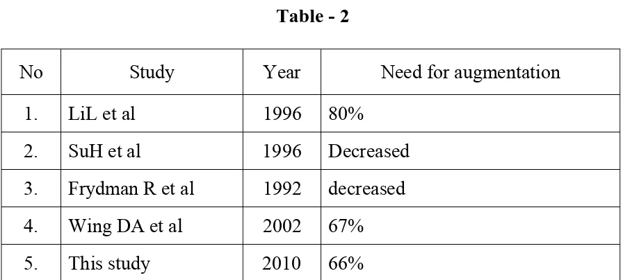
Table - 2
No Study Year Need for augmentation
1. LiL et al 1996 80%
2. SuH et al 1996 Decreased
3. Frydman R et al 1992 decreased
4. Wing DA et al 2002 67%
5. This study 2010 66%
In this study 66% required oxytocin, which was consistent with prior
studies.
In this study mean duration of first stage was less than 8 hours and
second stage duration was less than 30 minutes. These results were
consistent with WHO standards.
In this study 36 (72%) women 32% primigravida and 40%
multigravida delivered vaginally within 24 hours and totally 44 (88%)
women 46% primigravida and 42% multigravida delivered vaginally within
[image:75.595.80.528.205.407.2]Table - 3
Sl.
No. Study Year Incidence of vaginal delivery
1. LiL et al 1996 80.88%
2. SuH et al 1996 22.58% (vs 4.84% of control group)
3. Giacalone PL et al 1992 80.5%
4. Wing DA et al 2002 87.5%
5. This study 2010 88%
In this study vaginal delivery rate was 88% (46% primigravida and
42% multigraavida) the results were consistent with above mentioned studies.
In this study intrapartum complications like hypertonus, tachysystole
or hyperstimulation were not encountered, which was consistent with Wing
DA et al study. Meconium passage was encountered in 4% and NICU
admission was 10%.
In this study the success of induction was vaginal delivery within 48
hours. Success rate was 88% which was consistent with 87.5% success rate
[image:76.595.82.528.114.373.2]