0022-538X/95/$04.0010
Copyrightq1995, American Society for Microbiology
Functional Interactions between Herpes Simplex Virus
Immediate-Early Proteins during Infection: Gene
Expression as a Consequence of ICP27
and Different Domains of ICP4
LORNA A. SAMANIEGO, ARTHUR L. WEBB,ANDNEAL A. DELUCA*
Department of Molecular Genetics and Biochemistry, University of Pittsburgh School of Medicine, Pittsburgh, Pennsylvania 15261
Received 17 April 1995/Accepted 20 June 1995
Two of the five immediate-early gene products, ICP4 and ICP27, expressed by herpes simplex virus type 1 have profound effects on viral gene expression and are absolutely essential for virus replication. Functional interactions between ICP4 and ICP27 may contribute to establishing the program of viral gene expression that ensues during lytic infection. To evaluate this possibility, viral mutants simultaneously deleted for ICP27 and defined functional domains of ICP4 were constructed. These mutant viruses allowed a comparison of gene expression as a function of different domains of ICP4 in the presence and absence of ICP27. Gene expression in the absence of both ICP4 and ICP27 was also examined. The results of this study demonstrate a clear involvement for ICP27 in the induction of early genes, in addition to its known role in enhancing late gene expression during viral infection. In the absence of both ICP4 and ICP27, viral early gene expression, as measured by the accumulation of thymidine kinase and ICP6 messages was dramatically reduced relative to the amounts of these messages seen in the absence of only ICP4. Therefore, elevated levels of early gene expression as a consequence of ICP27 occurred in the absence of any ICP4 activity. Evidence is also presented regarding the modulation of the ICP4 repression function by ICP27. When synthesized in the absence of ICP27, a mutant ICP4 protein was impaired in its ability to repress transcription from the L/ST promoter in the context of viral infection and in vitro. This defect correlated with the loss of the ability of this mutant protein to bind to its recognition sequence when produced in infected cells in the absence of ICP27. These observations indicate that ICP27 can regulate the activity of at least one domain of the ICP4 protein as well as contribute to elevated early gene expression independently of ICP4.
During lytic infection, herpes simplex virus type 1 (HSV-1) gene expression occurs in a regulated fashion commencing with the synthesis of the immediate-early gene products, fol-lowed by the synthesis of the early and late classes of gene products (24, 25). There are five immediate-early (IE) proteins synthesized by HSV-1, namely, infected cell polypeptides (ICP) 0, 4, 22, 27, and 47. Except for ICP47, these proteins are known to affect the expression of viral genes (reviewed in reference 56). The genes encoding ICP22 and ICP47 can be deleted from the viral genome with no effect on the growth and viability of the virus in most cell types (34, 48, 62). Mutant HSV-1 deleted for the ICP0 gene grows poorly in cell culture, particularly at low multiplicities of infection (MOIs), but re-mains viable (58, 73). In transient expression assays, the ICP0 protein acts as a promiscuous transactivator of HSV-1 as well as heterologous promoters (11, 14, 42, 50, 63). On the other hand, studies of viral mutants carrying deletions or tempera-ture-sensitive lesions in ICP4 and ICP27 demonstrated that these proteins provide essential functions during productive infection (5, 10, 35, 49, 57).
ICP4 is a 175-kDa phosphoprotein (3, 47) that directly acts as a transcriptional regulatory protein. It is required for the induction of early and late genes and acts as a repressor of its own expression and that of several other viral genes (4, 6, 10,
43, 44, 49). The ICP4 protein consists of several modular do-mains that encode specific functions including those for DNA binding, nuclear localization, dimerization, and transcriptional activation (8, 45, 46, 66). The protein has sequence-specific DNA-binding ability, with preference for the consensus
ATCGTCN4YGCRC present in its own promoter as well as
other HSV-1 gene promoters (13, 30, 38, 40). ICP4 represses promoters containing a strong ICP4 binding site located near the start site of transcription (6, 20, 37, 44, 55). On such promoters, ICP4 forms a tripartite complex with the basal transcription factors TATA-binding protein (TBP) and TFIIB, and this activity requires amino acid residues in the N-terminal half of ICP4 (69). While this region of the protein is important for activation, a quantitative and genetic correlation has been established between the ability of ICP4 to form tripartite com-plexes and its ability to repress transcription (19). Unlike re-pression, ICP4 does not require specifically located binding sites to activate transcription (12, 17, 68). Multiple contacts with cellular transcription factors are probably required for ICP4 to activate transcription since TBP will not suffice in place of TFIID for activation by ICP4 in vitro (17).
The activity of the 63-kDa ICP27 protein is associated pri-marily with the regulation of late gene expression (57). ICP27 is required for efficient DNA replication and appears to play a role in the shutoff of host protein synthesis (35, 52, 57). A significant component of the regulatory effects of ICP27 is mediated posttranscriptionally at the level of mRNA process-ing (22, 23, 61, 70). Repression by ICP27 is observed in target genes containing introns, while activation appears to correlate
* Corresponding author. Mailing address: E1257 Biomedical Sci-ence Tower, Department of Molecular Genetics and Biochemistry, University of Pittsburgh School of Medicine, Pittsburgh, PA 15261. Phone: (412) 648-9947. Fax: (412) 624-1401.
5705
on November 9, 2019 by guest
http://jvi.asm.org/
with different polyadenylation signals (61). ICP27 has also been shown to affect the transcription of viral genes during infection (52). Whether these effects can be ascribed to ICP27 directly or indirectly remains to be determined.
It is likely that the program of gene expression that ensues during infection results, in part, from the combined action of and possible interaction between ICP4 and ICP27, as well as other IE proteins. This notion is supported by the results of transient expression studies in which the responses of different target HSV-1 promoters to various combinations of ICP0, ICP4, and ICP27 were determined. Under certain conditions, ICP4 and ICP0 act synergistically in the activation of early genes (11, 14, 42, 43, 63). Depending on the target promoter, ICP27 inhibits or further enhances the ability of ICP4 and/or ICP0 to activate some early and late genes, respectively (51, 63, 74). ICP27 has been implicated in the posttranscriptional pro-cessing of the ICP0 message (22, 23) and posttranslational modification of ICP4 (36, 51, 74), both of which present po-tential mechanisms for the modulation of ICP0 and ICP4 ac-tivities by ICP27. Subcellular localization of the proteins in the presence of wild-type (wt) and mutant forms of ICP0, ICP4, and ICP27 provides another perspective in which to analyze the interplay between these IE gene products. The function of these proteins in transcriptional regulation requires their proper import into the nucleus, and this process can serve as a possible target for controlling their activities. ICP4 appears to have an enhancing effect on the nuclear localization of ICP0, whereas ICP27 inhibits the nuclear localization of both ICP0 and ICP4 (78, 79). In cells cotransfected with ICP0 and ICP4 genes, both proteins colocalize in punctate granules distributed in the nucleus, suggesting a possible direct interaction between these two IE proteins (39). The functional significance of the observed colocalization is unknown.
While the results of transient expression assays clearly dem-onstrate potential functional interactions among the HSV-1 IE proteins, very little is known about the consequences of such interactions on viral gene expression in the context of the virus. The studies described in this report were designed to examine how potential interactions between ICP4 and ICP27 affect gene expression during virus infection. Viral mutants contain-ing a deletion of the ICP27 gene, combined with deletions in defined functional domains of ICP4, were constructed and characterized for expression of IE, early, and late genes. It was determined that ICP27 affects early gene expression in the absence of any ICP4 activity and that ICP27 can regulate the activity of at least one domain of the ICP4 protein by affecting its ability to bind to DNA.
MATERIALS AND METHODS
Virus and cells.Vero cells and the complementing cell lines E5, E8, and E26 were maintained as previously described (4, 10). E5 cells were previously de-scribed (5), and E8 and E26 cells are dede-scribed herein. Viruses carrying muta-tions in ICP4 alone or in both ICP4 and ICP27 were propagated on the appro-priate cell lines. The ICP4 mutant viruses, d8-10 (66), n208 (8), nd8-10 (69), and
d120 (5) were previously described, as was the ICP27 mutant virus, 5dl1.2 (35). Transfection and construction of cell lines.The construction of transformed cell lines using pSV2neo and cloned HSV genes was performed as previously described (5, 72). Plasmid pSV2neo encodes the neomycin resistance gene from
Escherichia coli under the control of the simian virus 40 early promoter and
confers resistance to the antibiotic G418 (72). Approximately 43106
Vero cells were distributed on two 85-mm petri dishes 24 h prior to transfection. Three hours prior to transfection the medium was aspirated and 10 ml of fresh medium containing 10% fetal bovine serum was added. For transfection, calcium phos-phate precipitates were prepared essentially as described by Graham and van der Eb (16). One microgram of pSV2neo, 5mg of both pK1-2 (7) and pKHX-BH (2), encoding ICP4 and ICP27, respectively, and 30mg of salmon testis DNA were suspended in 1.0 ml of 23transfection buffer. The composition of transfection buffer was as previously given (16). One milliliter of 250 mM CaCl2was added
dropwise, and the precipitate was allowed to form at room temperature for 20
min. One milliliter of suspension was then dispensed to each of the two petri dishes of Vero cells, and the cells were incubated at 378C for 4 h. Following this incubation, the medium was removed and 3 ml of 15% glycerol in 13 transfec-tion buffer was added to each plate. The plates were incubated at 378C for 2 min, after which the glycerol was removed, the cells were washed with isotonic saline, and medium containing 10% fetal bovine serum was added. The cultures were incubated for 40 h at 378C; then, the cells were trypsinized and suspended in medium containing 700mg of G418 per ml. The single cell suspension was plated into new 85-mm petri dishes at a density of 53103cells per cm2and incubated
at 378C. After 3 to 5 days the G418 concentration was lowered to 250mg/ml, depending on the extent of cell death. G418-resistant colonies were isolated 14 days after G418 selection. Individual G418-resistant colonies were expanded in the presence of 250mg of G418 per ml by standard cell culture procedures.
Preparation of viral DNA and Southern blot analysis.Approximately 23106
cells plated in a 60-mm dish were infected at an MOI of 10 PFU per cell, harvested at 18 h postinfection, and lysed in buffer containing 0.6% sodium dodecyl sulfate (SDS) and 400mg of proteinase K per ml for 4 h at 378C. Following RNase treatment, phenol-chloroform extractions, and ethanol precip-itation, equal amounts of total DNAs in all samples were cleaved with the indicated restriction enzyme, fractionated by agarose gel electrophoresis, and transferred to nitrocellulose as previously described (71). Hybridization and the subsequent washing of the membrane were performed as previously described (60). Plasmids pKC-1 (65), pKBY (cloned wt BamHI-Y fragment), and pKHX-BH (2) were used as probes to verify the presence of mutations intro-duced into the viral genome. The probes were labelled with32P by using the Nick
Translation System (Gibco-BRL, Life Technologies) according to instructions provided by the manufacturer.
Isolation of infected-cell RNA and Northern (RNA) blot analysis. Approxi-mately 53106cells seeded into 100-mm plates were infected at an MOI of 10
PFU per cell and harvested at the indicated times postinfection. For infections performed in the presence of cycloheximide, the medium was supplemented with 100mg of cycloheximide per ml 1 h prior to and during infection. Total cellular RNA was extracted by using the Ultraspec RNA Isolation System (Biotecx Laboratories, Inc.) as suggested by the manufacturer. Isolated RNA was resus-pended in 20ml of diethylpyrocarbonate-treated water, and its concentration was determined spectrophotometrically.
Unless otherwise indicated, 20mg of total infected-cell RNA was fractionated in 1.3% agarose gels containing formaldehyde (60) with constant buffer recircu-lation. The conditions for blotting, hybridization, and washing were described previously (26). Nick-translated pKC-1 (65), pW3DHS8 (58), a gel-purified 1.6-kb ScaI fragment from pKX2-bG3 (15), and a SacI-SmaI fragment from the thymidine kinase (TK) coding region were used to detect ICP4, ICP0, ICP6, and TK messages, respectively. The probes were labelled with32
P by using the Nick Translation System as described above.
Analysis of viral proteins.Viral polypeptides were extracted (33) from cells infected at an MOI of 10 PFU per cell and separated by SDS-polyacrylamide gel electrophoresis (SDS-PAGE) (32). For Western blot (immunoblot) analysis, the separated proteins were transferred to nitrocellulose by electroblotting and the blot was subsequently analyzed for the presence of ICP4 by using a 1:400 dilution of N15 antibody (64). For detection of ICP0, the protein blots were incubated in a 1:200 dilution of the antibody CO25, a polyclonal rabbit antiserum raised against purified ICP0 (generously provided by Richard J. Courtney, The Penn-sylvania State University College of Medicine, Hershey). The blots were pro-cessed with the Protoblot Western blot alkaline phosphatase system (Promega) as specified by the manufacturer. For radiolabelling of viral polypeptides, ap-proximately 63105cells plated in 30-mm dishes were incubated with 100mCi of
[35S]methionine per ml for 1 h at the indicated times postinfection. The labelled
viral proteins were extracted from the infected cells and separated by electro-phoresis as described above.
In vitro transcription and primer extension.The in vitro transcription reaction mixtures (15ml) included 4.5ml of HeLa cell nuclear extract (;60mg of protein) prepared by the method of Dignam et al. (9), 16.7mg of pL/ST plasmid (19) per ml as template DNA, and the following buffer components as previously de-scribed (20): 40 mM N-2-hydroxyethylpiperazine-N9-2-ethanesulfonic acid (HEPES; pH 7.9); 60 mM KCl; 12% glycerol; 8.3 mM MgCl2; 0.6 mM each ATP,
GTP, CTP, and UTP; 0.3 mM dithiothreitol; and 12 U of RNasin (Promega). ICP4 protein (100 to 150 ng) was added to some reaction mixtures. The reaction mixtures were incubated at 308C for 80 min, and the reactions were stopped by the addition of 85ml of 0.15 M sodium acetate (pH 5.3) with 15 mM EDTA (pH 8.0). The reaction mixtures were extracted twice with phenol, and the synthesized RNA was precipitated with ethanol prior to primer extension.
The RNA transcribed in vitro was annealed to 2 to 3 ng of a 59-end-labelled oligonucleotide primer that hybridizes to the transcript 75 bases from the initi-ation site (19). In a similar manner, 12mg of total infected-cell RNA was annealed to 10 ng of a32
P-end-labelled oligonucleotide primer containing the sequence from nucleotide157 to191 of the noncoding strand downstream of the L/ST promoter (1, 77). DNA was 59 end labelled by incubation in the presence of [g-32
P]ATP and polynucleotide kinase (New England Biolabs). Primer annealing and extension were performed as previously described by Imbalzano et al. (26). The primer extension products generated by the Moloney murine leukemia virus reverse transcriptase (Gibco-BRL) were analyzed by electrophoresis in 5% denaturing polyacrylamide gels. The positions of the
on November 9, 2019 by guest
http://jvi.asm.org/
transcription start sites were verified by running a sequencing ladder next to the primer extension products. The sequencing reactions were performed by using plasmid pKBK (cloned wt BamHI-K fragment) as template with the Sequenase version 2.0 kit (USB Technologies), as recommended by the manufacturer. pKBK contains the joint-spanning BamHI fragment in pBR325.
Gel retardation assays.The wt and mutant ICP4 proteins used for the assays were purified from infected cells harvested at 12 to 15 h postinfection as previ-ously described (27, 65, 67). Bacterial expression and purification of recombinant human TBP and TFIIB were performed as described by Kao et al. (28) and Ha et al. (21), respectively, with minor modifications (69). The 135-bp BglII-NotI fragment corresponding to nucleotide positions2110 to125 of the L/ST pro-moter was used as probe following treatment with calf intestinal alkaline phos-phatase and incubation with polynucleotide kinase and [g-32P]ATP. The binding
reactions were performed in a total volume of 30ml containing 3.03105
cpm (;1 ng) of probe and the indicated mixture of ICP4 proteins and general transcription factors. TBP (50 ng), TFIIB (500 ng), and/or ICP4 (various amounts) were added. The reaction mixtures were incubated at 308C for 40 min in buffer conditions exactly as described by Smith et al. (69) except that 80 mM KCl was used in the reactions. DNA-protein complexes were separated on native 4% polyacrylamide gels prepared in 0.53Tris-borate-EDTA buffer and were run at 200 V. After electrophoresis, the gel was dried and exposed to Kodak XAR-5 film.
RESULTS
Construction of ICP4- and ICP27-complementing cell lines
and viruses simultaneously deficient in both functions.The
purpose of this study was to examine the effect of ICP27 on viral gene expression in different ICP4 backgrounds and also to possibly correlate this effect to biochemical differences of the ICP4 proteins synthesized in viral infection, in the presence and absence of ICP27. In order to accomplish this goal, it was necessary to construct cell lines that efficiently and simulta-neously complement defects in both ICP4 and ICP27. To this
end Vero cells were cotransfected with 5mg each of pK1-2 (7),
the ICP4-expressing plasmid, and pKHX-BH (2), the
ICP27-complementing plasmid, and 1mg of pSV2neo, as described in
the Materials and Methods. G418-resistant colonies were se-lected and amplified as previously described (5).
Sixty-seven G418-resistant cell lines were obtained, and each
was seeded into 0.8-cm2wells in triplicate. Subsequently, 100
PFU of d120 (ICP4 deletion), 5dl1.2 (ICP27 deletion), and KOS (wt) were applied separately to each of the three wells for a given cell line to determine the ability of the cell lines to host the different viral mutants. All 67 colonies were efficient hosts for KOS. Thirteen cell lines supported plaques of d120, but not 5dl1.2 (ICP4 cell lines), and only four cell lines supported plaques of 5dl1.2, but not d120 (ICP27 cell lines). Surprisingly, 19 of the 67 cell lines supported plaques of both d120 and 5dl1.2 (ICP4/ICP27 cell lines). This distribution was not ex-pected and may suggest that any possible cytotoxic effects of ICP4 or ICP27 alone may be reduced because of some bio-chemical interaction between the two proteins. All 4 of the ICP27 cell lines and the 19 ICP4/ICP27 cell lines were tested in burst size assays with d120 and 5dl1.2. The data in Table 1 show that only 2 of the 23 cell lines, E8 and E66, gave substantial burst sizes of 5dl1.2. Of these, only E8 cells grew at the same rate as Vero cells. A number of quite efficient ICP4/ICP27 cell lines were obtained, as indicated by the burst sizes for both
d120 and 5dl1.2 in excess of 300 PFU per cell (Table 1). E26
cells were chosen for further study because they grew at the same rate as Vero cells and gave wt size plaques. A number of the other cell lines would have been equally useful.
The viruses d8-10, n208, and nd8-10 all contain defined ICP4 alleles and express ICP4 proteins having previously described biochemical and biological activities (8, 66, 69). These are depicted in Fig. 1B along with d120 (5). d120 contains a 4.1-kb deletion in both copies of the ICP4 gene. d8-10, n208, nd8-10, and d120 were each used to coinfect E26 cells with 5dl1.2 at an MOI of 5 PFU of each virus per cell. At 18 h postinfection, the
monolayers were harvested and viral lysates were prepared. The progeny of the coinfections were plated out for plaques on E26 cells. Multiple plaques were picked from each coinfection and were frozen and thawed in 0.5 ml of medium. Twenty microliters of each plaque isolate was screened for the ability
to grow in wells of 105E26, E5, E8, and Vero cells. With the
exception of the d8-10:5dl1.2 progeny, isolates that grew only on E26 cells were chosen for further study. For d8-10, isolates that grew only on E26 and E8 cells were chosen for further study since d8-10 does not require that ICP4 be supplied in
trans to grow.
Isolates with the proper growth characteristics were used to
infect 105E26 cells for the preparation of infected-cell DNA.
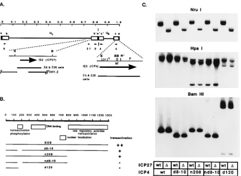
The isolated infected-cell DNA was used for restriction en-zyme analysis to verify the incorporation of the appropriate mutations in the genomes of the viral isolates. All of the mu-tations defining the ICP27 and ICP4 alleles in this study are easily assayed for by restriction enzyme and Southern blot analysis. Once the identity of the desired mutant was verified, the isolates were plaque purified an additional two times, and then a virus stock was prepared. A final Southern blot, probing for both the ICP4 and the ICP27 regions of the genome, was performed on all the multiple mutant isolates, wt virus, and the single mutant viruses used to construct the multiple mutants (Fig. 1C). The NruI digest probed with the ICP27 plasmid shows that the intended viruses possess the 1.2-kb deletion characteristic of 5dl1.2 (34). The HpaI digest probed with an ICP4-encoding plasmid (pKC1) shows the presence of the
HpaI site specifying the nonsense mutation in ICP4 at amino
acid 774 in nd8-10, n208, and their 5dl1.2 counterparts (8).
[image:3.612.315.555.89.334.2]d120 is deleted for 4.1 kb of ICP4-coding and 39sequences (5) and has the same HpaI pattern as that in d120:5dl1.2 (d120:
TABLE 1. Growth of ICP4 and ICP27 mutants on transformed cells
Cell line Initial screen resulta PFU/cell b
d120 5dl1.2
E1 4/27 1,110 150
E8c 27 ,10 740
E11d 4/27 830 750
E16 4/27 420 120
E18d 4/27 1,080 310
E20 4/27 240 360
E21d 4/27 960 320
E22 4/27 920 60
E23 4/27 490 150
E26d 4/27 810 310
E28 4/27 890 120
E29 4/27 520 80
E31 4/27 ,10 30
E36 4/27 ,10 340
E45d 4/27 550 500
E51 4/27 280 180
E53 27 ,10 240
E55d 4/27 460 550
E58 27 ,10 ,10
E57 4/27 870 220
E59d 4/27 390 380
E65 4/27 170 270
E66c 27 ,10 880
a
Efficient host for both ICP4 and ICP27 mutants (4/27) or only for the ICP27 mutant.
b
MOI, 5; incubation, 18 h. Progeny were assayed on E5 (d120) or E8 (5dl1.2) cells.
c
Considered an efficient host for ICP27 mutants.
dConsidered an efficient host for ICP4 and ICP27 mutants.
on November 9, 2019 by guest
http://jvi.asm.org/
D27). The BamHI digest probed for ICP4 is diagnostic of the 204-bp deletion defining the 8-10 mutation (66). The appro-priately shortened BamHI-Y fragment is clearly present in both d8-10 and nd8-10. The BamHI digest of d120 and d120:
D27 appears exactly as previously described (5).
The E26 cell line made it possible to obtain viruses simul-taneously deficient in ICP4 and ICP27. Total yields in viral stocks range from 100 to 500 PFU per cell depending on the
ICP4 allele. Therefore, standard stock titers in excess of 109
PFU/ml are routinely obtained. In the case of d120:D27, the
viral genome lacks the flanking homology with the DNA
in-serted in E26 cells at the 59end of ICP27 and at the 39end of
ICP4. This situation for the single mutants gives a frequency of appearance of wt virus from rescue due to growth on the cell
line of,1026. Therefore, since rescues of individual deletions
in d120:D27 should be independent events, the appearance of
wt recombinants would be ,10212. In practice, wt
recombi-nants have never been seen.
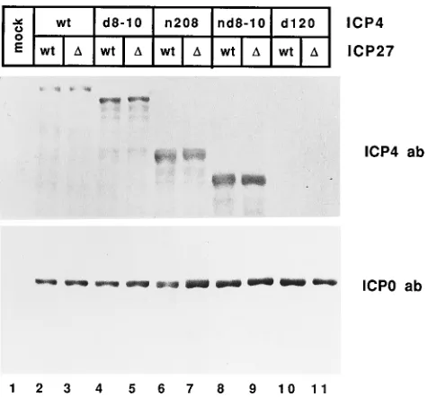
Expression of ICP4 and ICP0 proteins. Since ICP4 and
ICP0 are intimately involved in the regulation of HSV gene expression, it is likely that their levels may affect later gene expression. To determine if ICP27 affects the relative amounts of ICP4 or ICP0 in the different ICP4 backgrounds and to verify that the proper ICP4 protein was expressed in the pres-ence and abspres-ence of ICP27, Western blot analyses were per-formed on extracts from cells infected with the indicated viruses. ICP4 polypeptides of the expected sizes were observed as shown in Fig. 2. Comparison of each pair of mutant strains carrying a specific ICP4 allele in a wt or an ICP27 deletion background did not indicate any significant differences in the apparent sizes or levels of the ICP4 protein. The very small
d120 peptide (5) was not detected in this experiment. In
[image:4.612.63.554.70.428.2]addi-tion, the levels of ICP0 polypeptides for each pair of viruses appeared similarly unaffected by the absence of ICP27. These results cannot rule out subtle changes in the modification of the protein that were not resolved in this gel. A slight decrease
FIG. 1. Structures of viruses carrying deletions in ICP4 and ICP27. (A) HSV-1 genome, with the unique long (UL) and short (US) regions and the locations of ICP4,
ICP27, and ICP0 indicated (small arrows). The expanded maps of ICP4 and ICP27 are shown below with the relevant restriction sites BamHI (B), HpaI (H), and NruI (N). BamHI restriction fragments within the ICP4 region include Y, M9, and P. The short serine-rich transactivation domain deleted in d8-10 and nd8-10 is indicated (D). H*, HpaI site specifying the translational stop codon in n208 and nd8-10. The sequences used to generate the ICP4- (E5 and E26) and ICP27-complementing (E8 and E26) cell lines as well the sequence deleted from the ICP27 deletion mutant 5dl1.2 are also indicated. (B) The 175-kDa ICP4 protein is diagrammed at the top, with the numbers referring to amino acids. Important functional domains of the protein are indicated below (66). The coding sequences of mutant ICP4 proteins are shown relative to that of the wt (strain KOS). The relative transactivation by these proteins is indicated on right. (C) Viral DNAs from the different mutant viruses were isolated, cleaved with the indicated restriction enzyme, and analyzed by Southern blot analysis. The ICP4 and ICP27 alleles carried by the mutant strains are indicated at the bottom.D, deletion. The blots prepared from viral DNAs digested with NruI, HpaI, or BamHI were separately probed with the nick-translated plasmids pKHX-BH, pKC1, and pKBY, respectively.
on November 9, 2019 by guest
http://jvi.asm.org/
in the electrophoretic mobility of ICP4 in the absence of func-tional ICP27 has been noted previously (36, 51, 74). Prelimi-nary experiments indicate that all the mutant ICP4 proteins used in this study are also slightly reduced in electrophoretic mobility when synthesized in the absence of ICP27 (59). The unambiguous assessment of this observation will require the use of techniques with greater resolution.
Previous studies have described the inhibitory effect of ICP27 on the nuclear localization of ICP4 and ICP0 (78, 79). We find that, regardless of the presence or absence of ICP27, the wt and mutant ICP4 proteins localize predominantly to the nucleus of infected Vero cells as determined by immunofluo-rescence staining, in agreement with previous results (59, 79). Two of the mutants, n208 and nd8-10, do not have the se-quence (amino acids 820 to 1029) determined to be the target of the inhibition by ICP27 (79). Therefore, these observations are consistent with published observations. In the case of ICP0, diffuse nuclear staining and low levels of cytoplasmic staining were observed in cells infected with KOS. ICP0 was found to be predominantly localized to the cytoplasm of cells infected with nd8-10 and d120. ICP4 is thought to promote the nuclear import of ICP0 (78), and its absence in d120 may in part explain the cytoplasmic localization of ICP0. In agreement with the observations of Zhu et al. (78), nuclear staining of
ICP0 was observed in cells infected with 5dl1.2, nd8-10:D27,
and d120:D27 (59).
Expression of early and late proteins.As a preliminary
as-sessment of changes in viral gene expression as a function of both ICP27 and specific mutant ICP4 gene products, the polypeptide profiles of the infected cells were analyzed. Vero cells infected with the indicated mutant viruses were
pulse-labelled with [35S]methionine from 6 to 7 h postinfection, and
the labelled proteins were analyzed by SDS-PAGE and auto-radiography (Fig. 3). The presence and absence of ICP27 in the n208, nd8-10, and d120 pairs is readily seen in the polypep-tide profile. ICP27 is shut off at this time postinfection in the KOS and d8-10 pairs and is not seen in the profiles of these
mutants. Of the different ICP4 alleles tested, only the wt and
d8-10 can support viral replication well into the late phase.
This is evident from the polypeptide profile of cells infected with viruses carrying these alleles in which the early and late gene products (ICP5, ICP8, gB, and ICP25) are well repre-sented. Reduced expression of the late genes, most notably ICP5 and ICP25, can be observed in cells infected with the ICP4 mutant (n208) lacking the C-terminal half of the protein. These proteins were barely detectable or absent in nd8-10- and
d120-infected cells. This is consistent with previous
observa-tions (5, 8, 66).
[image:5.612.61.299.70.291.2]There was a clear effect of ICP27 in all the ICP4 mutant backgrounds. Comparisons of KOS and d8-10 with the corre-sponding viruses lacking ICP27 show a drastic reduction in the levels of ICP5 and ICP25, consistent with the observation that ICP27 function is important for late gene expression (57). The effect of ICP27 combined with the other ICP4 mutant back-grounds on early gene expression varied. At this level of res-olution, the effect of ICP27 is most pronounced on ICP6. In the background of viruses (nd8-10 and d120) expressing an ICP4 protein that has little or no activation function, the syn-thesis of ICP6 was dramatically reduced at this time postinfec-tion in the absence of ICP27. ICP27 had little effect on the accumulation of ICP6 in the n208 background, presumably because the activation function of n208 can compensate for the lack of ICP27 function. How ICP27 may be affecting ICP6 expression in the absence of ICP4 activation is addressed in Discussion. An important conclusion which can be drawn from
[image:5.612.321.552.73.351.2]FIG. 2. Expression of IE polypeptides in cells infected with viruses defective for ICP4 and ICP27 functions. Proteins extracted from cells at 6 h postinfection were separated on an SDS–10% polyacrylamide gel and electroblotted onto nitrocellulose. Two similar blots were separately probed with antisera (ab) against ICP4 (upper gel) and ICP0 (lower gel).D, deletion.
FIG. 3. Synthesis of viral polypeptides in cells infected with viruses defective for ICP4 and ICP27 functions. Cells were infected with the indicated viruses at an MOI of 10 PFU per cell, labelled with 100mCi of [35
S]methionine per 35-mm plate 6 to 7 h postinfection, and processed for SDS-PAGE. Labelled proteins extracted from mock-infected cells were included as a control. The positions of some viral proteins, including the mutant ICP4 proteins n208 and nd8-10, are
indicated on the right.D, deletion.
on November 9, 2019 by guest
http://jvi.asm.org/
this experiment is that ICP27 can affect the expression of genes in the absence of activation by ICP4.
Expression of IE and early messages.To further investigate
the effect of ICP27 on gene expression in the different ICP4 backgrounds, we determined the levels of accumulated mRNA transcribed from two IE genes, ICP0 and ICP4, and two early genes, ICP6 and the TK gene. Total cellular RNA was isolated from infected cells 6 h postinfection and subjected to Northern blot analysis as detailed in Materials and Methods. Replicate membranes were probed for the ICP4, ICP0, ICP6, and TK messages (Fig. 4A).
ICP27 had little effect on the accumulation of the ICP4 message in any of the ICP4 mutant backgrounds. The slight
reduction in the ICP4 message in the d8-10:D27 background
relative to the d8-10 background is not reproducible and there-fore not significant. An ICP4 message is not seen in the d120 backgrounds, because of the large deletion in the ICP4 coding sequences in these viruses. Likewise, ICP0 message abundance showed little variation as a function of ICP27. However, the
more intense and broader band in the n208:D27, nd8-10:D27,
and d120:D27 backgrounds relative to their wt ICP27
counter-parts is reproducible and may indicate some degree of
heter-ogeneity in the ICP0 messages accumulated in the absence of ICP27. Previous studies have indicated an involvement for ICP27 in the splicing of the ICP0 message (22, 23). The
lower-mobility forms of the ICP0 mRNA in n208:D27, nd8-10:D27,
and d120:D27 could represent alternatively processed
mes-sages. However, any functional significance of these observa-tions could not be explained in terms of different ICP0 protein levels since these did not appear to differ in the presence and absence of ICP27 for a given ICP4 background (Fig. 3).
The accumulation of the two early messages, ICP6 and TK analyzed in this experiment was affected similarly by the ab-sence of ICP27 in each of the ICP4 backgrounds. The different ICP4 alleles had relatively small effects on the level of ICP6 mRNA accumulation. This is consistent with the results in Fig. 3. However, it should be noted that ICP4 does have a stimu-latory effect on ICP6 transcription. The level of the ICP6 message in d120 was about fourfold lower than that in KOS-infected cells (lanes 1 and 9). Deletion of ICP27 in each of the ICP4 backgrounds resulted in relative reductions in the levels of the ICP6 message. Interestingly, the relative levels of ICP6 mRNA in KOS- and 5dl1.2-infected cells at 6 h postinfection were not reflected in the polypeptide profile in Fig. 3. The cells used to generate the samples in Fig. 3 were labelled from 6 to 7 h postinfection, and perhaps in the process of proceeding through the replication cycle ICP6 expression is rapidly being shut off at this time. The most pronounced reductions in ICP6 mRNA levels were seen with nd8-10 and d120 pairs of viruses. Surprisingly, in the absence of ICP27 in these backgrounds, the level of the ICP6 message is barely detectable (lanes 8 and 10). This is notable since ICP0 is abundantly expressed and
local-ized to the nucleus in the nd8-10:D27 and d120:D27
back-grounds. The same general trend is seen for the TK message. The only difference between TK and ICP6 is that, while ICP6 is induced approximately 3- to 4-fold by ICP4, TK is induced 30- to 40-fold. This is likely due to differences in the basal promoter strengths of the two promoters.
To more accurately evaluate the effect of ICP27 on the levels of the TK message synthesized by viruses carrying defective alleles of ICP4, a separate blot analysis in which the sensitivity of detection was increased was carried out (Fig. 4B). This was accomplished by loading about threefold more RNA in each lane and exposing the autoradiogram longer. The ratio of the
TK messages in n208 and n208:D27 (lanes 1 and 2)
approxi-mates the ratios observed for the virus pairs KOS and KOS:
D27 (Fig. 4A, lanes 1 and 2) and d8-10 and d8-10:D27 (Fig. 4A,
lanes 3 and 4). In contrast, the amounts of TK mRNA in
nd8-10:D27- and d120:D27-infected cells (Fig. 4B, lanes 4 and 6) represent 10- to 12-fold reductions from those in nd8-10-and d120-infected cells, respectively (lanes 3 nd8-10-and 5). To rule out the possibility that the differences in the levels of the TK message were due to gross variations in the amounts of input viruses, we analyzed RNA from cells infected in the presence of cycloheximide. Infections with the different viruses were performed at the same MOIs as those used with the untreated cells. In the absence of protein synthesis, the different viruses were expected to express the TK message at similar levels. As shown in Fig. 4B (lower panel), minor variations were ob-served, but these differences were not sufficient to account for the differences seen in the absence of cycloheximide.
[image:6.612.67.295.73.393.2]Expression of TK in the presence of cycloheximide is pre-sumably comparable to its expression in the absence of ICP4 and ICP27, as well as other IE functions. The results show that, under these conditions, TK is still expressed, albeit at very low levels. However, in the absence of the ICP4 activation func-tion, ICP27 has a significant effect on the levels of ICP6 and TK mRNA accumulation.
FIG. 4. Accumulation of IE and early mRNAs in cells infected with viruses defective for ICP4 and ICP27 functions. (A) Northern blot analysis of ICP4, ICP0, ICP6, and TK mRNA synthesis in Vero cells infected with the indicated viruses at an MOI of 10 PFU per cell. Ten micrograms of total infected cellular RNA prepared from cells harvested at 6 h postinfection was used per sample. (B) Expression of the TK message in the presence and absence of cycloheximide. For this analysis, 30mg of RNA was used for each sample to increase the sensitivity of detection and the blots were exposed for an extended period of time for autoradiography. Treated cells were incubated in media supplemented with 100 mg of cycloheximide per ml 1 h prior to and during infection.D, deletion.
on November 9, 2019 by guest
http://jvi.asm.org/
Effect of ICP27 on the repression function of the ICP4
pep-tides.To examine the effect of ICP27 on the repression
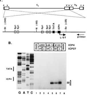
func-tion of the ICP4 peptides synthesized in the different back-grounds, we chose to determine the levels of expression of a gene, L/ST, which is normally repressed by ICP4. L/ST is a family of transcripts spanning the junction between the long and short regions of the HSV-1 genome (77). The L/STs are expressed late in infection from a promoter that achieves max-imum activity only in the absence of functional ICP4 (1a, 77). The promoter contains the VP16 recognition sequence, TA ATGARAT, characteristic of IE regulatory regions, an array of Sp1-binding sites, a TATA box, and a high-affinity ICP4 binding site (Fig. 5A), which is responsible for its repression (19).
To explore the regulatory role of ICP4 in the expression of the L/STs and the possible effect of ICP27 on this specific ICP4 function, we assayed the synthesis of the L/STs in cells infected with the mutant viruses by primer extension. Low levels of L/ST accumulation were seen in the KOS and d8-10 back-grounds at 12 h postinfection; these levels of accumulation
were not seen with theD27 counterparts of these viruses (59).
This is consistent with the previous report showing the low level accumulation of L/ST during the late phase of infection (77). In the absence of ICP4, with or without the additional
deletion of ICP27, the accumulation of L/ST was clearly evi-dent (Fig. 5B, lanes 6 and 7). The multiple bands correspond-ing to the primer extension products represent transcripts ini-tiated from three alternative transcription start sites, with the major site being the same as that previously reported (77). Repression of the L/ST promoter was observed with the virus carrying the n208 allele of ICP4 regardless of the presence or absence of ICP27 (lanes 2 and 3). As with n208, repression of the promoter was also observed with the nd8-10 virus (lane 4).
Interestingly, in the nd8-10:D27 background, repression was
relieved (lane 5). This observation suggests that ICP27 affects the ability of the nd8-10 mutant ICP4 protein to repress tran-scription of the L/ST promoter.
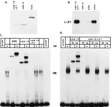
[image:7.612.158.462.77.411.2]In vitro transcription experiments using the L/ST promoter as template were conducted to determine if the differential effect on L/ST repression in the nd8-10 backgrounds was a direct reflection of altered activity of the nd8-10 protein (Fig. 6B). Previous in vitro transcription results demonstrated that KOS, n208, and nd8-10 ICP4s repressed the transcription of the L/ST promoter (19). As expected, the activity of the pro-moter was repressed by ICP4 purified from wt-infected cells (Fig. 6B). In agreement with results obtained in vivo (Fig. 5) and previous in vitro results (19), repression of L/ST was also observed with the nd8-10 ICP4. In contrast, the ICP4 protein
FIG. 5. Repression of the L/ST promoter in cells infected with viruses defective for ICP4 and ICP27 functions. (A) The genomic location of the L/ST gene is shown spanning the junction between the long (UL) and short (US) regions. The transcription control region of the gene is also shown along with the relevant promoter
elements including binding sites for Sp1, TBP (TATA box), and ICP4. Restriction sites: N, NotI; Sc, SacI; St, StuI. (B) Expression from the L/ST promoter was assayed by primer extension as detailed in Materials and Methods. RNA used for the analysis was isolated from Vero cells infected with the indicated viruses at 12 h postinfection. The sequence ladder on the left (GATC) was generated with plasmid pKBK as template. The positions of the TATA box and the ICP4 binding site are indicated on the left, and the nucleotides corresponding to the transcription initiation sites are indicated on the right.
on November 9, 2019 by guest
http://jvi.asm.org/
synthesized in nd8-10:D27-infected cells was no longer able to repress the L/ST promoter. This result indicates that, although the two viruses code for the same ICP4 protein, the mutant ICP4 protein synthesized by nd8-10 is biochemically distinct
from that expressed by nd8-10:D27.
To determine why nd8-10:D27 ICP4 was not able to repress
transcription, we compared the mutant ICP4 proteins made in
nd8-10- and nd8-10:D27-infected cells with respect to two properties of ICP4 that contribute to its repression activity: (i) the ability to bind to the ICP4 binding site and (ii) the ability to form a tripartite complex with TBP and TFIIB on the promoter. The requirement for these two activities in ICP4-mediated repression of several HSV-1 promoters has been previously reported (19). A segment of the L/ST promoter containing the ICP4 binding site and TATA box was used as probe for the gel retardation assays shown in Fig. 6C. Like the wt protein, the mutant ICP4 protein made by the nd8-10 virus binds to DNA and can participate in a tripartite complex for-mation with TBP and TFIIB, as evidenced by the forfor-mation of a more slowly migrating complex in the presence of TBP and TFIIB (Fig. 6C). On the other hand, the same protein made in
the absence of ICP27 (by the virus nd8-10:D27) is defective for
DNA binding and, consequently, the formation of tripartite complexes (Fig. 6C). While the amounts of the nd8-10 and
nd8-10:D27 proteins used in the reactions were approximately equal (Fig. 6A), we compared the binding activity of a given amount (20 ng) of the nd8-10 protein with those of increasing
amounts of the nd8-10:D27 protein to more rigorously control
for small variations in the amounts of the proteins and also to
see if nd8-10:D27 ICP4 could engage in tripartite complexes if
enough of the protein bound to DNA (Fig. 6D). Small
amounts of the nd8-10:D27 protein could be seen binding to
the probe and forming tripartite complexes when 40 ng, and, more evidently, 80 ng of ICP4 protein was included in the binding reactions. However, despite a fourfold increase in the amount of protein used in the binding reactions, the amount of
nd8-10:D27 protein observed binding to the probe was dramat-ically less than the amount of nd8-10 that bound.
[image:8.612.129.489.70.415.2]From these experiments we conclude that the nd8-10 protein can repress L/ST transcription but does not have this activity when expressed in viral infection in the absence of ICP27. This is presumably due to an as yet unknown modification of the
FIG. 6. The inability of nd8-10:D27 ICP4 protein to repress the L/ST promoter correlates with the inability to bind to DNA. (A) Western blot analysis of purified wt and mutant ICP4 proteins. wt and mutant ICP4 proteins were purified from the nuclei of infected cells by gel filtration and ion-exchange chromatography as previously described (64). Approximately 150 ng of each purified ICP4 protein was run on an SDS–9% polyacrylamide gel and transferred to nitrocellulose. The membrane was probed with the ICP4-specific antibody N15. (B) Repression of in vitro transcription from the L/ST promoter by ICP4. In vitro transcription reactions were conducted with equal amounts of wt and mutant ICP4 proteins with plasmid pL/ST as template. (C) Inhibitory effect of ICP27 on the ability of the mutant ICP4 protein nd8-10 to bind to DNA. The L/ST promoter sequence from2110 to125 was used as probe for the gel retardation assays. Twenty nanograms of purified wt or mutant ICP4 protein, together with recombinant TBP (rTBP) and rTFIIB, was incubated with the probe in the indicated combination. Band DB, complex containing rTBP and rTFIIB bound to DNA. (D) The assay was done as for panel C with the indicated amounts of the nd8-10 and nd8-10:D27 proteins.
on November 9, 2019 by guest
http://jvi.asm.org/
ICP4 protein that directly or indirectly involves ICP27 and renders the nd8-10 protein deficient in DNA binding.
DISCUSSION
The goal of this study was to evaluate the consequences of potential functional interactions between ICP4 and ICP27 for viral gene expression during viral infection. The regulatory effects of ICP27 were examined in different virus backgrounds bearing specific lesions in the ICP4 gene. To accomplish this, viral mutants simultaneously deleted for ICP27 and defined functional domains of ICP4 were constructed. This allowed us to compare the levels of gene expression as a function of different domains of ICP4 in the presence and absence of ICP27. Gene expression in the complete absence of ICP4 and ICP27 was also examined.
The results described here demonstrate a clear involvement of ICP27 in the activation of early genes in addition to its previously reported enhancing function for late gene expres-sion during viral infection (35, 36, 57). In the complete absence of ICP4 and ICP27, viral early gene expression as measured by the accumulation of TK and ICP6 messages was dramatically reduced relative to the amounts of these messages seen in the absence of only ICP4. Evidence regarding the modulatory ef-fect of ICP27 on the repression function of ICP4 was also presented. When synthesized in the absence of ICP27, a mu-tant ICP4 protein that normally represses but does not activate transcription was impaired in its ability to repress transcrip-tion. This defect correlated with a severe reduction in the ability of the mutant ICP4 protein to bind to its recognition sequence when synthesized in infected cells in the absence of ICP27.
Early regulatory functions of ICP27.The results of transient
expression assays indicate a modulatory role for ICP27 in the regulation of viral gene expression (35, 36, 51, 63). ICP27 was shown to modulate the regulatory effects of ICP4 and/or ICP0. ICP27 by itself had little or no detectable activity in affecting expression of the early and late promoters tested, with the possible exception of the gB promoter (51). In the context of a virus infection in which ICP4 is not activating viral gene ex-pression, the effect of deleting ICP27 amounts to about 10-fold decreases in the levels of TK and ICP6 mRNAs without af-fecting ICP0 expression (Fig. 4A). This result indicates that, in the absence of activation by ICP4, the level of early gene expression is influenced by the presence of ICP27.
Whether the elevated levels of early messages are mediated by ICP27 directly, possibly as a result of differences in mRNA processing, or by enhancing activation by ICP0 is not clear. Expression of TK in the absence of both ICP4 and ICP27 approaches that seen in the absence of prior viral protein synthesis (Fig. 4B). Therefore, the presence of ICP0 alone did not appear to greatly contribute to the expression of TK in this background, despite the observations that ICP0 is abundantly expressed and, as previously reported (78), localized in the nucleus. It is possible that ICP0 synthesized in the absence of ICP27 is not appropriately modified to efficiently activate gene expression in a viral infection. In the presence of ICP27, a significant portion of ICP0 remains in the cytoplasm (59, 78). Therefore, it remains possible that, in the presence of ICP27 in an ICP4-deficient background, early gene expression is low because a large amount of ICP0 remains in the cytoplasm and that, in the absence of ICP27, the nuclear form of ICP0 is not properly modified. Fine structural analysis of the ICP0 protein synthesized in the presence and absence of ICP27 will be needed to help resolve this question. Alternatively, ICP27 may affect the activity of cellular transcription factors or other viral
IE proteins, such as ICP22. ICP22 has been shown to affect the phosphorylation of cellular RNA polymerase II (53). However, since IE transcription was unaffected by ICP27, the possible involvement with cellular factors is less likely.
On the basis of the studies in this report, a modulatory effect of ICP27 on the activation function of ICP4 could not be ascribed. In each of the different ICP4 activation backgrounds, the elimination of ICP27 resulted in decreases in the amounts of TK and ICP6 messages and proteins (Fig. 3 and 4A). How-ever, since this effect was observed in the d120 background, any effects on ICP4 by ICP27 are masked by the direct or modu-latory effect of ICP27 on other viral or cellular proteins.
It is clear that ICP27 affects the levels of viral early gene expression in the absence of ICP4. Previous reports have shown that ICP27 mutants are delayed in the synthesis of viral early genes (35, 36). This observation could be a manifestation of the effect reported here. We propose that this effect is independent of ICP4 and either that ICP27 acts directly to enhance early gene expression or that it results in the modifi-cation of an IE protein, possibly ICP0, which is involved in early gene activation. Additional viral mutants, simultaneously deficient in other sets of IE genes will be needed to resolve these questions.
Modulation of ICP4-mediated repression by ICP27. The
data in this study did not reveal an effect of ICP27 on the activation function of ICP4 during viral infection. However, ICP27 was found to affect the ability of an ICP4 protein to repress transcription. The mutant ICP4 protein, nd8-10, was found to be an effective repressor of the L/ST promoter, both in vivo and in vitro, only when synthesized in the presence of ICP27 (Fig. 5B and 6B). ICP4 represses transcription by bind-ing to an ICP4 recognition site located near the start site (31). Repression also involves the formation of ICP4-TBP-TFIIB complexes (69) on the TATA box and ICP4 binding site of repressed promoters (19). The L/ST promoter contains an ICP4 binding site located near its transcription start site (1a, 77). The wt and nd8-10 protein can bind to this site, form tripartite complexes, and repress transcription (Fig. 5 and 6). The inability of the nd8-10 protein synthesized in the absence of ICP27 to bind to DNA and repress transcription suggests that it is biochemically distinct from the same protein made in a wt ICP27 background. This result supports the hypothesis that ICP27 is involved in the posttranslational modification of ICP4 (36, 51, 52) and demonstrates that such modifications may have physiological consequences.
During infection, ICP4 exists in multiple forms with slight variations in electrophoretic mobilities, presumably as a result of differential phosphorylation (75). ICP27 has been shown to subtly affect the electrophoretic mobility of the ICP4 protein (36, 51, 52) such that, in the absence of ICP27, there is a greater representation of the more slowly migrating forms of the ICP4, suggesting a greater extent of phosphorylation. In this study, we were not able to observe these multiple ICP4 species, and this may be due to the limited resolving capabil-ities of our gel system. Finer electrophoretic techniques will be necessary to unambiguously observe such changes. The obser-vations of Michael et al. (38) suggest that the different phos-phorylated species of ICP4 vary in their abilities to interact with DNA. The DNA binding domain of ICP4 produced in E.
coli is sufficient to bind to DNA (76). This domain is
presum-ably unphosphorylated. The nd8-10 protein produced in in-fected cells is phosphorylated (1). Perhaps, increased phos-phorylation of the nd8-10 protein in the absence of ICP27 reduces its ability to bind to DNA and hence repress transcrip-tion. This possibility is currently being tested.
The effect of ICP27 on the repression function of ICP4 was
on November 9, 2019 by guest
http://jvi.asm.org/
observed with the nd8-10 allele but not with the n208 (Fig. 5B) allele or the d8-10 and ICP4 alleles (59). One potential reason for this is that, with the exception of nd8-10, all the other ICP4 proteins possess considerable activation function. In the pres-ence of the activation function of ICP4 supplied by wt virus, the ICP4 promoter is still down regulated late in infection through a mechanism independent of the ICP4 binding at the start of transcription (54). In addition, other IE promoters of HSV are less active late in infection and do not possess ICP4 binding sites near their transcriptional start site. Therefore, the activity of IE promoters is reduced late in infection possibly as a consequence of the activation function of ICP4. This presum-ably does not occur in the nd8-10 background, which would then repress transcription solely through the binding-site mechanism.
It is possible that the regulation of the repression function of ICP4 may be of little consequence in lytic infection since ICP4 is activating transcription. It is interesting to note that wt ICP4 isolated from early and late times postinfection or isolated from ICP27 deletion mutant-infected cells will all activate tran-scription in vitro (18). The function, state, and activity of ICP4 in infected ganglia at various stages of latency are unknown. The ICP4 message has been detected in latently infected gan-glia (29). Therefore, it is possible that IE proteins, in conjunc-tion with cellular mechanisms, may regulate the various activ-ities of the IE proteins to allow or attenuate viral lytic gene expression. The differential regulation of the repressor and activator functions of ICP4 may be one aspect of this regula-tion.
These studies provide some insight into how ICP4 and ICP27 may act in concert or independently of each other to regulate viral gene expression. Additional mutants deficient in different sets of IE proteins will be necessary to fully under-stand the contributions and interactions between IE regulatory proteins of HSV. These studies are in progress. In addition, this study has provided an attractive background for the gen-eration of safe defective HSV vector strains. It has long been recognized that UV-irradiated virus itself is not toxic to cells (41), indicating that viral gene expression is responsible for the toxicity. The d120:5dl1.2 virus is very limited in viral gene expression and does not recombine with sequences in the com-plementing cell to generate wt virus. While this virus still re-tains some of the toxicity associated with mutant HSV strains (unpublished observations), it represents a considerable im-provement and the starting point for the development of more efficacious vectors.
ACKNOWLEDGMENTS
We thank Priscilla A. Schaffer for 5dl1.2, Richard J. Courtney for the ICP0 antibody, and Sandra K. Weller for the ICP6 clone. We also thank Patricia Bates, Mike Carrozza, Baohua Gu, and Ruhul Kuddus for discussions and for reading the manuscript.
This work was supported by NIH grants AI27431 and AI30612.
REFERENCES
1. Bates, P., and N. DeLuca. Unpublished observations.
1a.Bohenzky, R. A., A. G. Papavassiliou, I. H. Gelman, and S. Silverstein. 1993. Identification of a promoter mapping within the reiterated sequences that flank the herpes simplex virus type 1 ULregion. J. Virol. 67:632–642.
2. Bond, V. C., and S. Person. 1984. Fine structure physical map locations of alterations that affect cell fusion in herpes simplex virus type 1. Virology
132:368–376.
3. Courtney, R. J., and M. Benyesh-Melnick. 1974. Isolation and characteriza-tion of a large molecular weight polypeptide of herpes simplex virus type 1. Virology 62:539–551.
4. DeLuca, N. A., M. A. Courtney, and P. A. Schaffer. 1984. Temperature-sensitive mutants in herpes simplex virus type 1 ICP4 permissive for early gene expression. J. Virol. 52:767–776.
5. DeLuca, N. A., A. McCarthy, and P. A. Schaffer. 1985. Isolation and char-acterization of deletion mutants of herpes simplex virus type 1 in the gene encoding immediate-early regulatory protein ICP4. J. Virol. 56:558–570. 6. DeLuca, N. A., and P. A. Schaffer. 1985. Activation of immediate-early, early,
and late promoters by temperature-sensitive and wild-type forms of herpes simplex virus type 1 protein ICP4. Mol. Cell. Biol. 5:1997–2008.
7. DeLuca, N. A., and P. A. Schaffer. 1987. Activities of herpes simplex virus type 1 (HSV-1) ICP4 genes specifying nonsense peptides. Nucleic Acids Res.
15:4491–4511.
8. DeLuca, N. A., and P. A. Schaffer. 1988. Physical and functional domains of the herpes simplex virus transcriptional regulatory protein ICP4. J. Virol.
62:732–743.
9. Dignam, J. D., R. M. Lebovitz, and R. G. Roeder. 1983. Accurate transcrip-tion by RNA polymerase II in soluble extract from isolated mammalian nuclei. Nucleic Acids Res. 11:1475–1489.
10. Dixon, R. A. F., and P. A. Schaffer. 1980. Fine-structure mapping and func-tional analysis of temperature-sensitive mutants in the gene encoding the herpes simplex virus type 1 immediate early protein VP175. J. Virol. 36:189– 203.
11. Everett, R. D. 1984. Trans-activation of transcription by herpesvirus prod-ucts: requirements of two HSV-1 immediate-early polypeptides for maxi-mum activity. EMBO J. 3:3135–3141.
12. Everett, R. D. 1984. A detailed analysis of an HSV-1 early promoter: se-quences involved in trans-activation by viral immediate-early products are not early gene specific. Nucleic Acids Res. 12:3037–3056.
13. Faber, S. W., and K. W. Wilcox. 1986. Association of the herpes simplex virus regulatory protein ICP4 with specific nucleotide sequences. Nucleic Acids Res. 14:6067–6083.
14. Gelman, I. H., and S. Silverstein. 1985. Identification of immediate early genes from herpes simplex virus that transactivate the virus thymidine kinase gene. Proc. Natl. Acad. Sci. USA 82:5265–5269.
15. Goldstein, D. J., and S. K. Weller. 1988. Herpes simplex virus type 1-induced ribonucleotide reductase activity is dispensable for virus growth and DNA synthesis: isolation and characterization of an ICP6 lacZ insertion mutant. J. Virol. 62:196–205.
16. Graham, F. L., and A. J. van der Eb. 1973. A new technique for the assay of infectivity of human adenovirus type 5 DNA. Virology 52:456–467. 17. Gu, B., and N. A. DeLuca. 1994. Requirements for activation of the herpes
simplex virus glycoprotein C promoter in vitro by the viral regulatory protein ICP4. J. Virol. 68:7953–7965.
18. Gu, B., and N. A. DeLuca. Unpublished observations.
19. Gu, B., R. Kuddus, and N. A. DeLuca. 1995. Repression of activator-medi-ated transcription by herpes simplex virus ICP4 via a mechanism involving interactions with the basal transcription factors TATA-binding protein and TFIIB. Mol. Cell. Biol. 15:3618–3626.
20. Gu, B., R. Rivera-Gonzalez, C. A. Smith, and N. A. DeLuca. 1993. Herpes simplex virus infected cell polypeptide 4 preferentially represses Sp1-acti-vated over basal transcription from its own promoter. Proc. Natl. Acad. Sci. USA 90:9528–9532.
21. Ha, I., W. S. Lana, and D. Reinberg. 1991. Cloning of a human gene encoding the general transcription initiation factor IIB. Nature (London)
352:689–695.
22. Hardwicke, M. A., and R. M. Sandri-Goldin. 1994. The herpes simplex virus regulatory protein ICP27 can cause a decrease in cellular mRNA levels during infection. J. Virol. 68:4797–4810.
23. Hardy, W. R., and R. M. Sandri-Goldin. 1994. Herpes simplex virus inhibits host cell splicing, and regulatory protein ICP27 is required for this effect. J. Virol. 68:7790–7799.
24. Honess, R. W., and B. Roizman. 1974. Regulation of herpesvirus macromo-lecular synthesis. I. Cascade regulation of the synthesis of three groups of viral proteins. J. Virol. 14:8–19.
25. Honess, R. W., and B. Roizman. 1975. Regulation of herpes virus macromo-lecular synthesis: sequential transition of polypeptide synthesis requires functional viral polypeptides. Proc. Natl. Acad. Sci. USA 72:1276–1280. 26. Imbalzano, A. N., D. Coen, and N. A. DeLuca. 1991. Herpes simplex virus
transactivator ICP4 operationally substitutes for the cellular transcription factor Sp1 for efficient expression of the viral thymidine kinase gene. J. Virol.
65:565–574.
27. Imbalzano, A. N., A. A. Shepard, and N. A. DeLuca. 1990. Functional rele-vance of specific interactions between herpes simplex virus type 1 ICP4 and sequences from the promoter-regulatory domain of the viral thymidine ki-nase gene. J. Virol. 64:2620–2631.
28. Kao, C. C., P. M. Lieberman, M. C. Schmidt, Q. Zhou, R. Pei, and A. J. Berk. 1990. Cloning of a transcriptionally active human TATA binding factor. Science 248:1646–1650.
29. Kramer, M. F., and D. M. Coen. 1995. Quantification of transcripts from the ICP4 and thymidine kinase genes in mouse ganglia latently infected with herpes simplex virus. J. Virol. 69:1389–1399.
30. Kristie, T. M., and B. Roizman. 1986. DNA-binding site of the major regu-latory proteina4 specifically associated with promoter regulatory domains of agenes of herpes simplex virus type 1. Proc. Natl. Acad. Sci. USA 83:4700– 4704.