A Dissertation on
OCULAR MANIFESTATIONS IN PATIENTS
WITH CHRONIC KIDNEY DISEASE –
A HOSPITAL BASED STUDY
Submitted to
THE TAMILNADU DR. M.G.R. MEDICAL UNIVERSITY
CHENNAI
With partial fulfillment of the regulations
for the award of the degree of
M.S. (OPHTHALMOLOGY)
BRANCH - IIISTANLEY MEDICAL COLLEGE
CHENNAI -600 001
CERTIFICATE
Certified that this dissertation is the bonafide work of
Dr. P.SHOBHA on “OCULAR MANIFESTATIONS IN PATIENTS WITH CHRONIC KIDNEY DISEASE – A HOSPITAL BASED STUDY “ during her M.S.(Ophthalmology) course from June 2010 to April 2012 at the Government Stanley Medical College & Hospital,
Chennai .
Department of Ophthalmology
600 001.
DECLARATION
I , hereby declare that this dissertation entitled “OCULAR MANIFESTATIONS IN PATIENTS WITH CHRONIC KIDNEY DISEASE – A HOSPITAL BASED STUDY” is a bonafide genuine
research work carried out by me under the guidance of
Prof. Dr. K.Basker, M.S.,D.O., HOD, Department of Ophthalmology , Government Stanley Medical College and Hospital, Chennai – 600001.
Date : Signature
ACKNOWLEDGEMENT
I express my sincere thanks toProf .Dr .S.GEETHALAKSHMI
,M.D.,PHD. ,Dean, Stanley Medical College for permitting me to conduct this study.
I express my profound gratitude to Prof. Dr. K. Basker,
M.S.,D.O., Head of Department, for assigning me this interesting topic, guiding me with his vast knowledge and experience and providing me all the necessary facilities.
I wish to express my sincere thanks to Prof. Dr. R.Vijay Kumar
M.D.,D.M.,(Nephro) Former Head of Department, Nephrology, Stanley Medical College for permitting me to conduct this study. I express my gratitude to Prof. Dr .M. Edwin Fernando,M.D.,D.M.,(Nephro) ) Head of Department, Nephrology, Stanley Medical College for his valuable guidance for the study.
I am very grateful to Prof.Dr. K.Kanmani M.S.,D.O., and Prof. Dr.Thangarani M.S., for their continuous support and guidance.
I am very grateful to my Assistant professors Dr. B.Meenakshi M.S., Dr. P. Geetha M.S.,D.O., Dr. Nandhini M.S.,
Dr. Venkatesh M.S., Dr. Vinayagamoorthy M.S., for rendering their valuable advice and guidance for the study.
I wish to express my sincere thanks to all my colleagues who had helped me in bringing out this study.
CONTENTS
S.NO. TITLE PAGE. NO.
PART – I
1. INTRODUCTION 1
2. CHRONIC KIDNEY DISEASE 2
3. EPIDEMIOLOGY 5
4. ETIOLOGY OF CHRONIC KIDNEY DISEASE 7 5. PATHOGENESIS OF OCULAR FEATURES 10 6. OCULAR FEATURES OF CHRONIC KIDNEY
DISEASE
17
PART II
8. AIM OF THE STUDY 24
9. MATERIALS AND METHODS 25
10. OBSERVATION AND DISCUSSION 28
11. RESULTS 50
12. CONCLUSION 53
PART – III
13. PROFORMA
14. KEY TO MASTER CHART 15. MASTER CHART
INTRODUCTION
Chronic kidney disease (CKD) is a worldwide health problem.
There is a rising incidence of renal failure due to chronic kidney disease
and this phenomenon is common in both the developed and under
developed countries. There is a significant mortality and morbidity
associated with this condition and it drastically reduces the quality of the
patient’s life.
Normal functions of the kidneys can be affected by a variety of
diseases and medical conditions. These cause a reduction in GFR,
metabolic imbalances and retention of harmful waste products. A
majority of patients progress to end stage kidney disease and may require
dialysis or renal transplantation.
Chronic kidney disease leads to a lot of systemic effects that affects
a variety of systems in the body. The eye also shows changes due to long
standing kidney disease. Some systemic diseases such as diabetes,
hypertension and auto immune disorders affect the kidneys as well as the
eye. Ocular manifestations may arise as a result of the primary diseases
causing renal failure or as a result of the secondary effects of renal failure
itself. It is thus very difficult to ascertain whether the systemic effects are
changes caused by the kidney disease unless the patient is monitored
CHRONIC KIDNEY DISEASE
Definition
The National Kidney Foundation (NKF) Kidney Disease Outcome Quality Initiative (K/DOQI) Work Group set about to develop clinical practice guidelines to define chronic kidney disease (CKD) and to classify stages in its progression.
CKD was thus defined as the presence of kidney damage or decreased level of kidney function for three months or more, irrespective of diagnosis.
Calculation of Glomerular Filtration Rate (GFR)
Glomerular Filtration rate is an important element in making a diagnosis of chronic kidney disease as well as to assess the progression of the disease. Creatinine levels cannot serve as an estimate or indicator of the filtering ability of the kidney as it is both filtered in the glomerulus as well as secreted by the proximal tubule. Creatinine clearance also fails as an indicator of Glomerular filtration rate as it is known to overestimate GFR by around 40% in normal individuals.
for women ( with a standard deviation of approximately 20 ml / min / 1.73 m2). After age 30, the average decrease in GFR is 1 ml / min / 1.73 m2 per year.
Equations based on serum creatinine but factored for gender, age, and ethnicity are the best alternative for estimation of GFR. The most commonly used formula is the Cockcroft-Gault equation20. This equation was developed
to predict CrCl, but has been used to estimate GFR.
CrCl = [(140-age) x Weight (Kg)]/[S. Cr x 72] [x 0.85 in women]
The Cockcroft-Gault formula is reasonably accurate in mild renal impairment with a GFR of around 50 ml/min but can overestimate GFR by up to 100 per cent when GFR is less than 10 ml/min.
The abbreviated MDRD equation21,22 is recommended for routine use
and requires only serum creatinine, age, gender and race.
Stages of CKD
The Kidney Disease Outcomes Quality Initiative (K/ DOQI) guidelines have classified chronic kidney diseases into five stages.
Stage 1 : Normal or increased glomerular filtration rate (GFR) but some evidence of kidney damage reflected by microalbuminuria / proteinuria , hematuria or histological changes.
Stage 2 : Kidney damage with a mild decrease in GFR (60 -89 ml/min/1.73m2).
Stage 3 : Moderate decrease in GFR (30 – 59 ml/min/1.73m2).
Stage 4 : Severe decrease in GFR (15 – 29 ml/min/1.73m2).
EPIDEMIOLOGY OF CHRONIC KIDNEY
DISEASE IN INDIA
The exact data of the prevalence of chronic kidney disease in India is not
widely available. The first published study was from Apollo Hospital, Chennai by Dr. M. K. Mani24. It was a population based study, primarily at preventing kidney disease particularly by detection and treatment of diabetes and hypertension Prevalence of CKD in the surveyed community was 0.16%.
Another study was from All India Institute of Medical Sciences by Dr. Sanjay Agarwal23. This was a community based study done in urban Delhi
where 4972 subjects were screened with blood urea and serum creatinine estimation with a specific aim to find out the prevalence of CKD. The prevalence of CKD,was defined as serum creatinine > 1.8mg% persisting for more than 3 months in the absence of any reversible factor.CKD prevalence was calculaled to be 0.79% or 7852 per million population
Jha and Modi 26 reported a study from Bhopal from 2002-2005. They reported that the average crude and age adjusted incidence rates of End stage renal disease were 151 and 232 per million population respectively.
Screening and Early Evaluation of Kidney disease (SEEK) was started in 2006 by nephrologists from USA and India.This study reported a very high prevalence of 17.4% of CKD in the participants.
ETIOLOGY OF CHRONIC KIDNEY DISEASE
Renal insufficiency can ensue from a primary renal disease or a systemic disease which affects the kidneys. Chronic systemic diseases like diabetes mellitus and hypertension are some of the common causes of chronic kidney disease. The slow progression of these systemic diseases make it more difficult to identify the onset of kidney dysfunction. These are the preventable causes of renal failure. Thus continuous and rigorous monitoring of patients with such systemic diseases is essential for the early detection of involvement of kidneys.
CAUSES OF CHRONIC KIDNEY DISEASE 1
Most Common causes
Diabetes mellitus Hypertension Glomerulonephritis Interstitial nephritis
Hereditary / Congenital disease Analgesic nephropathy
Less common causes Metabolic Cystinosis Oxalosis Nephrocalcinosis Cystinuria Vascular
Ischemic renal disease Scleroderma
Hemolytic uremic disease Postpartum renal failure
Vasculitis
Wegeners granulomatosis Polyarteritis nodosa
Malignancy
Renal cell carcinoma Lymphoma
Structural
Cystic disease of kidney
PATHOGENESIS OF OCULAR FEATURES
Mild Chronic Kidney Disease patients are mostly asymptomatic although biochemical abnormalities may exist already. More severe manifestations develop as the GFR declines progressively. The following are the multisystemic complications that occur as a result of declining renal function and the accompanying ocular features.
Abnormal calcium and phosphate metabolism
There are several biochemical and hormonal abnormalities that occur in chronic renal disease that lead to abnormalities in the calcium and phosphate metabolism. Elevated levels of PTH1 in blood and hyperplasia1 of the parathyroid glands are seen early in the course of renal insufficiency. There is increased phosphorus retention by the failing kidney. Compensatory hyperparathyroidism results in increased phosphaturia thus maintaining serum phosphorus levels. Hyperphosphatemia becomes evident only when the GFR decreases to 20% of normal. Decreased production of 1,25- dihydroxyvitamin D in the kidney results in decreased absorption of calcium from the intestines. Persistent hypocalcemia is a powerful stimulus for the development of hyperparathyroidism. Thus hyperphosphatemia and persistant hypocalcemia leads to secondary hyperparathyroidism.
calcium – phosphate product. If the concentrations of calcium and phosphorus rise beyond a critical level, their solubility product is exceeded and precipitation occurs in tissues. Visceral deposits are an amorphous or microcrystalline compound composed of calcium, phosphate, or magnesium whereas arterial deposits consist of calcium hydroxyapatite crystals.2 In arteries, calcium is principally deposited in the tunica media and internal elastic lamina. Thus metastatic calcification explains the deposits of calcium in cases with persistent elevated calcium.
Calcification in extraskeletal tissues can also occur due to a process known as calciphylaxis. This refers to the development of local calcification after minor local trauma in the absence of markedly elevated calcium and phosphate levels. It is a condition of induced hypersensitivity in which tissues respond to an appropriate challenger with local calcification. Renal failure acts as an indirect sensitizing calcifier, as it gives rise to increasing levels of parathyroid hormone. Minor tissue injury to the eye being the local challenger represented by devitalisation of the interpalpebral limboconjunctival and corneal epithelium being the result of markedly decreased tearflow. In these cases the calcification decreases after hemodialysis and after renal transplant.
Posterior segment calcification is less common and tends to affect the sclera and choroid. Metastatic sclerochoroidal calcifications typically occur as bilateral, multifocal, yellow fundus lesions and are usually located superotemporally.5 Massive deposition of calcium hydroxyapatite in the previtreal space in a patient with Chronic Kidney Disease has also been reported. Retinal arteriolar calcification has also been reported in some patients.
Cardiovascular disease
Most of the patients with CKD have a higher blood pressure than the general population. Hypertension may be the primary inciting factor causing renal damage or it may be the consequence of the renal disease. Hypertensive retinopathy in varying stages is a common accompaniment in most cases of renal failure.
Anaemia
(hemolysis), and (3) abnormally low production of red blood cells by the bone marrow.
In Chronic Kidney Disease, anemia is almost always present, and can be a result of any of the mechanisms listed above. However, the typical “anemia of chronic renal insufficiency” is a result of a decreased production of red blood cells by the bone marrow.
This defect in red blood cell production is largely explained by the inability of the failing kidneys to secrete the hormone erythropoietin. This hormone is a necessary stimulus for normal bone marrow to produce red blood cells. In addition, other factors associated with renal failure, including the accumulation of so-called uremic toxins, may play a role in depressing bone marrow function. Excess stores of aluminum may accumulate in the bone marrow of long term dialysis patients and can contribute to anemia as well.
Excessive destruction of red blood cells is also seen in advanced renal failure. Normally, red blood cells survive for about four months before being destroyed. This life span is reduced in renal failure, probably because of chemical effects of uremia and decreased flexibility of the red blood cells. This hemolysis is usually mild and a person with a normal bone marrow could easily compensate for it by increasing red blood cell production. However, in renal failure, the bone marrow’s capacity to compensate is diminished.
Anaemia produces a variety of ocular symptoms and signs ranging from conjunctival pallor, retinal hemorrhages, disc pallor leading to nutritional amblyopia and can also cause retrobulbar neuritis.
Malnutrition
Malnourishment is very common in Chronic Kidney Disease. It is mostly due to inadequate intake due to anorexia , acidosis and insulin resistance. There is loss of muscle weight and muscle bulk. Low levels of serum albumin, transferring and cholesterol have been observed in renal failure.
Sodium and Water balance
Bleeding diathesis
The main factors that contribute the bleeding diathesis in Chronic Kidney Disease are anemia, changes in vascular wall and platelet abnormalities. Reduced red cell mass or increased vessel luminal diameter (mediated by vasodilating effects of prostacyclins and nitric oxide) decreases peripheral dispersion of platelets and their contact with the vessel wall.
In uraemia, factors in the plasma prevent normal platelet adhesion and aggregation by inhibiting platelet membrane receptor – ligand interplay. Increased nitric oxide synthesis, mediated by accumulation of guanidinosuccinic acid appears to play a central causative role in disrupting normal platelet function. These factors cause overt or occult bleeding in various systems. In the eye subconjunctival hemorrhage and retinal hemorrhages occur.
Hypertension
OCULAR FEATURES OF CHRONIC
KIDNEY DISEASE
Defective vision
Chronic Kidney Disease patients can manifest with defective vision due to a variety of reasons. The most common cause is cataract which may be due to the metabolic changes due to the causative factor or due to renal failure. It may also be due to the treatment modalities in the form of steroid which hasten cataract formation. The other reasons are advanced band keratopathy, secondary glaucoma, vitreous hemorrhage, retinopathy due to hypertension or diabetes 4 , optic neuropathy and infective endophthalmitis or panophthalmitis secondary to defective immunity.. Changes in the refractive status can also lead to defective vision. Most of the causes of defective vision are treatable.
Lid Edema
Conjunctival Congestion ( Red Eye)
Uremia refers to the complex and multi-organ clinical manifestations caused by accumulation of nitrogenous waste products due to renal failure. These waste products can be categorized into three main groups: 1. Small molecular weight substances eg. Urea and its subgroup 2. Middle molecular weight substances eg. beta2- Macroglobulin 3. Large molecular weight substances eg. p-cresol. Some of these uremic toxins can be exhaled through different mechanisms causing a fishy ammonia-like smell to the breath This uremic fetor is mainly attributable to the amines and more importantly, dimethylamine and trimethlamine. Increased level of some other gaseous compounds such as nitric oxide and hydrogen peroxide have also been traced in exhaled breath air of renal failure patients. All these volatile compounds have the potential ability to irritate the ocular surface and ultimately cause various ocular manifestations, especially the “red eye”13. The other reason could be the chronic inflammatory response to calcium accumulation in the conjunctiva4.
Conjunctival pallor
Conjunctival degeneration
Degenerative changes in the conjunctiva is also found in patients with Chronic Kidney Disease. These changes on biopsy appear to be of elastotic type of degeneration. They are at times accompanied by deposits of calcium. Pingecula4 along with a reduction in goblet cell density is a common finding.
Subconjunctival hemorrhage
Bleeding diathesis is found in many patients with renal failure. The multiple and complex mechanisms which are the cause of this has been discussed above. Due to the propensity for bleeding spontaneously is demonstrated by the frequent occurrence of recurrent subconjunctival hemorrhage in patients with renal failure. The degree of subconjunctival hemorrhage may vary from small innocuous spots to severe hemorrhage which involves all quadrants of the conjunctiva along with chemosis. The sclerosed conjunctival vessels secondary to hypertension may also rupture due to the fluctuating blood pressure leading to subconjunctival hemorrhage.
Dry Eye
degree of dryness may vary from mild to very severe dryness leading to secondary ocular infections. The systemic causative factors like Diabetes may also play a primary role in causation of dryness.
Corneal deposits
There is a chalk like material found to be deposited on the cornea. There is a clear deposit free zone between the deposit and limbal margin. They are predominantly near the area of conjunctival degeneration. These patients show an abnormal high serum calcium levels. The clinical picture is similar to white limbus girdle of Vogt Type I. it has been found that in patients who undergo hemodialysis or those who have underwent renal transplant the deposits regress. This could be because of the fact that the secondary hyperparathyroidism starts to regress once the inciting factors are altered. If allowed to continue the deposits progress more centrally to involve a big portion of the cornea and develop into a full fledged band keratopathy.
Cataract
whether the cataract is due to diabetes or due to the senile changes in patients. The other important factor that plays a significant role in cataract formation is the use of steroids and other cataractogenic medications in the treatment of renal disease. Of these cases of drug induced cataracts steroid induced cataracts are very common.
Glaucoma
Secondary glaucoma in the form of neovascular glaucoma may occur in patients with diabetes and also in hypertensives who have retinal vessel occlusion.
Retinal Detachment
Retinopathy
Diabetic and hypertensive patients show evidence of retinopathy. If these disease processes are the main reason for the renal failure then the severity of the retinopathy is more. If they are coexistent conditions then there is not much difference in the type of retinopathy except for the occurrence of more cotton wool spots in renal patients.
Vitreous hemorrhage.
Vitreous hemorrhage is not a common accompaniment in Chronic Kidney Disease. Most of the cases of vitreous hemorrhage are due to advanced proliferative diabetic retinopathy. Untreated hypertension or accelerated hypertension in these patients can also lead to vitreous hemorrhage. The bleeding tendency in Chronic Kidney Disease patients and the anemia along with the capillary changes can also lead to vitreous hemorrhage.
Optic neuropathy
Secondary ocular infection
AIM OF THE STUDY
MATERIALS AND METHODS
This is a Cross Sectional, Descriptive, Non interventional, Hospital based study. The period of study was for 15 months, from August 2010 to October 2011.
Institutional Ethical Committee approval for conducting the study was obtained.
Patients presenting to Department of Nephrology, Stanley Medical College diagnosed with Chronic Kidney Disease were examined for ocular manifestations at the Department of Ophthalmology, Stanley Medical College. Sampling technique was consecutive and 100 patients (200 eyes) were enrolled in this study.
Importance of ocular evaluation were explained to the patients. Evaluation procedures were explained and an informed consent was obtained. After obtaining consent 200 eyes of the enrolled patients were examined thoroughly.
The following ocular evaluation were conducted. 1. Relevant ocular history
2. Best corrected visual acuity
3. Detailed slit lamp examination of anterior segment
4. Posterior segment evaluated with indirect ophthalmoscope and Slit lamp biomicroscopy using 90D. Hypertensive retinopathy if present was graded using Keith, Wagner and Barker classification and diabetic retinopathy was graded using ETDRS system.
5. Schirmer’s test using Whatman filter paper strip.
6. Intraocular pressure was measured with Goldmann applanation tonometer.
7. Visual field analysis using Octopus perimeter, B scan ultrasonograghy and optical coherence tomography was done whereever indicated
.
The results thus obtained were tabulated and analysed. Statistical analysis was done using the Chi square test
Inclusion criteria:
2. Renal transplant recipients.
3. Duration of renal disease for more than 3 months. 4. Age group between 20 years to 70 years.
Exclusion criteria:
1. Cases with renal disease of unknown etiology 2. Cases with acute fulminant disease
OBSERVATION AND DISCUSSION
In this study, a total of one hundred cases of Chronic Kidney Disease who were referred from the Department of Nephrology, Stanley Medical College, Chennai to the Department of Ophthalmology were enrolled in the study after obtaining informed consent and were subjected for thorough ocular examination .
AGE INCIDENCE
[image:35.612.117.506.381.546.2]The Age groups of the patients included in the study were as follows.
Table. 1 : Age Incidence
Age Group No. of Cases %
20 – 29 16 16 30 – 39 25 25 40 – 49 21 21 50 – 59 18 18 60 – 69 20 20
SEX RATIO
[image:36.612.137.501.72.297.2]Male patients formed 72% of the total patients in the study group. The average male : female ratio was 2.6:1. The gender difference was more in the age groups 30- 39 and 40 – 49 which showed a ratio of 4:1 and 4.25:1 respectively. The ratio was almost equal in the age group 50 – 59 which showed a ratio of 1.25:1.
Table. 2 : Sex Ratio
Sex No. Of Cases %
STAGES OF CKD IN STUDY POPULATION
[image:37.612.130.506.74.278.2]The study group consisted of 31 patients with Stage V disease, 21 with Stage IV and 19 patients with Stage III disease. 29 patients were evenly distributed in groups of stage I,II and post transplant category.
Table. 3 : Stages Of CKD In Study Population
Stage of CKD Patients No. of %
The Study of Ocular evaluation in patients with chronic renal failure published in Nepal Medical College Journal in 2008 by L.Bajracharya et al4 showed an almost equal distribution of study population in the various stages of chronic kidney disease. But in this study , patients were predominantly placed in the advanced stages of the disease.
50.9% of patients had stage V disease and 23.67% had stage IV disease as per 5 year Cumulative report of Chronic Kidney Disease Registry of India25 2010
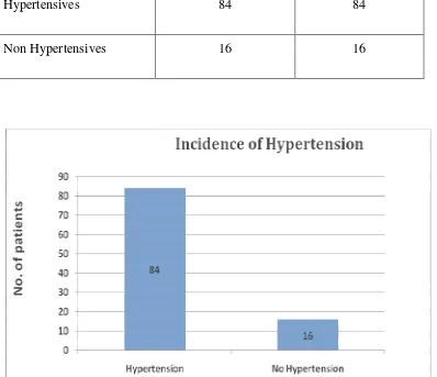
INCIDENCE OF HYPERTENSION
[image:39.612.118.516.305.648.2]The study group consisted predominantly of hypertensive patients. They constituted 84% of the patients. Of the 100 patients who were studied 34 were both hypertensive and diabetic.
Table 4. Incidence of Hypertension
Disease No. of patients %
74.7% of patients were hypertensives in the 5 year Cumulative report of CKD Registry of India25 2010, which is comparable to this study.
INCIDENCE OF DIABETES
[image:40.612.150.484.404.692.2]Diabetic patients constituted about 40% of the study group out of which 37% were suffering from type II diabetes and only 3% were suffering from type I Diabetes. 60% of the patients were nondiabetics. 17 diabetic patients were on Inj.Insulin and the rest were on oral hypoglycemic agents.
Table. 5 : Incidence of Diabetes
No. of Patients %
In the 5 year Cumulative report of Chronic Kidney Disease Registry of India25 2010, 38.4% of the study group had diabetes of which 9.9% belonged to type I DM and 90.1% belonged to type II group. This is similar to this study were type I diabetics constituted 7.5% of the diabetic population and type II constituted 92.5%.
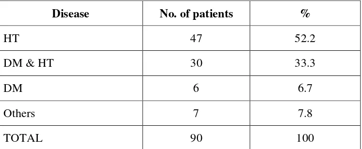
CAUSE OF CHRONIC KIDNEY DISEASE
[image:41.612.117.490.424.578.2]In this study, out of the 100 patients , 90 patients had chronic kidney disease and 10 patients were renal transplant recipients.
Table 6.Cause Of Chronic Kidney Disease
Disease No. of patients %
HT 47 52.2 DM & HT 30 33.3
DM 6 6.7
Others 7 7.8 TOTAL 90 100
7 patients had renal disease due to other causes like Glomerulonephritis, Analgesic nephropathy , IgA nephropathy etc.
Of the 10 post renal transplant patients, 3 had hypertension, 4 had diabetes and hypertension.
Hypertension was the single main cause of Chronic Kidney Disease in this study contributing to 52.2%. 33.3% of patients had both diabetes and hypertension.
In the Study of Ocular evaluation in patients with chronic renal failure published in Nepal Medical College Journal in 2008 by L.Bajracharya et al4 the commonest cause of Chronic Kidney disease was hypertension(36.1%).
Kidney Disease contributing to 31.2%. Hypertensive nephrosclerosis constituted 12.18% .
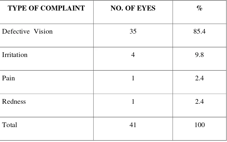
OCULAR SYMPTOMS IN PATIENTS WITH CKD
[image:43.612.117.505.375.618.2]With regards to the ocular complaints in patients screened, 59% of patients had no ocular complaints and only 41% complained of some form of ocular discomfort . 85.4 % of patients with ocular complaints , had defective vision, 9.8% of patients had ocular irritation ,2.4% of patients had pain and discharge each .
Table 7. Ocular Symptoms in Patients with CKD TYPE OF COMPLAINT NO. OF EYES %
Most of the patients with ocular complaints were having their eyes checked for the first time which showed the lack of awareness about the potential ocular complications in CKD.
In the study done by Easterbrook et al, published in the British journal of Ophthalmology (1970), all patients had excellent visual acuity. Whereas L.Bajracharya’s4 study of ocular evaluation in patients with chronic renal
BEST CORRECTED VISUAL ACUITY
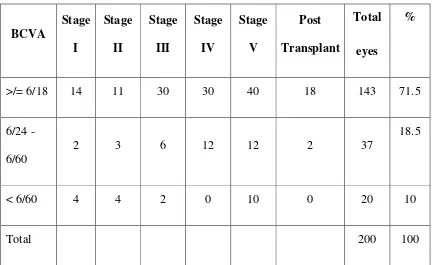
Best corrected visual acuity was tabulated .71.5% of the total patients enrolled were with vision 6/18 or better .In this study, according to WHO criteria ,18.5% were visually impaired (6/24 – 6/60) and 10% were in the category of legally blind (vision <6/60).
This finding is comparable to the Study of Ocular evaluation in patients with chronic renal failure published in Nepal Medical College Journal in 2008 by L.Bajracharya et al4 which showed that 76.6% of patients had good vision
[image:45.612.115.548.435.700.2]and patients with impaired vision were 11.7% and those who were legally blind were 11.7%.
Table. 8 : BEST CORRECTED VISUAL ACUITY
BCVA Stage I Stage II Stage III Stage IV Stage V Post Transplant Total eyes %
>/= 6/18 14 11 30 30 40 18 143 71.5 6/24 -
6/60 2 3 6 12 12 2 37
Patients with visual acuity less than 6/60 were significant with p value<0.05
ANTERIOR SEGMENT FINDINGS IN DIFFERENT STAGES OF CHRONIC KIDNEY DISEASE
[image:46.612.116.532.421.667.2]Of the 200 eyes included in the study 160 eyes showed changes in the Anterior segment . 57.5% of eyes had cataract , 16.9% of eyes showed lid edema and 17.5% of eyes had conjunctival pallor. The other ocular findings that were noticed were iridocyclitis, defective extraocular movements and pingecula.
Table. 9 : Anterior Segment Findings in Different Stages of CKD
Stage
I Stage II Stage III Stage IV Stage V Transplant Post Total eyes %
Lid edema 4 2 4 8 9 0 27 16.9
Conj. Pallor 6 2 8 4 8 0 28 17.5
Pingecula 2 0 0 2 4 0 8 5.0
Cataract 5 2 10 31 38 6 92 57.5
EOM
Restriction 0 0 0 0 1 0 1 0.6
Proptosis 0 0 0 0 1 0 1 0.6
Iridocyclitis 0 0 0 0 0 1 1 0.6
Band shaped
Keratopathy 0 0 1 1 0 0 2 1.3
PINGECULA
CONJUNCTIVAL PALLOR
PLASTIC IRIDOCYCLITIS IN A POSTRENAL TRANSPLANT
When the incidence of these symptoms in different stages of Chronic Kidney Disease were studied it was found that cataract was found in 38 eyes of patients with end stage or Stage V renal disease. Stage V group patients also exhibited the most number of anterior segment signs ( 61 eyes). In patients with stage IV and Stage III renal disease 46 and 23 eyes showed signs.
On analysing the findings ,the significant findings in the anterior segment were cataract (p <0.02) and conjunctival pallor(p <0.02) .
mucormycosis of the maxillary sinus with orbital involvement. The patient underwent surgical debridement of the sinus with appropriate medical therapy.
Plastic iridocyclitis was found in a renal transplant patient with transplant dysfunction. The patient was on steroids and other immunosupressive agents.
CATARACT IN PRESENILE PATIENTS
[image:53.612.114.497.483.650.2]An observation of the incidence of cataract in the presenile age group (<=50) was done. This showed that of the 65 patients (130 eyes) , 53eyes had some form of cataract and 11 eyes have undergone cataract extraction with a posterior chamber intraocular lens in place.
Table. 10 : Cataract in Presenile Patients
No. of eyes %
INCIDENCE OF OCULAR SURFACE DISEASE IN CKD
[image:54.612.164.483.271.393.2]A reduced Schirmer’s value was noted in 54 eyes and was normal in 146 eyes. The incidence of ocular surface disease in the study was 27%.
Table. 11 : Incidence Of Ocular Surface Disease in CKD
Schirmer's test No. of eyes %
Reduced 54 27 Normal 146 73
The presence of ocular surface disease in this study was significant with p value <0.01.
WHATMAN FILTER PAPER STRIP
INCIDENCE OF HYPERTENSIVE RETINOPATHY IN CKD
In this study there was a total of 84 patients (168 eyes) with hypertension, of which 92 eyes (46%) showed hypertensive changes.
43 eyes had Grade III retinopathy making it the most common hypertensive retinopathy. Higher grades of hypertensive retinopathy was found in advanced stages of CKD i.e 24 eyes in stage IV and 23 eyes in stage V.
[image:57.612.116.521.452.684.2]92 eyes (46%) out of the 200 under study showed hypertensive retinopathy In the study by L.Bajracharya et al4 48% of total patients had hypertensive retinopathy and retinopathy was more common in patients in stage IV and Stage V disease which is comparable to this study.
Table. 12 : Incidence of hypertensive retinopathy in CKD
Stage
I Stage II Stage III Stage IV Stage V Transplant Post
Total eyes
Grade I
HR 0 2 5 2 5 3 17 Grade II
HR 5 2 9 3 8 1 28 Grade III
HR 5 4 5 19 8 2 43 Grade IV
INCIDENCE OF DIABETIC RETINOPATHY IN CKD
Of the 40 patients (80eyes) with diabetes , 51 eyes i.e., 63.75% exhibited some degree of diabetic retinopathy.
Diabetic retinopathy was most common in patients with Stage V renal disease with 25 eyes out of the 51 eyes belonging to this group followed by stage IV having 13 eyes with diabetic retinopathy.
CKD similar to what was observed in the WESDR study where prevalence of PDR was much higher in patients with persistent microalbuminuria.
Severe NPDR was the most common stage of diabetic retinopathy which was found in 17 eyes. Second most common diabetic retinopathy was moderate NPDR and PDR having 12 eyes each.
Clinically significant macular edema was present in 6 eyes in various grades of Diabetic retinopathy. All of them belonged to stage V CKD. In the Wisconsin Epidemiologic study of Diabetic Retinopathy (WESDR)27,28 the
SEVERE NPDR WITH CLINICALLY SIGNIFICANT MACULAR EDEMA
ADVANCED DIABETIC EYE DISEASE
Table. 13 : Incidence of Diabetic Retinopathy In CKD
Stage
I Stage II Stage III Stage IV Stage V Transplant Post
Total eyes
Mild NPDR 0 0 0 0 2 0 2 Moderate
NPDR 0 0 0 6 4 2 12 Severe
NPDR 2 0 2 7 6 0 17 Very Severe
NPDR 0 0 0 0 0 0 0 PDR 0 1 2 0 9 0 12 Advanced
DR 0 3 1 0 4 0 8 Total 2 4 5 13 25 2 51
L.Bajracharya et al4 demonstrated that 88.3% of diabetics had retinopathy whereas in this study 63.75% of diabetics had retinopathy. Both the studies were similar in the fact that more number of patients in stage V showed Diabetic retinopathy.
In this study group of CKD patients with diabetes , the patients with diabetic retinopathy is 63.75% which is significantly more when compared to diabetic retinopathy prevalence in diabetic population without CKD.
MACULAR FINDINGS IN STUDY GROUP
31 eyes from the study group showed changes in the macula. The most common sign was macular edema which was found in 13 eyes. This group had a mixed population of diabetic and hypertensive patients. Other common findings in the macula were age related macular degeneration changes and clinically significant macular edema in diabetic patients. PED, subretinal precipitates and macular hole .
[image:66.612.116.545.476.688.2]Macular edema was more common in stage V CKD.
Table. 14 : Macular Findings
Stage
I Stage II Stage III Stage IV Stage V Transplant Post TOTAL EYES
ARMD 3 0 1 0 3 0 7 CSME 0 0 0 0 6 0 6 Macular Edema
(DM & HT) 0 0 2 1 8 2 13 PED 0 2 0 0 0 0 2 Sub Retinal PPT 0 0 0 2 0 0 2 Macular Hole 1 0 0 0 0 0 1
MACULAR EDEMA
OTHER POSTERIOR SEGMENT FINDINGS
[image:70.612.115.544.312.444.2]When diabetic and hypertensive retinopathies as well as macular symptoms were excluded there were some posterior segment findings exhibited by 11 eyes from the study group. 4 eyes had a raised CD ratio. BRVO was found in 2 eyes of 2 patients. 3 eyes had disc pallor all of them belonged to stage IV.
Table. 15 : Other Posterior Segment Findings
Stage
I Stage II Stage III Stage IV Stage V Transplant Total Post
BRVO 0 0 0 2 0 0 2 CRAO 0 0 0 0 2 0 2 RAISED CD
RATIO 2 0 2 0 0 0 4 Disc Pallor 0 0 0 3 0 0 3
Two patients had raised CD ratio. IOP and fields were
normal in both the patients. B scan was done in patients with m edia
opacities did not show any positive findings.
BRANCH RETINAL VEIN OCCLUSION
RE
RESULTS
The age distribution in the study group was more or less even, with the patients in the age group 30 – 39 slightly more than the rest. Mean age of the patients with chronic kidney disease is 44.2.
Male patients formed 72% of the total patients in the study group. The average male : female ratio was 2.6:1.
Out of 100 patients 90 had CKD in various stages &10 belonged to post renal transplant group. The sample size in each grade of CKD showed more patients in stage 5 (31%), & stage 4 (21%) followed by stage 3 (19%), stage 1(10%) and stage 2 (9%).
The commonest cause of CKD was hypertension in 47% pts (52.2%) followed by both diabetes and hypertension in 30 patients (33.3%). Patients with only diabetes were 6 patients (6.7%) & with other causes (like IgA nephropathy , Analgesic nephropathy etc.,) were 7 patients (7.8%).
10% of patients were legally blind with visual acuity <6/60.
Of the 200 eyes included in the study 160 eyes had Anterior segment changes. 92 eyes (57.5%) had cataract . Of the 92 eyes 38 eyes were in Stage V group and 31 eyes belonged to Stage IV group.
In this study, 65 patients ( 130 eyes) belonged to less than 50 years (presenile age group). 49.3% of presenile patients had cataract.
A reduced Schirmer’s value was noted in 54 eyes of the 200 eyes. The incidence of ocular surface disease in the study was 27%.
84 patients in the study had hypertension (HT alone or along with DM). 92 eyes out of 200 eyes studied showed hypertensive retinopathy. Higher grades of hypertensive retinopathy was more in advanced stages of CKD i.e 24 eyes in stage IV and 23 eyes in Stage V. Grade III hypertensive retinopathy was the most common grade of hypertensive retinopathy occurring in 43 eyes of the 92 eyes.
Prevalence of Diabetic retinopathy in CKD patients is significantly more when compared to diabetic patients without CKD.
31 eyes in the study showed changes in the macula. The most common sign was macular edema found in 13 eyes which had a mixed population of diabetic and hypertensive patients.
CONCLUSION
CKD is the end result of multiple systemic diseases or primary renal disease. During the natural course of the disease it affects multiple systems of the body including the eye. Detailed ocular examination was conducted in 100 patients in varying stages of CKD. In this study hypertension was the single main cause of CKD followed by DM and HT together.
Blurring of vision was the commonest ocular symptom. Most of the patients having complaints of blurring of vision were examined for the first time indicating the lack of knowledge about the potential ocular complications. Significant visual loss was due to cataract followed by Proliferative Diabetic retinopathy and macular edema.
Ocular findings that were present more in stage IV & stage V grades of CKD were cataract, lid edema, conjunctival pallor, hypertensive retinopathy, diabetic retinopathy, macular edema and CSME.
Retinopathy is often asymptomatic in its early stage. Delay in diagnosis can result in significant visual loss. Optimized control of risk factors like renal disease which affect onset and progression of retinopathy should be approached through an intensive, multidisciplinary health care which can markedly reduce the incidence of visual loss.
In this study most of the patients were having their eyes examined for the first time. This shows the lack of awareness of the importance of early ocular evaluation among the patients.
Ocular manifestations in patients with Chronic Kidney Disease – a hospital based study
PROFORMA
Serial no. :
Name :
Age :
Sex :
Occupation :
Address :
Ocular complaints : Renal status of patient
Stage :
Duration :
Treatment details : Diabetic status
Type :
Duration :
OCULAR EXAMINATION RE LE
Vision
Eyelids and lashes Extraocular movements
Slit lamp examination
Tear meniscus Conjunctiva Cornea Anterior chamber Iris Pupil Lens Fundus IOP Visual fields Schirmer’s test
INVESTIGATIONS
BloodsugarFasting Post prandial
Blood urea Serum creatinine urine : Albumin
KEY TO MASTER CHART
ARMD – Age Related Macular Degeneration BSK – Band Shaped Keratopathy
CAPD – Continuous Ambulatory Peritoneal Dialysis CCC – Circum Corneal Congestion
Ce – Cells
CVA – Cerebrovascular Accident DV – Defective vision
Ex – Exudate F – Female Gr – Grade
Mon – Months N – Normal
NPDR – Non proliferative Diabetic Retinopathy OHA – Oral Hypoglycemic Agents
P – Proptosis Pal – Pallor
PCIOL – Posterior Chamber IntraOcular Lens PCO – Posterior Capsule opacification PDR - Proliferative Diabetic retinopathy PED – Pigment Epithelial Detachment Pin – Pingecula
PL – perception of Light Red – reduced
Res – Restricted Ret – Retinopathy St – Stage
BIBLIOGRAPHY
1. Comprehensive Clinical Nephrology 2nd edition by Richard J Johndon MD FACP and John Feehally MA DM FRCP
2. G.q. Kazi M.B., F.R.C.S.(G) , C.I. Phillips Ph.d., M.D., D.P.H., F.R.C.S. (Edin, Lond) A.T. Lambie M.B., F.R.C.P. (Edin, Lond) , R.J. Winney M.B., M.R.C.P. (U.K.) - Hypocalcemic cataract as a presenting symptom of renal insufficiency – Postgraduate Medical journal (February 1984) 60, 166 – 167.
3. M. Easterbrook and C.B. Mortimer – Ocular signs in chronic renal failure – Brit. J. Ophthalmology. (1970) 54, 724.
4. L. Bajracharya, DN shah, KB Raut and S Koirala – Ocular evaluation in patients with chronic renal failure – a hospital based study – Nepal Medical college journal 2008 : 10(4): 209 – 214
5. Hari P, Singla IK, Mantan M, Kanilkar M, Batra B, Bagga B. – chronic renal failure in children – Indian pediatrics 2003; 40 : 1035 – 1042. 6. Lapco L, Weller JM, Greene JA – Spontaneously reversible retinal
7. Buchanan WS, Ellis PP- Retinal separation in chronic glomerulonephritis - Arch Ophthalmology 1964; 71:182-189.
8. Steiness IB – Reversible retinal detachment in renal insufficiency – Acta Med Scandinavia 1968; 183: 225 – 227.
9. Paris GL, Maacoul KL – reversible bullous retinal detachment in chronic renal disease – American Journal Ophthalmology 1969 ; 67: 249 – 251.
10. Wagdi S, Dumas J, Labelle P- Retinal detachment in renal insufficiency: a report of 3 cases – Canadian journal of Ophthalmology 1978; 13: 157 – 159.
11. Grass JD – Bullous retinal detachment and multiple retinal pigment epithelial detachments in patients receiving hemodialysis – Graefes Arch. Clin. Exp. 1992; 120 : 454 – 458.
12. Lemrini F, Dafrallah L, Kabbaj A – retinal detachment in children – Journal Fr. Ophthalmology 1993; 16: 159 – 164.
“Red eye” in renal failure patients.- Iranian journal of Medical Hypotheses and Ideas.
14. Obrador G, Pereira J. – Systemic complications of Chronic Kidney Disease, pinpointing clinical manifestations and best management. – Postgraduate Medicine , Minneapolis 2002: 115.
15. Duke-Elders S, Dohree JH. System of Ophthalmology, Vol X .1st ed. London: The CV Mosby Company 1967; chapter 4, 315-47.
16. Dursun D, Demirhan B, Oto S, Aydin P. Impression cytology of the conjunctival epithelium in patients with chronic renal failure. Brit J Ophthalmol 2000; 84: 1225-7.
17. Brenner Barry M. Brenner and Rector’s The Kidney Vol II.6th ed. Philadelphia: Saunders 2004; 2267-68.
18. Duane TD, Jaeger EA. Duane’s clinical ophthalmology Vol 5. Revised ed. USA: Harper and Row 1987; Chapter 31, 1-2.
20. Klahr S, Levey AS, Beck GJ, et al. The effects of dietary protein restriction and blood-pressure control on the progression of chronic renal disease. N Engl J Med. 1994;330:877–884.
21. Levey AS, Bosch JP, Lewis JB, et al. A more accurate method to estimate glomerular filtration rate from serum creatinine: a new
prediction equation. Ann Intern Med. 1999;130:461-70.
22. Coresh J, Wei GL, McQuillan G, et al. Prevalence of high blood pressure and elevated serum creatinine level in the United States: findings from the thirdNational Health and Nutrition Examination Survey (1988–1994). Arch InternMed. 2001;161:1207-16.
23. Agarwal SK. Chronic kidney disease and its prevention in India. Kidney Int. 2005;98:S41-5.62.
24. Mani MK. Prevention of chronic kidney failure at the community level. Kidney Int. 2003;63:Suppl 83:S86-9.
25. 5 year CUMMULATIVE Report CKD REGISTRY OF INDIA, INDIAN SOCIETY OF NEPHROLOGY , : Presented During : 41 Annual Conference of ISN (Trivandrum, December 2nd–5th 2010). 26. Clinical Nephrology Vol 74 – suppl 1/2010(S9 – S12) Burden of diseases
M.Dabhi, Department of Nephrology, Muljibhai Patel Urological Hospital, Nadiad, Gujarat- India.
27. The Wisconsin Epidemiologic Study of Diabetic Retinopathy published in Ophthalmology 2009 march ; 116(3): 497 – 503.
! "! ! ! # $ " ! $ ! " ! ! % " ! % ! & " ! ! '
!" # $ $ % & ' ( % & ' ( "" )* )! + !+
! , ', - . " / $ $ % . # 0 + $ - % . # 0 + $ - * 1 )1 )1 /+ /+
/ )2' . ' . "! 3 4. . # . 4,. " 5 5 1 1 3$ 3$ 6. . 6. . " )/ + 3
" 4 . , "1 $ . $' ! ! # #$ 3 $ 6. . + 0 6. . 0 0 /" /)" )1 3 3
' ,7 & . . ' .,% ,. .,7 ! 3 ) - 3 ) - #$ 3 #$ 3 $ 0 ) $ 0 ) "" ") 1 !+ !+
' , . /" ' . ' . / 1 1 $3- 0 $3- 0 0 0 ")1 /+ 3
* , . / , & 8 $'. .,7 1 1 $3- 0 $3- 0 6. . 6. . 1 /" *)1 1 /+ 3
$ / .,7 / $ 9#3 9#3 $ $ 6. . 6. . 0 0 1) *)! !+ +
1 $)# 4' " 3 $ 7 7 . .,7 1 1 $3- 0 $3- 0 6. . 6. . 11 1 /)/ *)! + 3
% ! ,7 . ! ! # $3- 0 $3- 0 6. . + . # 0 6. . + . # 0 /* ! *)1 + !+
. ' . // 7 . : , 7 ,. " .,7 1 $3- 0 $3- 0 6. . 6. . + 0 * ! 1)/ /+ 3
! ' . ' " . * .,7 3 ) - 3 ) - $3- 0 $3- 0 6. . 6. . ) * /+ 3
/ . , ', $, . .,7 ) / $3- 0 $3- 0 6. . 6. . * */ / 1 + 3
" ' ,7 , . * ' . " 3 ) - 3 ) - $ $ 6. . + 0 ' ( " " )1 *) /+ 0
$-: . .; / ' . ' . ,. ! .,7 ! $3- 0 $3- 0 6. . 6. . + , . ' * * * !+ 3
' ' /* 8 & . % . $' * $3- 0 $3- 0 6. . 6. . + 20 ! 1)* !+ 3
* #. ' ! . 9 . ! ! # $3- 0 $3- 0 1 11 ! ) /+ 3 5/ ,
3 ' . " $' " #$ 3 #$ 3 6. . 6. . 1" " )* !+ 3
1 3 ' " , & 8 < ' . .,7 1 # # $3- 0 $3- 0 6. . 6. . 1 )/ ) !+ 3
! 4 , . ' / $' * .,7 , 1 1 $3- 0 $3- 0 6. . 6. . ! / " ) + !+
! : !/ , 1 # $ $ 6. . + - 6. . 1! / 1 *) /+ 3
!! #,% . ! , $ 4 . / $ $ 6. . + - 6. . + - !* )" 3
!/ ' . = $' " .,7 ! $3- 0 $3- 0 # 0 & ' 0 # 0 & ' 0 0 0 !! * )1 * !+ /+
!" #. 4', ! 7 . # # #$ 3 #$ 3 # 0 & ' 0 # 0 / *)1 )/ /+ 3
! ! , & 8 ' .,% ,. / .,7 ! $3- 0 $3- 0 , . #- , . #- * " ")* )1 3 3
! ,4. /" :. ' 7 . / $3- 0 $3- 0 6. . 6. . 0 0 "/ 1 + 3
!* " , & 8 $' ! , #3 !" 3 ) - 3 ) - $ #$ 3 % & . , ' ..' 7 0 0 * *) 1) !+ !+
! : ',. , & 8 . .. / 1 ! $ $ /" / ") )1 + +
!1 ' % .7' / , 4 ,. / .,7 # # $3- 0 $3- 0 0 0 * !" )* *)" + 3
/ > ' / , & 8 =,. " $ $ 6. . 6. . 0 0 1 1 /)! *) + 3
/ '& ' / $ . : ' ,. $ $ 6. . 6. . 0 0 / )1 + 3
/! ' 4 ,. . $' .,7 / # # $ $ 6. . 6. . 0 0 " /)! )! + !+
// ' ., %, . , // $ . . .,7 ) ! ! $3- 0 $3- 0 6. . 6. . !/ " )! + 3
/" : . ' ! , & 8 = . 0 / .,7 / ! 2 : $ $ % & 6. . 1 ** ") )* !+ 3
/ # .% ' , & 8 : ' ,. !" !" $ $ 6. . + 0 ' ( 0 0 1/ /) 1) !+ 3
/ 3 ' / . ' .,% ,. / .,7 # # $3- 0 $3- 0 6. . 6. . 0 0 * ! * !+ 3
/* # ' ., " ' ', , .. / #3 $ $ . , ' ..' 7 6. . + # 0 0 0 "1 / 1) !+ 3
/ ' . "1 4 ,. . $' % . ! # $3- 0 $3- 0 6. . 6. . !! !/ ! * + 3
/1 ' ! , & 8 / ! ! # # $3- 0 $3- 0 6. . 6. . // !1 )1 * /+ 3
" > & ' . " % ) $ 7 , ! , 1 $ $ 6. . + % . # 0 6. . + % . # 0 + $ - 1 1 /) ? !+ 5/ #@ $-33
" % . "1 % ) $ :, 4 ! 1 1 3$ 3$ 6. . 6. . // ") )! + 3
"! ,. ' !/ , 6 '. ' 0#6 $ $ ! 1 )* !+ 3
"/ ,.,7 " % ) $ : ' ,. ! .,7 $3- 0 $3- 0 0 0 ! )! + 3
"" 6 , % . ' ! % ) $ # " .,7 7 ., '. ! 1 #$ 3 #$ 3 - - ! ! )! *) 3 3
" , ' . /1 $3-0: / ! , ! 3 ) - 3 ) - 2 : $ $ # 0 # 0 0 0 ! / ! * "+ 3 .4 !+
" / % ) $ % . / .,7 1 1 $3- 0 $3- 0 - - 1 // ! *)1 + 3
"* 0 ' / % ) $ # . " $ $ 6. . + 20 6. . 0 0 /+ 3
" ., ,7 /1 % ) $ ' .,% ,. # / .,7 , 1 1 $3- 0 $3- 0 6. . + . # 0 6. . + . # 0 ! / " 1 + !+
"1 ' 4 " 4 ,. . ' .,% " ! ! # # #$ 3 #$ 3 % . # 0 + $ - % . # 0 + $ - ! * )! 1) !+ 0
' % 7 " 4 ,. . $' " $ # ! , $ $ 6. . + - 6. . + - !"* /! ") 1) /+ !+
$ . ' / $3- 0 $3- 0 9 3 $0 9 3 $0 0 0 !1 ")/ 1)/ !+ 3
! > ' * . ' . 0 ' !" !" 3 ) - 3 ) - # # $3- 0 $3- 0 6. . 6. . 0 0 !/ ! ")1 ) /+ 3 3
/ 6 7 , & 8 $' " / # # $ $ ' ( ' ( 0 0 ! ! " *) )/ /+ 3
" :, , ! 4 ,. . ' .,% ,. ! , !" !" #$ 3 #$ 3 6. . + # + #$ 6. . + #+ #$ + # 0 ! )/ 3 +
> ' ! , $' # # $ $ * 1 ! ) /+ 3
, ', , . !" , $' $ $ $' . ' / 1) ) /+ 0
$-* : 7 ! , & 8 ' ., % " 7 '.!' ! $3- 0 $3- 0 !" " )1 1) /+ 3
4 7 " $ . $' .,7 $3- 0 $3- 0 6. . 6. . 1 1 /) 1 !+ 3
1 / $' '. . 1 3 ) - 3 ) - $ $ ! * 1* )1 "+ 3
" 4 ,. . : ' ,. # . ' .,7 1 1 3$ 3$ 6. . 6. . /" "! /) *)! !+ 3 .4 !+
' 7 " , & 8 $' / / $ $ % . # 0 % . # 0 * ")/ 1)! !+ /+
! ., , . /1 , & 8 $' .,7 1 $3- 0 $3- 0 / 1)/ !+ 3
/ 7 ' , & 8 $' . ! , 3 ) - 3 ) - # # $3- 0 $3- 0 * ! )! *)! !+ /+
" ' (' , & 8 . 4 ,. $ # ! , #3 # # $ $ 6. . + % . # 0 6. . + % . # 0 * 1 )* )1 + !+
% 1 . . ' 7 , / !" $ $ % & ' ( . ' !/ /! ) )" !+ 3
% * 8 . . =,. " $ $ . # 0 . # 0 ! 1 )* !+ /+
* ., /! , ' !" , 3 ) - 3 ) - $ $ . # 0 . # 0 " * )/ 1)" !+ !+
! "! ! ! # $ " ! $ ! " ! ! % " ! % ! & " ! ! '
1 > . 1 6 % .% $' " , ! ! $ $ . # 0 . # 0 !"/ 11 )! 1 3 3 #@ !+
* ' 4, % ! $ # / , 3 ) - 3 ) - $ $ # 0 & ' 0 # 0 0 0 * ! ) 1)" !+ !+
* 2 . ' . 7 % ) .% # .,. ! !! , #3 $ $ # 0 # 0 0 0 !// * /+ !+
*! 8( . ) ' ., . %,. / /! , #3 #3 #$ 3 #$ 3 . , ' ..' 7 . , ' ..' 7 0 0 / " /1 /1 /+ "?
*/ : " , & 8 $' ! !" !" $ $ 6. . + . # 0 6. . + % . # 0 + 0 0 0 !*! 1 )* )1 /+ 3
*" % ' 4 ,. . $' ! ! , !" !" $ $ # 0 . , ' ..' 7 0 0 ! 1 * 1 !+ 3
* $' . = ! 4 ,. . $' ! $ $ 6. . 6. . 1! !!* 1)/ 1 "+ 3
* ,.,7 " 4 ,. . 0 ' / 1 1 #$ 3 #$ 3 % . # 0 % . # 0 "/ ! )1 )1 3 3
** 6 % . ' ! , $' '. . 3 ) - 3 ) - $3- 0 $3- 0 $ 0 ) $ 0 )* *1 /1 /)! 1)1 !+ 3
* . % . : ' ,. $ # , #$ 3 #$ 3 ,4. , ,4. , /1 1 /)1 *)! !+ !+
*1 > , & 8 $' $ # 7 '.1 '1 #$ 3 #$ 3 " 1! /)* )" 3 3 5/ ,
, ' ! , ' # .,7 7 ., '. $3- 0 $3- 0 ! *) + 3
' / 4 ,. . : ' ,. # .,7 $3- 0 $3- 0 !! /" )" 1)" 3 3
! . /! 4 ,. . $' # " .,7 '. . $3- 0 $3- 0 " )" 3 3
/ #, ' 4 ,. . $' , " #$ 3 #$ 3 # 0 # 0 / ! )1 )! + 3 5!
" , ' /1 4 ,. . $' ! 1 $3- 0 $3- 0 11 1 /+ 0
$--7 . = ! 4 ,. . $' . .,7 7 '. ' $6 1 $3- 0 $3- 0 6. . 6. . / 1)! /+ +
6 0 # % . ! , ! $ $ 6. . + # 0 6. . + # 0 ! *1 1)/ !+ /+
* # % . !! , , '., ! , !" / $ $ # 0 # 0 0 0 1 *) /+ /+
:. ' . = "1 $ . . # .,7 ! / $ $ 8, , ' ( " / )1 1 3 3
1 >, " & . . . ,. # ! .,7 ! $3- 0 $3- 0 6. . 6. . * 1 + !+
1 2' . /1 $ . ' 7 . 1 3 ) - 3 ) - # # $3- 0 $3- 0 6. . 6. . 0 0 ! * )1 *) /+ 3
1 & ' 4 ,. . $' ! $ A , . ! #3 3 ) - A #0 $ $ ' ( ' ( 1 * ) *)/ !+ /+
1! , 8 / 4 ,. . $' $3- 0 $3- 0 !1 1)/ + +
1/ ' !! , ' ! # # $3- 0 $3- 0 6. . 6. . * /)/ )! + 3
1" % ' " , & 8 ' ! $ ! ! $3- 0 $3- 0 6. . 6. . 0 0 /" * )* *) !+ 3
1 ! , $' 7 ., '. !" !" 3 ) - 3 ) - $ $ - - 0 0 /! 1 ) ) !+ 0
$-1 -(' . ! , & 8 : ' ,. .,7 $3- 0 $3- 0 6. . 6. . * * )" *)! !+ 3
1* # . % " 4 ,. . ' ! / !" # $ $ % . # 0 % . # 0 + - "! * " *) /+ 3
1 , ' . 4 ,. . # .. " ! # $ $ 6. . 6. . ! 1)! *) + 3
11 . , ! 4 ,. . .,% ',. A # " ! $$$ $ "+A -; 3$ 3$ 1 *! " * !+ /+