DIABETIC RETINOPATHY IN END STAGE
RENAL DISEASE OF TYPE – II DIABETES
MELLITUS
Dissertation Submitted for
M.S.Degree(Branch III) Ophthalmology
April 2012.
THE TAMILNADU DR.M.G.R.MEDICAL UNIVERSITY
Dept. Of Ophthalmology Govt. Rajaji Hospital Madurai
CERTIFICATE
This is to certify that this dissertation entitled “DIABETIC RETINOPATHY IN END STAGE RENAL DISEASE OF TYPE -II
DIABETES MELLITUS” has been done under my guidance in Department of OPHTHALMOLOGY, MADURAI MEDICAL COLLEGE, MADURAI.
I Certify regarding the authenticity of the work done to prepare this dissertation.
Dr.P.THIYAGARJAN , M.S.,D.O., PROFESSOR & H.O.D
Dept. Of Ophthalmology GOVT. RAJAJI HOSPITAL MADURAI MEDICAL COLLEGE,
DECLARATION
I, Dr. P. SARAVANA SANKAR, Solemnly declare that the
dissertation titled, “DIABETIC RETINOPATHY IN END STAGE
RENAL DISEASE OF TYPE -II DIABETES MELLITUS” has been
prepared by me.
This is submitted to the “THE TAMILNADU DR.M.G.R.MEDICAL UNIVERSITY, CHENNAI, In partial fulfillment of the requirement for the award of M.S., (Ophthalmology) Branch-III degree examination to be held in APRIL 2012
Place: Madurai
ACKNOWLEDGEMENT
I am extremely grateful to Dr.P.THIYAGARAJAN, M.S.D.O.,
Professor and Head of the Department of Ophthalmology, Madurai Medical College, Madurai, for his guidance and help for executing my study.
I take this opportunity to express my deep sense of gratitude to
Dr.G.SRINIVASAN, M.S.,D.O., Professor of Ophthalmology, Madurai
Medical College, Madurai for his guidance and help for executing my study. I am extremely indebted to my beloved guide Dr.A.R.ANBARASI, Assistant Professor, Madurai Medical College, Madurai for her constant encouragement, throughout this dissertation. My sincere thanks to all my Assistant Professors for their valuable suggestions in carrying out this study.
I express my deep sense of gratitude to Dr.M.SHANMUGA
PERUMAL, Head of the Department of Nephrology.
I am grateful to Dr.D.EDWIN JOE., Dean, Madurai Medical College and Govt.Rajaji Hospital, Madurai for permitting me to utilize the clinical materials of this hospital.
CONTENTS
TITLE
PART
–
I
Page
No.
1. Introduction 1
2. Aim 5
3. Diabetes – Renal failure – Eye 6 4. Classification of diabetic retinopathy 12 5. Review of literature 18
PART – II
1. Patients and Methodology 34 2. Observation and Analysis 36 3. Discussion 49
4. Summary 55
5. Conclusion 57 6. Bibliography
7. Proforma
INTRODUCTION
Eye is a mirror that reflects pathological changes occurring in many organs of the body. Numerous systemic disorders affect both eye and kidney. Examination of eye is an in dispensable part of the clinical assessment of a patient with renal disorders.
CRF is irreversible and progressive process that result in END STAGE RENAL DISEASE(ESRD) where patient has to depend on renal replacement for survival.1
Richard bright in 1836 first associated renal disease with blindness.2 By ESRD 80% of pts will have secondary hypertension.3 Ocular morbidity may be due to coexisting risk factors like hypertension, diabetes, uremia and anemia. The ophthalmologist may be consulted for a variety of reasons about the patients whose problem appear renal.
On the other hand nephrologists may be aware of many ocular manifestations of renal disorders which are common and rare in nephrology and the potential toxicity of therapeutic agents.
Diabetes is the most common disorder with ocular and renal manifestations.7 Rubeosis iridis and neovascular glaucoma occur due to posterior segment pathology. Rising concentration of intracellular calcium might contribute to early cataractegenesis and calcium deposit in lens.8
Hypertension affects the eye and kidney in parallel and very often occurs along with diabetes.
Hypertensive changes are particularly severe in renal failure. This has been attributed to the effects of retained nitrogenous products.9 Accelerated Hypertension can result in optic disc edema.10
Renal disease and ocular complications in diabetes are frequently disturbing and destined to become one of the challenging problems of the future.
Blindness due to proliferative retinopathy or maculopathy is approximately 5 times in diabetic patients with nephropathy compared with non albuminuric patients.11In India majority of end stage renal disease patients are of type II diabetes . 5% of diabetic patients die of end stage renal disease.
has been recognized as a major cause of kidney involvement in diabetes. Diabetic nephropathy ultimately leads to end stage renal disease.
Both anterior and posterior optic neuropathy can occur in CRF, when hemoglobin level falls below 5gm% retinopathic features like retinal hemorrhages, hard and soft exudates and pallor of optic discs could be present .The retinal arterioles look pale and veins appear distended.
Retinopathy is often asymptomatic in its most treatable stage. Delay in diagnosis can result in significant increase in patients risk of visual loss. Type II DM accounts for 90% - 95% diabetes cases and differs from type I diabetes in average age of onset and Etiology. 13
Patients with type I diabetes, who are generally younger and are more likely to live long enough to benefit from tight glycemic control, than patients with type II disease, who face a shorter life expectancy, because of their age and risk of cardiovascular disease.14 For patients with co-existent disease, the delayed benefits of glycemic control may be offset by the more immediate inconvenience, complications, and costs of intensive treatment and by the health effects of co morbid conditions. Ocular condition is also an indicator of metabolic control of the disease process.
AIMS AND OBJECTIVES
a. To record the stage of retinopathy in end stage renal disease patients of diabetic origin and on treatment.
b. To record the progress of retinopathy at end of 6months and 12months in these patients.
c. To correlate the severity of retinopathy with renal failure.
DIABETES – RENAL FAILURE – EYE
Diabetes is defined as a major group of metabolic disease characterized by hyperglycemia, with disturbances in carbohydrate, fat and protein metabolism either due to defect in insulin secretion, action or both.15 It is the chronic hyperglycemia that causes most of the microvascular damage and contributes to the development of macrovascular disease.
In type I diabetes onset is symptomatic and after ten years seventy percent of the patients have retinal abnormalities and after twenty years 30-40% have clinical nephropathy. In type II diabetes the intial onset is asymptomatic and 15-20% have retinal abnormalities and 5-10% have sustained microalbuminurea at the time of diagnosis.16
MICROVASCULAR COMPLICATIONS
Nephropathy Retinopathy Neuropathy
Macrovascular Complications
KIMMELSTIEL – WILSON LESIONS
PATHOLOGY PICTURE - 1
Diabetic Nephropathy
The most frequent cause of renal involvement in disease affecting other organs is diabetes. Renal failure in those with Insulin dependent diabetes mellitus and disease duration of 5 years together with hypertension and retinopathy is highly suspicious of diabetic nephropathy.
In Non Insulin dependent diabetes renal involvement seems to occur as frequent as individuals with insulin dependent diabetes after the same duration of disease but in this age group other renal disease are common.
The kidney may be damaged in three ways16 1. Glomerular damage
2. Ascending infections
3. Ischemia due to hypertrophy of afferent and efferent arterioles.
DIABETIC GLOMERULAR SCLEROSIS
It is the leading cause of premature death in young diabetic patients. Older diabetics develop nephropathy but the proportion affective is much smaller. The intial structural lesion in the glomerulus is thickening of the basement membrane. Associated changes may result in disruption of protein cross linkages that make the membrane an effective filter. In consequence a progressive leak of protein into the urine occurs.
The earliest evidence of this is microalbuminuria which in turn in some years progressed to intermittent albuminuria followed by persistent proteinuria. Once overt proteinuria occurs renal function invariably declines with 50% of the people reaching end stage renal disease with in 7 years of onset of proteinuria.
CHRONIC RENAL FAILURE
More commonly it occurs at blood urea nitrogen level of more than 100mg / dl and serum creatinine level of more than 10mg/ dl. It is good medical practice to initiate replacement theraphy just before the onset of ureamic symptoms usually when GFR is 5-10ml/ dl minute.
END-STAGE RENAL DISEASE
ESRD is final common end despite the dissappearance of renal insult that initially lead to the loss of some nephrons. It is striking that 60% of ESRD is now due to diabetes and essential hypertension. The most common primary renal cause of failure is glomerulo nephritis.
MANAGEMENT
The under lying cause of renal disease should be treated aggressively wherever possible eg. Tight diabetic control.
The Guideline of treatment are :
- Blood pressure control - Treatment of hyperkalemia - Correction of acidosis
- Fluid restriction along with salt. - Treatment of hyperlipedemia - Correction of aneamia
- Patient education
RENAL REPLACEMENT THERAPY
- Haemodialysis - Haemofiltration - Haemodiafiltration - Peritoneal dialysis - Kidney transplantation
DIALYSIS IN ESRD OF DIABETES MELLITUS
No distinction is attempted in ESRD treatment outcome between type I and type II diabetes mellitus patients. Today it is generally accepted that the renal replacement therapy should be considered earlier in diabetic than non-diabetic ureamic patients.
CLASSIFICATION OF DIABETIC RETINOPATHY
- Duke – Elder - Airlic – House
This system proposes fundus photography, flourescein angiography, ophthalmoscopy, fundus diagrams and slit lamp biomicroscopy of the retina.
- Kanski Classification
This is the popular and widely accepted classification of diabetic retinopathy. It is basically treatment oriented ophthalmoscopic classification. - Modern ETDRS Classification17,18
The early treatment diabetic retinopathy study has developed systems to grade the severity of diabetic retinopathy at various stages by assessing the lesions seen through stereoscopic colour fundus photographs and fluorescein angiograms.
NON-PROLIFERATIVE DIABETIC RETINOPATHY
A.MILD NPDR
DIABETIC RETINOPATHY DISEASE SEVERITY SCALE
Mild NPDR
Microaneurysms only
Moderate NPDR
More than just Microaneurysms but
less than severe nonproliferative
diabetic retinopathy
Sereve NPDR
Any of the following
20 intraretinal hemorrhages in
each of 4 quadradants OR
Definite venus beading in 2+
quadrants OR
Prominent IRMA in 1+ quadrant
and no PDR
Proliferative DR (PDR)
1 or more of
Neovascularization
Vitreous/ preretinal
hemorrhage
B. MODERATE NPDR
Hemorrhages and/or microaneurysm- standard photograph 2A and / or soft exudates, venous beading or IRMAs definitely present. Definitions not met with C, D, E and F.
C. Severe NPDR
Hemorrhages and/or microaneurysms in all four quadrants. Venous beading in two or more quadrants. IRMA>Standard photograph # 8A in atleast one quadrant.
D. Very Severe NPDR
Any two or of C.
Definition not met with E,F.
PROLIFERATIVE DIABETIC RETINOPATHY
E. EARLY PDR New vessels. F. HIGH RISK PDR
4
‐
2
‐
1
Rule
4 quadrants H/MA ( STD 2A)
4 quadrants VB
NVE – ½ disc area and preretinal/vitreous hemorrhage
In retina, the primary sources VEGF – A are ganglion cells. Muller and colleagues have shown that the level of VEGF (A) –in ocular tissues correlates with new vessel formation 19.
PROLIFERATIVE DIABETIC RETINOPATHY
The exact pathogenesis of retinal neovascularization remains unclear. In 1948, observations led Michelsen to propose that there exists in the retina a risk factor or factors affecting the budding of new vessals. Later it was explained by Ashton and others that hypoxia was the primary stimulus for production of angiogenic factors.
The most frequently studied molecules include basic fibroblast growth factor (bfgf), vascular endothelial growth factor (VEGF) growth hormone, and more recently the angiopoietiens studies however indicate VEGF as the main predictor of angiogenesis.20,32,33
EPIDEMIOLOGY OF DIABETIC RETINOPATHY
NEO VASCULARISATION AT DISC (NVD)
NEO VASCULARISATION ELSEWHERE (NVE)
- Severity of retinopathy was related to the duration of diabetes ranging from 2% in subjects with diabetes for less than 2yrs to 98percent in subjects with diabetes for 15yrs or more32. The severity also increased with duration of diabetes.
- In contrast the older onset was likely to have retinopathy at the time diabetes was diagnosed.
The study also found out that elevated glycoslylated hemoglobin was associated with severe retinopathy in all age groups. Proterniuria was associated with severe retinopathy in both groups.
PROLIFERATIVE DIABETIC RETINOPATHY
NVD
Neovascularization is frequently found within 45degrees of the optic disc. It is observed at the disc as a cart wheel configuration radiating from the center best indentified by stereoscopic view, by either contact or precorneal lenses or stereoscopic photography.
NVE
wool spots and hemorrhagic microaneurysms. IRMA may be difficult to differentiate from early NVE. Flourescein angiography shows leakage in new vessels as a differentiating feature.
VITREOUS AND PRERETINAL HEMORRHAGE
Small hemorrhages may occur near growing tips of the vessel but they are usually subhyaloid. Progression of vitreous detachment starts on either side of vascular arcades of fovea. Vitreous usually remains attached to the disc by proliferating fibrovascular tissue. Vitreous hemorrhage may occur as a result of vitreous traction on new vessels.
VITREOUS TRACTION AND FIBROUS PROLIFERATION
Fibrous tissue develops along the vessels which may subsequently contract. Contraction of posterior vitreous face and fibrovascular proliferation leads to tractional retinal detachment. Retina along the temporal arcades is the first to detach and extends to involve the fovea. Contraction of fibrovascular tissue can also lead to distortion or horizontal displacement of macula.
RUBEOSIS
HYPERTENSIVE RETINOPATHY
CLINICALLY SIGNIFICANT MACULAR EDEMA
DIABETIC MACULOPATHY
Diabetic macular edema may be present at any level of retinopathy and alters the structure of macula in any of these manners.21
Macular edema, i.e., a collection of intraretinal fluid in the macula with or without lipid exudates and with or without cystoid changes
Non perfusion of parafoveal capillaries with or without intraretinal fluid
Traction in the macula by fibrous tissue proliferation causing surface wrinkling or macular detachment
Intraretinal or preretinal hemorrhage in the macula
Lamellar or full thickness retinal hole formation
Any combination of the preceding.
CSME as defined by ETDRS includes any of these
Retinal thickening at or within 500microns of the center of macula
Hard exudate at or within 500microns of the center of the macula, if there is the thickening of the adjacent retina
REVIEW OF LITERATURE
OCULAR COMPLICATIONS OF RENAL FAILURE
The association of blindness and end stage renal disease was first noted by Bright and fundus changes in uraemic patients was described by Leibrich.21,22,23 Subsequently this was named Bright disease or albuminuric retinitis. By the end of the nineteeth century and as a result of hypertensive changes, albuminuric was no longer consider as a separate entity, but as manifestation of hypertension in uraemic patients.
Since the introduction of effective anti hypertensive treatment, dialysis and kidney transplantation such patients with severe hypertension have become rare. Never the less hypertension is still an important problem in kidney diseases and complications of atherosclerosis is common in renal patients as a result of chronic hypertension and hyperlipidaemia.
VASCULAR LESIONS
BLINDNESS
In patients with severe and long standing hypertension a severe reduction in arterial pressure may cause infarction of optic nerve followed by blindness or visual loss. In occasional patients with cerebrocortical infarction occurs under these circumstances with cerebral blindness. Anterior ischemic optic neuropathy and retinal infarction have been described as complication of haemodialysis associated with hypotension. Uraemia, anemia and disc edema due to intracranial hypertension are other risk factors for optic neuropathy in patients with renal disease. In addition patients with chronic hypertension are predisposed to retinal arterial and venous obstructive disease leading to visual loss. Sudden blindness like purtschners like retinopathy with or without cotton wool spots may occur in patients in chronic renal failure with or without trauma, pancreatitis or autoimmune disease. The precipitating factors for this retinovaso-occlusive disorder remain unclear. The conditions have been described in renal transplant also.
SEROUS RETINAL DETACHMENT AND
CORTICOSTEROID INDUCED CHANGES IN RETINAL
PIGMENT EPITHELIUM
transplantation, haemodialysis or patients receiving steroid therapy. In severe forms bilateral bullous retinal detachment, multiple retinal pigment epithelial detachments and yellow fibrin like subneural exudates beneath the sensory retinal detachment may be observed.
Flourescein angiography shows wide spread retinal pigment epithelial changes and leakage’s. Several factors may play a role in precipitating serous detachment in patients with renal failure, including impaired fluid and electrolyte imbalance, choroidopathy associated with hypertension, thrombotic microangiopathy or immune complex vasculitiditis and dysfunction of overlying epithelium which may be associated with renal disease such as membranoproliferative glomerulonephritis type 2. Corticosteroid can damage the RPE and predispose a patient to serous retinal detachment. Where as stress may produce chronic serous retinopathy. The visual prognosis is unfavourable. A decrease of corticosteroid dosage may help in reducing visual symptoms and enhance resolution of retinal detachment. In refractory cases focal laser photocoagulation may be applied to leakage points.
GLAUCOMA
resistance to aqueous outflow. The increase in pressure is thought to be part of cerebral edema that occurs as a consequence of the rapid reduction in serum osmolality. Corticosteroid is another risk factor for an increased intraocular pressure. In patients who are beginning long term haemodialysis or corticosteroids intraocular pressure should be measured early as diagnosis and treatment of glaucoma may prevent visual loss.
CORNEAL CALCIFICATION IN RENAL FAILURE
In patients with renal failure and associated hyperparathyroidism soft tissue calcification are first detected first in the peripheral interpalpebral cornea and adjacent conjunctiva. The corneal calcification eventually spread to toward visual axis in patients on chronic intermittent haemodialysis producing band keratopathy with decrease in visual acuity epithelial erosions resulting in severe pain. Patients with renal failure and hypercalcemia may present with inflamed pingecula and diffuse inflammatory edema.
BROWN TUMORS OF THE ORBIT
DIABETIC RETINOPATHY AND RENAL FAILURE
Nephropathy and retinopathy are the major microvascular complications of diabetes.22,23 It is the chronic hyperglycemia that cause most of the microvascular damage and contributes to the development of the macrovascular disease. This microangiopathy affects nearly all diabetics and while most diabetics and while most diabetics may develop clinically evident retinopathy, nephropathy occurs in a subset.
In patients with nephropathy retinopathy is always present and proliferative retinopathy is common. However thirty five percent of the patients with proliferative retinopathy have no signs of diabetic nephropathy and these patients will probably never develop diabetic nephropathy.
disc edema and diffuse macular edema which may lead to massive macular edema.
Macular edema, glaucoma, cataracts and corneal diseases must be considered in diabetics facing blindness. Preservation of vision correlates well with blood pressure control and that patients with end stage renal disease suffering from diabetes now enjoy an equivalent visual prognosis whether treated by dialysis or undergo renal transplantation. It is important to consider laser photocoagulation for proliferative or pre-proliferative diabetic patients facing renal failure.
avoidable. In addition, hypertension accelerates the evolution of background retinopathy to proliterative retinopathy. Treatment of hypertension and renal disease will decrease retinopathy particularly macular edema and stabilize vision.
Watnabe at all followed two hundred and sixty eight Japanese diabetic on haemodialysis who had fifty percent survival at six months with stable visual activity in three hundred and sixty four of four hundred and eighteen eyes 87.7% while twenty of the four hundred and eighteen eyes 8% deteoriated.
ROLE OF PHOTOCOAGULATION
DIABETIC RETINOPATHY AND RISK FACTORS
Approximately 10% of diabetic population has type I (Insulin dependent) diabetes mellitus which is usually diagnosed before the age of forty years.31 The majority of diabetic patients, however have a type II (non insulin dependent) diabetes mellitus which is usually diagnosed at the age of forty years. These patients may or may not be treated with insulin.
Although these patients with type I diabetes mellitus experience a high incidence of severe ocular complications and are more likely to have significant ocular problems during their life times, those with type II diabetes mellitus make up the majority of the clinical patients with the diabetic eye disease.
RISK INDICATIONS OF RETINOPATHY
JOINT CONTRACTURE
Association of retinopathy with contractures has been established. Eye examination in cases of joint contracture is needed.
NEUROPATHY
Cardiovascular autonomic neuropathy is an independent risk factor for proliferative diabetic retinopathy.
CONDITIONS THAT MAY AFFECT THE COURSE OF
DIABETIC RETINOPATHY
HYPERTENSION
Appropriate medical treatment is indicated for prevention of cardiovascular disease, stroke and death. Hypertension itself may result in hypertensive retinopathy super imposed on diabetic retinopathy.
ELEVATED TRIGLYCERIDES
Appropriate management is important. Proper diet and reduced levels may result in less retinal vascular leakage.
PROTEINURIA
Aggressive management of elevated creatnine in renal disease is indicated to avoid renal retinopathy which may increase the risk of progression of diabetic retinopathy.
CARDIOVASCULAR DISEASE
closure of arterial system of retina.29 A decreased risk of hemorrhage into the vitreous may result but they may also be decrease in retinal function with an associated decrease in vision. Management of cardiovascular disease may help relieve some of the ischemic process in retina.
CLINICAL TRIALS
There are no clinical trials that have specifically shown that control of systemic conditions that may affect the eyes, prevents the progression of diabetic retinopathy. Clinical experiences suggest an association with the systemic benefits of appropriate treatment of these problems.
Effect of haemodialysis on diabetic macular leakage. Tokuyama T, Ikeda T, Satok26
Aim
To evaluate the effects of haemodialysis on macular oedema by flourescein angiography in patients with diabetic retinopathy and end stage renal disease.
Flourescein angiograms obtained at 4 weeks showed that macular leakage was unchanged (70%), decreased in 10% and increased in 20% when compared with the basic line appearance.
Conclusions
These results indicate that haemodialysis does not benefit macular leakage in diabetic patients receiving haemodialysis for end stage renal disease.
Renal pathology patterns in type II diabetes mellitus:relationship with retinopathy. The collaborative study group. (Schwartzmann, Lewis EJ, Leon, Martin T , Lewis JB, Battle D.)
Background
Results
Patients with Kimmelestial Wilson nodules had more severe overall retinopathy than those with mesanglial sclerosis lesion. Six of seven with proliferative retinopathy had kimmelstiel Wilson nodules and seven without retinopathy had mesanglial sclerosis lesions.
A co-relation of the eye and kidney in diabetes mellitus and hypertension. Yazdani I, Ahmed S, Channa A, Gayoori.
The study was undertaken to observe the co-relation between
the fundus. It was concluded that a correlation exists between the arterial changes in the fundus of the eye and the glomeruli of the kidney.
Clinical and epidemiological aspects of diabetic retinopathy and its relationship with diabetic nephropathy.
Ergrafov Vlu, Mamaeva, Bishele NA, Luidina LI. Results of clinical and laboratory examinations of 161 diabetes are presented. The main factors or risk of nonproliferative diabetic retinopathy are the duration and degree of compensation of diabetes mellitus, development and stage of diabetic nephropathy, the latter factor replacing in experiments with simulation of diabetic retinopathy the level of arterial hypertension, and the blood serum content of high-density lipoprotein cholesterol and ration of total cholesterol to high- density lipoprotein cholesterol.
Discordance between retinopathy and nephropathy in type 2 diabetes KanauchiM, Kawano T, Uyamo H, Shiki H, Dohi K.
as absent, nonproliferative, or proliferative. The incidence of advanced nephropathy without retinopathy for all 22 cases with 2.3%.
Retinal complications in diabetics with renal failure DufierJL, Nguyen HV, Funck- Brentano JL
Study concluded on a series of 27 patients that all diabetics with end stage renal failure had retinopathy excepting one. Retinopathy, its severity and the prevalence of complications are primarily related to the evolution of diabetes, whatever the type and severity of the nephropathy. The aim of the study was to investigate the relationship between the grade of retinopathy and the severity of glomerular lesions in patients with type 2 diabetes. Results: only 5 patients of the 221 cases had no retinopathy. The rest of the cases either had absent, nonproliferative or proliferative retinopathy. The findings of this study were consistent with the hypothesis that there are important differences in some aspects of the pathogenesis or retinopathy and nephropathy.
Proliferating retinopathy in diabetes mellitus. Clinincal and autopsy results. Fishcher F.
various manifestations only retinopathy, nephropathy, and arterial hypertension are present in all cases, all other forms receding into the background. Conclusion : Diabetes mellitus in connection with retinopathia proliferans turns out to be the most serious form of the general diabetic disease.
Clinical profile of Indian non-insulin – dependent diabetics with nephropathy and renal failure. John L, Ganesh A, John G, Raju JM, Kirubakaran MG, Shastry JC.
Study conducted on 296 non-insulin depend and diabetic NIDDM Patients with nephropathy and renal failure. Results : Male preponderance was striking in this group and the age of onset of diabetes was between 30 and 50 years in 75%. Retinopathy was present in 86% with proliferative changes in 20% and coronary artery disease in 40% of the people. It was observed that people with NDDM developing nephropathy and renal failure had early onset of the disease and are significantly more male.
Aggressive opthalmological management in diabetic end stage renal disease : A study of 31 consecutively referred patients: David H Berman, Eli A Friedman, Andrew P.Lurdin.
diabetic retinopathy stabilized or improved in all 18eyes of the transplant patients and 41 of 44 eyes of the dialysis patients.
Visual status in Diabetic patients following: Therapy for end-stage nephropathy.
Robert C.Ramsay, M.D., Herbert L.Cantrill, M.D.William H, Knobloch, M.D., Christina M, Comty, M.D.,28
PART –II
MATERIALS AND METHODS
PATIENTS AND METHODOLOGY
This is a prospective non randomized study of 50 patients who were diagnosed end stage renal disease of diabetic etiology with or without hypertension. The onset of end stage renal disease was restricted to be less than six months.
The following criteria were to be noted to fulfil the diagnosis of ESRD 1.Raised BUN about 100mg/dl at time of diagnosis.
2.Raised serum creatinine about 8mg/dl.
3.Signs and symptoms of Uraemia for more than 3 months. 4.Known diabetic for more than 5yrs with retinopathy. 5.Only NIDDM patients were included.
is bicarbonate and few patients underwent acetone dialysis. Where acidosis is present only bicarbonate dialysis is done.
OPTHALMOLOGICAL EXAMINATION
Opthalmological examination is made twice and whenever the patient reports with diminished vision.
Complete opthalmological evalution included
Best corrected visual acuity.
Slit lamp examination of both eyes.
Direct opthalmoscopy.
Fundus examination with 90D lens and slit lamp.
Fundus photographs and FFA in suspicion of neovascularization.
Diabetic retinopathy is classified as per ETDRS classification.
Table 1
OBSERVATION & ANALYSIS
AGE &
SEX DISTRIBUTION
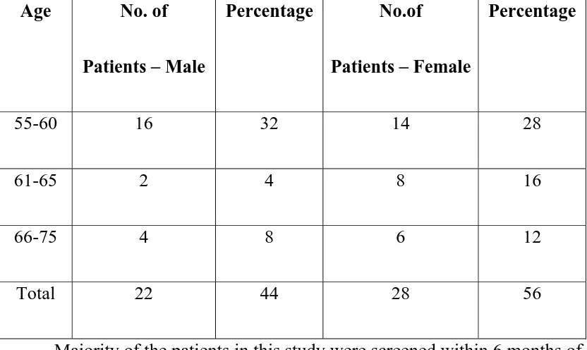
TOTAL NO OF PATIENTS - 50
Age No. of
Patients – Male
Percentage No.of
Patients – Female
Percentage
55-60 16 32 14 28
61-65 2 4 8 16
66-75 4 8 6 12
Total 22 44 28 56
Majority of the patients in this study were screened within 6 months of the start of symptoms of uremia patients were already under taking treatment for diabetes mellitus other with oral hypoglycaemic agents or insulin.
Majority of the patients were in the age group 55-60yrs, accounting to about 60%-men 32% & women 28%.
SEX DISTRIBUTION
No. of Patients – Male No.of Patients – Female Percentage
AGE DISTRIBUTION
0 5 10 15 20 25 30
No. of Patients – Male No.of Patients – Female
FUNDUS PICTURE OF JEYARAMAN – 55/ M – CASE- 1 (MODNPDR + CSME)
FUNDUS PICTURE OF CHINNASAMY – 57/ M – CASE- 4 (MODNPDR + GRADE 1 HTR CHANGES )
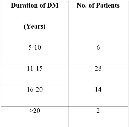
TABLE 2
DURATION OF DIABETES MELLITUS
IN RELATION TO ESRD
TOTAL NO OF PATIENTS -50
Duration of DM
(Years)
No. of Patients
5-10 6 11-15 28 16-20 14 >20 2
Only NIDDM patients were taken in the study
All patients were known to be diabetic for more than five years.
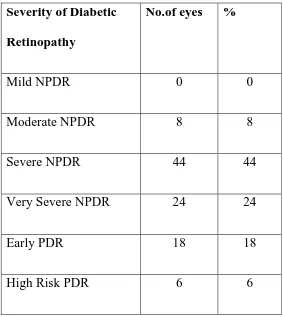
Table 3
SEVERITY OF DIABETIC RETINOPATHY
AT BASELINE
TOTAL NO OF EYES = 100
Severity of Diabetic
Retinopathy
No.of eyes %
Mild NPDR 0 0
SEVEIORITY OF RETINOPATHY 1
0 5 10 15 20 25 30 35 40 45
No.of eyes %
Mild NPDR Moderate NPDR Severe NPDR Very Severe NPDR Early PDR
High Risk PDR
All patients of NIDDM with ESRD had retinopathy.
Diabetic Retinopathy is graded under ETDRS classification.
Non proliferative retinopathy in the form of severe retinopathy is the commonest retinopathy.
FUNDUS PICTURE OF LAKSHMANAN – 56/ M – CASE- 6 (SNPDR + GRADE 1 HTR CHANGES )
FUNDUS PICTURE OF RAMESH – 59/ M – CASE- 9
(SNPDR + CSME )
BASE LINE RETINOPATHY
[image:54.612.127.481.331.647.2]FOLLOWUP RETINOPATHY
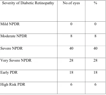
TABLE -4
SEVERITY OF DIABETIC RETINOPATHY
AT FOLLOW UP :
TOTAL NO OF EYES :100
Severity of Diabetic Retinopathy No.of eyes %
Mild NPDR 0 0
Moderate NPDR 8 8
Severe NPDR 40 40
Very Severe NPDR 28 28
Early PDR 18 18
0
5
10
15
20
25
30
35
40
No.of
eyes
%
Mild
NPDR
Moderate
NPDR
Severe
NPDR
Very
Severe
NPDR
Early
PDR
High
Risk
PDR
4 18 6 28 28 40 0
Follow up examination of retinopathy was done at the end of 6 months and one year.
Majority of the eyes was found to be stable at the end of 6months and one year.
Table 5
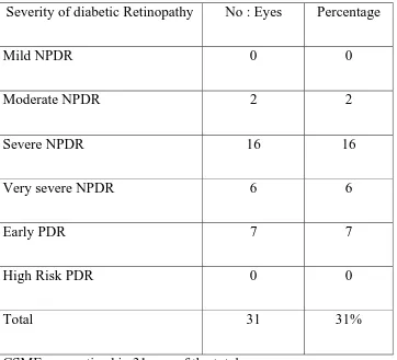
CLINICALLY SIGNIFICANT MACULAR EDEMA IN
DIALYSED ESRD AT BASELINE
TOTAL NO OF EYES : 100
Severity of diabetic Retinopathy No : Eyes Percentage
Mild NPDR 0 0
Moderate NPDR 2 2
Severe NPDR 16 16
Very severe NPDR 6 6
Early PDR 7 7
High Risk PDR 0 0
Total 31 31%
CSME was noticed in 31eyes of the total.
FUNDUS PICTURE OF VEILVENDAN -62 /F – CASE- 33 (VSNPDR )
FUNDUS PICTURE OF PANDI– 66/ M – CASE- 41
(EPDR + CSME)
TABLE 6
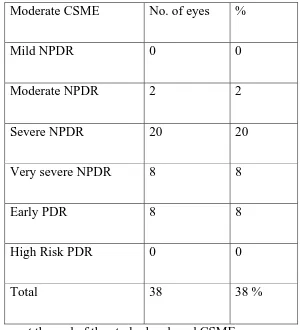
CLINICALLY SIGNIFICANT MACULAR EDEMA IN
DIALYSED DIABETIC ESRD AT FOLLOWUP
TOTAL NO. OF EYES = 100
Moderate CSME No. of eyes % Mild NPDR 0 0 Moderate NPDR 2 2 Severe NPDR 20 20 Very severe NPDR 8 8 Early PDR 8 8 High Risk PDR 0 0
Total 38 38 %
7 new eyes at the end of the study developed CSME No evidence of hole or cystoids changes occurred
TABLE 7
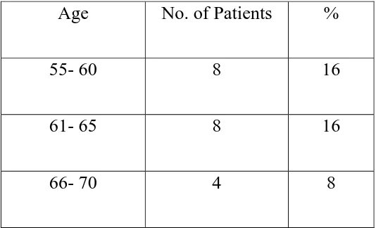
HYPERTENSION IN DIABETIC ESRD ON DIALYSIS
TOTAL NO. OF PATIENTS = 50
Age No. of Patients %
55- 60 8 16
61- 65 8 16
66- 70 4 8
Hypertension was presenting 40% of the study group -20 patients.
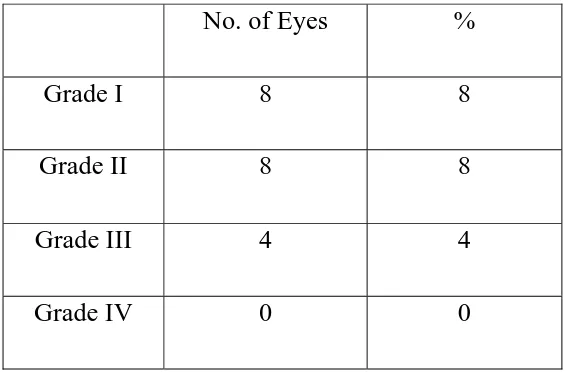
TABLE 8
INCIDIENCE OF HYPERTENSION RETINOPATHY
TOTAL NO. OF EYES = 100
No. of Eyes %
Grade I 8 8
Grade II 8 8 Grade III 4 4 Grade IV 0 0
FUNDUS PICTURE OF KAMAYE –67/F – CASE- 47 (HPDR)
FUNDUS PICTURE OF KAMATCHI –68/F – CASE- 50 (HPDR)
TABLE 9
VISUAL ACUITY IN DIABETIC ESRD ON DIALYSIS
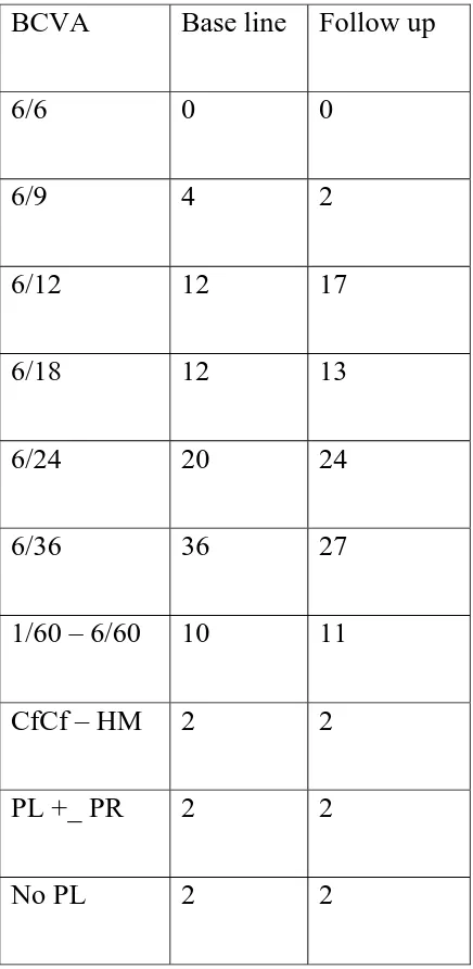
TOTAL NO OF EYES = 100 BCVA Base line Follow up 6/6 0 0 6/9 4 2 6/12 12 17 6/18 12 13 6/24 20 24 6/36 36 27 1/60 – 6/60 10 11
Visual acuity of no PL in two eyes was due to the presence of tractional retinal detachment majority of the visual acuity remained stable at the end of 6months. Few eyes showed progression of cataract.
Drop in visual acuity in three eyes was due to hard exudates near the macula.
PAN RETINAL PHOTO COAGULATION OF KAMATCHI CASE- 50
DISCUSSION
This
study was a nonrandomized prospective study including 50 patients with ESRD on dialysis of Diabetes mellitus origin conducted in a tertiary hospital in South India. Patients were referred from the department of nephrology for detailed opthalmological checkup. Renal failure was diagnosed with the criteria mentioned in patients and methodology. All patients underwent routine haemodialysis. Most of the patients due to reasons of inability to come for the ideal 3week schedule underwent 1 week dialysis schedule. No difference is tried to be made with respect to whether acetone or bicarbonate dialysis was done.AGE DISTRIBUTION
SEX DISTRIBUTION
Males and females were studied for their distribution in relation to retinopathy and nephropathy. It was found that ESRD of diabetic origin showed a slight female preponderance accounting for about 56%. John et al in the Indian study at CMC, Vellore showed a striking male preponderance especially with early nephropathy. As nephropathy proceeds renal failure it was not possible to conclude if males were effected earlier in this study. In the study conducted by Berman et al similar results were noted with females undergoing dialysis in upto 68% of the total. Robert C. Ramsamy et al showed almost equal distribution of males and females in their study. Three suppositions were given by Avram et al to explain why there are more women among the dialysed diabetic studied (1) women with diabetes mellitus live longer with the disease than men do. (2) women with diabetes may be more metabolically stable (3) More women are willing to accept dialysis and endure stress than are men.
CLASSIFICATION
Retinopathy, background retinopathy macular edema, nonproliferative retinopathy + macular edema, proliferative retinopathy, vitreous hemorrhage and tractional retinal detachment. More simpler classification have also been followed like in the Jean-Louis Brenato Fenck study mentioned in Diabetic Renal – retinal syndrome as early, edematous, mixed, ischemic and complicated. People with hypertension were classified in 4 groups as common arteriosclerosis, cross sign, exudates and papilledema.
RETINOPATHY AND NEPHROPATHY
Frequency of retinopathy and nephropathy is explained in the WESDR study. An increasing frequency of retinopathy was found with increasing duration of diabetes. Prevalence rose sharply from 13.61% in those with diabetes of less than 5 years to 95.3%. In those with diabetes hypertension are present in all cases with significant proportion of people dying of uremia when there is associated underlying proliferative retinopathy.
Bermal et al which showed a high percentage of background retinopathy and non-proliferative retinopathy in their study in the range of 52%.
Robert C Ramsamy et al in the study conducted on insulin dependent diabetes ESRD patients found proliferative diabetic retinopathy to be common at 59% at the outset of dialysis. Studies conducted by Engrafov et al showed that main risk factor or risk of non proliferative retinopathy are the duration and degree of compensation of diabetes mellitus and stage of diabetic nephropathy. In this study the underlying nephropathy is severe needing medical and renal replacement therapy hence the presence of very severe, severe and proliferative retinopathy being common. To increase the specificity of renal insult and retinopathy Schwartz et al showed histological evidence of Kimmelstiel Wilson nodules and sever retinopathy. No renal biopsy was however done in any of our cases.
HYPERTENSION
Incidence of hypertension and associated retinoapathy was 40% in our group of study. Epidemological studies mention hypertension as a risk factor for the development of diabetic retinopathy and may be superimposed on diabetic retinopathy.
common retinopathy graded was group 1& 2 of Keith wagner Barker classification. Hypertension could be the underlying risk factor for the development of severe retinopathy.
CSME
Clinically significant macular edema was noted in a significant proportion of patients at the out set of dialysis. It was noted to be 31%. The main visual morbidity in diabetes mellitus is CSME. The WESDR study noted significant also associations of proteinuria and macular edema was the commonest cause of visual morbidity in the Brener et al study.
VISUAL ACUITY
Berman et al noted 7eyes in background retinopathy and 8eyes of non proliferative retinopathy to have macular edema. Both these groups constituted a majority of the total retinopathy. Prompt treatment in the form of focal, panretinal photocoagulation or both resulted in stabilization of vision. Whereas in out study photocoagulation was not included.
Deterioration in visual acuity in our study demonstrated coincident with CSME and in some to the appearance of hard exudates on macula. Kline et al has suggested that type II DM is a group prone to macular edema. Baseline visual acuity was noted to be stabilized in a majority but this was just a I year follow up which needs to be further followed up for long time. 15eyes underwent photocoagulation for macular edema and showed improvement at the end of six months in the form of improved visual activity of 1-2 lines.
Other patients with clinically significant macular edema were advised to undergo photocoagulation.
SUMMARY
End stage renal disease is a microvascular insult of diabetes where retinopathy is also common. In this study of 50patients all end stage renal disease patients had retinopathy as it was one of the inclusion criteria. Common retinopathy seen was severe, very severe NPDR and PDR.
All patients had NIDDM, in whom renal failure occurs after 5 years of the duration of the disease. Nephropathy is more common among IDDM but as the number of NDDM far exceeds the number of IDDM hence the total number of patients with ESRD is also greater. In India 5% of the diabetic population die of end stage renal disease.
Hypertension was seen in 40% of the study population. Whether it is the primary pathology or secondary result of ESRD is not known. Hypertension is by itself known to be associated with progression of non proliferative to proliferative retinopathy macular edema and retinopathy stabilization need good control of hypertension.
Progression of retinopathy was seen in minority of 4 eyes at the six months.
Macular edema was seen in 31 eyes at the onset of dialysis. Seven new eyes developed macular edema at the end of six month follow up.
Macular edema is a pathology that needs prompt treatment. 15% eyes that underwent photocoagulation showed 1-2 line improvement.
Better understanding of importance of control of hypertension, improvement in hemodynamic monitoring, increased accessibility of dialysis units have improved the quality of life of people on dialysis and provide an opportunity for timely opthalmological intervention.
CONCLUSION
End stage renal disease of NIDDM is always associated with retinopathy as both are microvascular damage of the same disease of the same disease process.
Severe NPDR is the most common retinopathy.
Hypertension occurs in 40% of ESRD patients of NIDDM.
Combined retinopathy accounts for about 40% of the retinopathy.
Patients between 55-60yrs who are known to be diabetic are commonly associated with ESRD and retinopathy.
ESRD is found to be commonly associated with females.
Macular edema is seen in 31% of the patients with retinopathy.
Macular edema stabilizes with control of hypertension and dialysis.
New eyes without macular edema could however also develop macular edema during hemodialysis.
Diabetic retinopathy stabilizes on a short term basis in people with ESRD on dialysis.
Macular edema accounts for a subset of visual morbidity.
Prompt treatment in the form of photocoagulation and vitrectomy is utomost importance to reduce the visual impairment and improve quality of life.
It is also important for patients with diabetic ESRD to undergo opthalmological evaluation regularly.
Bibilography
1. Weather et all oxford text book of medicine Vol.III oxford Univ Press 1996; 3294 -5.
2. Duke elders S.Dohree JH.System of ophthalmology Vol X.1st ed chapter 4, 315 – 47.
3. Stein, JH et al. Internal Medicine 3rd .ed USA little brown and comp 1990; 809 – 10 .
4. Klassen – Broekman, Van Bijsterveld OP.Red eyes in renal failure Brit J. Ophthalmol 1992; 76: 268-71.
5. Klassen – Broekman. Van Bijsterveld. The role of serum calcium in development of acute red eye in CRF – EVR J OPHTHALMOL 1995; 5: 7-12.
6. Cohen SL – et al. Pingueculae – an association with renal failure. Queensland Jmed 1974; 43: 281-91.
7. Duane JD, Duanes Clinical ophthalmology Vol.5. Revised ed USA; harper and Row 1987; Chapter 31:1-2.
9. Peyman GA, sanders DR principles and practice of ophthalmology. Newdelhi – Jaypee brother’s 1987; 1205-35.
10. Schier RW, GOHchalki; Disease of Kidney Vol. I-II little brown and camp 1993; 364:1563.
11. Schmechel H, Heinrich U. Retinopathy and nephropathy in 772 msulin – treated diabetic patients in relation to the type of diabetes, Diabetes metabol; 1993:19:135-42.
12. Janka HV, Zlegler AG, Impact of blood pressure on diabetic retinopathy Diabetic metabol 1989;15:333-7.
13. Ley’s AM, Eye fundus of diabetic patient with nephropathy and hypertensive retinopathy Bull Soc Belge ophthalmol 1995;256:49-59. 14. Schelter F; Brass H, Morbidity of 565 type 2 diabetic patients according
to the stage of nephropathy, J diabetes complication 1998;12:103-9. 15. Stephen F.Ryan, Ann K. Danson, Huntel L.Little. Retinal diseases
P911-934.
17. ETDRS group – Grading diabetic retinopathy from stereoscopic color fundus photographs. An extension of modified Airlie House classification ETDRS REPORT No 10. opthal vol.98 may 1991 P.786-806.
18. ETDRS group – classification of Diabetic retinopathy from fluorescein Angiograms : ETDRS REPORT 11 – ophthal Vol.98 May 1991, P.807-822.
19. Miller JW, Adamis AP.Vascular endothelial growth factor/vascular permeablitic factor AMJ Pathol 1994; 145: 574-84.
20. Jaakols P mole DR, Jian. Tangeting of HIF to the von hippel lindau complex by 02 regulated prolyl hydroxylation science 2001; 292:4.
21. Scheilter T.Holkenh, Brass H, morbidity of 505 type 2 diabetic patients according to the stage of nephropathy, J diabetes complication
1998;12:103-9.
22. The Diabetic retinopathy study group. Relationship of adverse treatment effects to retinopathy severity diabetic retinopathy study report No.5. Dev Ophthalmol 1981; 2:248 (Kragel, Basel).
24. Yazdani I, Ahmed S, Channa A, Gagoor I – A correlation of the eye and kidney the diabetes mellitus and hypertension.
25. Tokuogama T, Ikedat, satok – effects of hemodialysis on macular leakage BJO.84 (12) 2000 Dec:1397-1400.
26. David S.Hall bell, MB Jimmy Alele, MD- Dealing with diabetic nephropathy Vol.105 1999 February. Post Graduate Medicine.
27. Jalai S. Das JP. Augmented panretinal photocoagulation for proliterative diabetic retinopathy. Afro Asian J, Opthalmol 1993;12. 257-59.
28. Ramsay RC, Cantrill HL et al visual status in diabetic Patients following therapy for end. Stage nephropathy; in friedman EA, L’s Esperance FA (Cds). Diabetic renal –retinal syndrome, or lands, Grune and Stratton 1986 Vol3 PP 443 – 451.
29. Kohner Km, Aldington SJ, Nugent Z.Retinopathy at entry m united kingdom. Prospective diabetes study (UKPS) of maturity onset diabetes. Diabetes 1987;36:42A.
31. Williams G.A, Scott IY, Hallen. A report by academy of Ophthalmology 2004; 111:1055-62
32. Nguyen Q, D. Vascular endothelial growth factor is a critical stimulus for Diabetic macular edema. Am J Ophthalmol 2006; 142:961-9.
PROFORMA
Name Age Sex
Mrd.No. Diagnosis
History of Diabetes Mellitus - Duration Hypertension - Duration Renal Parameters BUN
Creatinine Treatment History
Haemodialysis Frequency Type of Dialysis Duration
Ophthalmological evaluation. RE LE Best corrected visual acuity.
Anterior segment evaluation.
Lids
EOM
Pupil
Lens
Slit lamp biomicroscopy with 90D Fundus Direct Opthalmoscopy
Fundus Indirect Opthalmoscopy Fundus photographs
Fundus flourescein angiography Grading of retinopathy
81 MASTER CHART
S.
NO Name Age Sex OP.No
H/o DM Yrs H/o DM Yrs V/A"0"
Months Retinopathy "0" Months
V/A"0"
Months Retinopathy "6" Months
RE LE RE LE RE LE RE LE
1 Jeyaraman 55
M
470121 7 6/9 6/9
Mod NPDR+ CSME
Mod NPDR
+ CSME 6/9 6/18
Mod NPDR+ CSME
Mod NPDR + CSME
2 Muthuraman 55
M
471341 8 7 6/12 6/12
Mod NPDR+ CSME+ GrIHTR Mod NPDR+ CSME+ GrIHTR 6/9 6/18 Mod NPDR+ CSME+ GrIHTR Mod NPDR+ CSME+ GrIHTR
3 Lakshmanan 56
M
471432 12 8 6/12 6/12
Mod NPDR+ GrIHTR
Mod NPDR+
GrIHTR 6/12 6/12
Mod NPDR+ GrIHTR
Mod NPDR+ GrIHTR
4 Chinnaswamy 57
M
471741 13 10 6/12 6/12
Mod NPDR+ GrIHTR
Mod NPDR+
GrIHTR 6/12 6/12
Mod NPDR+ GrIHTR
Mod NPDR+ GrIHTR
5 Jegan 57
M 471934 11 8 6/12 6/12
SNPDR+ GrIHTR
SNPDR+
GrIHTR 6/12 6/12
SNPDR+ GrIHTR
SNPDR+ GrIHTR
6 Lakshmanan 56
M
482130 12 10 6/12 6/12 SNPDR+
GrIHTR
SNPDR+
GrIHTR 6/12 6/12
SNPDR+ GrIHTR
SNPDR+ GrIHTR
7 Rajesh 58
M
482242 9 6 6/12 6/12
SNPDR+ CSME- GrIHTR SNPDR+ CSME+ GrIHTR 6/12 6/12 SNPDR+ CSME+ GrIHTR SNPDR+ CSME+ GrIHTR
8 Ramaiah 59
M
482450 8 7 6/12 6/12
SNPDR+ CSME+ GrIHTR SNPDR+ CSME+ GrIHTR 6/12 6/12 SNPDR+ CSME+ GrIHTR SNPDR+ CSME+ GrIHTR
9 Ramesh 59
M
482452 11 5 6/36 6/36 SNPDR+
CSME
SNPDR+
CSME 6/12 6/12
SNPDR+ CSME
82
10 Suresh 59
M 484350 11 6/36 6/36
SNPDR+ CSME
SNPDR+
CSME 6/12 6/12 SNPDR
SNPDR+ CSME
11 Selvaraj 60
M 484431 10 6/36 6/36
SNPDR+ CSME
SNPDR+
CSME 6/12 6/18 SNPDR
SNPDR+ CSME
12 Ganesh 60
M 484521 10 6/36 6/36
SNPDR+ CSME
SNPDR+
CSME 6/18 6/18 SNPDR
SNPDR+ CSME
13 Vasan 60
M
484630 11 6/36 6/36 SNPDR+
CSME
SNPDR+
CSME 6/18 6/18
SNPDR+ CSME
SNPDR+ CSME
14 Pandian 60
M 484680 11 9 6/36 6/36
SNPDR+ CSME
SNPDR+
CSME 6/18 6/18
SNPDR+ CSME
SNPDR+ CSME
15 Seetha 56
F 484757 12 9 6/24 6/24
SNPDR+ GrIHTR
SNPDR+
GrIHTR 6/24 6/24
SNPDR+ GrIHTR
SNPDR+ GrIHTR
16 Ambika 57
F 484850 12 3 6/24 6/24
SNPDR+ GrIHTR
SNPDR+
GrIHTR 6/24 6/24
SNPDR+ GrIHTR
SNPDR+ GrIHTR
17 Alamelu 58
F
484900 13 4 6/24 6/24 SNPDR+
GrIHTR
SNPDR+
GrIHTR 6/24 6/24
SNPDR+ CSME+ GrIIHTR SNPDR+ CSME+ GrIIHTR
18 Rajammal 58
F
484971 13 7 6/24 6/24 SNPDR+
GrIHTR
SNPDR+
GrIHTR HM HM
SNPDR+ CSME+ GrIIHTR SNPDR+ CSME+ GrIIHTR
19 Sumathi 59
F
484980 12 6/24 6/24 SNPDR SNPDR 6/24 6/24
SNPDR+ CSME+ GrIIHTR SNPDR+ CSME+ GrIIHTR
20 Laksmi 58
F 490000 12 6/24 6/24 SNPDR SNPDR 6/24 6/24
SNPDR+
CSME SNPDR
21 Muthu 59 M 491321 11 6/12 6/12 SNPDR SNPDR 6/18 6/18 SNPDR SNPDR
83
23 Veeralakshmi 58 F 494231 12 6/24 6/24 SNPDR SNPDR 6/24 6/24 SNPDR SNPDR
24 Karpagam 57 F 494421 12 6/24 6/24 SNPDR SNPDR 6/24 6/24 SNPDR SNPDR
25 Kumari 60 F 494700 15 6/36 6/36 SNPDR SNPDR 6/36 6/36 SNPDR SNPDR
26 Velammal 59 F 495200 15 6/36 6/36 SNPDR SNPDR 6/36 6/36 SNPDR SNPDR
27 Radhika 55
F 496111 14 6/36 6/36
VSNPDR+
CSME VSNPDR 6/36 6/36 VSNPDR VSNPDR
28 Sundari 57
F 13 6/36 6/36
VSNPDR+
CSME VSNPDR 6/36 6/36 VSNPDR VSNPDR
29 Kathayee 56 F 496321 12 6/36 6/36 VSNPDR VSNPDR 6/36 6/36 VSNPDR VSNPDR
30 Ramaselvi 56 F 496421 12 6/36 6/36 VSNPDR VSNPDR 6/36 6/60 VSNPDR VSNPDR
31 Keerthi 62
M
496520 12 8 6/36 6/36
VSNPDR+ CSME GrIIHTR VSNPDR+ CSME GrIIHTR
6/36 6/60 VSNPDR+
GrIIHTR
VSNPDR+ GrIIHTR
32 Kumaran 63
M
497324 12 7 6/18 6/18
VSNPDR+ CSME GrIIHTR VSNPDR+ CSME GrIIHTR
6/36 5/60 VSNPDR+
GrIIHTR
VSNPDR+ GrIIHTR
33 Veilvandan 62 F 497338 12 6/18 6/18 VSNPDR VSNPDR 2/60 6/18 VSNPDR VSNPDR
34 Kannammal 62 F 497426 12 6/18 6/18 VSNPDR VSNPDR 6/18 6/18 VSNPDR VSNPDR
35 Laksnmi 62
F 497440 16 6/18 6/18
VSNPDR+ GrIIHTR
VSNPDR+
GrIIHTR 6/12 6/18
VSNPDR+ GrIIHTR
VSNPDR+ GrIIHTR
36 Periammal 62
F 497523 16 6 6/36 6/36
VSNPDR+ GrIIHTR
VSNPDR+
GrIIHTR 6/24 6/24
VSNPDR+ GrIIHTR
VSNPDR+ GrIIHTR
37 Perumaye 65
F
497538 16 8 6/36 6/36 VSNPDR+
GrIIHTR
VSNPDR+
GrIIHTR 6/36 5/60
VSNPDR+ GrIIHTR
84
38 Subbu 65
F
498420 16 5 6/36 6/36 VSNPDR+
GrIIHTR
VSNPDR+
GrIIHTR 5/60 5/60
VSNPDR+ GrIIHTR
VSNPDR+ GrIIHTR
39 Petchi 61
F
499804 17 3 6/36 6/36 EPDR+
GrIIHTR
VSNPDR+
GrIIHTR 6/24 6/24
EPDR+ GrIIHTR
VSNPDR+ GrIIHTR
40 Chandra 62
F
499916 17 6 6/36 6/36 EPDR+
CSME
VSNPDR+
GrIIHTR 6/24 6/24
EPDR+ CSME
VSNPDR+ GrIIHTR
41 Lakshmi 66
M
503424 17 8 6/36 6/36 EPDR+
CSME
EPDR+
CSME 6/24 6/24
EPDR+ CSME
EPDR+ CSME
42 Sundaram 67
M
513632 17 9 1/60 5/60 EPDR+
CSME
EPDR+
CSME HM 5/60
EPDR+ CSME
EPDR+ CSME
43 Veerasamy 67
M
513754 17 10 5/60 2/60 EPDR+
CSME
EPDR+
CSME 5/60 HM
EPDR+ CSME
EPDR+ CSME
44 Sundaram 67 M 516432 17 8 6/36 6/36 EPDR VSNPDR 6/36 6/36 EPDR VSNPDR
45 Meenakshi 66 F 527436 17 8 1/60 3/60 HPDR EPDR 1/60 3/60 HPDR EPDR
46 Raasathi 67 F 538327 16 1/60 3/60 HPDR EPDR 1/60 3/60 HPDR EPDR
47 Kamaye 67 F 549438 16 2/60 Cfcf HPDR EPDR 2/60 Cfcf HPDR EPDR
48 Saraswathi 67 F 553674 16 2/60 Cfcf HPDR HPDR 2/60 Cfcf HPDR HPDR
49 Revathi 67 F 564384 20 Nopl Pl+Pr HPDR HPDR NOP1 Pl+Pr HPDR HPDR
85
MASTER CHART KEY
DM- Diabetes Mellitus HR - high risk
PR - projection of ray of light
HT-Hyper tension NPDR - non proliferatvie diabetic retinopathy HM - hand movement
Yrs - Years PDR - proiferative diabetic retinopathy
RE-Right Eye PL - perception of light
LE- Left Eye Mod - moderate
V/A - Visual acuity VS - very severe
S- Severe