A Study of Percutaneous
Nephrolithotomy (PCNL) and Grading
of Complexity of PCNL procedures using
“Guy’s Stone Score”
Dissertation submitted in partial fulfillment
of the requirements for the degree of
M.Ch (Urology) – Branch IV
STANLEY MEDICAL COLLEGE & HOSPITAL
THE TAMIL NADU DR. M.G.R. MEDICAL UNIVERSITY
CHENNAI, INDIA
CERTIFICATE
This is to certify that this dissertation entitled “A Study of
Percutaneous Nephrolithotomy (PCNL) and Grading of Complexity
of PCNL procedures using “Guy’s Stone Score” is a bonafide work
done by Dr. K.Karthikeyan in partial fulfillment of the requirements of
The TAMIL NADU DR.M.G.R. MEDICAL UNIVERSITY, Chennai
for the award of M.Ch Urology Degree.
Prof. Dr.S.GEETHALAKSHMI.
M.D, Ph.D
DEAN
Stanley Medical College & Hospital, Chennai - 600001
Prof. Dr. V. SELVARAJ,
M.S, M.Ch, (Urology)
Professor and Head of the Department of Urology
Stanley Medical College & Hospital Chennai - 600 001
DECLARATION
I, Dr.K.Karthikeyan solemnly declare that the dissertation
titled “A Study of Percutaneous Nephrolithotomy (PCNL) and Grading
of Complexity of PCNL procedures using “Guy’s Stone Score” is a
bonafide work done by me at Govt. Stanley Medical College & Hospital
during February 2012 to March 2013 under the guidance and
supervision of Prof. Dr.V.Selvaraj, M.S., M.ch. (Urology) Professor
and Head Of The Department, Department of Urology.
The dissertation is submitted to Tamil Nadu, Dr.M.G.R
University, towards partial fulfillment of requirement for the award of
M.Ch. Degree( Branch-IV) in urology three years course
Place: Chennai
Date: Dr. K. Karthikeyan
GUIDE: Prof.Dr.P.Govindarajan. M.S., M.ch.(Urology)
ACKNOWLEDGEMENT
I owe my thanks to Dean, Government Stanley Medical College
and Hospital, Prof. Dr. S. Geetha Lakshmi M.D., PhD, for allowing
me to avail the facilities needed for my dissertation work.
I immensely thank my Professors Dr. V. Selvaraj, M.S., M.Ch.
H.O.D Department of Urology, Dr. P. Govindarajan , M.S., M.Ch. for
the valuable guidance and help rendered in completing this dissertation.
I extend my thanks to Dr.M.Deepak M.S., M.Ch.,
Dr A.R.Balaji M.S., M.Ch., Dr.Periasamy M.S., M.Ch., and
Dr. P. V. Thiruvarul M.S., M.Ch. for their valuable support and
guidance during the dissertation work.
I would like to thank my fellow post graduates who gave me
excellent cooperation during the dissertation work. A special thanks to
biostatistician Mr. Boopathy for having helped me with the statistics.
I also express my gratitude to all the patients who were subjects
CONTENTS
S.NO TOPIC PAGE NO
1. INTRODUCTION 1
2. AIM OF THE STUDY 3
3. REVIEW OF LITERATURE 4
4. MATERIALS AND METHODS 47
6. RESULTS 50
7. DISCUSSION 65
8. CONCLUSION 68
9. BIBLIOGRAPHY 69
APPENDIX
i) PROFORMA
INTRODUCTION
During the last two decades , the management of kidney stones
has vastly changed. Prior to these modifications all the kidney stones
were managed by open pyelolithotomy or nephrolithotomy which
caused a significant morbidity for the majority of patient. Percutaneous
Nephrolithotomy (PCNL) has now largely replaced open surgery as a
safe and effective treatment for renal stones 1.
It is now well recognized among surgeons that PCNL procedures
have different degrees of complexity which affects stone clearance.
The “Guy’s Stone Score” proposed by Thomas K and Smith et al 2,3,
is a valuable tool to stratify the complexity of PCNL procedures into
four groups based on the stone burden and the anatomy of both patient
and renal tract.
Grade I : Solitary stone in mid / lower pole or solitary stone in
pelvis with simple anatomy
Grade II : Solitary stone in upper pole or multiple stones in
patient with simple anatomy or solitary stone in patient with abnormal
Grade III : Multiple stones in a patient with abnormal anatomy or
stones in a caliceal diverticulum or partial Staghorn calculus
Grade IV : Staghorn calculus or any stone in a patient with spina
bifida/spinal injury.
AIMS AND OBJECTIVES
1) To evaluate patients with renal stones at our institution.
2) To study the indications for PCNL and to assess the outcome of
procedure in patients with renal stones.
3) To study the grading and complexity of PCNL procedures using
REVIEW OF LITERATURE
HISTORY
Dr Thomas Hillier MD was the first to publish a method of
percutaneous nephrostomy in 1865, he repeatedly drained a congenitally
obstructed kidney in a 4- year old boy. Goodwin and Casey in 1955
placed a trocar percutaneously in the collecting system. Later, the
Seldinger method of nephrostomy placement was adopted. The rst
percutaneous nephrolithotomy (PCNL) via a nephrostomy tract created
for the sole Purpose of stone removal was performed in 1976 by
Fernstrom and Johansson1 .
Untreated large staghorn calculi had a high 10 year mortality of
(28%)in comparison with surgery(7.2%).In 2005, the clinical practice
guideline report for the management of staghorn calculi by the
American Urological Association guidelines4 panel confirmed that
percutaneous treatment of staghorn calculi should be first-line treatment
RELEVANT ANATOMY
The kidneys are paired organs lying retroperitoneally on the
posterior abdominal wall. Each kidney is of a reniform shape, having a
upper and a lower pole, a convex border placed laterally, and a concave
medial border. The medial border has a marked depression the hilum
containing the renal vessels and the renal pelvis.
RENAL MORPHOMETRY
In adults, it is found that left kidney is larger than the right
kidney, and this finding is in agreement with morphometric findings in
fetal kidneys. The right kidney presented a mean length of 10.97 cm,
while the left kidney presented a mean length of 11.21 cm. The right
kidney presented 3.21 cm of mean thickness at the hilum, and the left
kidney presented mean thickness of 3.37 cm.1
An interesting and worthwhile finding is that, in the same kidney,
the superior pole has a greater width (mean = 6.48 cm) than the inferior
pole (mean = 5.39 cm). They also found a statistically significant
correlation between the kidney length and the stature of the individuals.
The anterior and the posterior renal arteries are the two main
from anterior division which supply the anterior and polar regions of
kidney. The remainder of kidney is supplied by the posterior segmental
artery. The segmental arteries give rise to the interlobar arteries beyond
the renal sinus and form the arcuate arteries at the corticomedullary
junction. The interlobular arteries branch from the arcuate arteries at
right angles and run to the periphery giving rise to the afferent arterioles
of the glomeruli. Brödel's line separates an avascular plane between
[image:16.595.177.414.351.472.2]the anterior and posterior segmental blood supplies.
Figure: 1
Arterial supply to the kidney. The kidney is supplied by the
anterior and posterior segmental branches of the main renal artery. The
anterior segmental artery supplies both the anterior half of the kidney
and the polar regions. The posterior segmental artery supplies only the
posterior aspect of the kidney (represented by the shaded region).
An avascular plane, known as Brödel's line, separates the anterior and
Figure:2
Location and orientation of kidneys in the retroperitoneum
The intrarenal veins do not follow a segmental structure. Unlike
the arteries, the venous system is freely interconnected. Multiple
anastomotic arcades between the veins prevent parenchymal congestion
and ischemia from venous injury.
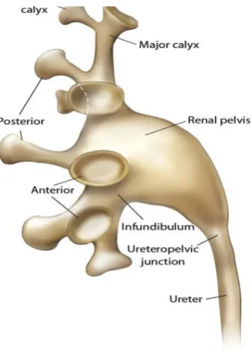
Collecting System Anatomy
The anatomic landmarks dividing the renal parenchyma from the
collecting system are the renal papilla. Calyces in direct apposition to
the renal papilla are defined as minor calyces and vary in number from
5 to 14 (mean: 8). A minor calyx may be single (draining only one
papilla) or compound (draining two or three papillae)5. Minor calyces
which then drain into an infundibulum ( Fig. 3 ). The infundibula are the
principal divisions of the pelvicalyceal system, draining directly into the
renal pelvis. There are usually three renal calyceal groups: the superior,
midzone, and inferior major calyces. Barcellos Sampaio and
Mandarim-de-Lacerda5 (1988) analyzed 140 three dimensional polyester resin
corrosion endocasts of human kidneys and contributed significantly to
our understanding of the intricate anatomy of the pelvicalyceal system.
They observed that the superior and inferior major calyces usually
consist of compound calyces that project toward the polar regions at
various angles. The midzone calyces, on the other hand, are generally
arranged in paired sets of anterior and posterior calyces. These paired
calyces have been observed to display one of two configurations
(Fig. 4). In the Brödel type configuration, the anterior calyx is short and
medially directed (forming a 70-degree angle to the frontal plane of the
kidney), whereas the posterior calyx is longer and more laterally
directed (positioned only 20 degrees from the frontal plane of the
kidney). The second configuration is the Hodson type in which the
posterior calyx is shorter and more medially directed and the anterior
calyx is longer and closer to the lateral edge of the kidney. It has been
shown that 69% of right kidneys exhibit the Brödel configuration and
Basic pelvicalyceal anatomy Figure:3
A Brodel configuration
[image:19.595.210.389.119.367.2]
B Hodson configuration
Calyceal orientations in the Brödel and Hodson configurations. In
the Brödel-type kidney (A), the longer posterior calyx is positioned 20
degrees from the frontal plane of the kidney and the shorter anterior
calyx forms a 70-degree angle with the frontal plane. In the Hodson-type
kidney (B), the shorter posterior calyx is positioned 70 degrees from the
frontal plane of the kidney and the larger anterior calyx forms a
20-degree angle with the frontal plane. Figure: 4
In studying the pelvicalyceal endocasts, Barcellos Sampaio 5
(1988) noted significant variability in the drainage patterns of the three
calyceal groups. The midzone calyceal group was variably found to
have drainage dependent on one of the polar calyceal groups (62%) or to
drain directly into the renal pelvis independent of either polar group
(38%). In 18% of the endocasts studied, the kidney midzone was
drained simultaneously by crossed calyces, one drains into the superior
calyceal group and the other drains into the inferior calyceal group. In
addition, a perpendicular minor calyx which drains directly to the pelvis
was noted in 11% of the endocasts. The only consistently noted findings
were that the superior calyceal group was drained by only one midline
infundibulum in 99% of the endocasts and paired calyces drain the
midzone. They are found to lie in two rows (anterior and posterior)
Clinical Relevance of Intrarenal Anatomy
A thorough understanding of intrarenal anatomy is essential for a
safe percutaneous puncture and minimize complications. Appreciation
of the anterior and posterior segmental blood supply of the kidney can
allow the urologist to utilize Brödel's line during percutaneous puncture.
A needle traversing the renal parenchyma posterolaterally through this
avascular plane avoids damage to any major blood vessels. More medial
punctures in the superior calyx may injure the posterior segmental
artery. The posterior segmental artery is the most commonly injured
vessel in endourologic procedures. Knowledge of the Hodson and
Brödel configurations of calyceal anatomy is crucial for precise
preoperative localization of a stone or other lesion on intravenous
pyelogram. Awareness of the great variability in calyceal drainage
patterns can aid greatly during intraoperative decision making for
appropriate puncture sites. The results of the Sampaio endocast studies
imply that it is easy to puncture a polar region which is drained by a
single infundibulum than a polar region drained by paired calyces.
Furthermore, the anatomic relationships of the intrarenal vessels to the
kidney collecting system predict a high rate of vascular injury for
percutaneous entry which is direct into the fornix of a calyx is the safest
route. 6
Preview of any renal access involves examination of the desired
calyx. The calyx is inspected for three factors: relation to the 12th rib,
extent of hydronephrosis, and presence of malrotation. Whether the
desired calyx resides above or below the 12th rib has critical
significance for the technique chosen for renal access and the possibility
of pleural injury. The degree of dilatation influences the difficulty of
renal puncture. Improper technique may still result in failure even in a
well dilated system. Finally, the unusual case of the malrotated or
ectopic kidney may necessitate minor adjustments in the access
technique.
INDICATIONS FOR PCNL:
Percutaneous stone extraction is the primary modality to treat
patients with large stones size more than 2 cms, obstructing kidney
stones (e.g., staghorn calculi) or stones with composition resistant to
fragmentation with extracorporeal lithotripsy 1. In addition, for patients
with concomitant renal stones and distal narrowing (e.g., infundibular
stenoses and coexisting calyceal stones, stones in calyceal diverticula, or
percutaneous route allows a convenient approach to address both
problems simultaneously. For patients with UPJ obstruction (even in the
absence of stones), percutaneous endopyelotomy provides an effective
alternative to laparoscopic or open pyeloplasty with acceptable success
rates.
IMAGING MODALITIES FOR PERCUTANEOUS ACCESS
Ultrasonography
Percutaneous ultrasound-guided nephrostomy7 is perhaps the
simplest and most direct technique to access and drain a hydronephrotic
collecting system. It is most often utilized to place a temporary urinary
diversion in the instances of an obstructing stone or pyonephrosis and
has also been used successfully to relieve obstruction secondary to
malignant compression. Although the technique has been especially
popular among interventional radiologists, it has gained popularity
among endourologists who are comfortable with ultrasonography.
Allergies to topical or injectable local anesthetic and coagulopathy are
the only relative contraindications to ultrasound-guided renal access.
The ultrasound guided access has no radiation hazard and allows to
image the structures across skin and kidney. Ultrasound access is safe in
Computed Tomography and Magnetic Resonance Imaging
CT-guided percutaneous access, is useful in special situations8.
A CT- or MRI-guided approach is a time-consuming and expensive
method that is not practical for most patients and needs to be considered
only if the aforementioned techniques are not feasible or do not provide
good results or if sophisticated preoperative planning is necessary.
Patients with a retrorenal colon or a abnormal anatomy due to spinal
anamoly predictably require cross-sectional imaging to facilitate safe
access before percutaneous nephro lithotomy 9. In addition, the CT
guided approach may be useful in obtaining renal access in patients with
ileal conduits, renal uric acid stones, or nephrolithiasis in the presence of
angiomyolipomas at risk for bleeding . Three-dimensional CT has been
described as a valuable tool for obtaining percutaneous access in the
morbidly obese with malrotated kidneys and large staghorn calculi 8.
There are no specific indications for MRI-guided percutaneous
nephrostomy, although the technique has been shown to be feasible and
FLUOROSCOPY
Endourologic procedures most often rely on fluoroscopy 10. Although
the risk is relatively small, everyone involved are exposed to radiation
which includes the patient, surgeon and other staff. The endourologist,
in particular, must undertake protective measures because he or she will
have radiation exposure regularly. Likewise, children with
nephrolithiasis secondary to cystinuria may be subjected to repeated
fluoroscopy-based procedures. Children are more vulnerable to
radiation. The two major risks associated with fluoroscopy are,11
Radiation-induced injuries to the skin and underlying tissues
(“burns”), and
The small possibility of developing a radiation-induced cancer
some time later in life.
Benefits outweigh the risks involved with fluoroscopy when one
has a medical need. Time and exposure should be kept to a minimum
required.
Skin injuries caused by fluoroscopy include the following 11
Early transient erythema
Telangiectasis
Moist desquamation
Late erythema
Dermal necrosis and Secondary ulceration
Table 1 -- Radiation Safety for Urologists 12
Put fluoroscopy beam under table.
Use lead apron and thyroid shields.
Use minimal fluoroscopic time: “Think before or after fluoroscopy, not during.”
Collimate.
Wear radiation detection devices.
Wear lead-impregnated glasses.
Use lead gloves?
The principle for radiation safety is ALARA: as low as
reasonably achievable. The maximum yearly whole-body exposure
allowed by the National Council on Radiation Protection is 5000 mrem,
or 5 rem12. Though the risk with radiation above standard limit is a small
health risk ,it is substantial over lifetime of a surgeon
antioxidants and glutathione elevating agents may protect tissues from
radiation's mutagenic effects. The use of such antioxidant preparations
may soon extend the concept of ALARA from dose to biologic damage.
Time, distance, and shielding are important factors for safe radiation.
Reducing fluoroscopy time during endourologic procedures is of
primary importance, because the exposure time determines the radiation
dose to the operating room personnel. Newer fluoroscopic equipment
features, including under-table fluoroscopic sources, timer alarms,
collimated x-ray beam, and last-image-holding/memory capability help
the urologist limit the fluoroscopy time. Grid-controlled fluoroscopic
technique may also reduce overall radiation dose by decreasing the
selected film frame rate13. Use of this technique, as opposed to
continuous fluoroscopy, has led to substantial dose reduction for the
patient and fluoroscopy operator without sacrificing image quality or
diagnostic confidence. The major source of radiation to the
endourologist is scatter from the patient's body 14. Radiation is emitted
from a source in all directions, and decreases with distance. Because
scattered radiation follows the inverse square law, operators near the
radiation beam can make significant reductions in exposure by
increasing their distance from the patient 14. For example, if one stands
radiation exposure, or an approximate 89% dose reduction. At a distance
of 12 feet, however, the dose will approximate natural background
levels, not registering on radiation monitoring devices (dosimeters).
During fluoroscopy, the kilo voltage and milliamperage are adjusted
automatically and the operator can control only the duration of the
exposure. Exposure doses may be reduced significantly by minimizing
the total “active” fluoroscopic time for a procedure through cautious
use of the exposure switch to ensure that irradiation occurs only when
there is a need for active viewing of the image. The use of a
last-image-hold feature is of great importance in reducing the overall irradiation
time13. With this feature, anatomic details can be scrutinized without a
competing concern about additional radiation dose. Thus, all
fluoroscopes used for percutaneous surgery should have a
last-image-hold feature so the urologist does not need to “think with a foot on the
pedal.” The irradiated site of the patient affects the scatter rate to the
endourologist. When the field is closer to the midline of the patient, less
radiation is scattered to the operator because it is attenuated through a
greater thickness of overlying tissue. When the field is more lateral, the
radiation is less attenuated by the patient and thus there is more radiation
scatter. Furthermore, in obesity there is a need for more radiation to
surgical drapes composed primarily of bismuth, specialized urologic
radiation shields, and special radioprotective gloves can be used to
substantially reduce scattered radiation dose .Collimation narrows the
beam and limits the imaging area to the exact position of interest, thus
reducing the scattered radiation to and from the patient. Keeping the
image receiver nearer the patient minimizes the distance between the
focal spot and the image receptor, keeps beam intensity as low as
possible, decreases image blur, and is useful as a scatter barrier between
the operator and the patient. In addition, the direction of the beam
significantly influences the amount of scattered radiation reaching the
operator 12. When the tube is above the operating table, there is a
combination of leakage and scattered radiation. However, when the
image intensifier is placed superiorly, radiation leakage is minimized, as
an additional layer of material shields the emission tube. There is a
reduction of scattered radiation to the operator. Shielding involves the
use of flexible protective clothing such as aprons, skirts, thyroid shields,
eyeglasses, and gloves. The basic protection for every urologist during
percutaneous surgery is a lead apron, thyroid shield, and eyeglasses. The
use of protective glasses is prudent, even though there is debate about
their absolute necessity. Nevertheless, approximately 1100 mrem/hr
radiation scatter, which certainly suggests that the use of eye protection
may be beneficial . The standard flexible material for protective clothing
is lead-impregnated rubber. The goal is to provide a barrier between the
radiation source and the operator so that radiation is attenuated by the
shield. Lead aprons are heavy and can become uncomfortable when the
operator is wearing them for a protracted period of time. The amount of
lead required for efficacy has been established as 0.5 mm, and it has
been estimated that the weight of aprons with this much lead ranges
from 2.5 to 7 kg13. Today, however, some aprons are made of
composites of lead with elements of lower atomic numbers so that the
weight can be reduced but the efficacy maintained (Castaneda, 1996)12.
Finally, all personnel exposed to radiation should wear
dosimeters positioned where the operator receives the maximal
radiation15. It has been estimated that the radiation exposure to the
underlying body is as little as 1% of the measured value. In modern
radiation protection practices, active personal dosimeters are absolutely
Percutaneous Access without Imaging—“Blind Access”
Attempting percutaneous access without the aid of imaging is
reserved for the rare instances when retrograde or intravenous
opacification is precluded, the pelvicalyceal system cannot be opacified,
or imaging machinery such as a fluoroscopic unit or sonography is
inaccessible 16. Poor renal function in the presence of ureteral
obstruction, for example, may represent such a situation, especially if
emergent collecting system decompression is required (i.e., urosepsis
from pyonephrosis). Percutaneous access without imaging relies on
anatomic landmarks and the assumption that anatomy is not aberrant.
The lumbar notch, bounded medially by the sacrospinalis and the
quadratus lumborum muscles, and laterally by the transversus abdominis
and the external oblique muscles, superiorly by the latissimus dorsi
muscle and the 12th rib, has been shown to be a useful anatomic
window for successful blind percutaneous calyceal puncture. An
18-gauge access needle can be inserted into the notch at a 30-degree angle
directed cephalad under the 12th rib to a depth of 3 to 4 cm
PREPARATION BEFORE PCNL
A good imaging is necessary. X ray KUB, IVU were used earlier
it will also show retrorenal colon. Urine should be sterile before PCNL.
Bleeding tendencies should be corrected. Medications like aspirin
,NSAIDS should be stopped
DURING PCNL
Broad-spectrum parenteral antibiotics are given to all patients
before surgery 18. After inducing general anaesthesia, the patient is
placed in lithotomy position, cystoscopy and retrograde catherisation
done with 5fr ureteric catheter. Alternatively patient is placed in prone
flexible cystoscopy can be used for RGC. With utmost care to the face
and extremity pressure points, padding of all pressure points ensured.
Site Selection
It is necessary to select the percutaneous nephrostomy tract that is
most suited for a particular procedure.Puncturing the posterior calyx is
preffered because it is straight, gives stability with transparenchymal
path and avoids major vascular structures. In addition, access from an
anterior calyx to the renal pelvis is technically demanding because it
requires directing the wire backward. Posterior segmental artery may be
injured if the pelvis is punctured directly. In general, the risk of injuring
lacks parenchymal support. Collecting system is visualized by injecting
contrast through ureteric catheter. Alternatively, a small amount of air
may be injected to provide an air pyelogram 10. The advantage of air is
that it is lighter than urine or contrast material and therefore identifies
the posterior calyces first, with the patient in the prone position. The
typical appearance of air in a posterior calyx filled with contrast agent
has been described as “Mickey Mouse ears.” With a single stone in the
renal pelvis or when the anatomy is unclear, the use of contrast material
is recommended to precisely delineate the intrarenal anatomy. However,
in the case of multiple radiopaque calyceal or complete staghorn calculi,
an air pyelogram outlines the collecting system satisfactorily and will
not interfere with the evaluation of residual stones or fragments due to
retained or extravasated contrast material. In general, anterior calyces
are more laterally located and posterior calyces are more medially
located (mnemonic LAMP: Lateral-Anterior, Medial-Posterior) 5.
Subcostal approach
With the C-arm in the vertical position, the collecting system is
inspected and the appropriate calyx is identified. The ideal site provides
the shortest tract to the calyx from below the 12th rib. With the C-arm at
vertical plane of the calyceal entry 10. The C-arm is next moved 30
degrees towards the urologist. This places the axis of the C-arm in the
same central posterior plane of the kidney, providing a straight end-on
view of the posterior calyces. After the calyx is chosen, the overlying
skin site is marked with a curved hemostat.
With the C-arm in the 30-degree position an 18-gauge
translumbar angiography needle is advanced in the plane of the
fluoroscope beam. The diamond tip prevents deflection by sharply
cutting through muscle and fascia while causing minimal shearing . In
general, the shorter the needle (11 to 15 cm) the easier it is to control.
Longer needles are necessary for obese patients or when triangulation is
utilized, because this latter technique may require a longer tract or more
flexibility to “bend around” a rib. The appropriate direction for needle
advancement is determined by obtaining a “bull's-eye sign” on the
fluoroscopic screen 10. This effect can be seen only when the needle hub
is superimposed on the needle shaft and is evident when the plane of the
needle is the same as that of the x-ray beam. If the axis of the needle
advancement is not parallel to the axis of the C-arm beam, a segment of
the needle shaft is visible. After determination of the appropriate plane
the hemostat held needle is advanced in 1to 2cm increments. Use of
avascular line of Brödel, because this provides the safest access to the
posterior calyceal system. A transparenchymal route avoids the hilar
vessels and seals the nephrostomy tract from urine leakage. The depth of
needle penetration is monitored by moving the C-arm back to the
vertical position. With the C-arm in the vertical position, the
approximation of the tip of the needle to the predetermined calyx can be
seen and guided fluoroscopically. For example, the needle is too deep if
it appears to be past the calyx on the fluoroscopic screen. Periodically, it
is important to evaluate the correct direction of needle advancement by
rotating the C-arm 30 degrees toward the surgeon and observing for the
bull's-eye effect. Both the appropriate axis and the needle depth are
prerequisites for a successful percutaneous access. The needle has
reached its intended target when its tip is in the desired calyx on both
planes of fluoroscopy. When the needle seems to be in a calyx, the stylet
can be removed and the correct needle position is verified by aspiration
of urine or air, or both. A 0.038-inch floppy-tip J-shaped guide wire is
inserted into the needle and either advanced across the UPJ or coiled
within the renal pelvis. With the needle left in place, a 1-cm skin
incision is made. The needle is then removed and the tract is dilated over
Intercostal Approach.
The risk of hydrothorax and hemothorax is increased when
percutaneous access to the calyces is performed above the 12th rib.
Various techniques to access the superior calyces while minimizing
complications have been described. The direct intercostal approach,
triangulation, indirect access by way of lower calyces, and retrograde
percutaneous nephrostomy have all been described.
Access to an superior pole calyx can be difficult by a subcostal
approach, and the endourologist needs to be familiar with the intercostal
approach6, 10. Many urologists favor this approach for gaining access to
the upper pole and suggest that it is straight and gives viable access to
most staghorn calculi, even though it carries minimal increase in
morbidity . Contemporary series, in contrast to older literature, indicate
that with caution intercostal puncture may be safe and effective. In
particular, care should always be taken to maintain that the access sheath
is secure in the collecting system.
A technique for minimizing the potential morbidity of the
intercostal approach by displacing the kidney caudally has been
described. This is achieved by placing an Amplatz sheath through a
which causes caudal displacement of the kidney that can be viewed
fluoroscopically. A second distinct puncture or a Y-tract is created into
the upper pole. This method was successful in majority of cases without
complications . Also, an occlusion balloon catheter can be used to apply
gentle caudal traction and displace the kidney downward and below the
costal margin during the initial access approach. Alternatively, the
needle can be advanced gradually only when the kidney is at its lowest
excursion point, either incrementally during consecutive
end-inspirations or while the patient is made to perform a Valsalva maneuver
by the anesthesiologist.
Another frequently used technique for access to a superior calyx
is triangulation. The C-arm is placed over the patient in the 90 degrees.
A retrograde pyelogram is obtained, and the skin over the desired calyx
is marked with a hemostat while the C-arm is maintained in the vertical
position. Medial extent of needle penetration for access to the desired
calyx is defined by this plane8.Then end –on view of posterior calyx is
seen with C-arm in 30 degrees. With the C-arm at 30 degrees, the skin
site over the calyx is marked lateral to the first site. The surgeon uses
this point on the skin surface to move in a vertical line inferiorly until a
site 1 to 2 cm below the 12th rib is reached. This third site is marked and
to the junction of the vertical plane and the 30-degree plane. Access is
achieved at the junction of all three axes, hence the term triangulation .
In the latter approach, the bull's-eye sign does not exist; and thus the
axis for needle advancement is based on the surgeon's observation of the
principles of two-plane fluoroscopic viewing, especially regarding the
needle tip and calyceal position. It is also very important to be familiar
with the orientation of the angle of advancement of the needle as it
relates to the depth of puncture along the medially defined plane
determined already. This approach is technically more demanding and
requires more experience with percutaneous punctures. This procedure
can place some torque on the renal parenchyma and should only be used
when the normal renal excursion allows the superior calyces to be close
to the level of the 12th rib.
Special Circumstances
Percutaneous access to anomalous kidneys for endourologic
procedures requires excellent radiographic imaging for guidance. CT or
MRI is imperative to properly define anatomy and guide puncture19.
In some instances laparoscopic guidance may be needed .
Malrotated kidneys and horseshoe kidneys are relatively easy to
facing posteriorly while the renal pelvis is anterior. In general, the more
medial the calyx, the more likely it is to be posterior. Because of the
possible aberrant vasculature, however, preoperative CT is extremely
helpful in deciding which calyx is best to access in terms of safety and
efficacy (being able to reach the pathologic site). One advantage of
horseshoe kidneys is that their embryologic ascent is limited by the
inferior mesenteric artery, resulting in an inferior location compared
with orthotopic kidneys. This results in a low incidence of pulmonary
complications because the tract is almost always subcostal. The tract
may be long, however, because these kidneys are more anterior; and in
obese patients extra-long dilators and nephroscopes may be necessary.
Also, these kidneys tend to have supernumerary calyces, making
maneuvering from one calyx to another difficult. Access is more
difficult with pelvic kidneys and cross-fused ectopic kidneys. The very
anterior location of these kidneys with surrounding bowel often
precludes safe access . Laparoscopic displacement of bowel with
subsequent combined laparoscopic and fluoroscopically guided puncture
has been used successfully. Cross-fused ectopic kidneys associated with
UPJ obstruction may be able to be accessed through the anterior
abdominal wall providing there are no intervening bowel segments. This
intraoperative ultrasound and/or cross-table lateral fluoroscopy to guide
the puncture.
Guide Wires and Catheters
In general, the wire preferred by most surgeons for initial access
is the J-wire. This wire has the benefit of being nonperforating, because
its distal end is in the shape of a soft J. It has a tendency to coil in the
calyx of access or in the renal pelvis and can be maneuvered in the
collecting system with low risk of injury. J-wires come in various
lengths, coatings, and stiffness, each having distinct advantages and
disadvantages. Hydrophilic coated wires are also commonly used for
initial access, because they are very slippery and are most likely to find
their way through a tight infundibulum, past an impacted stone, or
through the UPJ. The major advantages of these wires are their ability to
find their way through obstructions, to coil generously in the collecting
system or bladder, and to have innate resistance to kinking. Their four
disadvantages are their extreme slipperiness when wet, which can result
in inadvertent loss of access; their blunt tip, which can cause perforation
of the collecting system; their high coefficient of friction when dry,
which can cause difficulty passing catheters over them; and their lack of
A third wire commonly used for access as well as for manipulating
down the UPJ is the coaxial wire. This wire has an inner movable core,
allowing the end of the wire to be flexible or stiff, depending on the
desire of the surgeon and the particular situation. Once access is
obtained to the collecting system with the distal end of the wire being
flexible, the shaft of the wire leading into the collecting system can be
stiffened, allowing for easier dilation and preventing kinking and loss of
access.
Catheters are necessary once guide wire access has been obtained
to the collecting system. The tract initially should be serially expanded
to 10 to 12 Fr. This can be achieved using short fascial dilators 20. These
are tapered, Teflon-coated, and malleable but stiff enough to go over a
guide wire and dilate through fascia, muscle, and renal capsule. If a
guide wire gets kinked during passage of a dilator, the kinked portion
can be pulled into the dilator and the dilator is then advanced with back
tension on the wire. Once the dilating catheter is in the collecting
system, the guide wire can be changed to a stiffer wire or an attempt
can be made to maneuver a new wire down the UPJ. Other catheters,
such as a coudé-tipped catheter, Kumpe catheter, or a Cobra catheter,
catheters have tapered ends that are curved to varying degrees, allowing
access around corners or tortuosities.
Previously operated patients or those who have scarring from
infections, the fascia may be too fibrotic to dilate with a Teflon-coated
catheter or a balloon. In these situations a fascial incising needle may be
helpful. This device is a butterfly-shaped needle that goes over a guide
wire. The wings have cutting surfaces that can slice through the scar,
allowing subsequent catheter placement.
Dilation of the Nephrostomy Tract
Needle entry into the desired location of the pelvicalyceal system
represents the first step of a successful percutaneous intervention. The
tract also must be secured and dilated to allow for the passage of
nephroscopic equipment or drainage catheters. In the early experience
with percutaneous techniques, dilation of existing nephrostomy tracts
was carried out gradually using sequentially larger telescopic dilators
over a period of 8 days 20. Acute dilatation of the nephrostomy tract in a
single session with no untoward effects has been described. Since then,
multiple techniques have been developed that allow for safe, rapid
nephrostomy tract dilation so that percutaneous access and intrarenal
Guide Wire Introduction
The main principle of acute tract dilation is that it must always be
performed over a guide wire. After needle enters into the collecting
system it is confirmed by return of urine after removal of the stylet, the
Seldinger technique is used to advance a guide wire through the needle
into the collecting system 21. The wire should be stiff enough to support
the subsequent dilation. Passage of the wire down the ureter into the
bladder should be attempted to minimize the risk of wire dislodgement
during fascial dilation. In situations in which this is not possible
(e.g., impacted ureteral stone, narrow UPJ), the wire should be
positioned in a calyx that is far from the initial puncture tract to prevent
dislodgement during dilation. In patients with complete staghorn calculi,
the guide wire may coil within the punctured calyx because it cannot
pass into the renal pelvis. In this case, dilation must be performed very
gently because the guide wire can be easily displaced. Its better to place
a second safety guide wire. The safety wire is inserted immediately
alongside the working wire and serves to protect access to the
nephrostomy tract in case the working wire becomes kinked or
displaced. Insertion of the safety guide wire requires the use of a
double-lumen catheter or a coaxial system to accommodate two wires. This
guide wire and an outer sheath. After the inner dilator is removed, the
external sheath allows the safe insertion of the second guide wire,
ensuring its correct positioning within the ureteral lumen. Various safety
guide wire introducers are available .
Types of Dilators
A variety of techniques exist for acute dilation of the
nephrostomy tract. The most commonly used systems include
progressive fascial dilators, malleable dilators, metal coaxial dilators,
and high-pressure balloon dilators 20. The decision of which type of
dilation system is used varies among urologists on the basis of personal
preference and experience. Multiple investigators have found no
differences in renal parenchymal damage among the various dilation
methods. It should be noted, however, that when comparing balloon
dilators and malleable dilators several groups of investigators observed
lower renal hemorrhage rates and lower transfusion rates in patients
undergoing balloon dilation ( Davidoff and Bellman, 1997) 22.
Fascial Dilators
The fascial dilator system consists of progressively larger
polytetrafluoroethylene (Teflon) tubes designed to slide over a
rotating, screw-type fashion with the entire dilation procedure
performed under fluoroscopic control. The main advantage of this
system is that it is safe. Once the 8-Fr catheter is in place, subsequent
dilation is unlikely to kink the guide wire. The stability conferred by the
firm polytef composition also makes fascial dilators ideal for dilation of
fibrous tracts such as may be seen in patients with a history of
retroperitoneal surgery, percutaneous surgery, or inflammatory
processes of the kidney. The main drawback of this system is its
dependence on the integrity of the guide wire . In addition, despite their
purported safety, caution must be exercised when introducing fascial
dilators because their tips can perforate the renal pelvis medially,
causing excessive blood loss or extravasation of irrigating fluid into the
retroperitoneum.
Malleable Dilators
Malleable dilators were developed in 1982 by Kurt Amplatz to
improve upon some of the weaknesses of the older fascial dilators and
are now widely referred to as Amplatz dilators. A tapered 8-Fr
angiographic catheter is initially inserted down the ureter over the
working guide wire, and progressively larger polyurethane catheters are
stability conferred by the tapered 8-Fr catheter facilitates the entire
dilation process by preventing the guide wire from kinking and by
allowing the larger dilating catheters to slide more easily. These dilating
catheters range in diameter from 12 to 30 Fr in increments of 2 Fr. The
nephrostomy tract either can be dilated in a stepwise fashion with the
full set of dilators or some sizes can be skipped. The dilators must be
advanced over the working guide wire until they enter the calyceal
lumen. However, further insertion may damage the integrity of the
pelvicalyceal system and should be avoided. Thus, to avoid collecting
system tears, the distal end of the dilators should not be advanced across
the UPJ. When nephrostomy tract dilation is performed to treat large
renal stones, the dilators should be advanced only to the peripheral edge
of the stone. Calyceal or infundibular lacerations have been reported
when large dilators were forced past stones that were impacted in the
pelvicalyceal system. Once the tract is adequately dilated, an outer
sheath is passed in coaxial fashion over the polyurethane dilators. The
external sheath secures access to the kidney and allows the repeated
introduction and withdrawal of endourologic equipment. The sheaths
range in size from 28 to 34 Fr, and the outer diameter exceeds the inner
30-Fr dilator. The sheaths are impregnated with polytef to reduce the
coefficient of friction and to minimize buckling.
Complications that may occur with the malleable dilators include
perforation of the pelvicalyceal system, hemorrhage, extravasation, and
trauma to the renal capsule. Nephrostomy tract dilatation must always
be done under fluoroscopic observation. If excessive force is used
during the insertion of the dilators, the renal pelvis may be perforated
despite the presence of the 8-Fr catheter. When the medial segment of
the renal pelvis is perforated, there is the possibility of extravasation of
irrigation fluid into the retroperitoneum. Trauma to the renal capsule
with resultant perirenal hematoma can be caused by irregularities on the
leading edge of the Amplatz dilator. The disposable dilator sets ensure a
smooth leading edge on the sheath each time .
Metal Coaxial Dilators
Metal coaxial dilators are made of stainless steel and are mounted
together in a telescopic fashion, mimicking a collapsible radio antenna.
Progressively larger dilators are added until the tract is dilated to the
desired size ( Alken20, 1981 ). The metal telescopic dilators consist of an
8-Fr hollow guide rod that slides over a guide wire and a set of six metal
the lumen of the next dilator. A bulge at the end of the rod represents the
endpoint for the progression of the dilators, ensuring that they cannot be
advanced farther. After all dilators have been advanced, their tips are in
the same horizontal plane, close to the tip of the guide rod. The metal
coaxial dilation system is rigid and theoretically is excellent for patients
with previous surgery and associated perirenal fibrous tissue. However,
several notable drawbacks have limited its use. The main disadvantage
is that it is difficult to control the pressure exerted during dilation. The
central core of the apparatus must be held firmly while the outer dilator
is advanced to avoid untoward events such as perforation of the renal
pelvis and the resultant risks of extravasation and hemorrhage .
Balloon Dilation Catheters
For the fascial, malleable, and metal coaxial dilation systems, the
major risk of injury stems from the uncontrolled repetitive passage of
progressively larger dilators. In an attempt to minimize the morbidity of
nephrostomy tract dilation, balloon dilation catheters capable of
achieving tract dilation in a single step were developed. Before inserting
the balloon catheter, a 30-Fr polytef working sheath is backloaded
behind the uninflated balloon. The catheter is then inserted over the
The tip of the balloon, indicated by the radiographic marker, is advanced
just inside the calyx. Passing the balloon tip beyond the calyx or stone
may result in infundibular tears or urothelial injury from the impaction
of the stone. Once appropriately positioned, the balloon is inflated to
acutely dilate the tract. Pressures of 15 to 20 atm can easily be reached
with the balloon catheter. In patients with no previous renal surgery,
pressures of 4 to 5 atm are usually enough to dilate a nephrostomy tract.
In those who have had surgery, higher pressures are required to achieve
the final dilatation. As the balloon is inflated, in areas of high resistance
a characteristic “waist” appears, such as the renal capsule or a previous
operative scar. With persistent inflation, the balloon expands fully and
the waist disappears, allowing the backloaded sheath to be advanced
into the collecting system in a rotating fashion. This sheath is advanced
into the tract to the end of the balloon, not the end of the catheter. The
balloon is then deflated and retrieved from the tract. The working sheath
provides the access for further endourologic manipulations. The
purpose of balloon dilation is to achieve tract formation in a single step,
avoiding the need for serial dilation 22. Among the major advantages of
the balloon dilation system is its ease of use. Also, unlike serial dilators,
which repetitively generate angular shearing forces, the balloons
Theoretically, balloon dilation should generate less hemorrhage, but this
has yet to be definitively proved. Among the drawbacks of the balloon
dilation system are the relative inability to dilate dense fascial tissue or
scar tissue and the greater expense compared with other dilation
systems.
Novel Dilation Methods
Several groups have reported on alternative techniques of
nephrostomy tract dilation to avoid the morbidity associated with
repetitive insertion and withdrawal of malleable dilators. In contrast to
the traditional method, which employs sequential insertion of dilators of
increasing size, a “one-shot” method consisting of a single dilation of
the tract with a 25- or 30-Fr Amplatz dilator has been described . Similar
to the “one-shot” method,21 single-step dilation using an expanding
malleable sheath preloaded on a laparoscopic trocar has also been
described ( Goharderakhshan et al, 2001 ) 23. Preliminary results using
these novel methods suggest that they may be feasible and perhaps less
time consuming than some of the traditional methods of tract dilation (
Goharderakhshan et al, 2001) 23.
The indication for percutaneous access and the size of the
dilation. With the access tract dilated, either endourologic equipment or
a nephrostomy tube is introduced. When simple renal drainage is
needed, a 10-Fr nephrostomy tube may be sufficient and there is no need
for greater tract dilation. The final diameter of the tract should exceed
the tube or instrument size by 2 to 4 Fr, to allow adequate flow of fluid
around the instrument. When percutaneous access is needed for the
management of stone disease, the tract is usually dilated to 30 Fr to
accommodate a rigid nephroscope. Various authors have investigated
the use of a “mini-perc” technique in which the tract is dilated between
13 and 20 Fr. The early literature suggests that a smaller volume of renal
parenchyma is dilated, leading to a corresponding decrease in blood loss
and postoperative pain . However, the only randomized study in the
literature comparing the mini-perc and standard techniques showed no
advantage with the mini-perc technique suggesting instead that poorer
visualization and more difficult instrument handling may even place the
mini-perc technique at a slight disadvantage.
COMPLICATIONS OF PERCUTANEOUS RENAL SURGERY
Eventhough PCNL is a minimal invasive procedure for renal
management of complications are critical. Equally important are
prevention and minimization of these complications.
KEY POINTS: COMPLICATIONS OF PERCUTANEOUS
RENAL SURGERY 24
The risk of hemorrhage is increased by more medial punctures, multiple punctures, and punctures into kidneys with abnormal anatomy.
A tamponading balloon catheter (Kaye catheter) should be readily available in the surgical suite in case brisk bleeding or bleeding refractory to a large-bore nephrostomy catheter is encountered.
Delayed bleeding after percutaneous procedures usually indicates the presence of a pseudoaneurysm or an arteriovenous fistula.
If the renal pelvis is perforated during percutaneous surgery, maximal decompression with a ureteral stent and a nephrostomy tube should be accomplished and the procedure should be discontinued.
Because the risk of injury to the lungs or pleura increases with more superior punctures, a postoperative chest radiograph should be obtained for all patients in whom an intercostal puncture is performed.
Sepsis although rare can sometimes complicate PCNL even
though the urine culture is negative. 25% of stones particularly staghorn
calculi harbor bacteria 25.
Death is a very rare complication after PCNL mostly due to
cardiovascular causes 26
GUY’s Stone Scoring (GSS) 2,3
PCNL has a very good success rate open stone surgery is now
rarely needed. Although a minimally invasive option, PCNL is a major
operation and always cannot make a patient stone free. There is clear
benefits in having a standardized protocol of predicting the stone free
rate after PCNL. Patients could be informed before surgery about the
possibility of being stone-free after PCNL. It would help to objectively
assess the technical improvements. Surgeon can then compare their
results with predicted Stone Free Rate.
A scoring system that is simple, reproducible, easy and has a good
correlation with Stone Free Rate would be the ideal method of
Previously a number of approaches were used to classify
complexity. Tefekli et al.,27 divided stones in to simple and complex
(staghorn).
No consistent correlation between severities of the complications
and complexity but did find a non-statistically significant greater
success rate for “simple” stones compared with “complex” ones..
Progress in the field of technical refinements for PCNL would be easier
to monitor with a reliable grading system. Thomas K et al proposed a
scoring system Guy’s Stone Score which is validated and predicts stone
free rates after PCNL.
Guy’s Stone Score 2,3
Grade I : Solitary stone in mid / lower pole or solitary stone in
pelvis with simple anatomy
Grade II : Solitary stone in upper pole or multiple stones in
patient with simple anatomy or solitary stone in patient with abnormal
anatomy
Grade III : Multiple stones in a patient with abnormal anatomy
Grade IV : Staghorn calculus or any stone in a patient with spina
bifida/spinal injury.
Stone Free Rates for grade I, II, III & IV were 81%, 72%, 35% &
29%, respectively 2,3. Multivariate linear regression analysis (SPSS)
revealed that the Guy’s stone score was the only factor that significantly
and independently predicted the Stone Free Rate (P=0.01)2,3. None of
the other factors (i.e., stone burden, operating surgeon, patient’s weight,
age, comorbidity, and urine culture) correlated statistically significantly
with the Stone Free Rate.
Modified Clavien Grading System 28
Earlier there was no consensus on how to define complications
and stratify them by severity. This hampered comparison of outcome
data generated difficulties in informing patients about complications.
A new classification (modified Clavien System) has been proposed to
grade perioperative complications of general surgery and has been
validated in a cohort of 6336 patients 28. The same classification system
has recently been used by urologists to grade perioperative
complications following radical prostatectomy, laparoscopic live donor
nephrectomy, laparoscopic pyeloplasty, laparoscopic and open partial
Results of this new classification to grade complications after PCNL
have also been described.
Classification of Surgical complications according to the
Modified Clavien System 28
Grade 1: Any deviation from the normal postoperative course
without the need for pharmacologic treatment or surgical, endoscopic,
and radiologic interventions. Allowed therapeutic regimens include
drugs such as antiemetics, antipyretics, analgesics, diuretics,
electrolytes, and physiotherapy.
Grade 2: Complications requiring pharmacologic treatment with
drugs other than allowed for Grade 1 complications. Blood transfusions
and total parenteral nutrition are also included.
Grade 3a: Intervention not under general anesthesia
Grade 3b: Intervention under general anesthesia
Grade 4: Life-threatening complications, urosepsis (including
central nervous system complications) requiring intensive care unit stay
Grade 4a: Single-organ dysfunction (including dialysis)
Grade 4b: Multiorgan dysfunction
MATERIAL AND METHODS
Study Design: Prospective study
Duration : February 2012 to March 2013
Setting: Govt. Stanley Medical College and Hospital,Chennai.
Inclusion Criteria:
1) Patients with renal stone undergoing surgery-Percutaneous
nephrolithotomy
Exclusion Criteria:
1) Patients not fit for surgery – bleeding diathesis, high cardiac risk,
infection/ sepsis
Methodology:
50 patients with symptomatic renal stones presenting to Urology
OPD are evaluated and included in the study after informed consent.
The indications for surgery are studied and patient is taken up for the
same after anaesthetic fitness. PCNL is done using standard techniques.
The complexity of procedure is graded using radiological studies and the
outcome assessed based on “Guy’s Stone Score” and modified Clavien
PATIENTS AND METHODS:
Our study included 50 patient who had symptomatic renal stones
who underwent pcnl during the period between February 2012 to march
2013 All patients had basic investigations renal function test, urine
cultures, xray KUB, ultrasound examination intravenous urogram and
CT urogram in some All patients graded with Guy’s stone score. Stone
burden was de ned as the maximum diameter of the stone on Kidney
ureter bladder radiograph or CT urogram. Under general anesthesia, a
5F ureteral catheter was inserted in a retrograde fashion with cystoscope
allowing the injection of saline or contrast media through a infant
feeding tube attached to the ureteric catheter, no 16F foleys catheter was
deployed. Patient is put in prone and PCNL is done. Puncture was done
with 18 – gauge needle. A 0.035-inch terumo guide wire was then
inserted. Then, skin incision of 10mm was made on the puncture site.
This step was followed by multiple incremental Amplatz dilator set from
8F to 28 F dilators. Then, the dilators, were taken out and a 28F dilator
was passed and a 30F Amplatz sheath was advanced over it, in some
patients a single reusable 28F Amplatz dilator was advanced over an
central guide rod after dilation with a 9F dilator. This single passage
allowed the insertion of the 30F Amplatz working sheath. Amplatz
that sheath is in the collecting system, terumo guide wire was retained in
the system. Nephroscope was then introduced into the sheath, once the
stone was seen,. Lithotripsy was done with the help of pneumatic
lithoclast, fragments were retrieved out with the help of biopsy forceps
or the water pressure itself. At any time if the amplatz sheath was
displaced out of the system then methylene blue was injected into the
ureteric catheter and it was followed. Once all the stone fragments are
removed which we conform with fluoroscopy, 20fr percutaneous
nephrostomy drain was deployed, amplatz sheath removed. In patients
having residual stones a DJ stent was placed, PCN was removed on the
post-operative day 1 and foleys catheter with the retrograde catheter
removed on the second post op day.
At the end of the study demographic data, as well as
intraoperative findings For each patient noted. Hemoglobin was done
before the surgery and after surgery. Bleeding was considered a
complication when it was severe enough to lead to procedural
termination or requiring blood transfusion. All post operative
complications like fever ,transient raise in creatinine, sepsis other organ
injury, death due to procedure were noted and graded using modified
clavien grading. Presence of significant residual calculus more than
INTRAVENOUS UROGRAM OF A PATIENT WITH RIGHT STAGHORN CALCULUS.
PUNCTURE UNDER FLUOROSCOPY
RESULTS
50 patients underwent PCNL during the period February 2012 to
March 2013. There were 25 males and 25 females, According to Guy’s
stone score grade I, II, III, IV there were 32 , 9, 6 & 3 patients
respectively.
Grade I patients had solitary pelvic, lower or middle calyceal
calculus.
In Grade II, 5 patients had multiple calculi, 3 patients had upper
pole calculus and 1 patient had a horseshoe kidney with solitary
calculus . All Grade III patients had partial staghorn calculus while
Grade IV patients had a complete staghorn calculus.
Lowest Age of Patient was 11 years and highest was 70 years.
The mean age was 42.48years.
There were a total of 25 complications noted in the 50 patients
studied.
Residual stones that are defined as those of size more than 4 mm
were noted in 19 patients. These patients had to undergo ancillary
We usually did only a single puncture which were mostly
subcostal. We did supracostal puncture in one patient who developed
pleural injury who needed intercostal drainage and intensive care for
recovery.
The complications were graded and stratified using modified
clavien grading. (Grade 4a and 4b that is life threatening complications
sepsis, organ injury, intensive care are commonly Graded 4 in our study)
Fever was the presenting complaint in 8 patients; bleeding
requiring only blood transfusion was seen in 11 patients and 2 patients
needed open conversion for tackling the hemorrhage.
A transient raise in serum creatinine was noted in one patient,
which recovered with conservative management.
Sepsis was seen in 2 patients who needed Intensive Care.
1 patient recovered after intensive care.
1 patient died of sepsis and associated cardiovascular condition
(decreased Left ventricular function) in the whole series.
The mean operating time overall for all GUY grades was
GRADE I: Mean 80.3 Min
GRADE II : Mean 94.4 Min
GRADE III: Mean 111.67 Min
GRADE IV: Mean 110 Min
AGE DISTRIBUTION
Age group (years) Frequency Percent
<= 20 3 6.0
21 - 30 5 10.0
31 - 40 12 24.0
41 - 50 20 40.0
51 - 60 6 12.0
>60 4 8.0
Total 50 100.0
Gender was equally distributed in our series with 25 patients each
noted in the male and female categories. Mean age of the study group
THE PATIENTS CLASSIFIED WITH GUY’S STONE SCORE
Guy Grade Frequency Percent
Gr I 32 64.0
Gr II 9 18.0
Gr III 6 12.0
Gr IV 3 6.0
Total 50 100.0
The commonest Guy’s stone score grade noted in our study was
Grade I (64%). This was followed by Grade II (18%) and Grade III
( 12%). The least number of patients in the study belonged to Grade
COMPLICATIONS AFTER PCNL
52% 32%
4%4% 8%
Complications (n=25)
Bleeding Fever Pluera RFT Increase Sepsis
Complications Percent
Bleeding 52
Fever 32
RFT Increase 4
Pleura injury 4
Sepsis 8
Complications were noted in 50% ( 25 patients) of the population
studied. Among the complications, the most commonly noted one was
bleeding ( 52%) followed by fever (32%). Sepsis was noted in 8% of
the patients of which one person died in the post operative period in the
TOTAL NUMBER OF RESIDUAL CALCULUS AFTER PCNL
As there is increase in GUY’s Grade Residual Stones Increase
Guy Grade
Total Gr I Gr II Gr III Gr IV
N % N % N % N % N %
Residual
Stones
No 28 87.5 2 22.2 1 16.7 0 .0 31 62.0
Yes 4 12.5 7 77.8 5 83.3 3 100.0 19 38.0
Total 32 100.0 9 100.0 6 100.0 3 100.0 50 100.0
Stone free rate (SFR) was 87.5% for patients with Guy’s stone
Grade I , 22.2% for Grade II, 16.7% for Grade III and O% for Grade IV.
Hence it can be noted that as the Guy’s stone grade increased the stone