R E S E A R C H A R T I C L E
Open Access
Expression of
M
.
tuberculosis
-induced suppressor
of cytokine signaling (SOCS) 1, SOCS3, FoxP3 and
secretion of IL-6 associates with differing clinical
severity of tuberculosis
Kiran I Masood
1, Martin E Rottenberg
2, Naseem Salahuddin
3, Muhammad Irfan
1, Nisar Rao
4, Berit Carow
2,
Muniba Islam
1, Rabia Hussain
1and Zahra Hasan
1*Abstract
Background:Appropriate immune activation of T cells and macrophages is central for the control of
Mycobacterium tuberculosisinfections. IFN-γstimulated responses are lowered in tuberculosis (TB), while expression of Suppressor of Cytokine Signaling (SOCS) molecules–1 and 3 and CD4+CD25+FoxP3+T regulatory cells is increased. Here we investigated the association of these molecules in regard to clinical severity of TB.
Methods:Peripheral blood mononuclear cells (PBMCs) were isolated from patients with pulmonary TB (PTB, n = 33), extra-pulmonary TB (ETB, n = 33) and healthy endemic controls (EC, n = 15). Cases were classified as moderately advanced or far advanced PTB, and less severe or severe disseminated ETB.M.tuberculosis-stimulated IFN-γ, SOCS1, SOCS3 and FoxP3 gene expression and secretion of Th1 and Th2 cytokines was measured. Statistical analysis was performed using Mann–Whitney U, Wilcoxon Rank and Kruskal Wallis non-parametric tests.
Results:In un-stimulated PBMCs, IL-6 (p = 0.018) and IL-10 (p = 0.013) secretion levels were increased in PTB while IL-10 was also increased in ETB (p = 0.003), all in comparison with EC.M.tuberculosis-stimulated IL-6 (p = 0.003) was lowered in ETB as compared with EC. SOCS1 mRNA expression inM.tuberculosisstimulated PBMCs levels in moderately advanced PTB (p = 0.022), far advanced (p = 0.014) PTB, and severe ETB (p = 0.009) were raised as compared with EC. On the other hand, SOCS1 mRNA titers were reduced in less severe ETB, in comparison with severe ETB (p = 0.027) and far advanced PTB (p = 0.016). SOCS3 mRNA accumulation was reduced in far advanced PTB (p = 0.007) and FoxP3 mRNA expression was increased in less severe ETB as compared with EC (p = 0.017). Conclusions:The lowered SOCS1 mRNA levels in patients with less severe extra-pulmonary TB as compared to those with more severe ETB and PTB may lead to elevated IFN-γpathway gene expression in the latter group. As localized ETB has shown to be associated with more effective Th1 immunity and adaptive responses, this suggests a role for SOCS1 in determining disease outcome in extra-pulmonary TB.
Keywords:SOCS molecules, Cytokine regulation, Tuberculosis
* Correspondence:[email protected] 1
Department of Pathology and Microbiology, Aga Khan University, P.O. Box 3500, Stadium Road, Karachi 74800, Pakistan
Full list of author information is available at the end of the article
Background
In 2010 the Global Tuberculosis (TB) report indicated 8.8 million incident cases of TB with 1.1 million reported deaths from TB amongst HIV-negative individuals [1]. In Pakistan, the incidence of TB is 231/100,000 annually with pulmonary TB (PTB) accounting for the majority of all TB cases, while extra-pulmonary TB (ETB) contributes up to 18% of all TB cases [2].
Protection againstM.tuberculosisinfection is dependent upon the interaction between host T cells and macro-phages and is coordinated by cytokines. CD4+T cells play a central role in containment ofM.tuberculosisinfection by secreting Interferon-gamma (IFN-γ) [3]. The enhanced susceptibility to mycobacterial infection of IFN-γ knock-out mice [4,5], and of patients with genetic defects in IL-12/ IFN-γpathway [6], provide strong evidence that IFN-γ is required in defense againstM.tuberculosis. Coordinated Tumor necrosis factor alpha (TNF-α), and Interleukin-12 (IL-12) secretion by macrophages and dendritic cells res-pectively, is required for protection againstM.tuberculosis [7]. It has been observed that IFN-γresponses in patients with TB and further, pulmonary (PTB) patients with far advanced disease are lowered as compared to those with moderately advanced PTB cases [8]. Similarly, patients with disseminated forms of extra-pulmonary (ETB) disease such as miliary disease have defective IFN-γ dependent responses as compared to patients with loca-lized ETB such as, pleural TB [9]. IL-10 is a down-regulatory cytokine which can balance the effect of IFN-γ [10] and a decreasing IFN-γ/IL-10 ratio across the spectrum of TB is associated with progressive disease [11]. Hence, the clinical severity of TB may be determined by the regulatory balance between activating and inhibitory cytokines in the host.
T regulatory cells (Tregs) are a sub-population of CD4 T cells involved in inhibition of T cell responses and‘FoxP3” is a classical marker for these cells [12]. T regs are known to secrete IL-10 and TGF-βwhich restrict T effector cell responses [13]. Increase in CD4+CD25+ FoxP3+ cells has been shown to decrease Th1 cell responses in patients with TB [14].
Host gene expression profiling studies have identified changes in genes regulating lymphocyte trafficking, growth and proliferation and immune signaling pathways during M. tuberculosis infection [15,16]. Expression levels of the Suppressor of Cytokine Signaling (SOCS) molecules are found to be raised in patients with active TB [17] and are thought to play a role in regulation of cytokine secretion and responses [18]. SOCS1 inhibits STAT1 activation and thereby the response to IFN-γ. The importance of SOCS1 is illustrated by the death of SOCS1-/- mice within three weeks after birth due to uncontrolled IFN-γsignaling [18]. SOCS3 another member of SOCS family, inhibits STAT3 activation by gp130, a receptor chain of IL-6R
family molecules, amongst other receptors. SOCS3 is preferentially expressed in Th2 cells and hampers the differentiation of Th17 cells [19] and also attenuates the anti-inflammatory effects of IL-6 in macrophages [20].
A differential cytokine activation profile has been asso-ciated with the varying clinical severity of patients with TB [21-23]. We hypothesized that the expression of SOCS1, SOCS3 and FoxP3 affects both innate and adaptive im-mune responses in patients with TB. Therefore, we studied the expression of these molecules and of different cyto-kines across a clinical spectrum of TB patients as com-pared to that of EC by measuring their levels in both un-stimulated and M. tuberculosis-stimulated peripheral blood cells.
Methods
Subject selection
Sixty six patients with TB were recruited from Aga Khan University and Hospital (AKUH); Ojha Institute for Chest Diseases, DOW University of Health Sciences (DUHS), and Indus Hospital, Karachi using a cross-sectional study design. The study was approved by Ethical Review Com-mittees of the participating organizations and written informed consent was obtained from all participants. Inclu-sion criteria were: patients with a confirmed diagnosis of TB who had not received anti-tuberculous therapy (ATT); male or female; between 15–65 years of age; unrelated study subjects. Exclusion criteria were: pregnancy; co-morbid conditions (such as HIV infection, diabetes mellitus, chronic renal failure, chronic liver disease or corticosteroid therapy) and patients with relapsed TB.
Patients were classified as PTB and ETB as per WHO guidelines for treatment of TB [24]. All PTB patients were diagnosed by clinical examination, chest X-ray and had a positive sputum acid-fast bacillus (AFB) microscopy and/ or AFB culture [25]. Severity of PTB was classified as moderately advanced mod) or far advanced (PTB-adv) disease using a modified classification of the National Tuberculosis Association of the USA based on extent of lung tissue involvement [25]. All ETB patients were diag-nosed by clinical examination, radiological imaging or a positive histopathological staining result suggestive of granulomatous inflammation of site specific biopsy or FNAC (fine needle aspirate cytology). Severity of ETB was also assessed by WHO guidelines for treatment of TB, according to which cases with tuberculous lymphadenop-athy and unilateral pleural effusion were classified as less-severe ETB (L-ETB) and spinal, abdominal, ovarian and bilateral pleural effusion TB were classified as severe disse-minated ETB (D-ETB) [24].
after 48 h. An induration of < 10 mm was used as a cut-off for negative responses. Only TST negative EC were selected as the un-infected control group for the study.
The age of TB patients and ECs was comparable (mean ± SD; EC, 30.4 ± 10.1 y; TB, 31 ± 14.9 y) as was their gender distribution (Male/Female; EC, 08/07; TB, 23/43). Characteristics of study subjects with PTB and ETB and the specific disease site for each TB case is listed in Table 1. Total leukocyte (TLC) counts and neu-trophils were found to be significantly raised in all TB groups as compared with EC, while lymphocyte counts were reduced in the PTB and ETB groups as compared with EC (Additional file 1: Table S1).
Mycobacterium culture
M. tuberculosis H37Rv (ATCC) was cultured in a 7H9 Middlebrook medium supplemented with 0.02% glycerol, 10% albumin-dextrose-catalase Middlebrook enrichment, and 0.5% Tween 80 (Difco Laboratories, Detroit, MI) up to logarithmic phase. Aliquots of myco-bacteria were frozen in growth medium containing 15% glycerol and were stored at _70°C. For the infection assay, aliquots of mycobacteria were freshly thawed, washed three times in PBS, and diluted to MOI of 2.5. Bacterial viability was greater than 80% in each case. To prevent clumping of the mycobacteria, the cell suspen-sion was sonicated prior to use as described previously [26].
Infection of peripheral blood mononuclear cells (PBMCs) withM.tuberculosisH37Rv
Ten ml of venous blood was used to obtain a buffy coat layer containing peripheral blood mononuclear cells (PBMCs) using Ficoll-histopaque density gradient.
PBMCs plated were 106 cells per well and were co-incubated with M. tuberculosis H37Rv (ATCC) culture at MOI-2.5. PBMCs were cultured for 18 hours in RPMI 1640 medium, L-glutamine 2 mM (Sigma Aldrich, USA) with 10% autologous serum at 37°C after which cellular supernatants were collected and total RNA was isolated as described previously [27].
Real time PCR
Total RNA was isolated from PBMCs using Trizol reagent (Invitrogen, USA). RNA (1 μg) was reverse transcribed using MulV reverse transcriptase (Invitrogen, USA) as described previously [28]. Real time PCR was performed in duplicate 20μl reactions containing PlatinumWSYBRW Green qPCR Supermix-UDG (Invitrogen), 150 nM for-ward and reverse primers, and 2μl of cDNA on an ABI PrismW7500 sequence detection system (Applied Biosys-tems, Foster City, CA). HuPO (human acidic ribosomal protein) primer sequences were obtained from published reports [29]. IFN-γ, SOCS1, SOCS3 and FoxP3 primer sequences were designed using Primer Express software (version 3.0, Applied Biosystems, Foster City, CA). Se-quence specific primers used were
HuPO Forward 50-GCTTCCTGGAGGGTGTCC-30 HuPO Reverse 50GGACTCGTTTGTACCCGTTG-30 IFN-γForward 50- TATGATTCTGGCTAAGGA-30 IFN-γReverse 50-CCCCAATGGTACAGGTTTCT-30 SOCS1 Forward 50-TTTTTCGCCCTTAGCGTGA-30 SOCS1 Reverse 50-AGCAGCTCGAAGAGGCAGTC-30
[image:3.595.56.538.538.690.2]SOCS3 Forward 50-TGAGCGCGGCTACAGCTT-30 SOCS3 Reverse 50-TCCTTAATGTCACGCACGATTT-30 FoxP3 Forward 50-CACCTGGCTGGGAAAATGG-30 FoxP3 Reverse 50-GGAGCCCTTGTCGGATGAT-30
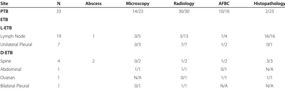
Table 1 Characteristics of the study subjects
Site N Abscess Microscopy Radiology AFBC Histopathology
PTB 33 14/23 30/30 10/16 2/23
ETB
L-ETB
Lymph Node 19 1 0/5 3/13 1/4 16/16
Unilateral Pleural 7 0/3 7/7 1/2 0/1
D-ETB
Spine 4 2 0/2 1/2 1/2 3/3
Abdominal 1 1/1 1/1 0/1 N/A
Ovarian 1 N/A 0/1 1/1 1/1
Bilateral Pleural 1 0/1 1/1 N/A N/A
PTB, pulmonary TB; ETB, extra-pulmonary TB; L-ETB, less severe ETB; D-ETB, disseminated ETB; N, number of subjects; Microscopy, acid fast bacillus smear using Ziehl Neelsen staining; Radiology, XRay/ CT scan/ or MRI; AFBC, acid fast bacilli culture by MIGIT system (Becton Dickinson, USA); histo-pathological staining of biopsy material and or FNAC (fine needle aspirate cytology) where relevant.
Two-fold dilutions of cDNA samples were amplified to control amplification efficiency and to determine the opti-mal concentration required for each primer pair. HuPO was used as a control gene to calculate theΔCtvalues for individual samples. The relative amount of cytokine/ HuPO transcripts was calculated using the 2-[ΔΔCt] method as described [30]. These values were then used to calculate the relative expression of cytokine mRNA in each of the samples tested.
Measurement of Th1/Th2/Th17 cytokines
Cellular supernatants were collected for cytokine measure-ments 18 hours post stimulation, spun to collect cellular debris and stored at -70°C until tested. Concentrations of IFN-γ, TNF-α, IL-2, IL-4, IL-6, IL-10 and IL-17 were mea-sured in cell supernatants with a Th1/Th2/Th17 Human Cytokine Flow Cytometric Bead Array kit (CBA) from BD Biosciences Ca, USA as described previously [31].
Statistical analysis
Data is depicted as median values for each group with the IQR (inter quartile range 25thto 75th percentile) indicated in each case. Comparison of non-parametric data between the groups was performed using the Mann–Whitney U, Kruskal Wallis and Wilcoxon Rank non-parametric tests. Analysis was performed and data plotted using GraphPad PRISM Version 5 (GraphPad Software, San Diego, CA, USA).
Results
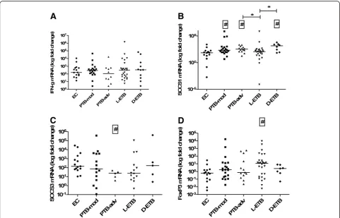
[image:4.595.60.539.327.667.2]DifferentialM.tuberculosis-induced SOCS1 in patients with pulmonary and extra-pulmonary tuberculosis We first compared the mRNA expression levels of IFN-γ, SOCS1, SOCS3, and FoxP3 in peripheral blood cells from patients with PTB, ETB and EC in either un-stimulated cells or after stimulation with M. tuberculosis. IFN-γ mRNA expression levels in un-stimulated and M.
tuberculosisstimulated PBMCs isolated from EC, PTB and ETB groups were similar (Figure 1A). SOCS1 mRNA levels were comparable in un-stimulated cells from EC, PTB and ETB groups. However, M.tuberculosis-stimulated SOCS1 mRNA titers differed between EC, PTB and ETB groups (p = 0.019, using Kruskal-Wallis ‘KW’ analysis). Further-more,M.tuberculosis-induced SOCS1 mRNA titers were higher in PTB as compared with EC (Mann Whitney-U test
‘MWU’p = 0.0067, Figure 1B).
SOCS3 mRNA levels were comparable between EC, PTB and ETB groups in un-stimulated and M. tuberculosis -stimulated PBMCs (Figure 1C). However, we observed that FoxP3 mRNA expression levels differed between un-stimulated PBMCs from EC, PTB and ETB (KW p = 0.0035), whereby FoxP3 levels were raised in PTB (MWU p = 0.014), and ETB (MWU p < 0.001) as compared with ECs. InM.tuberculosis- stimulated cells, FoxP3 expression levels were significantly higher in ETB (MWU p = 0.021) as compared with EC, but not in the case of PTB (Figure 1D). The direct effect of M.tuberculosis –stimulation was studied by comparing mRNA expression titers for each
gene in un-stimulated and M. tuberculosis-stimulated peripheral blood cells using the Wilcoxon rank test but it was observed that there was no difference in the titers of IFN-γ, SOCS1, SOCS3 or FoxP3. This may be due to the already raised mRNA titers in TB patients as a con-sequence of endogenous stimulation in the M. tubercu-losisinfected host.
Altered IFN-γ, IL-6 and IL-10 levels in tuberculosis
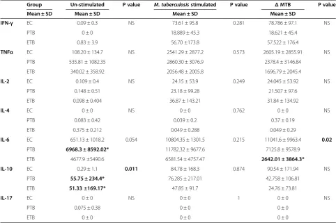
To further investigate the cytokine secretion profile in PTB and ETB cases as compared with EC we measured IFN-γ, TNF-α, IL-2, IL-4, IL-6, IL-10 and IL-17 in supernatants of un-stimulated and M. tuberculosis – stimulated PBMCs. IFN-γ, TNF-α, IL-2, IL-4 and IL-17 levels in supernatants of un-stimulated cells from TB and EC groups were similar (Table 2), whereas IL-6 levels were raised in PTB as com-pared with EC (MWU p = 0.018). IL-10 levels were raised in PTB (MWU, p = 0.013) and ETB (MWU, p = 0.003) as compared with EC.
[image:5.595.56.539.370.691.2]Subsequently, we measured M.tuberculosis-stimulated cytokine secretions in each group. No differences were
Table 2 Increased TNFα, IL-6 and IL-10 with down-regulated IFN-γin tuberculosis along with disease spectrum
Group Un-stimulated P value M. tuberculosisstimulated P value ΔMTB P value
Mean ± SD Mean ± SD Mean ± SD Mean ± SD
IFN-γ EC 0.09 ± 0.3 NS 73.61 ± 95.8 0.281 78.786 ± 97.1 NS
PTB 0 ± 0 18.889 ± 45.3 18.621 ± 45.4
ETB 0.83 ± 3.9 56.70 ±173.8 57.522 ± 176.4
TNFα EC 108.20 ± 134.7 NS 2541.29 ± 2877.2 0.573 2605.19 ± 2855.91 NS
PTB 535.81 ± 1082.35 2860.30 ± 3076.9 2378.4 ± 3146.84
ETB 340.02 ± 358.92 2056.48 ± 2005.8 1696.79 ± 2045.4
IL-2 EC 0.109 ± 0.4 NS 24.15 ± 53.9 0.249 24.045 ± 53.92 NS
PTB 0.148 ± 0.51 23.18 ± 99.28 21.507 ± 97.6
ETB 0.098 ± 0.404 36.87 ± 143.21 31.84 ± 134.92
IL-4 EC 0 ± 0 NS 0 ± 0 0.762 0 ± 0 NS
PTB 0.083 ± 0.42 0.039 ± 0.2 0.37 ± 0.19
ETB 0.375 ± 0.212 0.049 ± 0.288 0.049 ± 0.29
IL-6 EC 651.13 ± 1018.2 0.054 10804.35 ± 1301.5 0.215 11041.6 ± 9963.4 0.02
PTB 6968.3 ± 8592.02* 11782.32 ± 9677.6 7125.8 ± 9578.9
ETB 4677.9 ±5490.6 6581.54 ± 4757.47 2642.01 ± 3864.3*
IL-10 EC 0.29 ± 1.1 0.011 84.78 ± 168.3 0.874 90.54 ± 171.94 NS
PTB 55.75 ± 234.4* 76.285 ± 217.01 42.758 ± 106.81
ETB 51.33 ±169.17* 47.85 ± 91.7 24.76 ± 73.81
IL-17 EC 0 ± 0 NS 0 ± 0 1 0 ± 0 NS
PTB 0.075 ± 0.38 0 ± 0 0 ± 0
ETB 0 ± 0 0 ± 0 0 ± 0
EC, endemic control (n = 15); PTB, pulmonary TB (n = 33); ETB, extra-pulmonary TB (n = 33). IQR, interquartile range between 25th and 75th percentile.
* Denotes significant difference (p≤0.05) as compared with EC using Mann–Whitney U non-parametric test. P value calculated using Kruskal Wallis test. NS denotes non-significant P value (p > 0.05).
observed in response to M. tuberculosisin IFN-γ, TNF-α, IL-2, IL-4, IL-10 and IL-17 secretion between EC, PTB and ETB groups (Table 2).M. tuberculosis-induced IL-6 (MWU p = 0.003) was reduced in ETB as compared with EC.
DifferentialM.tuberculosisinduced SOCS1 expression with varying severity of tuberculosis
Since SOCS1 and FoxP3 gene expression levels differed between PTB and ETB groups we further investigated whether they associated with the clinical severity. There-fore, we compared gene expression levels between mod-erately advanced mod) and far advanced (PTB-adv) PTB, less severe (L-ETB) and more severe (D-ETB) ETB cases with ECs. In un-stimulated cells, SOCS1 mRNA levels were raised in PTB-adv as compared with PTB-mod (MWU p = 0.008) and EC (p = 0.034).
FoxP3 mRNA levels were significantly higher in PTB-adv (MWU p = 0.002), L-ETB (MWU p = 0.002)
and D-ETB (MWU, p = 0.018) as compared with EC. There was no difference between IFN-γ and SOCS3 mRNA ex-pression titers between un-stimulated cells of the TB and EC groups studied (Additional file 2: Table S2).
[image:6.595.57.539.346.655.2]We then comparedM.tuberculosis-stimulated responses in the study subjects. IFN-γ mRNA levels did not differ between the groups studied (Figure 2A). However, M. tuberculosis induced SOCS1 mRNA gene expression levels were raised in mod (MWU, p = 0.022), PTB-adv (MWU, p = 0.014) and D-ETB (MWU, p = 0.009) groups as compared with EC (Figure 2B). Further, M. tuberculosis-stimulated SOCS1 mRNA titers were signifi-cantly greater in PTB-adv (MWU, p = 0.016) and D-ETB groups (MWU, p = 0.027) as compared with L-ETB (Figure 2B). M. tuberculosis –induced SOCS3 mRNA expression was reduced in PTB-adv (MWU, p = 0.007) as compared with EC (Figure 2C).M.tuberculosis–induced FoxP3 mRNA expression levels showed a higher accu-mulation in PBMCs from L-ETB (MWU, p = 0.017) as compared with EC (Figure 2D).
Discussion
Th1 immune responses (IFN-γ, IL-12) are required for protection against infection with M. tuberculosiswhile a raised Th2-like cytokine profile (such as, IL-10, IL-4 and TGF-β) is implicated with disease progression in TB. To our knowledge, this is the first study to show an associ-ation between cytokine profiles in TB with the differen-tial expression of SOCS1, SOCS3 and FoxP3 molecules in PTB and ETB. All SOCS1 and FoxP3 mRNA levels and IL-6 and IL-10 protein titers were increased in PTB. SOCS1 and FoxP3 mRNA and IL-10 protein titers were also raised in ETB. Further M. tuberculosis- stimulated moderate ETB showed decreased SOCS1 as compared with both severe ETB and far advanced PTB cases.
The raised IL-6 levels could also contribute to the increased SOCS1 expression in PTB. IL-6 might in-hibit Th1 type cell responses by hampering IFN-γ, TNF-α and IL1β responses [32]. IL-10 is now recog-nized to be produced by almost every type of cell of the immune system, including most lymphocyte populations and cells of the innate immune system, such as antigen-presenting cells (DCs and macro-phages) and granulocytes [33]. In murine models, increased IL-10 hampers protective anti- M. tubercu-losis T cell responses [34]. IL-10 levels in peripheral blood cells and serum of active pulmonary TB patients were shown to be raised [35]. High IL-10 levels in both PTB and ETB were associated with an increase in FoxP3 expression. We observed FoxP3 gene expression and IL-10 secretion to be increased in both PTB and ETB compared to EC. The secretion of IL-10 by Tregs can account for the inhibition of T cell responses [36]. Neutralization of endogenous IL-10 in PBMCs from pulmonary TB patients resulted in increased T-cell prolif-eration and IFN-γ production [33], with enhanced prolif-erative responses to PPD in patients with anergic TB [37]. Thus, increased IL-10 levels in TB patients may hinder T cell responses.
M.tuberculosis–induced IL-6 and IL-10 levels in ETB were reduced as compared with EC. The reduced re-sponsiveness of cells fromM.tuberculosis-infected indi-viduals with advanced disease is in line with previous reports which demonstrate raised endogenous levels of IL-8 and reduced Mycobacterium-stimulated IL8 in patients with advanced leprosy disease [38].
We measured all the cytokines simultaneously after 18 h of culture. This time point has been found to be suitable for measuring most cytokines [39] except IFN-γ responses which are increased in up to 6 days of culture [40]. Therefore it may be that we have underestimated the IFN-γ levels in our study.
All TB patients showed an increased total leukocyte but decreased lymphocyte numbers as compared with the control group. The increased SOCS1 mRNA
expression observed in these patients despite a reduced lymphocyte count could be attributed to SOCS1 expres-sion from macrophages [41,42]. We previously demon-strated that an increased SOCS1 mRNA expression associates with clinical severity in PTB patients [28]. Here we show that post M. tuberculosis stimulation, SOCS1 levels were lower in cases with less severe ETB as compared with more severe ETB and also as com-pared with advanced PTB. Previous studies have shown that mycobacterial antigen specific IFN-γlevels are high-est in the L-ETB group [43] as compared with other sites. As SOCS1 impacts IFN-γ regulation [44], the reduced SOCS1 expression in less severe ETB group may indicate more effective T effector cell responses leading to protective granuloma formation in these cases. These data fit with previous studies that have shown higher IFN-γ responses in patients with pleural TB than those with miliary disease [9]. Also that in patients with tuberculous lymphadenitis IFN-γresponses are raised as compared to those with severe dissemi-nated ETB [27].
M. tuberculosis stimulated SOCS3 mRNA levels were lower in PTB-adv as compared with EC. Loss of SOCS3 expression in macrophages is associated with reduced IL-12 and TNF levels [45]. Therefore, decreased SOCS3 expression in PTB-adv patients may contribute to an impaired bactericidal response toM.tuberculosis.
Conclusions
Our results show differential expression of SOCS1 and FoxP3 together with variable IL-10 and IL-6 se-cretion in PTB and ETB. In addition, we observed an association between reduced SOCS1 expression and more localized ETB. These data suggest that the balance between these cytokines and SOCS1 may de-termine the dysregulation of immune balance in the host thereby affecting clinical severity of disease.
Additional files
Additional file 1:Table S1.Hematological characteristics of study subjects.
Additional file 2:Table S2.Increased SOCS1 gene expression in patients with far advanced pulmonary TB.
Competing interests
The authors declared that they have no competing interest.
Authors’contributions
Conception and design: ZH and MR; Analysis and interpretation: ZH, MR, KM, RH; Drafting the manuscript for important intellectual content: ZH, MR, KM, RH. All authors read and approved the final manuscript.
Acknowledgements
a SIDA Asia Link Program Grant, Swedish Research Council, and a University Research Council Grant, The Aga Khan University, Pakistan.
Author details
1Department of Pathology and Microbiology, Aga Khan University, P.O. Box
3500, Stadium Road, Karachi 74800, Pakistan.2Department of Microbiology and Tumor and Cell Biology, Karolinska Institutet, Stockholm, Sweden.3Indus Hospital, Karachi, Pakistan.4OJHA Institute of Chest Diseases, DOW University of Health Sciences, Karachi, Pakistan.
Received: 16 June 2012 Accepted: 10 January 2013 Published: 15 January 2013
References
1. WHO:Tuberculosis Factsheet. Geneva: World Health Organisation; 2011. ISBN 978 924 1564380.
2. WHO:Global Tuberculosis Control. Geneva: World Health Organisation; 2011. ISBN 978 924 1564380.
3. Stenger S, Modlin RL:T cell mediated immunity toMycobacterium
tuberculosis.Curr Opin Microbiol1999,2(1):89–93.
4. Dalton DK, Pitts-Meek S, Keshav S, Figari IS, Bradley A, Stewart TA:Multiple defects of immune cell function in mice with disrupted Interferon-gamma genes.Science1993,259(5102):1739–1742.
5. Kaufmann SH:Protection against tuberculosis: cytokines, T cells, and macrophages.Ann Rheum Dis2002,61(Suppl 2):ii54–ii58.
6. Qiu L, Huang D, Chen CY, Wang R, Shen L, Shen Y, Hunt R, Estep J, Haynes BF, Jacobs WR Jr,et al:Severe tuberculosis induces unbalanced up-regulation of gene networks and overexpression of IL-22, MIP-1alpha, CCL27, IP-10, CCR4, CCR5, CXCR3, PD1, PDL2, IL-3, IFN-beta, TIM1, and TLR2 but low antigen-specific cellular responses.J Infect Dis2008,198(10):1514–1519.
7. Flynn JL, Chan J:Immunology of tuberculosis.Annu Rev Immunol2001,
19:93–129.
8. Hirsch CS, Toossi Z, Othieno C, Johnson JL, Schwander SK, Robertson S, Wallis RS, Edmonds K, Okwera A, Mugerwa R,et al:Depressed T-cell Interferon-gamma responses in pulmonary tuberculosis: analysis of underlying mechanisms and modulation with therapy.J Infect Dis1999,
180(6):2069–2073.
9. Sharma SK, Mitra DK, Balamurugan A, Pandey RM, Mehra NK:Cytokine polarization in miliary and pleural tuberculosis.J Clin Immunol2002,
22(6):345–352.
10. Sahiratmadja E, Alisjahbana B, de Boer T, Adnan I, Maya A, Danusantoso H, Nelwan RH, Marzuki S, van der Meer JW, van Crevel R,et al:Dynamic changes in pro- and anti-inflammatory cytokine profiles and gamma interferon receptor signaling integrity correlate with tuberculosis disease activity and response to curative treatment.Infect Immun2007,75(2):820–829.
11. Jamil B, Shahid F, Hasan Z, Nasir N, Razzaki T, Dawood G, Hussain R:
Interferon gamma/IL-10 ratio defines the disease severity in pulmonary and extra pulmonary tuberculosis.Tuberculosis (Edinb)2007,87(4):279–287. 12. Gazzola L, Tincati C, Gori A, Saresella M, Marventano I, Zanini F:FoxP3 mRNA
expression in regulatory T cells from patients with tuberculosis.Am J Respir Crit Care Med2006,174(3):356. author reply 357.
13. Shevach EM:Mechanisms of FoxP3+ T regulatory cell-mediated suppression.
Immunity2009,30(5):636–645.
14. Guyot-Revol V, Innes JA, Hackforth S, Hinks T, Lalvani A:Regulatory T cells are expanded in blood and disease sites in patients with tuberculosis.Am J Respir Crit Care Med2006,173(7):803–810.
15. Lesho E, Forestiero FJ, Hirata MH, Hirata RD, Cecon L, Melo FF, Paik SH, Murata Y, Ferguson EW, Wang Z,et al:Transcriptional responses of host peripheral blood cells to tuberculosis infection.Tuberculosis (Edinb)2011,91(5):390–399. 16. Berry MP, Graham CM, McNab FW, Xu Z, Bloch SA, Oni T, Wilkinson KA,
Banchereau R, Skinner J, Wilkinson RJ,et al:An Interferon-inducible neutrophil-driven blood transcriptional signature in human tuberculosis.Nature2010,
466(7309):973–977.
17. Almeida AS, Lago PM, Boechat N, Huard RC, Lazzarini LC, Santos AR, Nociari M, Zhu H, Perez-Sweeney BM, Bang H,et al:Tuberculosis is associated with a down-modulatory lung immune response that impairs Th1-type immunity.J Immunol2009,183(1):718–731.
18. Wormald S, Zhang JG, Krebs DL, Mielke LA, Silver J, Alexander WS, Speed TP, Nicola NA, Hilton DJ:The comparative roles of Suppressor of Cytokine Signaling-1 and−3 in the inhibition and desensitization of cytokine signaling.J Biol Chem2006,281(16):11135–11143.
19. Croker BA, Krebs DL, Zhang JG, Wormald S, Willson TA, Stanley EG, Robb L, Greenhalgh CJ, Forster I, Clausen BE,et al:SOCS3 negatively regulates IL-6 signaling in vivo.Nat Immunol2003,4(6):540–545.
20. Niemand C, Nimmesgern A, Haan S, Fischer P, Schaper F, Rossaint R, Heinrich PC, Muller-Newen G:Activation of STAT3 by IL-6 and IL-10 in primary human macrophages is differentially modulated by Suppressor of Cytokine Signaling 3.J Immunol2003,170(6):3263–3272.
21. Somoskovi A, Zissel G, Zipfel PF, Ziegenhagen MW, Klaucke J, Haas H, Schlaak M, Muller-Quernheim J:Different cytokine patterns correlate with the extension of disease in pulmonary tuberculosis.Eur Cytokine Netw 1999,10(2):135–142.
22. Hasan Z, Jamil B, Ashraf M, Islam M, Yusuf MS, Khan JA, Hussain R: ESAT6-induced IFNgamma and CXCL9 can differentiate severity of tuberculosis.
PLoS One2009,4(4):e5158.
23. Kellar KL, Gehrke J, Weis SE, Mahmutovic-Mayhew A, Davila B, Zajdowicz MJ, Scarborough R, LoBue PA, Lardizabal AA, Daley CL,et al:Multiple cytokines are released when blood from patients with tuberculosis is stimulated withMycobacterium tuberculosisantigens.PLoS One2011,6(11):e26545. 24. WHO:Treatment of Tuberculosis: Guidelines for National Programmes. 3rd
edition. Geneva: World Health Organisation; 2003. WHO/CDS/TB/2003.313. 2003.
25. Crofton J:Clinical features of tuberculosis. Crofton and Douglas’s respiratory diseases. 4th edition. London: Wiley-Blackwell scientific; 1991. ISBN 978 06320 19731.
26. Hasan Z, Jamil B, Ashraf M, Islam M, Dojki M, Irfan M, Hussain R:Differential liveMycobacterium tuberculosis-,M.bovisBCG-, recombinant ESAT6-, and Culture filtrate protein 10-induced immunity in tuberculosis.Clin Vaccine Immunol2009,16(7):991–998.
27. Hasan Z, Cliff JM, Dockrell HM, Jamil B, Irfan M, Ashraf M, Hussain R:CCL2 Responses toMycobacterium tuberculosisare associated with disease severity in tuberculosis.PLoS One2009,4(12):e8459.
28. Masood KI, Rottenberg ME, Carow B, Rao N, Ashraf M, Hussain R, Hasan Z:
SOCS1 Gene expression is increased in severe pulmonary tuberculosis.
Scand J Immunol2012,76(4):398–404.
29. Dheda K, Huggett JF, Bustin SA, Johnson MA, Rook G, Zumla A:Validation of housekeeping genes for normalizing RNA expression in real-time PCR.
Biotechniques2004,37(1):112–114. 116, 118–119.
30. Livak KJ, Schmittgen TD:Analysis of relative gene expression data using real-time quantitative PCR and the 2(−delta delta C(T)) method.Methods 2001,25(4):402–408.
31. Talat N, Shahid F, Perry S, Dawood G, Hussain R:Th1/Th2 cytometric bead array can discriminate cytokine secretion from endogenously activated cells in pulmonary disease, recent and remote infection in tuberculosis.
Cytokine2011,54(2):136–143.
32. Shiratsuchi H, Johnson JL, Ellner JJ:Bidirectional effects of cytokines on the growth ofMycobacterium aviumwithin human monocytes.
J Immunol1991,146(9):3165–3170.
33. Gong JH, Zhang M, Modlin RL, Linsley PS, Iyer D, Lin Y, Barnes PF:
Interleukin-10 downregulatesMycobacterium tuberculosis-induced Th1 responses and CTLA-4 expression.Infect Immun1996,64(3):913–918. 34. Beamer GL, Flaherty DK, Assogba BD, Stromberg P, Gonzalez-Juarrero M, de
Waal Malefyt R, Vesosky B, Turner J:Interleukin-10 promotes
Mycobacterium tuberculosisdisease progression in CBA/J mice.
J Immunol2008,181(8):5545–5550.
35. Verbon A, Juffermans N, Van Deventer SJ, Speelman P, Van Deutekom H, Van Der Poll T:Serum concentrations of cytokines in patients with active tuberculosis (TB) and after treatment.Clin Exp Immunol1999,
115(1):110–113.
36. Kursar M, Koch M, Mittrucker HW, Nouailles G, Bonhagen K, Kamradt T, Kaufmann SH:Cutting edge: regulatory T cells prevent efficient clearance ofMycobacterium tuberculosis.J Immunol2007,178(5):2661–2665. 37. Boussiotis VA, Tsai EY, Yunis EJ, Thim S, Delgado JC, Dascher CC,
Berezovskaya A, Rousset D, Reynes JM, Goldfeld AE:IL-10-producing T cells suppress immune responses in anergic tuberculosis patients.J Clin Invest 2000,105(9):1317–1325.
38. Hasan Z, Mahmood A, Zafar S, Khan AA, Hussain R:Leprosy patients with lepromatous disease have an up-regulated IL-8 response that is unlinked to TNF-alpha responses.Int J Lepr Other Mycobact Dis2004,72(1):35–44. 39. Antas PR, Sales JS, Pereira KC, Oliveira EB, Cunha KS, Sarno EN, Sampaio EP:
40. Ferrand RA, Bothamley GH, Whelan A, Dockrell HM:Interferon-gamma responses to ESAT-6 in tuberculosis patients early into and after anti-tuberculosis treatment.Int J Tuberc Lung Dis2005,9(9):1034–1039. 41. Egwuagu CE, Yu CR, Zhang M, Mahdi RM, Kim SJ, Gery I:Suppressors of
cytokine signaling proteins are differentially expressed in Th1 and Th2 cells: implications for Th cell lineage commitment and maintenance.
J Immunol2002,168(7):3181–3187.
42. Whyte CS, Bishop ET, Ruckerl D, Gaspar-Pereira S, Barker RN, Allen JE, Rees AJ, Wilson HM:Suppressor of Cytokine Signaling (SOCS)1 is a key determinant of differential macrophage activation and function.
J Leukoc Biol2011,90(5):845–854.
43. Hasan Z, Rao N, Salahuddin N, Islam M, Ashraf M, Rottenberg ME, Hussain R:
M.tuberculosissonicate induced IFN gamma, CXCL10 and IL-10 can differentiate severity in tuberculosis.Scand J Immunol2012,74(2):220–226. 44. Krebs DL, Hilton DJ:SOCS proteins: negative regulators of cytokine
signaling.Stem Cells2001,19(5):378–387.
45. Liu Y, Stewart KN, Bishop E, Marek CJ, Kluth DC, Rees AJ, Wilson HM:Unique expression of Suppressor of Cytokine Signaling 3 is essential for classical macrophage activation in rodents in vitro and in vivo.J Immunol2008,
180(9):6270–6278. doi:10.1186/1471-2334-13-13
Cite this article as:Masoodet al.:Expression ofM.tuberculosis-induced
suppressor of cytokine signaling (SOCS) 1, SOCS3, FoxP3 and secretion of IL-6 associates with differing clinical severity of tuberculosis.BMC Infectious Diseases201313:13.
Submit your next manuscript to BioMed Central and take full advantage of:
• Convenient online submission
• Thorough peer review
• No space constraints or color figure charges
• Immediate publication on acceptance
• Inclusion in PubMed, CAS, Scopus and Google Scholar
• Research which is freely available for redistribution