R E S E A R C H A R T I C L E
Open Access
Population seroprevalence of antibody to
influenza A(H7N9) virus, Guangzhou, China
Yong Ping Lin
1,2†, Zi Feng Yang
3†, Ying Liang
1†, Zheng Tu Li
3, Helen S. Bond
4, Huiying Chua
4, Ya Sha Luo
5,
Yuan Chen
1, Ting Ting Chen
3, Wen Da Guan
3, Jimmy Chun Cheong Lai
6, Yu Lam Siu
7, Si Hua Pan
3,
J. S. Malik Peiris
4,7,8, Benjamin J. Cowling
4,9*and Chris Ka PunMok
7,8*Abstract
Background:Since the identification in early 2013 of severe disease caused by influenza A(H7N9) virus infection, there have been few attempts to characterize the full severity profile of human infections. Our objective was to estimate the number and severity of H7N9 infections in Guangzhou, using a serological study.
Methods:We collected residual sera from patients of all ages admitted to a hospital in the city of Guangzhou in southern China in 2013 and 2014. We screened the sera using a haemagglutination inhibition assay against a pseudovirus containing the H7 and N9 of A/Anhui/1/2013(H7N9), and samples with a screening titer≥10 were further tested by standard hemagglutination-inhibition and virus neutralization assays for influenza A(H7N9). We used a statistical model to interpret the information on antibody titers in the residual sera, assuming that the residual sera provided a representative picture of A(H7N9) infections in the general population, accounting for potential cross-reactions.
Results:We collected a total of 5360 residual sera from December 2013 to April 2014 and from October 2014 to December 2014, and found two specimens that tested positive for H7N9 antibody at haemagglutination inhibition titer≥40 and a neutralization titer≥40. Based on this, we estimated that 64,000 (95 % credibility interval: 7300, 190,000) human infections with influenza A(H7N9) virus occurred in Guangzhou in early 2014, with an infection-fatality risk of 3.6 deaths (95 % credibility interval: 0.47, 15) per 10,000 infections.
Conclusions:Our study suggested that the number of influenza A(H7N9) virus infections in Guangzhou substantially exceeded the number of laboratory-confirmed cases there, albeit with considerable imprecision. Our study was limited by the small number of positive specimens identified, and larger serologic studies would be valuable. Our analytic framework would be useful if larger serologic studies are done.
Keywords:Avian influenza A(H7N9), Public health, Serology, Severity
* Correspondence:bcowling@hku.hk;ch02mkp@hku.hk †Equal contributors
4WHO Collaborating Centre for Infectious Disease Epidemiology and Control,
School of Public Health, Li Ka Shing Faculty of Medicine, The University of Hong Kong, Hong Kong Special Administrative Region, China
7HKU-Pasteur Research Pole, School of Public Health, Li Ka Shing Faculty of
Medicine, The University of Hong Kong, Hong Kong Special Administrative Region, China
Full list of author information is available at the end of the article
Background
A novel avian influenza A(H7N9) virus emerged in early 2013 [1, 2] and caused cases of human infections in China in spring 2013, and subsequent winter and spring periods. The first epidemic wave was concentrated in eastern China, but Guangdong province in southern China was one of the most heavily affected areas in the second epidemic wave of H7N9 between December 2013 and April 2014 [3]. In total, 124 severe cases were re-ported in the first wave and 273 severe cases in the second wave, across China [3]. Guangzhou, a city with population 12.8 million in 2013, is the provincial capital of Guang-dong province. The objective of this study was to estimate the number of human infections with H7N9 virus in Guangzhou, and infer the severity of human infections on a per-infection basis.
Methods
Recruitment of participants
We collected residual sera in the First Affiliated Hospital of Guangzhou Medical University from patients in non-respiratory diseases wards with no signs of influenza-like illness at presentation or during hospitalization, and patients from the routine body check center. Sera were collected in two phases: 2674 sera were collected from December 2013 to April 2014, and 2686 sera were collected from October 2014 to December 2014.
Laboratory methods
Residual sera were first tested in parallel in each phase, using haemagglutination inhibition (HI) assay against a pseudovirus containing the H7 and N9 of A/Anhui/1/ 2013(H7N9) using methods previously described [4]. Serum from a laboratory-confirmed H7N9 case was used as a posi-tive control. Then, 306 posiposi-tive samples with HI titer≥10 on the pseudovirus assay were further tested with HI and neutralization assays using live A/Anhui/1/2013(H7N9) vi-ruses in a BSL-3 laboratory according to the guidelines from WHO (HI: http://www.who.int/influenza/gisrs_laboratory/ cnic_serological_diagnosis_hai_a_h7n9_20131220.pdf; MN: http://www.who.int/influenza/gisrs_laboratory/cnic_ serological_diagnosis_microneutralization_a_h7n9.pdf ) using 1 % horse red blood cells. We defined a positive serum sample as a sample with a titer≥40 by HI and MN.
Statistical analysis
We analyzed the serological data to infer the cumulative incidence of influenza A(H7N9) infections, in a Bayesian framework. In the Bayesian framework, information brought by new data, in the form of the likelihood func-tion, is combined with prior information that is specified in a prior distribution to obtain a posterior distribution that represents updated knowledge.
We stratified our analyses by age group (0–14, 15–24, 25–54, 55–64 and 65+ years of age) to allow extrapola-tion of seroprevalence to the underlying populaextrapola-tion. We described the weekly probability of a serum being tested positive (i.e. infected) for each age group as a parameter that depended on the baseline seroprevalence plus the seroprevalence due to recent infections. We assumed that all infected individuals had a rise in titer to ≥40 after a lag of 2 weeks [5], and that titers following infec-tion waned after 6 months because that occurs following other infections including human influenza virus infec-tions [6, 7]. We did not have information on the timing of infections in the general population, but we did have information on the timing of laboratory-confirmed cases which were generally severe and required hospitalization. We assumed that the risk of severe disease remained a constant fraction of all infections over the course of the epidemic, so that the timing of severe cases was indica-tive of the timing of infections in the general popula-tion (Addipopula-tional file 1). Based on these assumppopula-tions, we described the seroprevalence due to recent infections as a product of the cumulative incidence of recent H7N9 infections and expected scaled seroprevalence based on the delay between possible infection dates and collection of sera. Yang et al. [5] reported that before the H7N9 epi-demic, 9/1129 specimens collected in April-May 2013 from the general population in Zhejiang province were positive for H7N9 at a HI titer of≥40. We used this infor-mation to construct informative priors for the baseline seroprevalence in each age group at a titer≥40 based on the population structure in Guangzhou (Additional file 1). We used a Jeffrey’s prior, i.e. a beta(0.5, 0.5) distribution, as a non-informative prior distribution for the cumulative incidence of infection in each age group. We also con-ducted a sensitivity analysis using a flat beta(1, 1) prior instead for the cumulative incidence of infection.
We fitted the model using Markov Chain Monte Carlo (MCMC) methods with a warm-up period of 30,000 iter-ations followed by a further 30,000 iteriter-ations and five chains. Posterior estimates of the cumulative incidence of infection in each age group were then combined using population weights to provide a single estimate of the over-all age-standardized cumulative incidence of H7N9 in-fection in Guangzhou. Given this estimate, we further estimated the total number of infections in the population.
cases. We used this information to estimate the risk of severe case and death following an H7N9 infection. We used trace plots and the potential scale reduction statis-tic [9] to confirm that MCMC chains converged and were well-mixed. All statistical analyses were conducted using R version 3.1.2 (R Foundation for Statistical Com-puting, Vienna, Austria).
Results
The age and sex distribution of patients from whom we collected residual sera is shown in Table 1 and the timing of sera collection in comparison to the timing of detection of severe cases in Guangzhou are shown in Fig. 1. Of 306 positive samples with HI titer ≥10 on the pseudovirus assay, two specimens further tested positive for H7N9 antibody at an HI titer of ≥40 and a neutralization titer≥40: a specimen collected on 14 February 2014 from a 38y woman with HI titer of 1:80 on pseudovirus assay was positive at an HI titer of 80 and a neutralization titer of 40, and a specimen collected on 4 April 2014 from a 68y man with HI titer of 1:80 on pseudovirus assay was positive at an HI titer of 80 and a neutralization titer of 80. In addition, a specimen collected on 7 March 2014 from an 83y woman had HI titer of 1:40 on pseudovirus assay and was tested positive at an HI titer of 40 but had a neutralization titer ≤10. There was no report of recent acute respiratory illnesses in the 3 individuals with titers≥40.
We used MCMC to fit our model with a total of 60,000 iterations and 5 chains, and in diagnostic checking we confirmed that the MCMC runs were well-mixed and converged. Based on the two specimens that tested positive for H7N9 antibody at an HI titer of≥40 and a neutralization titer≥40, the posterior distribution (Fig. 2) indicates that the most credible estimate of the overall age-standardized cumulative incidence of H7N9 infections between December 2013 and April 2014 was 0.50 % (95 % credibility interval, CrI: 0.06, 1.51 %), corresponding to 64,000 (95 % CrI: 7300, 190,000) infections in Guangzhou (Table 2). Using this as the denominator, and the 16 severe
cases and 11 deaths as the numerators respectively [8], we estimated that the risk of a severe illness following infection was 5.2 (95 % CrI: 0.72, 23) per 10,000 infec-tions while the risk of death following infection was 3.6 (95 % CrI: 0.47, 15) per 10,000 infections. Alternative assumptions about the waning in antibody titers after infection led to broadly similar estimates (Table 2). Sce-nario 3 gave a somewhat lower estimate of the cumula-tive incidence of infection and higher estimates of risk of severe case and death following an infection.
In a sensitivity analysis using a beta(1, 1) prior for the cumulative incidence of infection, we estimated that there were 59,000 (95 % CrI: 9300, 170,000) infections, and the risk of death following infection was 3.6 (95 % CrI: 0.60, 14) per 10,000 infections. In another sensitivity analysis in-cluding the three samples with HI titers of ≥40 as posi-tives, we estimated that there were 64,000 (95 % CrI: 7400, 190,000) infections, and the risk of death following infec-tion was 3.5 (95 % CrI: 0.47, 15) per 10,000 infecinfec-tions.
Discussion
[image:3.595.57.539.591.732.2]In this study we estimated that 64,000 (95 % credibility interval: 7300, 190,000) humans were infected with influ-enza A(H7N9) virus in Guangzhou between January 2014 and April 2014, which far exceeds the 20 laboratory-confirmed infections [8]. It was previously estimated that the number of cases of symptomatic H7N9 virus infec-tions could be around 500–1000 times the number of laboratory-confirmed cases [3, 10], and this is consistent with our estimates of the numbers of infections albeit with considerable uncertainty. We are the first to estimate the infection fatality risk for H7N9, and our estimate of 3.6 deaths (95 % CrI: 0.47, 15) per 10,000 infections places the severity of H7N9 higher than that of human influenza vi-ruses for which the fatality risk is around 1 per 10,000 in-fections but varies considerably by age [11]. The severity of H7N9 virus infections appears to increase with age [3, 10], but we did not have sufficient sample size to make robust age-specific estimates of incidence or severity. We would anticipate higher incidence of H7N9 infections in
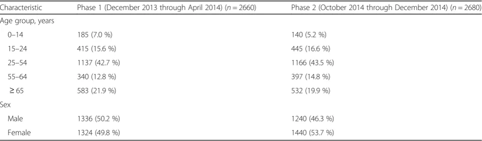
Table 1Age and sex distribution of residual sera collected from Guangzhou
Characteristic Phase 1 (December 2013 through April 2014) (n= 2660) Phase 2 (October 2014 through December 2014) (n= 2680)
Age group, years
0–14 185 (7.0 %) 140 (5.2 %)
15–24 415 (15.6 %) 445 (16.6 %)
25–54 1137 (42.7 %) 1166 (43.5 %)
55–64 340 (12.8 %) 397 (14.8 %)
≥65 583 (21.9 %) 532 (19.9 %)
Sex
Male 1336 (50.2 %) 1240 (46.3 %)
adult women and older adult men, based on patterns in exposure frequency in live poultry markets [12].
Our study is limited by the small number of positive samples. We used a statistical model to extrapolate from the small number of positives to a population estimate of the cumulative incidence of infection, which had very
considerable uncertainty. Because we did not collect de-tailed patient information such as exposures when obtain-ing the sera specimens, we were unable to explore risk factors for infection. Larger serological studies in affected areas would be worthwhile, and the methods we describe could provide a framework for analysis of such studies. However, since larger serologic studies (i.e. with tens of thousands or even hundreds of thousands of samples) have not been reported, and to the best of our knowledge have not been done, our data may provide the only avail-able population-based estimates of the cumulative inci-dence of H7N9 infection.
Several limitations concerning study design bear men-tion. Firstly, as most of our samples from the first sample collection were obtained before or during the peak inci-dence of H7N9 cases in Guangzhou and second sample collection was initiated in October 2014, the population exposure to influenza A H7N9 may not be optimally cap-tured. In hindsight, it would have been more ideal to ar-range collection of sera in April to June 2014, the period when maximal serological responses to H7N9 exposure might have been expected. We accounted for the overlap-ping of epidemic and sera collection in our analysis. Sec-ondly, instead of analyzing serial cross-sectional residual sera from hospitals, collecting paired sera before and after epidemics would allow better identification of recent in-fections based on rises in antibody titers. However such a study would be much more complex and challenging than the one we conducted. Thirdly, collection of sera directly from the general community, rather than through hos-pitals, would be preferable if feasible, to minimize
Fig. 2The prior and posterior distributions of the overall age-standardized
cumulative incidence of H7N9 infections, assuming that antibody titers wane exactly 6 months (26 weeks) after infection. The most credible estimate of the overall age-standardized cumulative incidence of H7N9 infections between December 2013 and April 2014 was 0.43 % (95 % credibility interval: 0.05, 1.32 %)
Fig. 1Timing of collection of residual sera from 5340 patients in Guangzhou in two phases (black lines), and the detection of severe cases of
[image:4.595.61.538.86.322.2] [image:4.595.57.290.445.663.2]potential selection biases. Fourthly, antibody titers fol-lowing laboratory-confirmed H7N9 virus infection can be low, and some infections might have been missed. One study that followed up severe cases reported that 65.8 % of non-fatal cases had detectable H7N9 antibody, 14 days after illness onset [5]. Finally, while a titer≥40 is not conclusive evidence of infection because of the poten-tial for a small number of people to have cross-reactive antibody following historical infections with other viruses [13], we accounted for this baseline seroprevalence in our model.
Our estimate of the total number of infections, including mild or asymptomatic infections, is consistent with the in-formation available from ILI surveillance in Guangzhou. In national surveillance in the first 10 weeks of 2014 there were 58,291 ILI cases in sentinel clinics, of whom 1090 cases were tested, and three were positive for H7N9 [8]. While it is not possible to make a population-based esti-mate of the number of mild cases based only on this infor-mation, it is clear that the occurrence of ILI due to H7N9 infection in early 2014 was around two orders of magni-tude lower than the expected occurrence of ILI due to sea-sonal human influenza activity in a typical epidemic when perhaps 30 % of ILI cases could test positive for human in-fluenza. At a population level, 10 to 20 % of persons might be infected with human influenza viruses during an epi-demic, and our estimate is consistent with the incidence of H7N9 infections being two orders of magnitude lower than this.
The large number of infections estimated in our analysis, based on a small number of positive sera, implies a sub-stantial risk of exposure to infection among the local popu-lation of Guangzhou. Visits to live poultry markets are common in Guangzhou, with one survey in May–August 2013 finding that 47 % of adult respondents had visited a live poultry market at least once in the preceding year [12].
A previous serological study in Shenzhen identified sero-logical evidence of high rates of infection (7.2 % in May 2013 and 14.9 % in December 2013) in poultry workers but not in the general population, consistent with repeated occupational exposures [14]. In that study, the absence of confirmed infections or severe illnesses in poultry workers with serological evidence of recent infection is also consistent with most infections being mild. A study in Beijing estimated very low risks of H7N9 in poultry workers in Beijing, and did not identify any infections in 1300 person-years of exposure among the general population from 2013 to 2015 [15].
Conclusion
In conclusion, by estimating the number of human infec-tions with influenza A(H7N9) virus in Guangzhou between January 2014 and April 2014, we were able to show that most infections are not associated with severe disease, which is consistent with the interpretation of mild H7N9 cases detected through ILI surveillance [3, 10, 16, 17]. However, it is concerning that so many mild infections have occurred, implying that H7N9 has had many oppor-tunities to acquire specific adaptations needed for efficient transmission of H7N9 between humans.
Additional file
Additional file 1:Technical Appendix (DOCX 51 kb)
Abbreviations
CrI:Credibility interval; HI: Haemagglutination inhibition; MCMC: Markov Chain Monte Carlo
Acknowledgements
[image:5.595.55.541.98.258.2]The authors thank Vicky Fang for technical advice. Table 2Results of main analyses and sensitivity analyses
Main analysis Sensitivity analysis
Alternative models including 2 specimens with HI and neutralization titers≥40
Main model with alternative prior for cumulative incidence
Main model including three specimens with HI titers≥40
Model 1 2 3 4 1 1
Estimated overall cumulative incidence,θ(%)
0.43 (0.05, 1.32) 0.52 (0.06, 1.59) 0.17 (0.02, 0.50) 0.41 (0.06, 1.22) 0.40 (0.06, 1.17) 0.43 (0.05, 1.32) Estimated total number of infections 55,385 (6503, 169,010) 66,441 (7572, 204,113) 21,638 (2910, 64,431) 52,507 (7387, 156,391) 51,644 (8091, 149,142) 55,481 (6576, 168,665)
ISR (per 10,000 infections) 5.90 (0.84, 25.11) 5.02 (0.69, 21.68) 14.17 (2.17, 56.43) 5.66 (0.90, 22.40) 5.82 (1.00, 21.71) 5.89 (0.83, 24.83)
IFR (per 10,000 infections) 4.07 (0.54, 17.41) 3.45 (0.44, 15.13) 9.75 (1.41, 39.48) 3.89 (0.58, 15.69) 4.11 (0.68, 15.68) 4.06 (0.54, 17.49)
The main analysis was repeated to compare four alternative assumptions of expected scaled seroprevalence,x'i. Sensitivity analyses aimed at comparing 1. main
Funding
The project was supported by the Theme Based Research Scheme from the Research Grants Council of the Hong Kong Special Administrative Region, China (project no. T11-705/14 N), the Area of Excellence Scheme of the Hong Kong University Grants Committee (grant no. AoE/M-12/06), the Health and Medical Research Fund of the Hong Kong Special Administrative Region, China (grant no. RRG-11), the Municipal Science and Technology Bureau Foundation of Guangzhou (grant no.2014Y2-00031), Science research project of the Guangdong Province (Grant no.2013B020224006 & Grant no.2016A050503047) and the Harvard Center for Communicable Disease Dynamics from the National Institute of General Medical Sciences (grant no. U54 GM088558). The funding bodies had no role in study design, data collection and analysis, preparation of the manuscript, or the decision to publish.
Availability of data and materials
All the data and material are provided in the Additional file 1.
Authors’contributions
YPL, ZFY, CKPM designed the study. YPL, ZFY, YL, ZTL, YSL, YC, TTC, WDG, JCCL, YLS, SHP, JSMP, CKPM performed the experiments. HSB, HC, BJC, CKPM analysed the data. BJC wrote the first draft. All authors edited the manuscript and approved the final version.
Competing interests
JSMP receives research funding from Crucell NV. BJC reports receipt of funding from MedImmune Inc. and Sanofi Pasteur, and consults for Crucell NV.
Consent for publication Not applicable.
Ethics approval and consent to participate
Approval for the study was obtained from the ethics committee of the First Affiliated Hospital of Guangzhou Medical University (ref no. 2015-8). Because this was a retrospective analysis of anonymized specimens, the ethics committee waived the need for informed consent from patients.
Author details
1Department of Laboratory Medicine, The First Affiliated Hospital of
Guangzhou Medical University, Guangdong, China.2Research Centre of Translational Medicine, The First Affiliated Hospital of Guangzhou Medical University, Guangdong, China.3State Key Laboratory of Respiratory Disease, National Clinical Research Center for Respiratory Disease, First Affiliated Hospital of Guangzhou Medical University, Guangdong, China.4WHO Collaborating Centre for Infectious Disease Epidemiology and Control, School of Public Health, Li Ka Shing Faculty of Medicine, The University of Hong Kong, Hong Kong Special Administrative Region, China.5Department of Laboratory Medicine, The Second Affiliated Hospital of Guangzhou University of Chinese Medicine, Guangdong, China.6Department of Pathology, Li Ka Shing Faculty of Medicine, The University of Hong Kong, Hong Kong Special Administrative Region, China.7HKU-Pasteur Research Pole, School of Public Health, Li Ka Shing Faculty of Medicine, The University of Hong Kong, Hong Kong Special Administrative Region, China.8Centre of Influenza Research, School of Public Health, Li Ka Shing Faculty of Medicine, The University of Hong Kong, Hong Kong Special Administrative Region, China.9School of Public Health, Li Ka Shing Faculty of Medicine, The University of Hong Kong, 21 Sassoon Road, Pokfulam, Hong Kong, China.
Received: 7 May 2016 Accepted: 27 October 2016
References
1. Gao R, Cao B, Hu Y, Feng Z, Wang D, Hu W, et al. Human infection with a novel avian-origin influenza A (H7N9) virus. N Engl J Med. 2013;368(20):1888–97. 2. Li Q, Zhou L, Zhou M, Chen Z, Li F, Wu H, et al. Epidemiology of human
infections with avian influenza A(H7N9) virus in China. N Engl J Med. 2014;370(6):520–32.
3. Feng L, Wu JT, Liu X, Yang P, Tsang TK, Jiang H, Wu P, Yang J, Fang VJ, Qin Y et al. Clinical severity of human infections with avian influenza A(H7N9) virus, China, 2013/14. Euro Surveill. 2014; 19(49):pii=20984.
4. Zhang A, Huang Y, Tian D, Lau EH, Wan Y, Liu X, et al. Kinetics of serological responses in influenza A(H7N9)-infected patients correlate with clinical outcome in China, 2013. Euro Surveill. 2013;18(50):20657.
5. Yang S, Chen Y, Cui D, Yao H, Lou J, Huo Z, et al. Avian-origin influenza A(H7N9) infection in influenza A(H7N9)-affected areas of China: a serological study. J Infect Dis. 2014;209(2):265–9.
6. Ng S, Ni MY, Fang VJ, Ip DK, Chan KH, Leung GM, et al. Characteristics of vaccine failures in a randomized placebo-controlled trial of inactivated influenza vaccine in children. Pediatr Infect Dis J. 2014;33(2):e63–6. 7. Zhao X, Fang VJ, Ohmit SE, Monto AS, Cook AR, Cowling BJ. Quantifying
Protection Against Influenza Virus Infection Measured by Hemagglutination-inhibition Assays in Vaccine Trials. Epidemiology. 2016;27(1):143–51. 8. Chen Z, Liu H, Lu J, Luo L, Li K, Liu Y, et al. Asymptomatic, mild, and severe
influenza A(H7N9) virus infection in humans, Guangzhou, China. Emerg Infect Dis. 2014;20(9):1535–40.
9. Gelman A, Rubin DB. Inference from iterative simulation using multiple sequences. Stat Sci. 1992;7(4):457–72.
10. Yu H, Cowling BJ, Feng L, Lau EH, Liao Q, Tsang TK, et al. Human infection with avian influenza A H7N9 virus: an assessment of clinical severity. Lancet. 2013;382(9887):138–45.
11. Wong JY, Kelly H, Ip DK, Wu JT, Leung GM, Cowling BJ. Case fatality risk of influenza A (H1N1pdm09): a systematic review. Epidemiology. 2013;24(6):830–41.
12. Wang L, Cowling BJ, Wu P, Yu J, Li F, Zeng L, et al. Human exposure to live poultry and psychological and behavioral responses to influenza A(H7N9), China. Emerg Infect Dis. 2014;20(8):1296–305.
13. Bai T, Zhou J, Shu Y. Serologic study for influenza A (H7N9) among high-risk groups in China. N Engl J Med. 2013;368(24):2339–40.
14. Wang X, Fang S, Lu X, Xu C, Cowling BJ, Tang X, et al. Seroprevalence to avian influenza A(H7N9) virus among poultry workers and the general population in southern China: a longitudinal study. Clin Infect Dis. 2014;59(6):e76–83. 15. Yang P, Ma C, Cui S, Zhang D, Shi W, Pan Y, et al. Avian influenza A(H7N9)
and (H5N1) infections among poultry and swine workers and the general population in Beijing, China, 2013–2015. Sci Rep. 2016;6:33877. 16. Ip DK, Liao Q, Wu P, Gao Z, Cao B, Feng L, et al. Detection of mild to
moderate influenza A/H7N9 infection by China's national sentinel surveillance system for influenza-like illness: case series. BMJ. 2013;346:f3693.
17. Xu C, Havers F, Wang L, Chen T, Shi J, Wang D, et al. Monitoring avian influenza A(H7N9) virus through national influenza-like illness surveillance, China. Emerg Infect Dis. 2013;19(8):1289–92.
• We accept pre-submission inquiries
• Our selector tool helps you to find the most relevant journal • We provide round the clock customer support
• Convenient online submission • Thorough peer review
• Inclusion in PubMed and all major indexing services • Maximum visibility for your research
Submit your manuscript at www.biomedcentral.com/submit