R E V I E W
Open Access
Perioperative cardiovascular monitoring of
high-risk patients: a consensus of 12
Jean-Louis Vincent
1*, Paolo Pelosi
2, Rupert Pearse
3, Didier Payen
4, Azriel Perel
5, Andreas Hoeft
6, Stefano Romagnoli
7,
V Marco Ranieri
8, Carole Ichai
9, Patrice Forget
10, Giorgio Della Rocca
11and Andrew Rhodes
12Abstract
A significant number of surgical patients are at risk of
intra- or post-operative complications or both, which
are associated with increased lengths of stay, costs,
and mortality. Reducing these risks is important for
the individual patient but also for health-care planners
and managers. Insufficient tissue perfusion and cellular
oxygenation due to hypovolemia, heart dysfunction or
both is one of the leading causes of perioperative
complications. Adequate perioperative management
guided by effective and timely hemodynamic
monitoring can help reduce the risk of complications
and thus potentially improve outcomes. In this review,
we describe the various available hemodynamic
monitoring systems and how they can best be used
to guide cardiovascular and fluid management in the
perioperative period in high-risk surgical patients.
Introduction
An estimated 230 million surgical procedures are
per-formed each year around the world [1], and a significant
number are in patients at risk of intra- or post-operative
complications or both. Although less than 15% of
in-patient procedures are performed in high-risk in-patients,
such patients account for 80% of deaths [2-4]. Even for
those patients who survive to leave hospital,
post-operative complications remain an important
determin-ant of functional recovery, long-term survival [5], and
health-care costs. Thus, mitigation of these risks is
im-portant not only for the individual patient but also for
health-care managers.
The risk of perioperative complications is related to
various factors, including patient status and comorbidities,
* Correspondence:jlvincen@ulb.ac.be 1
Department of Intensive Care, Erasme Hospital, Université Libre de Bruxelles, 808 route de Lennik, 1070 Brussels, Belgium
Full list of author information is available at the end of the article
the type of surgery performed and its duration, the degree
of urgency, the skills and experience of the operating and
anesthetic teams, and the post-operative management.
In-sufficient tissue perfusion and cellular oxygenation due to
hypovolemia, heart dysfunction or both is one of the
lead-ing causes of perioperative complications and poor
out-comes [6-9]. Thus, effective fluid management to prevent
and treat hypo/hypervolemia and titration of vasoactive
drugs for heart dysfunction are crucial to maintain
ad-equate oxygen delivery (DO
2) and prevent fluid overload
and its consequences [10-12]. Therefore, selecting the
most appropriate hemodynamic monitoring device (for
diagnosis and to guide therapies) may be an important
first step in reducing the risk of complications. The aims
of this review are to describe the available hemodynamic
monitoring systems and to evaluate the most appropriate
clinical setting for each.
Basic hemodynamic monitoring
Clinical examination remains an important initial step in
the hemodynamic assessment of high-risk surgical
pa-tients. However, individual vital signs often lack the
specificity and sensitivity that are needed to guide
hemodynamic management. For example, blood
pres-sure is a variable influenced by both cardiac output (CO)
and vascular tone; hence, blood pressure can remain
within the normal range in the presence of low-flow
states, including hypovolemia, as a result of increased
peripheral vascular resistance. Similarly, heart rate may
fail to reflect the development of hypovolemia under
anesthesia [13].
Combining and integrating parameters from various
hemodynamic monitoring systems may help improve
our understanding of hemodynamic status [14]. For
ex-ample, the combination of arterial pressure and the
par-tial pressure of end-tidal carbon dioxide (PetCO
2) can
help differentiate between vasodilation and low CO as a
cause of hypotension (PetCO
2transiently decreases when
CO decreases) and may prevent
‘
reflex
’
fluid administration
whenever blood pressure decreases. Similarly, a reduction
in the PetCO
2value for the same minute ventilation (in
the absence of hypothermia) suggests decreased
pulmon-ary blood flow (and thus CO) and may serve as a trigger
for more advanced hemodynamic monitoring.
Arterial pressure
Continuous invasive measurement of arterial pressure
helps identify the rapid fluctuations in arterial pressure
that may occur in high-risk patients. Artifacts (over- or
under-damping) should be carefully identified and
elimi-nated, especially when systolic-diastolic components and
waveform have to be analyzed. Non-invasive techniques
for continuous measurement of blood pressure are
usu-ally performed in peripheral arteries and may become
unreliable in case of vasoconstriction or low peripheral
flow. Non-invasive assessment of pressure waveforms
from more central measurement sites, such as the
bra-chial artery, may be a valuable option in the future.
Central venous pressure
A central venous catheter (CVC) is often used for
ad-ministration of fluids, vasopressors, and inotropes and
for measurement of central venous pressure (CVP).
Since transmural CVP is the only value related to right
ventricular (RV) preload but is not commonly
moni-tored, interpretation of CVP values must take into
ac-count intrathoracic pressure changes, which are largely
influenced by mechanical ventilation. Thus, changes in
CVP with concomitant CO variations give an indication
of RV function and potential peripheral venous
conges-tion, the latter of which is an important factor for organ
perfusion [15]. In addition, careful checking of the CVP
wave may help to diagnose tricuspid regurgitation with a
‘
v
’
wave during systole. When the CVP is low (<6 mm
Hg) with a concomitant low CO, there is almost
cer-tainly some degree of hypovolemia. Although changes in
CVP correlate poorly with changes in CO [16] (as for
pulmonary artery occlusion pressure), they can be used
to assess the dynamic response to a fluid challenge [17]
and to diagnose severe hypovolemia or cardiac
dysfunc-tion or both, especially where other monitoring systems
are not available.
Cardiac output monitoring
The perioperative period is characterized by large
varia-tions in whole body oxygen consumption (VO
2). The
main goal in this period is to maintain an adequate DO
2to meet the fluctuating tissue oxygen requirements.
Glo-bal DO
2is determined by CO and the oxygen content of
arterial blood, and so after correction of hypoxemia and
anemia (topics that will not be dealt with here),
main-tenance of an adequate CO is the next logical step to
improve DO
2. There are various methods available for
monitoring CO [18], although a survey indicated that
CO is routinely monitored in high-risk surgical patients
by only about 35% of practitioners in Europe and North
America [19] (Table 1).
Doppler echocardiography
Though difficult to use as a continuous monitor of CO
with conventional probes, transthoracic (TTE) or
trans-esophageal (TEE) echocardiography can provide
imme-diate point-of-care assessment of acute hemodynamic
changes in selected patients. Echo techniques can also
help to visualize the lungs, but this is beyond the scope
of this review. Obviously, it is not possible to use TEE in
all types of surgery. In addition to the estimation of CO
(usually easier with TEE than with TTE), Doppler
echo-cardiographic examination can provide an indication of
cardiac function because it allows visualization of the
cardiac chambers, valves, and pericardium [20]. It also
allows measurement of the ejected stroke volume (SV)
and derived left ventricular (LV) function parameters.
TEE provides several views, including the following:
[image:2.595.305.539.447.715.2]The LV short-axis view, which can be used to evaluate
LV function. Calculation of the LV fractional area
contraction, or the simpler
‘
eyeballing method
’
,
Table 1 What hemodynamic monitoring do you routinely
use for the management of high-risk surgery patients?
(Please mark all that apply)
ASA respondents
ESA respondents
(n = 237) (n = 195)
Answer options Response
percentage
Response percentage
Invasive arterial pressure 95.4% 89.7%
Central venous pressure 72.6% 83.6%
Non-invasive arterial pressure 51.9% 53.8%
Cardiac output 35.4% 34.9%
Pulmonary capillary wedge pressure 30.8% 14.4%
Transesophageal echocardiography 28.3% 19.0%
Systolic pressure variation 20.3% 23.6%
Plethysmographic waveform variation 17.3% 17.9%
Pulse pressure variation 15.2% 25.6%
Mixed venous saturation (ScvO2) 14.3% 15.9%
Central venous saturation (SvO2) 12.7% 33.3%
Oxygen delivery (DO2) 6.3% 14.4%
Stroke volume variation 6.3% 21.5%
Near-infrared spectroscopy 4.6% 5.1%
Global end-diastolic volume 2.1% 8.2%
informs about the kinetic (contractile) state and the
shape (volume) of the heart. Poor contractility may
indicate that inotropic support could help, and
‘
kissing
’
of the papillary muscle may indicate the need
for fluids if the right heart is functioning normally.
The short-axis view may also be used to identify septal
dyskinesia. The finding of an RV D-shape may suggest
the presence of RV dysfunction/failure, indicating a
non-adaptation to an acute increase in RV afterload
(pulmonary embolism) or RV myocardial ischemia.
The four-chamber view, which can help in assessing
LV and RV function by evaluation of the right-to-left
size ratio (normal <0.6).
In more advanced echocardiographic evaluation, fluid
status and fluid responsiveness can also be assessed in
mechanically ventilated patients by means of the
super-ior vena cava collapsibility index (TEE bicaval view) or
inferior vena cava distensibility index (TTE subcostal
view). In addition, echocardiography allows the rapid
and reliable estimation of SV. Finally, there are particular
and specific conditions in which diagnosis and treatment
are strictly related to the echocardiographic examination
(for example, pericardial effusion, valve disruptions, aortic
dissection, and systolic anterior motion of the mitral
valve).
A miniaturized, disposable monoplane TEE probe that
can be left in place for up to 72 hours (ClariTEE
™
, ImaCor
Inc., Garden City, NY, USA) was recently introduced and
has the potential to provide ongoing qualitative cardiac
as-sessment [21]. We believe that, where expert
echocardiog-raphy skills are not available, training programs should be
developed to ensure that clinicians taking care of the
high-risk patient are familiar with at least the basic
appli-cations of TTE and TEE.
Pulmonary artery catheter
Though criticized in recent years for its intrinsic
inva-siveness and no clear evidence of improved outcomes
[22-25], the pulmonary artery catheter (PAC) is the only
tool that provides continuous monitoring of pulmonary
artery pressure, right-sided and left-sided filling
pres-sures, CO, and mixed venous oxygen saturation (SvO
2).
Although the PAC can now be largely replaced by less
invasive hemodynamic monitoring techniques in many
cases, in some complex clinical situations (for example,
cardiac surgery, organ transplant surgery, and surgery
associated with major fluid shifts or high risk of
respira-tory failure or in patients with compromised RV
func-tion), the PAC still represents a valuable tool when used
by physicians adequately trained to correctly interpret
and apply the data provided [26,27]. In such patients,
the PAC can be inserted for limited periods of time and
removed when no longer necessary.
Other cardiac output monitoring devices
Pulse contour analysis
SV can be estimated continuously by analysis of the
ar-terial pressure waveform, usually derived from an
in-dwelling arterial catheter or by a non-invasive finger
pressure cuff. To calculate SV from a pressure trace, the
algorithms used by these devices have to compensate for
the overall impedance of the system on the basis of the
estimation of compliance and resistance of the
cardio-vascular tree. In this regard, optimization of the input
signal is imperative, and severe distortions of the arterial
waveform (for example, severe arrhythmias and multiple
ectopic beats) and inadequate response of fluid-filled
transducer systems (that is, over- and under-damping)
[28] can result in unreliable CO measurement.
Calibrated devices
–
The PiCCOplus
™
/PiCCO
2™
system (Pulsion Medical
Systems, Munich, Germany) consists of a
thermistor-tipped catheter which is usually placed in
the femoral artery, although catheters for radial,
axillary, or brachial applications are also available.
The PiCCO
™
device measures CO by transpulmonary
thermodilution, which additionally provides the
computation of volumetric preload parameters
—
global
end-diastolic volume (GEDV) and intrathoracic blood
volume
—
and extravascular lung water (EVLW). The
CO measured by the Stewart-Hamilton principle from
the thermodilution curve is used to calibrate a pulse
contour algorithm, which measures the area under
the systolic pulse pressure curve and calculates the SV
in order to provide beat-by-beat CO measurement.
The system has to be frequently recalibrated, at least
every 8 hours in hemodynamically stable patients and
more often if changes in vasoactive support are
provided [
29
]. The system has been validated in a
variety of clinical settings [
30
].
–
The EV1000
™
/VolumeView
™
system (Edwards
Lifesciences, Irvine, CA, USA) has been more
recently introduced and is analogous to the PiCCO
™
monitor, using pulse wave analysis to calculate CO.
A proprietary thermistor-tipped femoral artery
catheter and a separate sensor are the main
components of the system. This system requires
calibration by transpulmonary thermodilution. It has
been validated against the PiCCO
™
and transpulmonary
thermodilution in critically ill patients [
31
].
correction for vascular compliance, with calibration
using a transpulmonary lithium indicator dilution
technique performed via an indwelling arterial
catheter. It has been validated in critically ill
patients [
32
,
33
].
Uncalibrated devices (without external calibration)
With preloaded data
–
The PulsioFlex
™
system (Pulsion Medical Systems)
displays trends of estimated CO by using the
patient
’
s anthropometric and demographic
characteristics (necessary for internal calibration),
analysis of the arterial pressure tracing, and a
proprietary algorithm for data analysis. The system
requires a dedicated additional sensor, which can be
connected to a regular arterial pressure catheter.
Based on the same pulse contour algorithm used by
the PiCCO
™
, the device can be calibrated by
entering a CO obtained from an external source (for
example, Doppler echocardiography) or by the
system
’
s own internal algorithm.
–
The LiDCO
™
rapid
(LiDCO Ltd) device uses the
same algorithm as the LiDCO
™
plus
system, but
instead of lithium dilution, nomograms based on the
patient
’
s age, weight, and height are used to estimate
SV and CO (so-called
‘
nominal
’
SV and CO). An
externally estimated CO can be used to calibrate the
device.
–
The FloTrac
™
/Vigileo
™
system (Edwards
Lifesciences) consists of a proprietary transducer
(FloTrac
™
) connected to a standard (radial or
femoral) arterial catheter. Individual demographic
variables (age, sex, height, and weight) and a
database containing CO variables derived by using
the PAC are used to calculate impedance and a
‘
normal
’
SV against which the standard deviation of
the pulse pressure sampled during a 20-second
interval is correlated to estimate CO. Arterial
waveform analysis is used to calculate vascular
resistance and compliance. The algorithm used by
the Vigileo
™
device has been modified over time, and
recent studies evaluating the device in the perioperative
setting have shown an improved performance and a
significant reduction in the time needed to adapt to
vascular dynamics. In the intensive care unit (ICU)
setting, concerns remain regarding the accuracy in
situations of acute hemodynamic instability as well as
hyperdynamic conditions, although recent software
modifications seem to improve the reliability of CO
measurements. The FloTrac
™
/Vigileo
™
system has
been shown to be suitable for integration into
perioperative optimization protocols, resulting in
improved clinical outcomes [
34
,
35
].
Without preloaded data
–
The MostCare system (Vytech, Padua, Italy), powered
by the pressure recording analytical method, performs
a beat-to-beat estimation of SV and CO by analyzing
the pressure waveform, sampled at high resolution
(1,000 points per second = 1 kHz). The area under the
pressure wave is determined during the whole cardiac
cycle. In each phase, the method identifies specific
points (
‘
points of instability
’
) characterized by
modifi-cations in velocity and acceleration in relationship to
the previous and the subsequent point. All of these
‘
points of instability
’
, mainly caused by reflected waves
from the periphery (backward travelling waves), give
the arterial pulse its specific profile, which is analyzed
by MostCare for estimation of the vascular impedance
(Zt). The contribution of the reflected waves to the
forward travelling wave can be accurately identified
only with a very high sampling rate. The ability to
update the Zt during each heart beat makes the
system extremely reactive when abrupt changes in
impedance occur (for example, changes in vascular
tone) [
36
,
37
]. Although some promising clinical data
are available [
38
], larger validation studies are needed
to confirm these observations. A multicenter study
comparing MostCare with echo-Doppler for CO
measurement was recently completed
(ClinicalTrials.-gov identifier: NCT01678950).
Non-invasive pulse contour analysis
Doppler monitoring devices
–
Esophageal Doppler offers a minimally invasive
determination of CO. The CardioQ
™
/CardioQ-ODM
™
(Deltex Medical Ltd, Chichester, UK) is the most
commonly used device. Esophageal probes measure
blood flow in the descending part of the aorta. SV is
calculated by multiplying the cross-sectional area of
the aorta (from nomograms based on height, weight,
and age) by the blood flow velocity. Technical and
methodological concerns regarding probe positioning
and the use of nomograms have been raised.
–
A transthoracic continuous Doppler CO monitor,
the USCOM (Uscom, Sydney, Australia), is also
available for intermittent SV and CO assessment.
Using supra- or parasternal windows, flow velocity
at the level of the aortic or pulmonary valves can be
assessed non-invasively. Aortic diameter can be
loaded from another measurement (two-dimensional
echocardiography) or from a nomogram as
mentioned above. The technique is rapid and can be
used in adult and pediatric patients. Clinical
validation studies have shown conflicting results,
partially due to variations in signal acquisition
inherent in the technique [
45
].
Applied Fick principle and dye dilution
–
The NICO
™
System (Novametrix Medical Systems,
Wallingford, CT, USA), using the partial CO
2rebreathing method, applies the Fick principle to
CO
2in patients who are intubated and mechanically
ventilated via a disposable rebreathing circuit that is
added to the ventilator tubing. CO
2production is
calculated as the product of CO
2concentration and
airflow during a breathing cycle, and arterial CO
2content is derived from end-tidal CO
2and its
corresponding dissociation curve. The rebreathing
loop can induce an intermittent partial rebreathing
state in 3-minute intervals. This rebreathing cycle
results in an increased end-tidal CO
2, mimicking a
decrease in CO
2production. The differences in these
values can be used to calculate CO. The system is
not really clinically acceptable, because the assessment
of CO is possible only in patients with fixed ventilator
settings and good respiratory function with no
relevant pulmonary shunt [
46
,
47
].
–
DDG-30® pulsed dye densitometry (Nihon Kohden,
Tokyo, Japan) is based on the transpulmonary dye
dilution technique using indocyanine green (ICG).
Signal detection in the arterial blood is performed
by transcutaneous optical absorbance measurements
similar to pulse oximetry. CO is calculated from
the ICG-dye dilution curve according to the
Stewart-Hamilton principle. Factors that compromise signal
detection, such as vasoconstriction, interstitial edema,
movement, or ambient light artifacts, all limit the
reliability of CO assessment using this method [
48
].
Bioimpedance and bioreactance
–
Electrical bioimpedance estimates CO continuously
by detecting variations in thoracic or whole body
impedance induced by cyclic changes in blood flow.
Electrodes attached to the skin (BioZ, CardioDynamics,
San Diego, CA, USA) or an endotracheal tube
(ECOM
™
, Conmed Corporation, Utica, NY, USA)
provide electric current stimulation, and signal
variations are analyzed by using mathematical
algorithms. The reliability of these systems is
poor [
49
,
50
].
–
The Bioreactance® technique (NICOM®, Cheetah
Medical Ltd, Maidenhead, Berkshire, UK) analyzes
the variations in the frequency spectra associated
with delivery of an oscillating electrical current. This
technique may be somewhat superior to the
bioimpedance technique [
51
,
52
] but is dependent on
body size [
53
] and the aeration of the lung. It is less
accurate in diseased lungs in which reactance may
be affected by the amount of EVLW and alveolar
collapse or consolidation or both.
Pitfalls in the interpretation of cardiac output
Although CO can be measured with reasonable accuracy
and precision with some of these systems, it is difficult
to assess the optimal CO for an individual patient. A
‘
normal
’
or even high CO does not preclude the
pres-ence of inadequate regional and microcirculatory flow,
and a low CO may be adequate in a context of low
metabolic demand, especially during surgery under
gen-eral anesthesia. Moreover, simple identification of a low
CO does not tell us what to do about it. To correctly
in-terpret the data acquired by any of the described devices,
we need to combine/integrate several variables to help
decide whether the CO/SV is adequate and how it can
be optimized in the most effective manner.
How to select the best system
measurements of CO and less invasive but also less
ac-curate modalities.
Several questions can be raised when considering choice
of CO monitoring in the perioperative period:
1. Are we ready to accept a less accurate measurement
in order to limit invasiveness? (Figure
1
). A less
accurate measurement may be acceptable if the
trend analysis is reliable. Cost may also be an
important issue.
2. Do we need continuous, semi-continuous, or
intermittent measurements? Most complications
after surgery do not have a sudden onset (except
sudden cardiac failure due, for example, to myocardial
infarction or pulmonary embolism) or an obvious
cause (for example, massive bleeding during surgery)
but develop slowly; therefore, semi-continuous or
intermittent measurements may be acceptable.
However, it should be noted that only beat-by-beat
measurement of SV allows assessment of the response
to preload-modifying maneuvers, such as a fluid
challenge or passive leg raising (PLR) test.
3. Are calibrated or uncalibrated systems preferable?
Non-calibrated systems are acceptable for the
operating room (OR) or the post-anesthesia care
unit (PACU) but may not be suitable for more
complex cases, especially in the ICU. In unstable
patients, there is a necessity to
‘
re-calibrate
’
more
often because of frequent changes in vascular tone
and also because derived variables (for example,
EVLW and GEDV) need to be re-calculated. A
practical option may be to use an uncalibrated
system in the OR/PACU and replace it with a
calibrated system in the ICU.
4. What alarms do we need? A major problem for
patient surveillance by telemetric monitoring is
artifact robustness. Any system with too many false
alarms is prone to failure as personnel become
desensitized.
5. What kind of monitoring for what kind of patient?
This is not a
‘
one size fits all
’
decision; rather, the
optimal monitoring technique for each patient will
vary depending on the degree of risk and the extent
of the surgical procedure (Figure
2
).
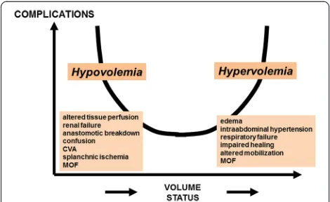
Fluid management
Inadequate fluid management may lead to reduced CO
and DO
2to injured tissues, which is associated with an
in-creased incidence of post-operative complications.
More-over, the systemic inflammatory response associated with
tissue injury results in capillary leak and tissue edema.
Fluid restriction and diuresis may decrease edema in
pa-tients with poor ventricular function but may also increase
the incidence of acute kidney injury. Meanwhile, excessive
fluid administration may lead to a range of adverse effects,
including coagulopathy and edema of lungs, gut, and
per-ipheral tissues (Figure 3). Retention of sodium and water
following surgery may reduce fluid requirements. Once
the patient is stabilized, fluids should be given only to
cor-rect deficit or continuing losses. Unfortunately, estimates
of fluid deficit that are based on traditional physiological
parameters, such as heart rate, blood pressure, and cardiac
filling pressures, are not sufficient.
Static indicators of preload
[image:6.595.303.537.479.687.2]–
CVP: Many high-risk surgical patients have a CVC
in place, and a CVC is a requirement for some
devices needing calibration by thermodilution.
Despite its limitations (vide supra), changes in
CVP over time may be helpful to guide fluid
[image:6.595.57.291.567.704.2]Figure 1The compromise between accuracy and invasiveness of monitoring systems. CO, cardiac output; PA, pulmonary artery.
Figure 2Possible choice of monitoring system in relation to a patient’s degree of perioperative risk. CO, cardiac output; PAC, pulmonary artery catheter; PPV, pulse pressure variation; ScvO2,
therapy, especially when the CVP is low and
associated with low flow. A CVP of more than
8 mm Hg might also be considered an
‘
alarm
’
for
potential venous congestion associated (or not)
with fluid overload [
15
].
–
GEDV and EVLW: These are volumetric parameters
derived from transpulmonary thermodilution and
are integrated into the PiCCO
™
plus
, PiCCO
2™
, and
EV1000
™
monitors. EVLW can help in the
identification of (cardiogenic or non-cardiogenic)
pulmonary edema and has the potential to increase
the safety of fluid therapy in patients with structural
lung disease, acute respiratory distress syndrome, or
congestive heart failure.
–
The end-diastolic area of the left ventricle may be the
most reliable static parameter of preload but is largely
dependent on LV diastolic compliance. Its ability to
accurately predict fluid responsiveness is limited.
Functional hemodynamic parameters
Positive pressure ventilation induces cyclical changes in
intrathoracic pressure, which affect preload by
decreas-ing venous return to the right heart and increasdecreas-ing
venous return to the left ventricle. The degree of the
resulting changes in LV SV (stroke volume variation;
SVV) and pulse pressure (pulse pressure variation; PPV)
better predict fluid responsiveness than do static
param-eters, when RV function is not a limitation and for a
fixed tidal volume. Most devices using pulse contour
analysis, including the current version of the
non-invasive ClearSight monitor, display SVV and PPV.
Des-pite the numerous validity criteria required to interpret
such variations, these variables may help predict fluid
re-sponsiveness at different thresholds and have been
inte-grated into hemodynamic optimization protocols [58].
Respiratory variations in the pulse oximeter
plethysmo-graphic waveform have been shown to predict fluid
responsiveness in mechanically ventilated patients, similar
to changes in the arterial pressure waveform [59]. The
Masimo
™
device (Masimo Corporation, Irvine, CA, USA)
provides automated calculation of the pleth variability
index (PVI) by measuring changes in perfusion index over
a time interval including at least one complete respiratory
cycle. The PVI has been shown to predict fluid
respon-siveness in various perioperative settings and has been
in-tegrated into fluid optimization algorithms. However, the
PVI has the same limitations as the other dynamic
param-eters and has limited accuracy in the presence of
vasocon-striction with or without the use of vasopressors [60-62].
Limitations
It is important to note that all of the dynamic variables
have significant confounding factors [58]. The reliability
of these indices is affected by spontaneous breathing
ac-tivity, arrhythmias, right heart failure, decreased chest
wall compliance, and increased intra-abdominal
pres-sure, although most of these limitations are uncommon
in the OR. Nevertheless, in the ICU, a relatively small
proportion of patients present suitable criteria for these
indices [63]. Another major limitation of dynamic
pa-rameters is that they are dependent on the size of the
tidal volume. Some authors have suggested that they
re-quire a tidal volume of at least 8 mL/kg body weight
[64], although they have been successfully used with
tidal volumes of 6 to 8 mL/kg body weight [61,62]. A
re-cent study and meta-analysis have indicated a decreased
rate of post-operative complications when low tidal
volumes are applied during anesthesia [65,66], and
in-creased use of protective ventilation (lower tidal
vol-umes) in the OR may reduce the usefulness of dynamic
parameters or at least require new interpretation rules.
Finally, within a range of PPV values of 9% to 13%, fluid
responsiveness cannot always be reliably predicted; there
is a
‘
gray zone
’
in which prediction of fluid
responsive-ness is difficult. One study [67] indicated that fluid
re-sponsiveness could not be reliably predicted by using
dynamic measures in as many as 25% of anesthetized
patients.
A PLR test has been suggested to overcome some of
these limitations in dynamic evaluation but should be
performed rigorously with simultaneous analysis of
con-tinuous CO monitoring. It is obviously impractical
dur-ing most operative conditions [68]. In addition, the
blood volume shift from the leg to the central
compart-ment is non-predictable. In a hypovolemic state, it is
reasonable to consider this volume shift less than that
generated in
‘
normal
’
volemic conditions.
[image:7.595.56.291.89.233.2]Despite these limitations and confounding factors,
whenever possible, one is advised to assess fluid
respon-siveness by using the available functional hemodynamic
parameters before attempting to increase CO with fluid
Figure 3Both hypo- and hypervolemia are associated with moreadministration. This approach can indicate if and when
CO can be further increased by fluids and can identify
when the flat portion of the cardiac function curve has
been reached, thus preventing unnecessary fluid loading
[58]. It is important to remember that, generally
speak-ing, fluid responsiveness is not an (absolute) indication
to give fluids. Decisions about fluid administration
should be based not only on dynamic parameters but
also on the likely risk associated with fluid
administra-tion. During surgery, systematic fluid administration in
the presence of fluid responsiveness may improve
post-operative outcomes [69].
Mixed venous oxygen saturation
Changes in SvO
2may reflect important
pathophysio-logical changes in the relationship between DO
2and
VO
2, both of which may fluctuate significantly during
the perioperative period.
Reorganization of the Fick equation shows that:
SvO
2¼
SaO
2–ð
VO
2=
½
CO
Hb
C
Þ;
where C is the amount of oxygen bound to 1 g of
hemoglobin (Hb). From this equation, it is clear that
SvO
2will decrease in the presence of hypoxemia,
hyper-metabolic states (increased VO
2), a decrease in CO, or
anemia. Therefore, changes in SvO
2are directly
propor-tional to those in CO but only when arterial oxygen
sat-uration (SaO
2), VO
2, and Hb concentration remain
constant. The SvO
2is around 75% in healthy patients
but is closer to 70% in acutely ill patients who have a
somewhat lower Hb concentration.
Central venous oxygen saturation (ScvO
2) is used as a
surrogate for SvO
2when a PAC is not
in situ
, but has
some limitations. Although the determinants of ScvO
2and SvO
2are similar, they cannot be used
interchange-ably [70-73]. Regional variations in the balance between
DO
2and VO
2result in differences in the Hb saturation
of blood in the superior and inferior vena cavae [74].
ScvO
2is affected disproportionately by changes in the
upper body and does not reflect the SvO
2of coronary
sinus blood [74]. In healthy individuals, ScvO
2may be
slightly less than SvO
2[75] because of the high oxygen
content of effluent venous blood from the kidneys [76],
but this relationship is reversed during periods of
hemodynamic instability as blood is redistributed to the
upper body at the expense of the splanchnic and renal
circulations [77]. In shock states, therefore, ScvO
2may
exceed SvO
2by up to 20% [72]. This lack of equivalence
has been demonstrated in various groups of acutely ill
patients, including not only those with shock [70,71,78]
but also patients undergoing general anesthesia for
car-diac [73,79] and non-carcar-diac [71,80] surgery. Even trends
in ScvO
2do not closely reflect those of SvO
2[70,73,78].
Lower values of ScvO
2have been associated with more
complications in patients undergoing cardiothoracic
sur-gery [81]. Therefore, some authors have proposed to
maintain SvO
2or ScvO
2above a cutoff value. In patients
undergoing elective cardiac surgery, administration of
intravenous fluid and inotropic therapy to attain a target
SvO
2of at least 70% in the first 8 hours after surgery was
associated with fewer complications and a shorter hospital
stay [82]. In patients undergoing major abdominal
(includ-ing aortic) surgery, achiev(includ-ing an oxygen extraction ratio of
less than 27% (from intermittent measurements of ScvO
2)
was associated with a shorter hospital stay [83].
During surgery, this measurement is less informative:
firstly, hypoxemia is generally corrected; secondly, under
anesthesia, especially with neuromuscular paralysis,
oxy-gen use decreases in all tissues, so that reductions in
ScvO
2are uncommon [84]. Nevertheless, low ScvO
2values imply first and foremost that CO may be
inad-equate. At the same time, very high ScvO
2values may
imply that oxygen extraction is low, purporting a worse
prognosis, at least during cardiac surgery [85].
Blood lactate concentrations
Lactate is a physiological substrate (carbohydrate)
pro-duced from pyruvate reduction during cytosolic glycolysis.
In stable conditions, lactate production and elimination
are equivalent (that is, 1,200 to 1,500 mmol per day),
lead-ing to a stable blood lactate concentration of 0.8 to
1.2 mmol/L. The net flux of lactate depends on the
differ-ence between release and uptake and varies among organs
and with their energetic conditions [86]. Hyperlactatemia
is associated with increased morbidity and mortality in
crit-ically ill patients [87-90]. Persistent hyperlactatemia is a
more relevant indicator of poor outcome than an isolated
elevated lactate value is. Hyperlactatemia is not always a
consequence of tissue hypoxia; sometimes, it stems from an
accelerated
‘
aerobic
’
glycolysis resulting from cytokine
influ-ence and catecholamine stimulation, a situation termed
‘
stress hyperlactatemia
’
. In practice, irrespective of the
different metabolic modifications, an elevated lactate level
indicates the presence of shock, and a decrease in lactate
levels over time is a good indicator of effective treatment.
Accordingly, repeated blood lactate measurements are
recommended to monitor lactate production and clearance
over time during surgery in high-risk patients.
Management strategies based on perioperative
monitoring
have been some important problems with many clinical
trials in the field, such as lack of blinding and suboptimal
management of the control group.
There are basically two options to optimize
periopera-tive cardiovascular management (Table 2), both of which
aim to increase SV/CO by means of fluid loading
crease in cardiac preload) or inotrope administration
(in-crease in contractility) or both:
One option is reactive, by applying a rapid
intervention only when a hemodynamic change
occurs. One should then individualize treatment
with fluid challenge techniques. The response to the
rapid administration of a fluid bolus (for example,
250 mL) can be evaluated during surgery (especially
in the presence of signs of fluid responsiveness). The
response can be monitored by evaluating the blood
pressure or heart rate, but the CO/SV response is
much more accurate. Inotropic agents are added in
the absence of an adequate response.
The other option is pro-active and is based on a
strategy of hemodynamic manipulation targeting
supranormal CO or DO
2values to minimize the risk
of tissue hypoperfusion. Adequate fluid administration
is the first element of this strategy. Several studies
have indicated that fluid management based on PPV,
SVV, and SV optimization may decrease
post-operative wound infections and possibly
post-operative organ dysfunction [
99
,
100
]. Inotropic
agents may be added if fluids alone are not sufficient
for this purpose. There is a risk of overtreatment as
excessive use of dobutamine has been associated with
increased rates of complications [
101
]. The use of
dopexamine as an alternative has given controversial
results [
102
,
103
].
Conclusions
Cardiovascular monitoring systems play an important role
in optimizing perioperative hemodynamic management.
Use of hemodynamic monitoring devices
per se
in the
perioperative setting has not been linked to improved
outcomes; however, appropriate measurement and
inter-pretation of cardiovascular variables may help guide
thera-peutic interventions, which in turn can improve patient
outcomes. The most appropriate system must be selected
for the individual patient prior to surgery, taking into
con-sideration the individual risks of the patient and the
pro-cedure. Appropriate interpretation of the information
offered by hemodynamic monitoring requires the
integra-tion of several variables. Echocardiography is increasingly
used as a first tool to identify a problem and help select
initial treatment. To improve patient management and
outcome, the clinician must understand the advantages
and the limitations of the various tools and parameters
used during perioperative care.
Abbreviations
CO:Cardiac output; CVC: Central venous catheter; CVP: Central venous pressure; DO2: Oxygen delivery; EVLW: Extravascular lung water; GEDV: Global
end-diastolic volume; Hb: Hemoglobin; ICG: Indocyanine green; ICU: Intensive care unit; LV: Left ventricular; OR: Operating room; PAC: Pulmonary artery catheter; PACU: Post-anesthesia care unit;
PetCO2: Partial pressure of end-tidal carbon dioxide; PLR: Passive leg raising;
PPV: Pulse pressure variation; PVI: Pleth variability index; RV: Right ventricular; ScvO2: Central venous oxygen saturation; SV: Stroke volume; SvO2: Mixed
venous oxygen saturation; SVV: Stroke volume variation; TEE: Transesophagel echocardiography; TTE: Transthoracic echocardiography; VO2: Oxygen
consumption; Zt: Vascular impedance.
Competing interests
RP has received equipment loans from LiDCO Ltd and has performed consultancy work for Edwards Lifesciences, Covidien (Dublin, Ireland), and Masimo. AP has received advisory board fees from Pulsion Medical Systems. AH has received lecture honoraria from Edwards Lifesciences and is an advisory board member without fees for UPmed (Munich, Germany). PF has received honoraria from Masimo for presentations in congresses. AR has received lecture and advisory board fees from LiDCO, Edwards Lifesciences, and Masimo. J-LV, PP, DP, SR, VMR, CI, and GDR declare that they have no competing interests.
Author details 1
[image:9.595.67.290.118.318.2]Department of Intensive Care, Erasme Hospital, Université Libre de Bruxelles, 808 route de Lennik, 1070 Brussels, Belgium.2AOU IRCCS San Martino-IST, Department of Surgical Sciences and Integrated Diagnostics, University of Genoa, Largo Rosanna Benzi 8, 16132 Genoa, Italy.3Adult Critical Care Unit, Royal London Hospital, Whitechapel Road, London E1 1BB, UK.4Department of Anesthesiology and Critical Care, Lariboisière Hospital, Assistance Publique-Hôpitaux de Paris, University of Paris 7 Denis Diderot, 75475 Paris, Cedex 10, France.5Department of Anesthesiology and Intensive Care, Sheba Medical Center, Tel Aviv University, Tel Aviv 52621, Israel.6Department of Anesthesiology and Intensive Care Medicine, University of Bonn, Sigmund-Freud-Str. 25, 53105 Bonn, Germany.7Department of Human Health Sciences, Section of Anesthesiology and Intensive Care, University of Florence, Azienda Ospedaliero-Universitaria Careggi, Largo Giovanni Alessandro Brambilla 3, 50139 Florence, Italy.8Department of Anesthesia and Intensive Care Medicine, University of Turin, S.Giovanni Battista Molinette Hospital, 10126 Turin, Italy.9Medico-Surgical Intensive Care Unit, Saint-Roch
Table 2 Options to optimize perioperative hemodynamic
management in high-risk patients
▪ ReactiveCorrect hypotension, tachycardia.
Give fluids in the presence of suspected hypovolemia with increased pulse pressure variation (PPV), systolic pressure variation, stroke volume variation (SVV), or pleth variability index (PVI).
Identify a reduction in cardiac output and react promptly with fluid challenge.
Identify a reduction in central venous oxygen saturation (ScvO2) and react promptly with fluid challenge.
▪ Pro-active
Maintain arterial pressure and heart rate within acceptable ranges.
Maximize stroke volume.
Maintain PPV or SVV at less than 12% or PVI at less than 14%.
Maintain cardiac index (CI) or oxygen delivery (DO2) in a desired range (for example, CI of more than 4.5 L/minute/m2and DO2of more than 600 mL/minute/m2).
University Hospital, University of Nice, 5 Rue Pierre Dévoluy, 06006 Nice, France.10Service d’Anesthésiologie, Cliniques Universitaires Saint-Luc, Institute of Neuroscience (IoNS), Université catholique de Louvain, Avenue Hippocrate 10, 1200 Brussels, Belgium.11Department of Anesthesia and Intensive Care Medicine, University Hospital, Medical School, University of Udine, P. le S. Maria della Misericordia 15, 33100 Udine, Italy.12Department of Intensive Care Medicine, St George’s Healthcare NHS Trust, Blackshaw Road, London SW17 0QT, UK.
References
1. Weiser TG, Regenbogen SE, Thompson KD, Haynes AB, Lipsitz SR, Berry WR, et al. An estimation of the global volume of surgery: a modelling strategy based on available data. Lancet. 2008;372:139–44.
2. Jhanji S, Thomas B, Ely A, Watson D, Hinds CJ, Pearse RM. Mortality and utilisation of critical care resources amongst high-risk surgical patients in a large NHS trust. Anaesthesia. 2008;63:695–700.
3. Pearse RM, Harrison DA, James P, Watson D, Hinds C, Rhodes A, et al. Identification and characterisation of the high-risk surgical population in the UK. Crit Care. 2006;10:R81.
4. Lobo SM, de Oliveira NE. Clinical review: What are the best hemodynamic targets for noncardiac surgical patients? Crit Care. 2013;17:210.
5. Khuri SF, Henderson WG, DePalma RG, Mosca C, Healey NA, Kumbhani DJ. Determinants of long-term survival after major surgery and the adverse effect of postoperative complications. Ann Surg. 2005;242:326–41. 6. Hamilton MA, Cecconi M, Rhodes A. A systematic review and meta-analysis
on the use of preemptive hemodynamic intervention to improve postoperative outcomes in moderate and high-risk surgical patients. Anesth Analg. 2011;112:1392–402.
7. Gurgel ST. do Nascimento P Jr. Maintaining tissue perfusion in high-risk surgical patients: a systematic review of randomized clinical trials. Anesth Analg. 2011;112:1384–91.
8. Cecconi M, Corredor C, Arulkumaran N, Abuella G, Ball J, Grounds RM, et al. Clinical review: Goal-directed therapy-what is the evidence in surgical patients? The effect on different risk groups. Crit Care. 2013;17:209. 9. Jhanji S, Lee C, Watson D, Hinds C, Pearse RM. Microvascular flow and tissue
oxygenation after major abdominal surgery: association with post-operative complications. Intensive Care Med. 2009;35:671–7.
10. Marjanovic G, Villain C, Juettner E, zur Hausen A, Hoeppner J, Hopt UT, et al. Impact of different crystalloid volume regimes on intestinal anastomotic stability. Ann Surg. 2009;249:181–5.
11. Kulemann B, Timme S, Seifert G, Holzner PA, Glatz T, Sick O, et al. Intraoperative crystalloid overload leads to substantial inflammatory infiltration of intestinal anastomoses - a histomorphological analysis. Surgery. 2013;154:596–603.
12. Nessim C, Sideris L, Turcotte S, Vafiadis P, Lapostole AC, Simard S, et al. The effect of fluid overload in the presence of an epidural on the strength of colonic anastomoses. J Surg Res. 2013;183:567–73.
13. Pizov R, Eden A, Bystritski D, Kalina E, Tamir A, Gelman S. Hypotension during gradual blood loss: waveform variables response and absence of tachycardia. Br J Anaesth. 2012;109:911–8.
14. Vincent JL, Rhodes A, Perel A, Martin GS, Della Rocca G, Vallet B, et al. Clinical review: update on hemodynamic monitoring - a consensus of 16. Crit Care. 2011;15:229.
15. Legrand M, Dupuis C, Simon C, Gayat E, Mateo J, Lukaszewicz AC, et al. Association between systemic hemodynamics and septic acute kidney injury in critically ill patients: a retrospective observational study. Crit Care. 2013;17:R278.
16. Marik PE, Baram M, Vahid B. Does central venous pressure predict fluid responsiveness? A systematic review of the literature and the tale of seven mares. Chest. 2008;134:172–8.
17. Vincent JL, Weil MH. Fluid challenge revisited. Crit Care Med. 2006;34:1333–7. 18. Thiele RH, Bartels K, Gan TJ. Cardiac output monitoring: a contemporary
assessment and review. Crit Care Med. 2015;43:177–85.
19. Cannesson M, Pestel G, Ricks C, Hoeft A, Perel A. Hemodynamic monitoring and management in patients undergoing high risk surgery: a survey among North American and European anesthesiologists. Crit Care. 2011;15:R197. 20. Repesse X, Bodson L, Vieillard-Baron A. Doppler echocardiography in
shocked patients. Curr Opin Crit Care. 2013;19:221–7.
21. Maltais S, Costello WT, Billings FT, Bick JS, Byrne JG, Ahmad RM, et al. Episodic monoplane transesophageal echocardiography impacts postoperative management of the cardiac surgery patient. J Cardiothorac Vasc Anesth. 2013;27:665–9.
22. Rhodes A, Cusack RJ, Newman PJ, Grounds RM, Bennett ED. A randomised, controlled trial of the pulmonary artery catheter in critically ill patients. Intensive Care Med. 2002;28:256–64.
23. Harvey S, Harrison DA, Singer M, Ashcroft J, Jones CM, Elbourne D, et al. Assessment of the clinical effectiveness of pulmonary artery catheters in management of patients in intensive care (PAC-Man): a randomised controlled trial. Lancet. 2005;366:472–7.
24. Harvey S, Young D, Brampton W, Cooper AB, Doig G, Sibbald W, et al. Pulmonary artery catheters for adult patients in intensive care. Cochrane Database Syst Rev. 2006;3:CD003408.
25. Shah MR, Hasselblad V, Stevenson LW, Binanay C, O’Connor CM, Sopko G, et al. Impact of the pulmonary artery catheter in critically ill patients: meta-analysis of randomized clinical trials. JAMA. 2005;294:1664–70.
26. Vincent JL, Pinsky MR, Sprung CL, Levy M, Marini JJ, Payen D, et al. The pulmonary artery catheter: in medio virtus. Crit Care Med. 2008;36:3093–6. 27. Vincent JL. The pulmonary artery catheter. J Clin Monit Comput. 2012;26:341–5. 28. Gardner RM. Direct blood pressure measurement - dynamic response
requirements. Anesthesiology. 1981;54:227–36.
29. Hamzaoui O, Monnet X, Richard C, Osman D, Chemla D, Teboul JL. Effects of changes in vascular tone on the agreement between pulse contour and transpulmonary thermodilution cardiac output measurements within an up to 6-hour calibration-free period. Crit Care Med. 2008;36:434–40. 30. Oren-Grinberg A. The PiCCO Monitor. Int Anesthesiol Clin. 2010;48:57–85. 31. Bendjelid K, Marx G, Kiefer N, Simon TP, Geisen M, Hoeft A, et al.
Performance of a new pulse contour method for continuous cardiac output monitoring: validation in critically ill patients. Br J Anaesth. 2013;111:573–9. 32. Cecconi M, Fawcett J, Grounds RM, Rhodes A. A prospective study to
evaluate the accuracy of pulse power analysis to monitor cardiac output in critically ill patients. BMC Anesthesiol. 2008;8:3.
33. Cecconi M, Dawson D, Grounds RM, Rhodes A. Lithium dilution cardiac output measurement in the critically ill patient: determination of precision of the technique. Intensive Care Med. 2009;35:498–504.
34. Senn A, Button D, Zollinger A, Hofer CK. Assessment of cardiac output changes using a modified FloTrac/Vigileo algorithm in cardiac surgery patients. Crit Care. 2009;13:R32.
35. Cecconi M, Fasano N, Langiano N, Divella M, Costa MG, Rhodes A, et al. Goal-directed haemodynamic therapy during elective total hip arthroplasty under regional anaesthesia. Crit Care. 2011;15:R132.
36. Romano SM, Pistolesi M. Assessment of cardiac output from systemic arterial pressure in humans. Crit Care Med. 2002;30:1834–41.
37. Scolletta S, Bodson L, Donadello K, Taccone FS, Devigili A, Vincent JL, et al. Assessment of left ventricular function by pulse wave analysis in critically ill patients. Intensive Care Med. 2013;39:1025–33.
38. Romagnoli S, Romano SM, Bevilacqua S, Ciappi F, Lazzeri C, Peris A, et al. Cardiac output by arterial pulse contour: reliability under hemodynamic derangements. Interact Cardiovasc Thorac Surg. 2009;8:642–6. 39. Penaz J. Criteria for set point estimation in the volume clamp method of
blood pressure measurement. Physiol Res. 1992;41:5–10.
40. Westerhof N, Lankhaar JW, Westerhof BE. The arterial Windkessel. Med Biol Eng Comput. 2009;47:131–41.
41. Stover JF, Stocker R, Lenherr R, Neff TA, Cottini SR, Zoller B, et al. Noninvasive cardiac output and blood pressure monitoring cannot replace an invasive monitoring system in critically ill patients. BMC Anesthesiol. 2009;9:6. 42. Bogert LW, Wesseling KH, Schraa O, Van Lieshout EJ, de Mol BA, van
Goudoever J, et al. Pulse contour cardiac output derived from non-invasive arterial pressure in cardiovascular disease. Anaesthesia. 2010;65:1119–25. 43. Broch O, Renner J, Gruenewald M, Meybohm P, Schottler J, Caliebe A, et al.
A comparison of the Nexfin(R) and transcardiopulmonary thermodilution to estimate cardiac output during coronary artery surgery. Anaesthesia. 2012;67:377–83.
44. Bubenek-Turconi SI, Craciun M, Miclea I, Perel A. Noninvasive continuous cardiac output by the Nexfin before and after preload-modifying maneuvers: a comparison with intermittent thermodilution cardiac output. Anesth Analg. 2013;117:366–72.
46. Gueret G, Kiss G, Rossignol B, Bezon E, Wargnier JP, Miossec A, et al. Cardiac output measurements in off-pump coronary surgery: comparison between NICO and the Swan-Ganz catheter. Eur J Anaesthesiol. 2006;23:848–54. 47. Tachibana K, Imanaka H, Takeuchi M, Takauchi Y, Miyano H, Nishimura M.
Noninvasive cardiac output measurement using partial carbon dioxide rebreathing is less accurate at settings of reduced minute ventilation and when spontaneous breathing is present. Anesthesiology. 2003;98:830–7. 48. Hofer CK, Buhlmann S, Klaghofer R, Genoni M, Zollinger A. Pulsed dye
densitometry with two different sensor types for cardiac output measurement after cardiac surgery: a comparison with the thermodilution technique. Acta Anaesthesiol Scand. 2004;48:653–7.
49. Ball TR, Culp BC, Patel V, Gloyna DF, Ciceri DP, Culp Jr WC. Comparison of the endotracheal cardiac output monitor to thermodilution in cardiac surgery patients. J Cardiothorac Vasc Anesth. 2010;24:762–6.
50. Gujjar AR, Muralidhar K, Banakal S, Gupta R, Sathyaprabha TN, Jairaj PS. Non-invasive cardiac output by transthoracic electrical bioimpedence in post-cardiac surgery patients: comparison with thermodilution method. J Clin Monit Comput. 2008;22:175–80.
51. Squara P, Denjean D, Estagnasie P, Brusset A, Dib JC, Dubois C. Noninvasive cardiac output monitoring (NICOM): a clinical validation. Intensive Care Med. 2007;33:1191–4.
52. Raval NY, Squara P, Cleman M, Yalamanchili K, Winklmaier M, Burkhoff D. Multicenter evaluation of noninvasive cardiac output measurement by bioreactance technique. J Clin Monit Comput. 2008;22:113–9.
53. Garisto C, Favia I, Ricci Z, Romagnoli S, Haiberger R, Polito A, et al. Pressure recording analytical method and bioreactance for stroke volume index monitoring during pediatric cardiac surgery. Paediatr Anaesth. 2015;25:143–9.
54. Critchley LA, Critchley JA. A meta-analysis of studies using bias and precision statistics to compare cardiac output measurement techniques. J Clin Monit Comput. 1999;15:85–91.
55. Cecconi M, Rhodes A, Poloniecki J, Della Rocca G, Grounds RM. Bench-to-bedside review: the importance of the precision of the reference technique in method comparison studies - with specific reference to the measurement of cardiac output. Crit Care. 2009;13:201.
56. Squara P, Cecconi M, Rhodes A, Singer M, Chiche JD. Tracking changes in cardiac output: methodological considerations for the validation of monitoring devices. Intensive Care Med. 2009;35:1801–8.
57. Critchley LA, Lee A, Ho AM. A critical review of the ability of continuous cardiac output monitors to measure trends in cardiac output. Anesth Analg. 2010;111:1180–92.
58. Perel A, Habicher M, Sander M. Bench-to-bedside review: Functional hemodynamics during surgery - should it be used for all high-risk cases? Crit Care. 2013;17:203.
59. Desebbe O, Cannesson M. Using ventilation-induced plethysmographic variations to optimize patient fluid status. Curr Opin Anaesthesiol. 2008;21:772–8.
60. Sandroni C, Cavallaro F, Marano C, Falcone C, De Santis P, Antonelli M. Accuracy of plethysmographic indices as predictors of fluid responsiveness in mechanically ventilated adults: a systematic review and meta-analysis. Intensive Care Med. 2012;38:1429–37.
61. Forget P, Lois F, de Kock M. Goal-directed fluid management based on the pulse oximeter-derived pleth variability index reduces lactate levels and improves fluid management. Anesth Analg. 2010;111:910–4.
62. Forget P, Lois F, Kartheuser A, Leonard D, Remue C, de Kock M. The concept of titration can be transposed to fluid management but does is change the volumes? Randomised trial on pleth variability index during fast-track colonic surgery. Curr Clin Pharmacol. 2013;8:110–4. 63. Mahjoub Y, Lejeune V, Muller L, Perbet S, Zieleskiewicz L, Bart F, et al.
Evaluation of pulse pressure variation validity criteria in critically ill patients: a prospective observational multicentre point-prevalence study. Br J Anaesth. 2014;112:681–5.
64. Marik PE, Cavallazzi R, Vasu T, Hirani A. Dynamic changes in arterial waveform derived variables and fluid responsiveness in mechanically ventilated patients: a systematic review of the literature. Crit Care Med. 2009;37:2642–7.
65. Futier E, Constantin JM, Paugam-Burtz C, Pascal J, Eurin M, Neuschwander A, et al. A trial of intraoperative low-tidal-volume ventilation in abdominal surgery. N Engl J Med. 2013;369:428–37.
66. Serpa Neto A, Cardoso SO, Manetta JA, Pereira VG, Esposito DC, Pasqualucci Mde O, et al. Association between use of lung-protective ventilation with
lower tidal volumes and clinical outcomes among patients without acute respiratory distress syndrome: a meta-analysis. JAMA. 2012;308:1651–9. 67. Cannesson M, Le Manach Y, Hofer CK, Goarin JP, Lehot JJ, Vallet B, et al.
Assessing the diagnostic accuracy of pulse pressure variations for the prediction of fluid responsiveness: a‘gray zone’approach. Anesthesiology. 2011;115:231–41.
68. Monnet X, Teboul JL. Passive leg raising. Intensive Care Med. 2008;34:659–63. 69. Michard F. Long live dynamic parameters! Crit Care. 2014;18:413. 70. Chawla LS, Zia H, Gutierrez G, Katz NM, Seneff MG, Shah M. Lack of
equivalence between central and mixed venous oxygen saturation. Chest. 2004;126:1891–6.
71. Dueck MH, Klimek M, Appenrodt S, Weigand C, Boerner U. Trends but not individual values of central venous oxygen saturation agree with mixed venous oxygen saturation during varying hemodynamic conditions. Anesthesiology. 2005;103:249–57.
72. Reinhart K, Rudolph T, Bredle DL, Hannemann L, Cain SM. Comparison of central-venous to mixed-venous oxygen saturation during changes in oxygen supply/demand. Chest. 1989;95:1216–21.
73. Lorentzen AG, Lindskov C, Sloth E, Jakobsen CJ. Central venous oxygen saturation cannot replace mixed venous saturation in patients undergoing cardiac surgery. J Cardiothorac Vasc Anesth. 2008;22:853–7.
74. Glamann DB, Lange RA, Hillis LD. Incidence and significance of a ‘step-down’in oxygen saturation from superior vena cava to pulmonary artery. Am J Cardiol. 1991;68:695–7.
75. Barratt-Boyes BG, Wood EH. The oxygen saturation of blood in the venae cavae, right-heart chambers, and pulmonary vessels of healthy subjects. J Lab Clin Med. 1957;50:93–106.
76. Dahn MS, Lange MP, Jacobs LA. Central mixed and splanchnic venous oxygen saturation monitoring. Intensive Care Med. 1988;14:373–8. 77. Lee J, Wright F, Barber R, Stanley L. Central venous oxygen saturation in
shock: a study in man. Anesthesiology. 1972;36:472–8.
78. Ho KM, Harding R, Chamberlain J, Bulsara M. A comparison of central and mixed venous oxygen saturation in circulatory failure. J Cardiothorac Vasc Anesth. 2010;24:434–9.
79. Turnaoglu S, Tugrul M, Camci E, Cakar N, Akinci O, Ergin P. Clinical applicability of the substitution of mixed venous oxygen saturation with central venous oxygen saturation. J Cardiothorac Vasc Anesth. 2001;15:574–9.
80. Reinhart K, Kersting T, Fohring U, Schafer M. Can central-venous replace mixed-venous oxygen saturation measurements during anesthesia? Adv Exp Med Biol. 1986;200:67–72.
81. Collaborative Study Group on Perioperative ScvO2 Monitoring. Multicentre study on peri- and postoperative central venous oxygen saturation in high-risk surgical patients. Crit Care. 2006;10:R158.
82. Polonen P, Ruokonen E, Hippelainen M, Poyhonen M, Takala J. A prospective, randomized study of goal-oriented hemodynamic therapy in cardiac surgical patients. Anesth Analg. 2000;90:1052–9.
83. Donati A, Loggi S, Preiser JC, Orsetti G, Munch C, Gabbanelli V, et al. Goal-directed intraoperative therapy reduces morbidity and length of hospital stay in high-risk surgical patients. Chest. 2007;132:1817–24.
84. Van der Linden P, Schmartz D, Gilbart E, Engelman E, Vincent JL. Effects of propofol, etomidate, and pentobarbital on critical oxygen delivery. Crit Care Med. 2000;28:2492–9.
85. Perz S, Uhlig T, Kohl M, Bredle DL, Reinhart K, Bauer M, et al. Low and ‘supranormal’central venous oxygen saturation and markers of tissue hypoxia in cardiac surgery patients: a prospective observational study. Intensive Care Med. 2011;37:52–9.
86. Fuller BM, Dellinger RP. Lactate as a hemodynamic marker in the critically ill. Curr Opin Crit Care. 2012;18:267–72.
87. Meregalli A, Oliveira RP, Friedman G. Occult hypoperfusion is associated with increased mortality in hemodynamically stable, high-risk, surgical patients. Crit Care. 2004;8:R60–5.
88. Bakker J, Coffernils M, Leon M, Gris P, Vincent JL. Blood lactate levels are superior to oxygen-derived variables in predicting outcome in human septic shock. Chest. 1991;99:956–62.
89. Jansen TC, van Bommel J, Schoonderbeek FJ, Sleeswijk Visser SJ, van der Klooster JM, Lima AP, et al. Early lactate-guided therapy in intensive care unit patients: a multicenter, open-label, randomized controlled trial. Am J Respir Crit Care Med. 2010;182:752–61.
subscores, and 28-day mortality during early and late intensive care unit stay: a retrospective observational study. Crit Care Med. 2009;37:2369–74. 91. McKendry M, McGloin H, Saberi D, Caudwell L, Brady AR, Singer M.
Randomised controlled trial assessing the impact of a nurse delivered, flow monitored protocol for optimisation of circulatory status after cardiac surgery. BMJ. 2004;329:258.
92. Pearse R, Dawson D, Fawcett J, Rhodes A, Grounds RM, Bennett ED. Early goal-directed therapy after major surgery reduces complications and duration of hospital stay. A randomised, controlled trial [ISRCTN38797445]. Crit Care. 2005;9:R687–93.
93. Bundgaard-Nielsen M, Holte K, Secher NH, Kehlet H. Monitoring of peri-operative fluid administration by individualized goal-directed therapy. Acta Anaesthesiol Scand. 2007;51:331–40.
94. Wilson J, Woods I, Fawcett J, Whall R, Dibb W, Morris C, et al. Reducing the risk of major elective surgery: randomised controlled trial of preoperative optimisation of oxygen delivery. BMJ. 1999;318:1099–103.
95. Lobo SM, Salgado PF, Castillo VG, Borim AA, Polachini CA, Palchetti JC, et al. Effects of maximizing oxygen delivery on morbidity and mortality in high-risk surgical patients. Crit Care Med. 2000;28:3396–404.
96. Lopes MR, Oliveira MA, Pereira VO, Lemos IP, Auler Jr JO, Michard F. Goal-directed fluid management based on pulse pressure variation monitoring during high-risk surgery: a pilot randomized controlled trial. Crit Care. 2007;11:R100.
97. Pearse RM, Harrison DA, MacDonald N, Gillies MA, Blunt M, Ackland G, et al. Effect of a perioperative, cardiac output-guided hemodynamic therapy algorithm on outcomes following major gastrointestinal surgery: a randomized clinical trial and systematic review. JAMA. 2014;311:2181–90. 98. Morris C. Oesophageal Doppler monitoring, doubt and equipoise: evidence
based medicine means change. Anaesthesia. 2013;68:684–8.
99. Scheeren TW, Wiesenack C, Gerlach H, Marx G. Goal-directed intraoperative fluid therapy guided by stroke volume and its variation in high-risk surgical patients: a prospective randomized multicentre study. J Clin Monit Comput. 2013;27:225–33.
100. Goepfert MS, Richter HP, Zu EC, Gruetzmacher J, Rafflenbeul E, Roeher K, et al. Individually optimized hemodynamic therapy reduces complications and length of stay in the intensive care unit: a prospective, randomized controlled trial. Anesthesiology. 2013;119:824–36.
101. Fellahi JL, Parienti JJ, Hanouz JL, Plaud B, Riou B, Ouattara A. Perioperative use of dobutamine in cardiac surgery and adverse cardiac outcome: propensity-adjusted analyses. Anesthesiology. 2008;108:979–87. 102. Pearse RM, Belsey JD, Cole JN, Bennett ED. Effect of dopexamine infusion
on mortality following major surgery: individual patient data meta-regression analysis of published clinical trials. Crit Care Med. 2008;36:1323–9. 103. Takala J, Meier-Hellmann A, Eddleston J, Hulstaert P, Sramek V. Effect of