Vol. 53, No. 2 JOtJRNALOFVIROLOGY, Feb. 1985, p.624-633
0022-538X/85/020624-10$02.00/0
Copyright © 1985, American Society for Microbiology
Murine
Sarcoma
Virus
tsllO
RNA Transcripts:
Origin
from
a
Single
Proviral
DNA
and
Sequence of
the
gag-mos
Junctions in Both the
Precursor
and
Spliced Viral RNAs
MICHEALA. NASH, BILL L. BRIZZARD, JADE L. WONG, AND EDWIN C. MURPHY, JR.*
Department ofTumorBiologylVirology Section, UniversityofTexas M. D. Anderson Hospital, Houston, Texas 77030 Received 22August1984/Accepted 11October1984
Our previous studies have argued persuasively that in murinesarcomavirus ts110(MuSVts110) thegagand mosgenes arefusedoutof frame duetoa -1.5-kilobase (kb) deletion of wild-type murinesarcomavirus 349 (MuSV-349) viral information. As a consequence of this deletion, infected cells grown at 39°C appear morphologically normal, producing a 4-kb viral RNA and a truncatedgaggene product, P58gag. At 33°C, however, MuSVtsllO-infected cellsappear transformed, producingtwo viral RNAs, about 4 and 3.5 kb in length, and two viral proteins, P58gag and P85gag-mos. Recent SI nuclease analyses (Nash et al., J. Virol. 50:478-488, 1984) suggested strongly that at 33°C about 430 bases surrounding the out-of-frame gag-mos junction and bounded byconsensussplice donor andacceptorsitesareexcised from the4-kb RNA to form the 3.5-kb RNA. Asaresult of thisapparentsplicingevent,thegagandmosgenesseemed to be fused in frame and allowed the translationofP85gag-ms. In thepresentstudy, DNA primershybridizingtotheMuSVts11O 4- anid 3.5-kb RNAs just downstream of thegag-mosjunction pointswereused tosequence these junctions by the primer extension method. We observed that, relativetowild-type MuSV-349 5.2-kb RNA, the MuSVtsll0 4-kb RNA had suffered a 1,488-base deletion as a result of the fusion of wild-type gaggene nucleotide 2404 to wild-typemos gene nucleotide 3892. Thisgag-mosjunction is outof frame, containing both TAG and TGA termination codons in the reading frame 42 and 50 bases downstream of thegag-mosjunction, respectively.
Thus, theMuSVts110 4-kb RNAcanonlybe translated intoatruncatedgagprecursorcontaininganadditional C-terminal 14 amino acid residues derived fromanalternatemosgenereading frame. Similaranalyses of the MuSVtsllO 3.5-kb RNA showedafurther loss of bothgagandmossequences overthose deleted in theoriginal 1,488-base deletion. In the MuSVtsllO 3.5-kb RNA, we found that gag nucleotide 2017 was fused to mos nucleotide 3936(nucleotide 2449 in the MuSVtsllO 4-kb genome). This 431-base excised fragment is bounded exactly by in-frameconsensus splice donor and acceptorsequences. Asaconsequence of thissplice event, the TAG codon is excised and the restoration of the originalmosgenereading frame allows the TGA codon to be bypassed. Asacomplement tothe above sequencedata, blot hybridization studies showedunequivocally that MuSVtsll0-infected nonproducer 6m2 cells contain asingle, approximately 4.4-kb MuSVtsll0-related viral genome. The restriction mapof this proviruswasconsistent witharelationship towild-type MuSV-349 viral DNA bywayofa 1.5-kb deletion between thegagandmosgenes.No 3.5-kb provirus could be detected in 6m2 cells, necessitating that the 3.5-kb RNA be derived from the transcription product ofthe 4.4-kb genome. In MuSVtsllO producer 206-21C cells chronically superinfected with Moloney murineleukemia virus, however, anintegrated 3.9-kb MuSVtsll0-related genome wasreadily apparent. Fromitsrestrictionmap, this206-2IC unique viral DNAseems tohave arisenby integration ofahelper virusreverse transcriptase-mediated cDNA copy of the spliced3.5-kb RNA.
Transformation of cells by wild-type Moloney murine sarcoma virus (Mo-MuSV) is mediated by p37mos, the full-size polypeptide product of the v-mos gene. p37mos is
apparently translated from full-size viral RNA by internal initiation at the beginning of the mos gene reading frame situated near the 3' end of the viral RNA (4, 8, 12).
MuSVtsllO,
atemperature-sensitive (ts) transformation mu-tant of MuSV (2), differs from wild-type MuSV in that its transforming polypeptide is a gag-mos fusion protein, des-ignatedP85gag-mos,
whichisproduced in infectedcells grown at 33°C butabsent when these cells are grown at 39°C (6, 21). Studies in our laboratory have centered on the mechanism by which the differential expression of P85gag-mos is gov-erned. It was first observed that, in infected cells grown at the nonpermissive temperature of 39°C, a truncated gag gene precursor polypeptide, P58gag, and a 4-kilobase (kb) viral RNA were readily observed (6). Within 2 to 3 h of a* Corresponding author.
shift to the
permissive growth
temperature of33°C,
how-ever,P85gag-rns
together witha 3.5-kb viral RNAcould befound ininfected cells (6,
11).
Their appearance correlatedwell with the induction of the transformed phenotype (3).
Initial
heteroduplexing experiments performed
todetermine the structure oftheMuSVtsllO
viral RNAspecies
estab-lished that, relative towild-type viralRNA, both the 4- and 3.5-kb MuSVtsllO RNAs contained centraldeletionswhichspanned the
region
between apoint
within thep30
coding
sequence and the 5' end of the v-mos gene(7).
In furtherexperiments,
using
S1 nucleasemapping (11),
itwas estab-lished that theMuSVtsllO4-kb
RNAcontainedadeletion of about 1,475bases whichjoinedthe 3' end of thep30
coding
region to a joint just downstream of the first mos gene initiation codon. Similar S1 analyses on the MuSVtsllO
3.5-kb RNA revealed that a consensus
splice
donorsite in the 5' end of the p30coding region
(at nucleotide2017)
seemed tobejoined
in frameto asplice
acceptorsite in the 5' end(nucleotide
3936) of the v-mos gene.624
on November 10, 2019 by guest
http://jvi.asm.org/
pi X
Ligase
G 1~~IHlnd III/Xhol H ) 2.Pol pKC7 X
gag-mosjunction. The polypeptide product of this RNAis predictedto be atruncated gag precursorpolypeptide
con-tainingashort C terminustranslatedfroman
alternative
mos genereadingframe. Furthersequencing experimentsshowed that the MuSVtsllO 3.5-kb RNA is derived from the 4-kbRNAbyatemperature-dependent
splicing
mechanism which excises 431 bases surrounding the out-of-frame gag gene-mosgenejunctionin the 4-kb RNA andrejoins thegagandmosgenes inframeto allow thetranslation of
P85gag-mos
Thesecondpoint addressed in this study is theimportantquestion of the number of MuSVtsllO proviruses in 6m2 cells. We present evidence here that MuSVtsllO-infected
6m2 cells containasingle 4.4-kb viral DNAwhich, relative
to wild-type MuSV-349 DNA, contains an
approximately
1.5-kb central deletion between the gag and mos genes. Thus, separate origins for the MuSVtsllO 4- and 3.5-kb
RNAs seem extremely unlikely and the proposed splicing mechanism seemsall themoreattractive.
Bs
B.
Xbal/Aval
Bs D
1.Bst N I/Dde I
2.Pol I l
H
pKC l
I1.Hindl III
I2.Pol I
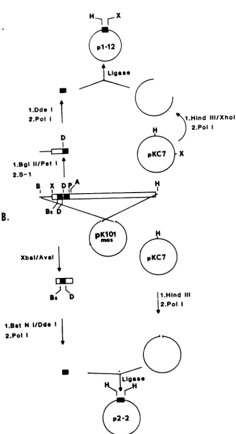
FIG. 1. Construction of primers for DNA sequencing. As de-scribed in detail in the text, primer 1 (A) was constructed by
insertion ofa52-bp DdeI fragment of thewild-type MuSV-124mos
geneintoHindIll-digested pKC7 DNA. Beforeligation, all
restric-tionsiteswerebluntended by "filling in" withthe Klenow fragment
of E. coli DNA polymerase I. Primer 2 (B) was constructed by
insertion ofa56-bp DdeIlPstIfragment of the wild-type MuSV-124
mos geneinto HindIII/XhoI-digested pKC7 plasmid DNA. Before ligation, all restriction siteswereblunt endedby filling in with the
Klenowfragmentor weretrimmed by S1 nuclease digestion. Thus, ourprevious studies in this system suggested that P58gagwastheproduct ofanout-of-frame fusion ofthegag and mos genes and that a possibly temperature-sensitive
splicingeventpermittedthealignment of thesetwogenesin a continuous open reading frame. These suggestions have awaited theresolution oftwopoints. First,itwas necessary thatthegaggene-mos genejunctionsinthetwoMuSVtsllO RNAs besequenced totestthe assumptions regarding their proposed compositions.Inthepresentstudywehave shown
by primerextensionnucleotidesequencing that the primary transcriptof MuSVtsllO viral DNA isa4-kb RNA in which thegagandmosgenes are, infact,joinedout offrame such thattermination oftranslationwouldoccurshortly after the
MATERIALS AND METHODS
Cells and viruses. Mo-MuSVtsllO wasderived from Mo-MuSV-349,asubclone ofMo-MuSV-124(1),asdescribedby Blairet al. (2). Nonproducer normalratkidney (NRK)cells infectedwithMo-MuSVtsll0, termed6m2,weremaintained at 33°C in McCoy5a mediumcontaining 15%(vol/vol)fetal calfserum. MuSVtsllOproducer cells, designated 206-21C,
were generated by superinfection of 6m2 cells with the IC strain of Moloney murine leukemia virus (Mo-MuLV) (6). These cells were maintained in the above culture medium and used for virusproductionin 2-quart (ca. 1.9-liter)roller bottles. Aspontaneous revertantof the 6m2 cellline, desig-nated 54-5A4, was also maintained as above at 37°C. The 54-5A4 cells exhibited a transformed phenotype at both 33 and39°C.
DNAprimerconstruction. Theconstruction of DNA
prim-ers from wild-type MuSV-124 DNA is depicted in Fig. 1.
Briefly, primer 1 wasconstructed by excisionofa 342-base
pair (bp) DNA fragment between an upstream BglII site (nucleotide 3698) andaPstI site(nucleotide 4040)withinthe wild-typemosgeneofMuSV-124 DNA (generously givento
us by Dino Dina). This fragment was blunt ended by
digestion with Si nuclease and further digested with DdeI. The56-bp DdeIlPstI fragmentwasfilledin withthe Klenow
fragment ofEscherichia coli DNA polymerase I, ligatedto
agarose gel-purified pKC7 (13) plasmid DNA doubly di-gestedwithHindIIIIXhoI and then filled in with the Klenow fragment, and used to transform E. coli RRI. The 56-bp insert in theplasmid, designated pl-12, couldbereleased by HindIIIlXhoI double digestion.
Primer 2 was constructed exactlyas described by Dono-ghue and Hunter (5). A DNA fragment extending from an XbaI site 10bp upstream(nucleotide 3863) ofthe beginning of the mosgenetoanAvaI site 194bpdownstream (nucleo-tide4057)wasexcised fromwild-type MuSV-124DNA.This
fragmentwasdoublydigestedwithBstNIIDdeI, filledinwith the DNApolymeraseI Klenowfragment, ligatedto HindIII-digested/Klenow-filled-in pBR322 DNA, and used to trans-form E. coli RRI. By using this subcloning strategy, the 56-bp insert in the plasmid, which we designated p2-12, could be excised by HindIII digestion. This plasmid is identicalto pDD39 described by Donoghueand Hunter(5). Purification of plasmid DNA. The various inserts and fragments described above were excised by digestion with theappropriate restriction enzyme(s), sizefractionatedon a 0.8% agarosegel,and stained with ethidiumbromide. DNA
A.
1.Dde I
2.Pol D
1.BgI II/Pat I 2.S-1I
B
on November 10, 2019 by guest
http://jvi.asm.org/
[image:2.612.60.293.63.490.2]626 NASH ET AL.
was purified from agarose by adsorption to glass powder (18).
In cases where small DNA fragments were purified from polyacrylamide gels, gel slices were crushed to a pastein 2
ml ofTNEbuffer(10 mM Tris [pH 8], 100 mM NaCl, 1 mM EDTA) and agitated overnight. Acrylamide granules were removed by centrifugation, and the supernatants were
fil-teredthroughIsolab QS-P columns to remove the remaining
acrylamide. The column effluent containing the DNA was phenol extracted, ethanol precipitated, and dissolved in water.
Isolation of cellular RNA. Intracellular RNA was isolated
from tissue culture cells by a guanidine hydrochloride
ex-traction procedure (20). Briefly, cellular pellets were
dis-solved in solution A, 8 Mguanidine hydrochloride-100 mM
sodium acetate (NaOAc; pH 5), and subjected to vigorous
Douncehomogenization. After centrifugation ofthe extracts at 10,000 rpm for 15 min, nucleic acids were precipitated fromthe supernatant byaddition of0.5 volumeof ethanol at
-20°C for 30 min and collected by centrifugation at 7,000 rpm for 20 min. The nucleic acid pellets were dissolved in 0.25 to0.50 the original volume of solution B (solution A
plus0.1 volume of200 mM EDTA) and
again
precipitatedwith0.5 volume ofethanol. This last step was repeated twice more,decreasingthevolumeof solution B byhalf each time.
Thefinalpellet wasdissolved in20 mM EDTA (pH 7.5) and
extracted with chloroform-isobutanol (4:1). The aqueous
phase was mixed with 2 volumes of4.5 M NaOAc (pH 6),
and the RNA was precipitated at -20°C overnight. The RNAwascollectedbycentrifugation,washed once with 3 M
NaOAc and once with 70% ethanol, anddissolvedinwater. Preparation of cellular DNA (13). Cells growing in roller culture were rinsed withan isotonicbufferandgently lysed
in
2%
sodium dodecyl sulfate(SDS)-8
M urea-0.35 MNaCl-1 mM EDTA buffered with 10 mM Tris (pH 8). The
cell lysate was extracted with an equal volume of phenol-chloroform (1:1) saturated with the above SDS-urea buffer
and ethanol precipitated. Nucleic acids from each roller culture were dissolved in 3 to 5 ml of water, digested sequentially with50
gig
ofRNase per ml at37°C for60minand 100
gig
ofproteinase K per ml at 37°C for 120 min, re-extracted withphenol-chloroform,
precipitated withethanol,and dissolved inwater.
Blot hybridization of DNA. Routinely, 15 ,ug of cellular DNA, digested with a given restriction enzyme as recom-mendedbythesupplier,wasanalyzedon a0.8%agarosegel
in50 mMTris(pH 8.3)-40 mM NaOAc-2 mM EDTA at 40 V
for16 to18h and transferredtonitrocelluloseessentiallyas
describedby Southern (15)andmodified byWahl et al.(19). The nitrocellulose was prehybridized at 37 to 42°C in 50% formamide-5x SSC
(SSC
= 0.15 M NaCl plus 0.015 Msodium
citrate)-5x
Denhardt solution(0.1%
bovine serumalbumin,
0.1% Ficoll, 0.1%polyvinylpyrrolidone)-0.
1% SDS, containing 100gig
ofsheared salmonsperm DNA and 2 mg of base-hydrolyzed yeast RNA per ml. Hybridizationwascarried out at 37 to 42°C in the samesolution containing
1x Denhardt solution and 2 x 10⁶
cpm
of32P-labeled
probe(1 X
108
to 3 x10⁸ cpm/gig)
per ml. Afterhybridization, thefilters were washedfourtimes with 3x
SSC-0.1%
SDS and twice with 0.lx SSC-0.1% SDS at 37°C, dried, and auto-radiographed.5' End labeling. Restriction fragments were first
de-phosphorylated with calf intestinal phosphatase (Boehringer
MannheimCorp.). Briefly, DNA was adjusted to 50 mM Tris
(pH9), 1 mM
MgC92,
0.1 mMZnCl2,and 1 mM spermidine containing 10U of calf intestinalphosphatase at37°C. After30min, an additional 10 U of calf intestinal phosphatase was
added for another 30 min. The digest was mixed with 0.1 volume of1Ox TNE and 0.05volume of 10% SDS for 15min
at 68°C to halt the reaction. After phenol-chloroform (1:1)
extraction, thenucleic acids were ethanol precipitated. For
5' endlabeling,dephosphorylated DNA was adjusted to 100 mM Tris (pH 7.6), 20 mM MgCl2, 10 mM dithiothreitol, 0.2 mMspermidine, and 0.2 mMEDTA, to which wasadded 10 to 20 U of T4 kinase and 150
,iCi
of[y-32P]ATP for 1 h at37°C. After the addition of 20 gig of carrier DNA, the
reaction mix was desalted by passage over a G-50 column
equilibrated inTNEbuffer. Radioactivity in the void volume was pooled, ethanolprecipitated, and dissolved in TE buffer (10 mM Tris [pH8], 1 mMEDTA).
Si nuclease analysis. S1 nuclease analyses were done
essentially as described previously (11). Briefly, the 5'
end-labeled 359-bpinsertfrompBA.36 washybridized to 10
gig oftotalcellular RNA from 6m2 cells grown at 39, 33, or 28°C, as indicated in the text, in 80% formamide-40 mM
PIPES [piperazine-N,N'-bis(2-ethanesulfonic acid)] (pH
6.8)-400
mM NaCI-1 mM EDTA at 56°C for 3 h, digested with S1 nuclease (200 U/ml) at 37°C for 30 min, phenolextracted, and ethanol precipitated. The digests were dis-solved in 20
gil
of10 M urea-1 mM EDTA-0.015% bromo-cresolgreen, heated to 50°Cfor5min, and analyzed on a 4%polyacrylamide gel containing 8 M urea. After
electropho-resis, the gels were fixed with 50% methanol-7%
HOAc,
dried, andautoradiographed.
Primerextension. 5' end-labeled primer 1 or 2 was mixed
individually with 10gigof total cellularRNA from 6m2 cells grown at 39, 33, or28°C,asindicatedin the text, and ethanol
precipitated. The pellets were thoroughly dissolvedin 30
gIl
of80% formamide-40 mMPIPES (pH6.8)-400mMNaCl-1
mM EDTA, immersed in an 85°C water bath for 10 min,
transferred to a water bath at65°C, and allowed to hybridize during cooling to room temperature overnight (5). After hybridization, the reaction mixeswere ethanol precipitated
and dissolved in 25 gil of 10 mM Tris (pH 7.4). Primer
extension in the presence of dideoxynucleoside
triphos-phates(ddNTPs)wascarriedoutessentiallyasdescribed by
Donoghue and Hunter(5). A5-,ul portion ofthehybrid mix
wasdispensed into fourseparate tubes, to each of which was
added 1 gil ofa 200 giM dNTP mix (dATP, dCTP, dGTP, dTTP),1
gIl
ofaddNTP mix calculatedtoproducetheratios given in parentheses (ddATP/dATP = 1.3; ddCTP/dCTP = 0.4; ddGTP/dGTP = 0.3; ddTTP/dTP = 0.9), and 1 gil ofavianmyeloblastosis virusreversetranscriptase(Seikagaku;
10
U/gil).
Thesemixtureswereincubated at 40 to 42°Cfor60 min to extend the primers, after which they were ethanol precipitated aftertheadditionof10gig
ofcarrieryeast RNA and 10mMMgC12.Sequence analysis. Theextendedprimersweredissolved in
75% formamide-20 mM EDTA (pH 8.0)-0.01%
bromo-phenol blue-xylene cyanol, heated to 100°C for 2 min, and
analyzed on a 6%
polyacrylamide-8
M urea gel in TBE buffer (50 mM Tris [pH 8.3], 50 mM boric acid, 2 mMEDTA)at2,000Vfor105 min. Afterelectrophoresis,thegel
was either subjected to autoradiography directly, using
Kodak XAR-2 film andDupont CronexLightning-plus
inten-sifying screens, or fixed with10% HOAc-10%methanolfor
20 min, dried,andautoradiographed.
RESULTS
Relative abundance ofMuSVtsllO-related mRNA species. Asreported previously (11), we have developed asensitive assay for the detection of both the 4.0- and the 3.5-kb
J.VIROL.
on November 10, 2019 by guest
http://jvi.asm.org/
MuSVtsllO RNA TRANSCRIPTS 627
wtMuSV 349 RNA -t1
9a9
MuSVts110 4kb RNA mos
splice
MuSVtsl10 3.5 kb RNA 1 MOS
B A
___
pBA.36-359_
175.
FIG. 2. S1 nucleaseassayforMuSVtsllO viral RNA. Theassay
depends on the differential hybridization of the 359-bp insert in
pBA36 towild-type MuSV-349 RNA, MuSVtsllO 4-kb RNA, and MuSVtsllO 3.5-kb RNA. As shown, wild-type MuSV-349 RNA, MuSVtsllO 4-kb RNA, andMuSVtsllO 3.5-kb RNA, respectively, willprotect359, 175, and 122 bp of the 5' end-labeledpBA36359-bp insertfromdigestion by S1 nuclease.
MuSVtsllORNAs in the presenceof each other. Theassay
relieson theincompletehybridization ofa359-bp wild-type viral DNA fragment(designatedpBA.36)tobothMuSVtsllO RNAs(Fig. 2). Because of the -1.5-kbcentral deletion, the MuSVtsllO 4-kb RNA willprotectapproximately 175bases ofthepBA.36insertfromdigestion with Si nuclease. Since the 3.5-kb RNA has a larger deletion in its mos gene than does the 4-kb RNA, hybrids between it and pBA36 can protectonly 122 bases of pBA36 from Si digestion. Thus, as shownpreviously (11), and in the accompanyingpaper(5a),
both the4-and the 3.5-kb RNAscan readily be detected in 6m2 cells grown at 33°C. Within 2 h of a shift to 39°C, however, only the 4-kb RNA canbe detected in 6m2 cells. Interestingly, if6m2 cells are shifted to 28°C, the spliced 3.5-kb RNA becomes predominant at the expense of the
unspliced 4-kb RNA (Sa; G. E. Gallick, R. Hamelin, S. Maxwell, D. Duyka, and R. B. Arlinghaus, Virology, in press). Thus, by simple temperature shifts, we can select conditions under which either the 4.0- or the 3.5-kb RNA predominates in6m2 cells.
gag-mosjunctionsintheMuSVts110RNA species. InFig. 3 are illustrated the regions of the MuSVtsllO 4- and 3.5-kb
RNAstowhich primers 1 and 2 will hybridize. The approx-imate positions of the gag-mos junctions in both of these
RNAs, asdetermined by Si nuclease mapping experiments suchastheone described above, arealso indicated (nucleo-tide3883 in the4-kb RNAand nucleotide 3936 in the 3.5-kb RNA). Note that primer 2 will hybridize along its entire length onlytothe 4-kb RNA;hybridization of primer2tothe 3.5-kb RNA is interrupted at its 3' end because of the deletion in the mos gene, and thus this hybrid cannot be extended with reverse transcriptase. Hence, an
unambigu-oussequenceforthegag-mosjunction inthe4-kbRNAcan beobtained withprimer 2. Incontrast, primer1will hybrid-ize completely with both RNAs. Hence, its extension will result in two superimposed and thus uninterpretable
se-quences ifone usesRNA from 6m2cellsgrown at 33°C. As discussed below, however,we wereabletocircumventthis problem by the extension of primer 1 hybridized to RNA
from 6m2 cells grown at28°C. In thesecells, the3.5-kb RNA is the dominant MuSVtsllO species (Sa). Extension of these primer 1 hybrids produced a clear sequence of the gag-mos junction in the 3.5-kb RNA.
As a control on the method and as a check on whether the
wild-type MuSV-349 sequence was the same as the
pub-lished sequence of MuSV-124 in the region under analysis, primers 1 and 2 were individually hybridized to MuSV-349 RNA and extended in the presence of dideoxynucleotides. An example of the sequence data obtained from these two experiments is shown in Fig. 4. Using both primers, we found that the MuSV-349 RNA sequence wasidenticaltothe published MuSV-124 sequence in the region analyzed
(nu-cleotides 3815 to 3972 of the MuSV-124 sequence; data not completely shown).
We next hybridized 5' end-labeled primer 2 to cellular RNA from 6m2 cells grown at either at 33 or 39°C and extended the primer as was done with MuSV-349 RNA. From the nucleotide sequence obtained (Fig. 5), we could
ascertain that the MuSVtsll0 4-kb RNA had suffered a
1,488-base deletion relative to wild-type RNA (Fig. SB),
resulting in the fusion of gag gene nucleotide 2404, located 71basesupstream of the p30-plO junction in wild-type viral RNA, to mos gene nucleotide 3892, found 17 bases down-stream from the 5' end of the mos gene. The position of this
deletion is in good agreement with our data, using the S1 nuclease method (11), and with the results of an earlier
heteroduplexing study (7). By using S1 nuclease, the dele-tion waspredicted to joinnucleotide 2409 to nucleotide 3883, located 25 bases upstream of the 3' end of the primer 1-RNA hybrid (nucleotide 3908). The actual junction point was found to be only 12 to 13 nucleotides upstream of nucleotide 3908 (Fig. 5). This 1,488-base deletion has resulted in the
removal of the first mos gene initiation codon (cf. Fig. 7A and B ) and would cause the mos gene to be translated out of frame for 14 amino acids before reachinga TAG and then a TGA stop codon 42 and 54 bases downstream of the gag-mosjunction point. Interestingly, the 1,488-base deletion is
bracketed by the 6-base direct repeat AACGCC found at
nucleotides 2399 to 2404 and 3886 to 3891 in the wild-type gag and mos genes, respectively. One of these repeats was
retained after the deletion. The calculated size for this deleted RNA is 3,820 nucleotides (wild-type 5,320 bases minus a 1,488-base deletion), in good agreement with the 4-kb size previously estimated by blot hybridization (6, 11, 16).
To obtain aninterpretable gag-mosjunction sequence for the MuSVtsllO 3.5-kb RNA, we hybridized primer 1 to RNA from 6m2 cells grown at 28°C. The bulk of the viral RNA in6m2 cells grown under these conditions is 3.5 kb in size. The sequence obtained from such anextendedprimer 1 hybrid is shown in Fig. 6. From this sequence it could be
ascertained (Fig. 6B) that nucleotide 3936 in the wild-type mos,gene (2488 in the MuSVtsllO 4-kb RNA), located 44 basesdownstreamof thegag-mos junction in the 4-kb RNA, was fused to nucleotide 2017 in the gag gene, located 387 basesupstream of thegag-mosjunction in the 4-kb RNA. As expected, this fusion point is about 40 bases upstream of the
3'end ofthe primer 1/viral RNAhybrid. Thesequenceof the 3.5-kb RNA is unambiguous up to the intron/exon border (Fig. 6). Upstream of this border, however, the presence of low levels of 4.0-kb RNA in the RNApreparation decreased
the resolution obtainable. If, however, one considers only
the majorbands in the sequence ladder, it is clear that, as suggested by ourpreviously published work (11), the splice donor site (9) surrounding nucleotide 2017 and the splice VOL.53, 1985
on November 10, 2019 by guest
http://jvi.asm.org/
[image:4.612.58.291.75.255.2]628 NASH ET AL.
3883
MuSVtsYI10 4kb RNA MOS
primer2
3908 3960
primer1 3981' 4036
MuSVtsI103.5kbRNA g
a-3936
FIG. 3. Hybridization of primers 1 and 2 to theMuSVtsllO 4-and 3.5-kb RNAs. Primer1 will hybridize to a56-base stretch of both the4.0- and 3.5-kbMuSVtsllORNAs about 50 bases down-stream of the proposed splice site. Primer 2 was constructed to
hybridize completely to a 52-base stretch of the MuSVtsllO 4-kb RNA approximately 25 bases downstream of the proposed gag gene-mos genejunction at nucleotide 3883. Because its upstream terminus extends across the proposed splice junction in the MuSVtsllO 3.5-kb RNA, the hybrid formed with this particular RNA cannotbeextended.
acceptorsurrounding nucleotide3936(9)are
joined
inframeas (GCA G/TG TCT). The fusion of nucleotides 2017 and
3936(2448 inthe4.0-kbRNA),
removing
431 basesfromthe 4-kb RNA, excises the TAG stop codon found at the3820 - TGGAGACCT G
GCGGCACCCTAC
-C AGAACCT
- -CGACCATCCTCTA
G-ACTGACATGGC
- - GCATTCAACCCC
___ ATGCTCCCAAACT
6-u',
~ ~ ~
/904
__ _
C G T A
FIG. 4. Nucleotidesequencingofwild-type MuSV-349 RNAby
use of primer 2. Primer 2 was 5' end labeled, hybridized to MuSV-349RNA,and extended in thepresenceofddNTPs,and the productwasanalyzedon a6%polyacrylamidesequencing gel.The sequenceobtainedwasidentical tothatpublished byVan Beveren etal.(17).
intron/exon border and restores the original mos reading
frame, allowing the bypass of the TGA stop codon (Fig.
7B). Thus, an in-framegag-mosfusionprotein, presumably P85gag-mos, canbetranslatedfromthe
resulting
3.5-kbRNA.Calculating from the established MuSV-124 sequence, this
fusion protein would have atheoretical molecularweightof
about72,000composed of325 gag geneamino acidsand 353 mos gene amino acids. The RNA from which this
protein
would be translated is 3,389 bases, 431 bases shorter thanthe3,820-base RNA,ingoodagreementwiththe3.5-kb size previously estimated by blothybridization (6, 11, 16).
MuSVtsllO-relatedprovirusesincells.Itwasof
fundamen-tal importance to determine the number and structural
organization of MuSVts11O-related
proviruses
in 6m2 cellssincethe validity ofourinterpretation ofourdata rests on theassumptionthat there should be
only
onetranscription-ally active MuSVts1lO
provirus
in 6m2nonproducer
cellsand thattranscription of this viralDNAshould
produce
only
the unspliced 4-kb RNA as theprimary transcript.
The predicted size of this DNA should be about onelong
A
*A DN\A AT AT
AT AT
G ^
AT
AT
GC
AT
AT
vGN
JUNCTW34-4
:IA
iii
G :
B AGA GAGGAA
AAG
GAA GAACGC
CGAG
2-SEOUENCE-
--L AACGCCA TGCTCC -3892 MOS
SEQUENCE
DELETKON
24043391GAAGAA CGC CAT GCTCCCAAA- -TAG- -TGA
2404 +47stop stop
+54
FIG. 5. Nucleotide sequenceofthe gag gene-mos genejunction
inMuSVts110 4-kbRNA. 5'end-labeledprimer2washybridizedto 10,ugofcellular RNAfrom6m2cellsgrownat33°Cand extendedby
reversetranscriptase in the presence ofddNTPs,and the product wasanalyzedon a6%polyacrylamide sequencinggel. (A) Nucleo-tide sequence of extended primer 2. Alongside the sequence is shownthe actual DNA sequenceread from thegel (complementary
tothe RNAplusstrand)and theplus-strandRNAsequence. In(B) the gag gene-mos genejunction in the MuSVtsllO 4-kb DNA is showndiagrammatically.
J. VIROL.
on November 10, 2019 by guest
http://jvi.asm.org/
[image:5.612.63.301.71.167.2] [image:5.612.325.559.291.634.2] [image:5.612.96.271.344.668.2]MuSVtsllO RNA TRANSCRIPTS 629 DN~~~~~C RNA
*4-A OC GJT AO-t AA
JJCTO SEOJENCTEO
W _ ~~~CG
C T C ATGTU C
V O ..,:;V30 U K 0
_i332
~~~~~~~~T
~~~~~GEA
FG6. Nuclotid seuec ofteslc
iei h
uVs
celuaRN
rm62 cel
rw
t 28C
xeddiUh
C G
_
-\\~~~~~C
G.>>_ 1 ~~~~~Gc
|~~~~~~~ G
!~~~~~~~~
U"CS
G ~~~CCACT
9T
TC C-ATCCAT TCT CCA A/G T T G
C^G [email protected]"
presenceofddNTPs,andanalyzedon a6%polyacrylamide
sequenc-ing gel. (A) Nucleotide sequence of extended primer 1. Alongside
the sequence is shown the actual DNA sequence readfrom thegel
and thecomplementary plus-strandRNA sequence. In(B),thegag gene-mos gene splice junction in the MuSV-349 3.5-kb RNA (as DNA)is showndiagrammatically.
terminal repeat
(LTR)
(-590bases)
larger
than the3,820-base RNA
transcript,
orabout 4.4 kb.Previously
we havepresented
preliminary
evidence that 6m2 cells contain asingle
candidate MuSVtsllOprovirus
residing
in anapproximately
12-kb mos+ BamHl DNAfragment
(11). Inthe datapresented
below,wereaffirm and extend theprevious
evidence.Digestion
of 6m2 cellular DNA with BamHlyields
two mos+ DNAfragments,
ap-proximately
21 and 12 kb in size(Fig.
8A, lane 2). SinceBamHl-digested
DNA from uninfected NRK-2 cells con-tainsonly
the 21-kb mos+species
(Fig.
8A, lane 1), the 21-kb DNA represents the c-mos gene and the MuSVtsllOprovirus probably
resides inthe 12-kbfragment.
DNAfromother MuSVts11 -infected cell lines, such as
producer
206-21C and revertant 54-5A4
cells,
asexpected,
alsocon-tain a 12-kb mos+ DNA (Fig. 8A, lanes 3 and 4). As the 21-kb rat c-mos gene is a single-copy gene, the equivalent hybridization intensity observed with the 12-kb viral DNA suggests that it also is present at the level ofone copy per genome. In contrast to this simple situation,
BamHI-di-gestedDNAfrom wild-type MuSV-349-infected cells shows a more complex
pattern
of mos+ proviral DNAs, rangingfrom 8 to 20 kb in size (Fig. 8A, lane 5).If6m2cellularDNA
is digested with
BamlSstI
orSstI
alone(SstI
is expected to cut oncewithin each viral LTR, yielding a DNAexactly the size expected for the primary transcript), the 12-kb mos+BamHI
fragment isreduced to about 3.85 kb in size in6m2,54-5A4, and 206-21C cellular DNA (Fig. 8B, lanes 2, 3, and 4). In contrast,
SstI
digestion of DNA from cells infectedwith wild-type MuSV-349 yielded a major 5.2-kb mos+ DNAfragment(Fig. 8B, lane 5). In uninfected NRK-2 cells,
the 21-kb
BamHI
c-mosfragmentis cutbySstI to2.65 kbin size (Fig. 8B, lane 1). Thus, the 3.85-kbviral DNA is exactly the size expected for theMuSVtsll0
viral DNA, about 1.5 kb shorter than 5.2-kb wild-type DNA and distinct from the c-mos gene.A partial restriction map of wild-type MuSV-349 DNA is shown at the topof Fig. 9. Fromthis map it can be seen that the 1.5-kb deletion in the
MuSVtsll0
genome should have removed the unique Sall andXbaI
sites present in the wild-type DNA. A comparison of 6m2 DNA and wild-type MuSV-349 DNA cut withSstI, SstlISalI,
and SstlXbaIshowed that, aspredicted, the 3.85-kb provirus was lacking the
SalI
andXbaI restriction sites (Fig. 10A, lanes 1, 3, and 5) and that the wild-type MuSV-349 5.2-kb viral DNA contained the uniqueSalI
and XbaI restriction sites (Fig.10B,
lanes 2 and 3) at the positions established by other investigators (14, 17). To establish the approximate limits of the deleted region in the 3.85-kb viral DNA, double digests were performed withSstIlXhoI
and SstIHindIII. The size of the virus-derived mos+ fragments produced by these di-gests, 2.4 and 3.4 kb, respectively (shown in Figure 10A, lanes 7 and 9), were about 1.5 kb shorter than the 3.7- and 4.6-kb fragments produced bySstIlXhoI
or SstIIHindIIIdigests of wild-type MuSV-349 DNA (Fig.lOB, lanes 4and 5). The 2.65-kb fragments seen in Fig. 10A, lanes 1, 2, 3, 4, 7, 8, 11, and 12, are derived from the rat c-mos gene, as are the -1.6-kb fragments seen in Fig. 10A, lanes 5, 6, 9, and 10. Taken together, these data suggest that the 3.85-kb viral DNA contains a -1.5-kb deletion mapping roughlybetween the XhoI site and the HindIll site. However, since an
SstIIBgiI
digest of the 3.85-kb viral DNA yielded a -1.6-kb mos+ fragment exactly the size observed in BgiI-digestedwild-type viral DNA (cf. Fig. 10A, lane 11, with Fig.lOB, lane 6), the deletion in the 3.85-kb DNA could be further localized to the region between the XhoI site (located at wild-type nucleotide 1982) and the BglI site in the 5' end of the v-mos gene (nucleotide 4120). By Si mapping andprimer extension sequencing, the actual MuSVtsllOdeletion maps between wild-type nucleotides 2404 and 3892 (see above). Thus, the organization of the 3.85-kb viral genome in 6m2 cells as estimated by restriction mapping correlates quite closely with its predicted structure based on these previous data.
Viral genomes in206-21CMuSVtsllOproducer cells. Inthe course of these experiments we were initially surprised to find that the 206-21C producer cell line chronically superin-fected with Mo-MuLV, unique among all
MuSVtsllO-infected cell lines examined, contained a second virus-re-lated mos+ DNA fragment. In an SstI digest of 206-21C
VOL. 53, 1985
on November 10, 2019 by guest
http://jvi.asm.org/
630 NASH ET AL.
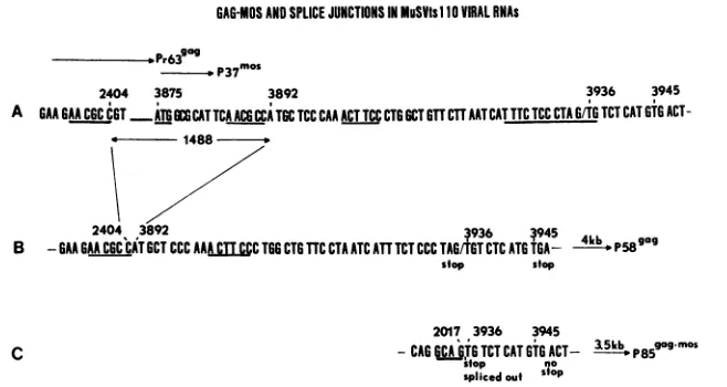
GAG-MOSANDSPLICE JUNCTIONS IN MuSVtsl O VIRAL RNAs
D.P37 o
2404 3875 3892 3936 3945
A GAAGAAC6CC6T
ATGCATTCAACGCiCTGCTCCCAA
ACTTCCCTGGCTGTTCTTAATCATTTCTCC CTAGIT TCT CAT GTGACT-* - 1488
-2404 3892 3936 ~945 411 gag0
B -GAAGAACGCCATGCTCCC AAA cTT CCTOG CTG TTC CTAATCATTTCT CCCTAGGT CTCATG TGA_ - p58
stop stop
C
2017 3936 3945
-CAG
iCA
TG TCT CAT GTG ACT- 35b P8gogmos sfop nosplicedouf Sfop
FIG. 7. Comparison of the MuSV-349 5.2-kb RNA, MuSVtsllO 4-kb RNA, and MuSVtsllO 3.5-kb RNA sequences. (A) Sequence of MuSV-349 RNA between nucleotides 2395 and 3949, showing nucleotides 2404 and 3892 bounded by the AACGCC repeats, the firstmos initiation codon at nucleotide 3875, the intron/exon border at nucleotide 3936, and the out-of-frame TAG and TGA termination codons. (B) Sequence of theMuSVtsll04-kbRNA at the gag gene-mos gene junction, showing the deletion of 1,488 nucleotides including one of the AACGCC repeats, the out-of-frame mos sequence, and the in-frame stop codons at nucleotides 3933 and 3945. (C) Sequence of the MuSVts110 3.5-kb RNA at the gag-mos splice junction, showing the splice junction between gag gene donor nucleotide 2017 and mos gene acceptor nucleotide 3936,which restores the mos genetoits original readingframe, avoiding the TGA stop codon at nucleotide 3945.
DNA, thissecondfragmentwasestimated to be about 3.35 kb insize(Fig. 8B, lane 4;Fig. 10A, lane2). Tocharacterize
this 3.35-kb DNA, a series of double digests similar to those done on 6m2 DNA (Fig. 10A) was performed, with the
followingresults. As did the 3.85-kb viralDNAinboth 6m2
and 206-2IC cells, the 3.35-kb viral DNA lacked Sall and
XbaI restriction sites (Fig. 10A, lanes 4 and 6). Also,
SStI/XhoI and SstI/HindIII double digests of the 3.35-kb
DNAgenerated mos+ DNA fragments 400 to 500 bp shorter than thoseproduced by digestion of the 3.85-kb viral DNA
(cf. lanes 7 and 8 and 9 and 10, Fig. 10A). However, the
SstIIBglI
digests of both the 3.8- and 3.35-kb DNAs (Fig.10B, lanes 11 and 12) produced mos+ DNA fragments of
identical size (1.6 kb), suggesting that both DNAs have an
identicalstructurefromaposition very near the 5' end of the v-mos gene downstream through the 3' LTR. From these
mapping data, it seems clear that the 400 to 500 bases present in the 3.85-kb DNA and missing from the 3.35-kb DNA mustbe located in the approximately 650-base region
between theXhoI andBglI sites in the3.85-kbDNA. Since
this is approximately the region which is spliced out of the 4-kb DNA to form the 3.5-kb RNA, wefavor the interpre-tationthat the 3.35-kb DNA represents a DNA copy ofthe
spliced 3.5-kb RNA which was reverse transcribed by the
helper virus-associated reversetranscriptase andintegrated intothegenomic DNA ofthe
206-21C
cell line.DISCUSSION
The data presented in this study argue forcefully that a
single, centrally deleted, 4.4-kb (calculated size including both LTRs) viral DNA is transcriptionally active in
MuSVtsllO-infected nonproducer6m2cells. When 6m2 cells are grown at 39°C, this 4.4-kb viral DNA is transcribed to forman approximately 3.8-kb viral RNA. If, as is
reasona-ble, one assumes the polyadenylate tail on this RNA to be 100to 150nucleotides, the full size of this RNA should be 3.9 to 4.0 kb, in very close agreement with the 4.0-kb size estimated inourpreviouswork (6, 11, 16). This 4.0-kb RNA is related to wild-type 5.2-kb MuSV-349 RNA by way of a 1,488-base deletion which hadasits result thefusion of gag
genenucleotide 2404to mosgenenucleotide 3892(usingthe Van Beveren et al. [17] numbering system). Although the
mechanism by which this deletion was generated as a consequence of UV irradiation is unclear, it is probably
significant that the deletion is bounded by the 6-bp direct
repeat AACGCC found at nucleotides 2399 to 2404 in the wild-type gag gene and at nucleotides 3886 to 3891 in the wild-type mos gene. As as consequence of the MuSVtsllO deletion,oneofthe6-base repeats is lost andoneisretained.
Because the p30 region of the gag gene in the 4.0-kb MuSVtsllO RNA is fused out of frame to a point just
downstreamofthe 5'end ofthev-mosgene,thepolypeptide product ofthis RNA isnecessarily atruncated gag precur-sor,P58gag, whichveryprobably containsashort(14-amino acid residue) C-terminal peptide encoded by an alternate,
apparently normally unused,v-mosreadingframe.
Termina-tionofthetranslationof thispolypeptide is dictatedbytwo
closely spaced stop codons found in this particular mos
reading frame 42 and 54 bases downstream from the gag-mosjunctionpoint.Afurther consequenceofthis deletion is theremovalofthe firstmosgeneinitiation codon locatedat wild-type nucleotide 3875, apoint17bases upstream of the
3' end ofthe deletion. Thus, the MuSVtsllO 4.0-kb RNA cannot be translated to form wild-type p37mos. Previously published in vitro translation experiments (10) have shown
unequivocally that, incontrast to
similarly
translated MuSV-349 5.2-kbRNA, the MuSVtsllO4.0-kbRNAdoesnotgiverise top37mos but instead isquite capableofsupporting the translation ofP33m"s and
P23mos
from the initiation codons found further downstream.Our previous data obtained by S1 nuclease mapping
stronglysuggested that the gag-mosjunctionin the smaller
MuSVts1103.5-kb RNAwas anin-framejunctionformedby
theunion ofconsensussplicedonor and acceptor sites found
surrounding nucleotides 2017 and 3936 (11). The present sequence data support this previous suggestion. Hence, it now seems clear that in 6m2 cells grownat 33°C 431 bases
surroundingthe out-of-frame gag-mosjunctionin the 4.0-kb RNA canbesplicedout,joininggagnucleotide 2017to mos nucleotide 3936(nucleotide2448in the 4-kb RNAsequence).
J. VIROL.
on November 10, 2019 by guest
http://jvi.asm.org/
[image:7.612.149.478.77.252.2]A
23-9..' ~ ~
] w~~2
..'e.War O...L~~1
43~~~
2
-3.85
-3135
-2.65
FIG. 8. ProviralDNAin MuSVtsllO-infected cells. High-molec-ular-weight DNA from 6m2 cells, 54-5A4 revertant cells,206-2IC producercells,MuSV-349-infectedmouseTBcells, and uninfected NRK-2 cells was digested with BamHI or doubly digested with BamHI and SstI, analyzed on a 0.8% agarosegel, transferred to nitrocellulose, and hybridizedtoa32P-labeledv-mos-specific probe. (A)v-moshybridizationtoBamHIdigests of (lane 1) NRK-2, (lane 2) 6m2, (lane 3) 54-5A4, (lane 4)206-21C, and(lane 5) MuSV-349 DNAs. (B) v-mos hybridization to BamHIISstI double digests of (lane 1) NRK-2, (lane 2) 6m2, (lane 3) 54-5A4, (lane 4)206-21C,and (lane 5) MuSV-349 DNAs.
This splice event excises an
in-frame
TAG stop codon andrestores the original mos gene reading frame 9 bases up-stream ofthe previously in-frame TGA termination codon. Theresulting 3.35-kb RNA(originally estimatedas3.5 kb in
length) could code for afusion protein containing 325 gag gene amino acidsand353 mosgeneamino acids.
The
validity
ofourinterpretation of the abovedata restson the
fundamental
point that there should beonly
onetranscriptionallyactive MuSVtsllO-related provirus in 6m2 cells. The blot hybridization experiments presented in this
study establish that this is almost surely the case. Using
enzymes that cut once within each viral LTR, we found a
single 3.85-kbviral DNA (about4.4 kb with the other LTR addedin)in 6m2 cells. The intensity ofthe signalobserved was equivalent to that of the single-copy rat c-mos gene.
Also, we found that enzymes which would cut wild-type viral DNA within the expected 1,488-bp deletion
(nucleo-tides2404to 3892) do not cutthe 3.85-kb DNA. Moreover, by meansof enzymatic double digests, itwasfound thatthe 3.85-kb viralDNAwasrelatedtowild-typeDNA by wayof
adeletion roughly spanning the region between anXhoIsite at gag gene nucleotide 1982 and a BglI site at mos gene
nucleotide4120.These nucleotideaddresses arewithinafew
hundred bases of the nucleotide 2404 to 3892 deletion mapped by S1nucleaseassays(11) and theprimer extension sequencing in the present study. Thus, the 3.85-kb viral
DNA in 6m2 cells is almost surely the source of the MuSVtsllO 4.0-kb RNA.
Although there is no trace in 6m2 cells ofa viral DNA
from which the MuSVtsllO 3.5-kb RNA could be
tran-scribed, producer 206-2IC cells
chronically
superinfected withMo-MuLVcontaina3.35-kb DNAwhoseorganizationveryclosely resembles the known structureofthe
approxi-mately 3.35-kb RNA. Since this viral DNA can be readily detected in 206-21C DNA, this observation reinforces our
finding that 6m2 cells lack such a viral DNA. Our present
interpretation of thisfinding is thatthis 3.35-kb DNA repre-sents an integrated cDNA copy of the 3.5-kb RNA
tran-scribed by the helper virus reversetranscriptase presentin
the 206-2IC cell line. The presence of a transcriptionally active provirus producing the 3.5-kb RNA at any tempera-turewithoutthe needforsplicing isanattractivepossibility inthisonecellline since 206-21C cellsappear to producethe 3.5-kbRNA(Sa)andP85gag-mosatboth33and 39°C(6). The
production of the 3.5-kbRNA at39°C by 206-21C cells was
originally thought to bepossibly due to acomplementation of thesplicing defectinMuSVtsll0 mediatedby viral factors provided by superinfecting Mo-MuLV. Recent data, how-ever (5a), argue against this possibility since 6m2 cells acutely infected withMo-MuLV cannot producethe 3.5-kb
Xhol SailI Bl I I BgI Hindlil
LTR .05LTR~~~No MuSV- 349
DNA
SstI d \ / Xba I SstI
a1
Xo
/Bgl
I llindIIII05 LR MuSVtsllo
A L DNA
dlOS 113
spice(330)
MuSVtsllO 4kbRNA
MuSVtsi1O
3.5kb RNA
FIG. 9. Partialrestriction mapsshowing thestructural relation-shipofwild-type MuSV-349 viral DNAto MuSVtsllO viralDNA and itstranscriptionproducts. Basedontheanalysesshown inFig. 8and10,theproposedstructureofwild-type MuSV-349 viralDNA isshowninthe topmostdrawing.Accordingto ourdata,MuSVts110 wasgenerated as a consequenceof thedeletion of1,488 bases of wild-typesequenceinformation(-4 ).Transcription ofMuSVtsllO DNAateither 33or39°C yieldsa4-kbRNAin which thegag and
mosgenesarefusedoutof frame.At33°C, splicing of431bases of MuSVtsllO 4-kbRNA information( 3 ) between in-frame splice donor(d)and acceptor (a) sitesproducesa3.5-kb RNAfromwhich
agag-mosfusionproteincanbetranslated.
on November 10, 2019 by guest
http://jvi.asm.org/
[image:8.612.94.261.75.432.2] [image:8.612.315.555.425.617.2]632 NASH ET AL.
3.85.i
t
3 35
2.65.
-*-*
X,
. E
:. 2..
12. 4.6
X 2 3 4 5 6
FIG. 10. Structure of proviral DNA in MuSVtsllO nonproducer 6m2cells, MuSVtsllO producer 206-21C cells, and wild-type !IuSV-349-infectedmouseTB cells. DNA from 6m2, 206-21C, of MuSV-349-infectedTBcellswasdigestedwithvarious restrictionenzymnes,
analyzed on a 0.8% agarose gel, and hybridized to a 32P-labeled v-mos-specific probe. (A) 6m2 DNA is in lanes 1, 3, 5, 7, 9, and il. 206-2IC DNA is in lanes 2, 4, 6, 8, 10, and 12. (Lanes 1, 2) SstI-digested DNA; (lanes 3, 4) SstIISalI-digested DNA; (lanes 5, 6) SstIIXbaI-digested DNA; (lanes 7, 8) SstIXhoI-digested DNA;tlanes
9, 10) Sst/HindIII-digested DNA; (lanes 11, 12) Sst/BglI-digested
DNA. (B) MuSV-349 DNA digested with (lane 1) SstI, (lane 2), SstIlSalI,(lane 3)SstIlXbaI,(lane 4)SstIlXhoI,(lane5) SstIIHindIII,
or(lane 6)SstIlBglI.
RNA at 39°C, although the spliced Mo-MuLV envelope mRNAcanbeformedatboth 33
asqd
390C. Thus, Mo-MuLV superinfection cannot promote splicing of the MuSVtsllO 4.0-kb RNAat390C. Instead, productionof the3.5-kb RNA at 390C in chronically superinfected 206-2IC cells seems to bedueto direct transcription froma newproviral DNA.Molecular cloning experiments are presently under way withthe object of obtaining MuSVtsllO viral DNA clones to
answer some of the remaining important questions in this system. It will beofgreat interesttodetermine whether the temperature-dependent splicing is an inherent property of the viral RNA or whether other as yet unappreciated host factorsare involved.
ACKNOWLEDGMENTS
We thank James Syrewicz forexpert technical assistance, Re-becca Bertrandand Martha Trinkle for manuscript preparation, and
Tania Busch forphotographic expertise. We also thank Gary Gal-lick, Richard Hamelin, and Ralph B. Arlinghaus for sharing data
withuspriortopublication.
This work wassupportedinpartby Public Health Servicegrants
CA-34734 and CA-16672 from the National Cancer Institute and grant G-854 from the Robert A. Welch Foundation. B.L.B. was
supported byaRosalie B. HiteFoundationPostdoctoralFellowship
and M.A.N. was supported by an American Legion Auxiliary PredoctoralFellowship.
LITERATURECITED
1. Ball, J. K., J. A. McCarter, and S. M. Sunderland. 1982. Evidenceforhelper-independent murinesarcomavirus. I. Seg-regation ofreplication-defective viruses. Virology 56:268-284. 2. Blair, D. G., M. A.Hull,and E. A. Finch. 1979. The isolation
andpreliminary characterization of temperature-sensitive trans-formation mutants of Moloney sarcoma virus. Virology 95:303-316.
3. Brown, R., J. P. Horn, L. Wible, R. B. Arlinghaus, and B. R. Brinkley. 1981. Analyses of the sequences of events in the transformationprocessincells infected withatstlansformation mutant ofMoloney murine sarcoma virus. Proc. Natl. Acad. Sci. U.S.A.78:5593-5597.
4. Cremer, K., E. P. Reddy,and S. A. Aaronson. 1981. Transla-tionalproductsof Moloney murinesarcomavirus RNA: identi-fication ofproteins encoded by the murine sarcoma virus src gene.J. Virol. 38:704-711.
5. Donoghue, D., and T. Hunter. 1982. A generalized method of subcloning DNA fragments by restriction site reconstruction: applicationto sequencing the amino-terminal codingregionof thetransforminggeneofr3azdar murinesarcomavirus.Nucleic Acids Res. 10:2549-2564.
5a.Hamelin, R.,B. L. Brizzard,M. A. Nash,E. C. Murphy,Jr., and R. B.Arlinghaus. 1985. Temperature-sensitive viral RNA expression in Moloney murine sarcoma virus tsllO-infected cells. J. Virol.53:616-623.
6. Horn,J.P., T. G.Wood, E. C.Murphy,Jr., D. G.Blair, and R.B.Arlinghaus.1981.Aselectivetenmperature-sensitivedefect inviralRNA.Expression in cells infected withatransformation mutantof murinesarcoma virus. Cell 25:37-46.
7. Junghans, R. P., E. C. Murphy, Jr., and R. B. Arlinghaus.1982. Electronmicroscopic analysis of tsllO Moloneymouse sarcoma virus, avariant of wild-type virus withtwo RNAs containing large deletions. J. Mol. Biol. 161:229-255.
8. Lyons, D. D., E. C. Murphy, Jr., S.-M. Mong, and R. B. Arlinghaus. 1980. The translation products of Moloneymurine sarcomavirus-124RNA. Virology105:60-70.
9. Moudt, S. M. 1982. A catalogue of splice junction sequences. Nucleic AcidsRes. 10:459-472.
10. Murphy, E. C., Jr., and R. B. Arlinghaus. 1982. Comparative tryptic peptide analysis of candidate P85gag-mos oftsllO Mo-loney murine sarcoma virus and P38-P23 mos gene-related proteins of wild-type virus. Virology 121:372-383.
11. Nash, M.,N. V.Brown, J.L.Wong,R. B.Arlinghaus, and E. C. Murphy,Jr. 1984. S1 nucleasemapping of viralRNAs froma temperature-sensitive transformationmutantof murinesarcoma virus. J. Virol. 50:478-488.
12. Papkoff,J., M.-H.Lai,T.Hunter, andI. Verma. 1981.Analysis oftransforminggene products from Moloney murine sarcoma
virus. Cell27:109-119.
13. Rao, R. N., and S. G. Rogers. 1979. Plasmid pKC7: avector containingtenrestriction endonuclease sites suitableforcloning DNA segments.Gene7:79-82.
14. Reddy,E.P., M.J.Smith,andS. A. Aaronson.1981.Complete nucleotide sequenceand organization ofthe Moloney murine
sarcomavirusgenome.Science 214:445-450.
15. Southern, E. M. 1975. Detection ofspecificsequences among DNAfragments separated by gelelectrophoresis.J. Mol. Biol. 98:503-517.
16. Stanker,L.H., J. P. Horn, G. E.Gallick,W.S.Kloetzer, E. C. Murphy, Jr.,D.G.Blair,andR. B.Arlinghaus. 1983. gag-mos polyproteins encoded by variants of the Moloney strain of
mousesarcomavirus. Virology 126:336-347.
17. VanBeveren, C., F. VanStraaten, J. A. Galleshaw, andI. M. Verma. 1981. Nucleotidesequenceofthe genomeofamurine
sarcomavirus.Cell 27:97-108.
18. Vogelstein,B., and D.Gillespie.1979.Preparativeandanalytical purification of DNA from agarose. Proc. Natl. Acad. Sci. U.S.A. 76:615-619.
J.VIROL.
on November 10, 2019 by guest
http://jvi.asm.org/
[image:9.612.61.302.73.344.2]19. Wahl, G. M., M. Stern, and G. R. Stark.1979. Efficient transfer oflarge DNA fragments fromagarosegelsto diazobenzyloxy-methyl-paper and rapid hybridization by using dextran sulfate. Proc. Natl. Acad. Sci. U.S.A. 76:3683-3687.
20. Westin, E. H., F. Wong-Staal, E. P. Gelmann, R. D. Favera,
T.S. Papas, J. A. Lautenberger, A.Eva, E.Premkumar Reddy, S. R. Tronick, S. A. Aaronson, and R. C. Gallo. 1982.
Expres-sion of cellular homologues of retroviral oncgenes in human hematopoietic cells. Proc. Natl. Acad. Sci. U.S.A. 79:2490-2494. 21. Wood, T. G., J. P. Horn, W. G. Robey, D. G. Blair, and R. B. Arlinghaus. 1980. Characterization of viral specified proteins present in NRK cells infected with a temperature-sensitive
transformation mutantof Moloney murinesarcoma virus.Cold
Spring Harbor Symp. Quant. Biol. 44:747-754.