0022-538X/90/031290-08$02.00/0
CopyrightC) 1990, American Society for Microbiology
Virus-Neutralizing Monoclonal Antibody
to
a
Conserved Epitope
on
the
Duck Hepatitis
B
Virus Pre-S
Protein
V. LAMBERT,' D. FERNHOLZ,2 R. SPRENGEL,2 I.
FOUREL,1
G. DELEAGE,3 G.WILDNER,2
C. PEYRET,1 C. TREPO,1 L. COVA,1AND H. WILL2*Laboratoire de Recherche surle hepatites, InstitutNational de la Santeetde la Recherche Medicale U271, 69003Lyon,' andLaboratoire dePhysicochimie-Biologique LBTM, Centre National de la Recherche Scientifique UMR9, 69622 Villeurbanne Cedex,3France, and Max-PlanckInstitutfur Biochemie, AmKlopferspitz, 8033 Martinsried/Munchen,
FederalRepublic of Germany2
Received 21 August1989/Accepted 10November 1989
In this study we used duck hepatitis B virus (DHBV)-infected Pekin ducks and heron hepatitis B virus
(HHBV)-infected herontissue tosearch for epitopes responsible for virus neutralization onpre-S proteins.
Monoclonal antibodies wereproduced by immunizingmice withpurifiedDHBVparticles. Of 10 anti-DHBV
specific hybridomas obtained, 1 was selected forthis study. This monoclonal antibody recognized in both DHBV-infected liversandviremicsera amajor (36-kilodalton) proteinand several minorpre-S proteinsin all
seven virus strains used. Incontrast, pre-S proteinsof HHBV-infected tissue or viremic seradid not react.
Thus, the monoclonal antibody recognizes a highly conserved DHBV pre-S epitope. For mapping of the
epitope, polypeptidesfromdifferentregions ofthe DHBVpre-S/Sgene wereexpressedinEscherichia coli and usedasthesubstrateforimmunoblotting.Theepitopewasdelimitedtoasequenceofapproximately 23 amino acids within thepre-S region, which ishighlyconserved infour clonedDHBVisolates andcoincideswith the main antigenic domainaspredicted bycomputeralgorithms. Inin vitro neutralizationassaysperformedwith
primaryduckhepatocytecultures, theantibodyreduced DHBVinfectivity byapproximately75%. These data demonstrateaconservedepitopeof the DHBVpre-Sproteinwhich is locatedonthe surfaceof the viralenvelope
and isrecognized byvirus-neutralizing antibodies. HumanhepatitisB virus(HBV),theprototypemember of the hepadnavirus family (23, 55, 61), is a small enveloped
DNAvirus which inducesacuteand chronicliver diseases in humans. Related viruses have been isolated from wood-chucks (63), ground squirrels (40), ducks (DHBV) (41, 69), and herons (HHBV) (58). Three HBV envelope proteins, designed pre-Si (P39/GP42), pre-S2 (GP33/GP36), and S (P25/GP27), have been identified andareencodedwithin the
pre-S/S open reading frame of the viral genome (43). They aresynthesized by initiation oftranslation at three distinct AUG codons and are coterminal. They carry epitopes in-volved in immune defense mechanisms (10, 21, 24, 28, 35, 42, 46)andplayarolein viruscell interaction (12, 45, 46, 49,
53). During infection, antibodies to pre-S proteins develop early, whereas antibodies to S are late markers and signal viruselimination(24, 28, 42, 46).Theprotective characterof antibodies to S protein is well documented, and epitopes responsible for virus neutralization have been mapped (10, 21). Since there is no established cell line which can be infectedwith HBVand since chimpanzeesaretheonly other
animals susceptibleto HBV, therole ofpre-S antibodiesin virus neutralization is less well studied. Immunization of chimpanzees withpre-S-specific synthetic peptides orHBV
preincubated with antibodies to a synthetic peptide was
showntobeprotective (15, 27, 44). Whether the neutralizing activityinthese experiments wasdue solely toneutralizing antibodiesoralsotocellular immuneresponsemechanisms
isnotclear.Tocircumvent the problems associated withthe functional analysis of pre-S proteins of HBV in immune defensemechanisms,we usedDHBVas a model.
Ducks infected withDHBVrepresentthemostconvenient animalsysteminwhich therole of envelope proteinscanbe
* Correspondingauthor.
studied both in vitro and in vivo. Viruses with defined nucleotide sequences can be produced by transfection of animals with cloned viral DNA (59), which facilitates the interpretation of neutralization studies. Primary hepatocyte cultures canbe reproducibly established and areinfectable
with virus(64), permittingan investigation intothe
neutral-izing activity of antibodies independent of the effects of other immune defense mechanisms.
HBV and DHBV (orHHBV)arethe least closelyrelated hepadnaviruses,and this hastobe taken intoaccountwhen DHBV-infectedanimals ortissueculture cellsareused. The major envelope protein of DHBV is only 17 kilodaltons (kDa),incontrast tothat of HBV(25 kDa),and antibodiesto humanSproteindonot cross-reactwith the DHBV Sprotein (DHBs) (17, 36, 40, 58),presumablybecause the correspond-ing antigenic regionislackinginthe DHBV Sgene(60).Also incontrast to HBV, sofaronly onemajor pre-S protein (of 36kDa)has beenconsistentlyidentified in seraand livers of infected ducks by several laboratories (17, 39, 50, 54, 58). Morerecently,asecondmajor putativeDHBVpre-S protein (of28 kDa)hasbeen discovered ininfected liver and serum
(68).The synthetic pathwayforthisproteinand its function
arenotyetclear. In thispaper,wedescribeaDHBV-specific
monoclonal antibody withneutralizing activity which binds
toaconservedepitopepresentonthemajor36-kDaprotein and several smaller DHBV pre-S proteins located on the
surface of viral particles.
MATERIALS ANDMETHODS
Viruses. Several DHBV isolates originating in different geographicareas wereexperimentallytransmittedto domes-tic French Pekin ducklings by inoculation with DHBV-positivesera. The viruses usedfor infectionwerefrom(i)a
domestic French Pekin duck (DHBV Fp) (5), (ii)a French
1290
on November 10, 2019 by guest
http://jvi.asm.org/
wildmallard (DHBV Fm) (6), (iii)anAmericanwild mallard
(DHBV Am) (32),(iv) twoChinesebrown ducks(DHBV C and DHBV 26), (v) a goose (DHBV 1), and(vi) aGerman
domestic Pekinduck (DHBV 3) (60). The viremic seraand infected liver tissues containingDHBV26 and DHBV 3were
from ducks previously transfected with the corresponding cloned DHBV DNA of known sequence (59; unpublished
data). From both the DHBV 1- and DHBV 26-containing
sera, oneDHBV DNAmolecule wascloned andsequenced (R. Sprengel and H. Will, unpublished data). For immuno-blot analysis, virus particles were concentrated from the
serum by centrifugation througha 10to20% sucrose gradi-ent as described previously (19). Heron serum containing
hightitersof HHBVfrom whichaHHBVgenomehas been cloned andsequenced,and thecorrespondinginfected liver originatedfrom northern Germany (58).
Hybridoma production. Hybrid cell lines producing anti-bodiestothe DHBVparticleswereobtainedfollowingfusion
ofmousemyeloma Sp2/0 Ag14cells(56)withmousespleen
cellsbythemethodof Kohler and Milstein(29).Thespleen cell donor was a BALB/c mouse (Charles River Breeding Laboratories, Saint-Aubin-les-Elbeuf, France) immunized twice at 2-week intervals by intraperitoneal injection of purifiedDHBV Fmparticles (50,ug of proteininincomplete Freund adjuvant). For mouse immunisation, DHBV parti-cleswerepurifiedfrom theserumofahighlyviremic French mallard duckling (DHBV Fm) by CsCl gradient centrifuga-tion aspreviously described (19). Mouse cellswerefused 3
daysafteranintravenous boosterinjectionofantigen (50 ,g
of protein in phosphate-buffered saline [PBS] solution). Hybridoma supernatants were screened by the
enzyme-linked immunosorbent assay (ELISA) and an
immunofluo-rescence assay. The antibody-producing hybridoma cells
were cloned by limiting dilutions at least twice, and cells from the last cloning were injected into BALB/c mice
pretreated by an intraperitoneal injection ofPristane (Ald-rich Chemie, Steinheim, Federal Republic ofGermany) to make ascites fluid. The class and subclass of monoclonal antibodies were determined by using antibody capture on
antigen-coated plates with a streptavidin-biotin system/
MonoAb SP Mouse kit (Zymed Laboratories, Inc., South San Francisco, Calif.). For production of a monoclonal
antibody to the amino-terminal region of the polymerase protein ofbacteriophage MS2, an MS2 fusion protein was
purified and usedforimmunization.
ELISA. Direct ELISA was carried outby the method of Engvall and Perlmann (16). Optimal dilutions of antigens (0.25to2.5,ugofpurifiedvirusper100 ,ul)weredetermined by checkerboard titration (66) with a polyclonal antiserum
from a mouse previously immunized with DHBV. As a
negativecontrol, proteins concentrated froma
DHBV-neg-ativeduckserum wereused. Theantigendilution incoating buffer(100 ,ulin0.1 M carbonate buffer[pH 9.6])wascoated overnightatroomtemperature into 96-well microtiterplates (Falcon Pro-bind;Becton DickinsonLabware,Oxnard, Cal-if.).Theplates werethen washedthree times in PBS buffer containing0.05% Tween20(PBS-Tween),and then 100,ulof hybridoma cell supernatants diluted in PBS-Tween was
incubated for 2 hat 37°C. Alkaline phosphatase-conjugated goat anti-mouse total immunoglobulins (Zymed Laborato-ries, Inc., South San Francisco, Calif.) diluted 1:750 were
addedto each well and incubated for1 h at 37°C. After the wells had been washed withPBS-Tween, substrate solution [1 mgofbis(p-nitrophenyl)phosphate (SigmaChemical Co., St. Louis, Mo.) in 10% diethanolamine buffer (pH 9.8) per
ml] was added. The optical density was read at405 nm on a TitertekMultiskan (FlowLaboratories, Inc., McLean, Va.). Immunofluorescence. Indirectimmunofluorescence studies werecarried out asdescribedpreviously (65). Briefly, frozen liver specimensfrom DHBV-infected ducks and uninfected controls were cut on a cryomicrotome. The
4-p,m-thick
liver sections obtained were incubated with hybridoma superna-tants, washed three times in PBS, and then incubated with fluorescein isothiocyanate-labeled anti-mouse immunoglob-ulins (Nordic). Finally, the slides were washed with PBS, mounted in 50% glycerol-PBS, and examined bymicros-copy.
Immunoblotting procedure. Duck liver homogenates pre-pared as described previously (68) orconcentrated DHBV particles were heat denatured in 1% sodiumdodecyl sulfate (SDS)-100mM dithiothreitol in 100 mM Tris hydrochloride (pH 6.8). Proteins were separatedonSDS-12.5% polyacryl-amide gels by the method of Laemmli (31) and electrotrans-ferred onto nitrocellulose filters (BA85; Schleicher & Schuell, Dassel, Federal Republic of Germany) (30). The filters were saturated with 10% dry milk in
PBS-0.65%
Tween 20 for 2 h and then incubated with diluted cell supernatants or ascites fluid for at least 12 h. After five washes in PBS-Tween, the blots were incubatedfor 1 h at room temperature with horseradish peroxidase-conjugated goat anti-mouse immunoglobulins (TAGO; Biosoft, Cam-bridge, United Kingdom) at a 1:1,000dilution. After exten-sivewashing withPBS-Tween,thefilterwasdevelopedwith diaminobenzidine (Sigma) until visible bandsappeared. For the immunoblots with the recombinant viral proteins, a previously describedprocedure wasused (22).
In vitro neutralization assay on primary duck hepatocyte cultures. Duck hepatocytes from aDHBV-negative animal wereobtained by two-step collagenaseperfusion of the liver (18). Briefly, cells were suspended in mediumcomposedof 75% minimumessential medium and 25% medium 199 sup-plemented with0.1% bovine serumalbumin, 5 mgof bovine insulin per ml, and 7 x
10'
M hydrocortisone hemisucci-nate. Cells (7 x 10⁶ per 75-cm2 Falcon flask) were seeded andmaintainedat37°C.The invitro neutralizationassaywas performed 1dayafterplating. Twomultiplicities ofinfection (MOI; ratio of viral particles to hepatocytes of280:1 and 28:1, respectively) were tested. Sera from DHBV-infected ducklings (diluted to2 x 109and 2 x 10⁸ virus particlesper ml[final dilution]) werepreincubated for 1.5h at37°C with 1:200-diluted ascitesfluid from anti-DHBV secreting clones orfrom mouse myelomacellsSP2/0,
respectively,
and then used to infect cells(sixflasks perMOI).Asapositivecontrol of infection, the same MOI was used without previous incubation with monoclonalantibody. The inoculum wasin contactwith the cellsfor1 hat roomtemperature, and then itwas removed, cells were washed with PBS, and 10 ml of fresh mediumwasadded. Themediumwaschangedevery 2 days and savedforfurther studies.Detection of viral DNA. The DHBV DNA level both in hepatocytes and in the corresponding supernatants was measured. DNA was detected by dot blot hybridization of culture medium spotted onto nitrocellulose by usinga Hy-briDot apparatus (Bethesda Research Laboratories, Inc., Gaithersburg, Md.).Afterdenaturation and neutralization of the fixed DNA (5), filters were hybridized as described below. Total DNA ofhepatocytes was prepared from cells harvested on day 9 postinfection as
previously
described (18). Briefly, afterincubation of the cellssuspended
inTEN solution (10mM Trishydrochloride[pH
7.4],
1 mMEDTA,
100mMNaCl)withproteinaseK(200
p,g/ml)
in the presenceon November 10, 2019 by guest
http://jvi.asm.org/
of 0.1% SDS, proteins were removed by extraction with phenol-chloroform and nucleic acids were precipitated with ethanol. After gel electrophoresis of 10 ,ug of total nucleic acid, DNA was denatured and transferred to nitrocellulose by the procedure of Southern (57) as modified by Wahl et al. (67). DHBV DNAof infected cells and virus particles or cell culture supernatants was quantified by dot spot hybridiza-tion and counting of the radioactivity.
For probe preparation, genome-length DHBV DNA was excised from a plasmid containing cloned DHBV 16 DNA (a gift from W. Mason, Fox Chase Cancer Center, Philadel-phia, Pa.), purified from low-melting-point agarose gel, and labeledby nick translation (52) to aspecific activity of 0.8 x 10⁸ cpm/,ug in a reaction containing [a-32P]dCTP (3,000 Ci/mmol).Hybridization was performedat42°Casdescribed previously (38).
Computer analysis of the protein sequence. DNA se-quencesfrom several cloned DHBV DNAs (36, 60;Sprengel and Will, unpublished data) and one HHBV DNA (58) were compared. Secondary structuresofproteinswerepredicted by usingthe computerpackage ofDeldageetal. (7,8) based ontheprediction methodsof Chou and Fasman(4),Garnier et al. (20), and Levin et al. (33). The solvent accessibility, hydrophilicity, andantigenicity were predicted (13, 25, 47). The methodfor predicting theantigenicityof the protein (47) is based on factors such as flexibility and hydrophilicities. The most probable antigenic siteswerepredicted(13,14, 25, 47).
Expression of DHBV pre-S proteinsinEscherichiacoli. For expression of DHBV pre-S proteins inE. coli, afull-length DHBV genome was isolatedfrom plasmid DHBV16-t-27 (59) by BamHI digestion and cloned intoa procaryotic expres-sion vector, pEx34a (62), previously linearized withBglII. Deletion mutants of the pre-S/S open reading frame were created by linearization ofpEx34aDHBV16 at the unique BglII
shte
in DHBV 16. The DNA was incubated with nuclease BAL 31 at 32°C in 0.6 M NaCl-20 mM Tris hydrochloride (pH8.0)-12 mMMgCl2-12mMCaCl2-1 mM EDTA, and thereaction was stopped with the samevolume of 0.1 M ethylene glycol-bis(,-aminoethyl ether)-N,N,N', N'-tetraacetic acid (EGTA). To remove all DHBV se-quencesbetween the PstI site in thepolylinkerand theBglII site in DHBV, we digested the mixture with PstI. The resulting DNA fragmentswereblunt ended with the exonu-clease activity ofKlenowpolymerase, electroeluted froma 1%agarosegel, ligated, andtransformed intoE.coli 2136by establishedprocedures(38).Plasmid DNAwasisolatedfrom colonies which expressed hybrid proteins visible by Coomassie blue stainingafterseparation by SDS-polyacryl-amidegel electrophoresis orvisibleby immunoblotting only, and the vector/DHBV-DNA junction was determined by dideoxy sequencing with a synthetic oligonucleotideasthe primer (22). Forimmunoblotting, protein extracts ofE. coli cellswere preparedasdescribed previously (22).RESULTS
Generationof anti-DHBVpre-Smonoclonal antibodies. Ten anti-DHBV-positive hybridomas as determined by ELISA were obtained in the fusion of SP2/0 with immune spleen cellsof the mouse immunized with DHBV particles. One of six monoclonal antibodies which gave a similar pattern in immunoblotting (designated SD20) was selected for this study. This clone secreted immunoglobulinGI lambda class and reacted specifically with DHBV particles in ELISA (datanotshown). Furthermore, in immunofluorescence
as-A
1 2 3 4 NO
.92
_62 B 1 2 3 4 s 6 7 a
Md
-43
-30
_20
-14
-62
-43
-30
-.20
-'4
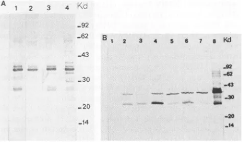
FIG. 1. Immunoblot analysis ofenvelope proteins of different avian hepadnavirus strains with monoclonal antibody SD20. (A) Partiallypurifiedviralparticlesof DHBVFm, Fp,C,and Am(lanes 1 to4,respectively); (B) proteinextractsoflivertissueinfected with HHBV and with DHBV Fm, Fp, C, 26, 3, and 1 (lanes 1 to 7, respectively). Forcomparison, viral particles ofserumcontaining DHBV Fp wereloaded in lane8.
says with liver tissue sections it recognized only DHBV-infected liver, and recognition could be inhibited
by prior
incubation ofthemonoclonalantibodywithpurifiedDHBV particles but not with control proteins. The fluorescent staining was cytoplasmic (data not shown). When purified DHBV particles were analyzed by immunoblotting, SD20 alwaysrevealedamajorproteinof 36 kDa(P36)andusually alsoseveral minor bands(Fig. 1A). Occasionally, aprotein of 28 kDa was also reactive (Fig. 1A). The sizes of all immunoreactive proteins are similarto those described by DHBV pre-S proteins (17, 39, 50, 54, 58). This strongly suggeststhat SD20recognizes anepitope ofpre-S proteins. This interpretation is also consistent with the cytoplasmic localizationof theantigenasindependentlyshownby immu-nohistology with a polyclonal antibody to apre-S protein expressed inE. coli(datanotshown).
SD20 recognizes a pre-S epitope conserved in DHBV iso-lates.TotestwhetherSD20 recognizesanepitopeconserved among avian hepadnaviruses, we used immunoblotting to analyzethereactivityof thepre-S proteinsinseraand livers of different DHBV isolates and from an HHBV-infected heron.
Onemajorprotein (36 kDa)and,with variableintensity,a
protein of 28 kDa reacted when we used concentrated DHBV virionpreparations fromseraofducks infectedwith DHBVFm,DHBVFp, DHBVC,and DHBV Am(Fig. 1A). Inaddition, several minor bandsrangingfrom 28to 38 kDa were observed (Fig. 1A). In the liver samples, the pre-S protein pattern was slightly different, as both the 36- and 28-kDa bandswerepresent asmajorimmunoreactive bands (Fig. 1B). In contrast, pre-S proteins of HHBV-infected liverswerenotdetectedby SD20(Fig. 1B). Infection of the heron liverwasindependentlydemonstratedbythe presence oflarge amounts ofnucleocapsid protein asdetermined by immunoblottingwith anti-core antibodies and by detection ofviral DNA by Southern blot analysis (data not shown). This suggests that SD20 recognizes a pre-S epitope thatis conserved in DHBVs but absent in HHBVpre-Sprotein.
Localization of thepre-S epitope. To map thebindingsite of SD20, wetested its reactivity by immunoblottingtoseveral MS2-pre-S fusion proteins expressed in E. coli (Fig. 2). Some of the fusionproteinswereefficiently expressed inE.
on November 10, 2019 by guest
http://jvi.asm.org/
[image:3.612.318.555.71.210.2]2
3 4
5
6
7
9
44 6
59
77
100
154
44
44
Pre-S
223
_ 185
163
DHBs
285
285
285
285
285
285
1 9 28 53 161 328
FIG. 2. DHBVpre-S/Sproteinsexpressedin E.coli.Fragments of clonedDHBV 16 DNA (60)insertedintoexpressionvector pEx 34awith the amino acid positions of the pre-S/S gene. Numbered black dotsinthepre-S/SgeneindicatepositionsoftheAUGcodons. Thereaction patternof thecorrespondingfusion proteins with the SD20 monoclonal antibodyisindicatedintherightpanel. Theboxed numbersdenoting fusion proteins(left-hand side)correspondto the lanes ofthe immunoblot of Fig. 3 into which thecell extracts were loaded.
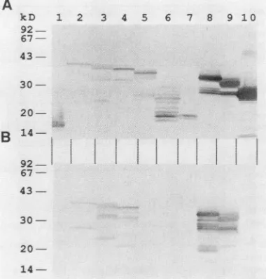
coliand could be visualized by Coomassie blue staining (data not shown); others were detected only by immunoblotting with an anti-MS2 monoclonal antibody (Fig. 3A). The de-duced molecular weights of the fusion proteins with the lowestelectrophoreticmobility were in close agreement with those predicted from the sequence. Proteins with higher electrophoretic mobility which were also detected probably derive from degradation during synthesis in E. coli or premature termination of translation or from partially de-graded mRNA. SD20 reacted with five of the eight fusion proteins tested (Fig. 3B). The reaction pattern observed indicates that the epitope is localized between amino acids 77and 100ofthepre-Spolypeptide(Fig. 2). This region is in a highly conserved pre-S domain of all DHBV isolates, containing onlyonepoint mutation in one isolate(Fig. 4).
In vitro neutralizing activity. To examine whether SD20 would block infectivityof DHBV, we performed an in vitro neutralization test. Primary duck hepatocytes were infected withDHBV-positive duck serum (MOI of 280 and 28) with or without previous incubation with SD20 ascites fluid. The ascites fluid was used in excess (1:200 dilution) as deter-mined by the method of limiting dilution (datanotshown). Intracellular viral DNA was examined 9 days after infection, and DNA from secreted virions was determined every 2 days.
A
kD 1 2 3 4 5 6 7 8 91 0
92 67- 43- 30-
20-B
1492I
67-
43-+
+ 30-
-j+
+
20-
14-FIG. 3. Mapping oftheepitoperecognized by monoclonal anti-bodySD20. MS2 fusionproteins expressedin E. coli,separated by SDS-polyacrylamide gel electrophoresis, andblottedto nitrocellu-+ lose were visualized by an MS2-specificmonoclonal antibody(A) + and tested for reactivity with monoclonal antibody SD20 (B). As negative controls, the MS2protein expressed bythe vector alone and an MS2-DHBVpolymerase fusionproteinwereloadedinlanes 1and10,respectively.In lanes 2 to 9MS2-pre-S/S fusion proteinsas
indicated in Fig. 2 wereloaded.
AtanMOIof28, several bandsofintracellularviralDNA characteristic of viral replication were detected (Fig.
5A,
lane c), indicating that active DHBV replication occurs in thesecells. When the viruswaspreincubatedwithSD20,the amount of intracellular viral DNA was strongly reduced (73.4 ± 5.1%) (Fig. SA,lanes cand d) as quantified by dot spot hybridization and radioactivity counting (data not shown). Neutralizationwas as effective(73.6 ±
1.4%)
when a10-fold-higher MOIwastested(Fig. 5A, lanesaandb).In themedium, the release ofDHBV particleswasdetectable from day 5 until day 9 for both MOIs tested. Similarly, as observed inside the cells, DHBV DNA released into the medium was decreased by approximately 75% (Fig. SB). Incubationof the inoculum withanunrelated ascitesfluiddid notaffect the DHBV DNA titer in cellsorsupernatants(data not shown), which demonstrates the specificity of the in vitroneutralizationassay.Theneutralization capacityof the other anti-DHBV monoclonal antibodies is currentlybeing investigated.DISCUSSION
Inthis papera monoclonal antibody, SD20, with neutral-izing activity against pre-S protein of DHBV has been described. Although the neutralizing activity ofSD20 was high(75%), complete neutralizationwas neverachieved. A constant residual infectivity of 25% was observed under several assay conditions and was not changed by 10-fold dilution of the inoculum. Interference of very abundant duck serum proteinsin the bindingofthe SD20 antibody to viral particles can therefore not explain the residual infectivity. The concentration of the SD20 was not a limiting factor, since the neutralization assay had been
performed
in an excessofantibody. Ourobservationofonly
partial
neutral-ization isconsistent with related studies which showed that44
on November 10, 2019 by guest
http://jvi.asm.org/
[image:4.612.73.292.73.389.2] [image:4.612.345.535.79.278.2]HHV4 - ImlISISL S EF SRARISL KS R . ..i HTQ TID V QH PEFSPI TA Q M IDSV
DNSV26- F S Q ... V X
OWI6- T I ... L
DIMVi - NPTS KS Nil .
ON3
- r KQESFISGLNI VSNLKVSLII GNSNTLSINI KFL4IQHPAK SMDVRRIEGGELLLNQLAGR NIP...KGTL TVSGKFPTID IIVLDHVQTNELR AGN E TA L DQR.TP PIT E K KFKQ N KPAE PITELAA EPQWKISPEDP KAKAIPV K E I
A T K N S
N ~ ]SP
R...Z.LLR~DVI~U~JARL&~RYQ
TNH
EITLQQQGAWPACAGRRVG S API POWTPEE 111RYOEERPPETTTI PPTSPTQWKL QPGDDPLLGN QSLLETHPL. YQ.TEPAVPV
L V K T GA G T K E L N T V L
..R T V
.. ~~~~~~~~~~~~~~~~~~~~V
.. ~~~~~~~~~~~~~~~~~~V
IKTPPL..K K SGTFGGIL AGLIGLLVSF FLLIKILEIL RRLDVWWISLSSPKGKNQCA FQDTGAQISP NYAGSCPWGC PCFLWTYLRL FIIFLLILLV
F E K FE E S Y E F T V V FNSCG
R E L S N L
V
ACLLYLTDNGSTILGKLQWASVSALFSSI SSLLPSDPKS LVALTFGLSL IWMTSSSATQTLVTLTQLAT
LSALFYKS-PRE-S
s
FIG. 4. Pre-S/S protein sequencealignment of several avian hepadnaviruses. The pre-S/Sproteinsweretranslated from the nucleotide sequenceof HHBVand of various cloned DHBVs (seeMaterials and Methods for sources).The DHBVisolate of Germanorigin (DHBV3
[60]) is givenonthebottom line, and amino acid differences from the other avian hepadnavirusesareshown inthecorrespondingupperrows.
To maximize the number of matches, a few amino acid insertions hadtobe made (indicated by dots). The AUG believedto be usedas
translation initiation codon for themajor 36-kDa pre-S protein (2) is numberedas1. Theantigenic regiontowhich themonoclonalantibody SD20 binds isindicated by the striped box.
for virtuallyall viruses a small fraction escapes neutraliza-tion even when a large excess of neutralizing antibodies is
used (37). Several factors such as neutralization escape
variants, aggregation of viral particles, and retention of virus infectivity despite the presence of bound antibody, have
beenproposedtoexplain thepersistentfraction ofinfecting virus (9, 26, 37). Furthermore, since the neutralizationtest requires9days, thereis timefor horizontalspread of virus, which may prevent the measurement of more effective
neutralization.
During preparation ofthisarticle, anotherreport describ-ing DHBV pre-S monoclonal antibodies with neutralizing activitycametoourattention(3). Inthisstudy, neutralizing
monoclonal antibodies recognizing three nonoverlapping epitopeswereidentified onthe DHBV pre-Sprotein,butno
precise localisationwasattempted. LikeSD20, theyreacted
with threepre-S proteins of 34, 36, and 37 kDa, but unlike SD20, the 28-kDa pre-S protein was not detected. This suggests that SD20 does not bind to the same epitope,
although itmay bind within thesame region.
Twomajor conclusions canbe drawn fromboth indepen-dently performed neutralizationstudies. First, DHBVpre-S proteins are targets for neutralizing antibodies; second,
somepre-S sequences are locatedataccessible sitesonthe
surface of viral particles. Similar conclusions have been drawnforpre-S proteins ofHBV (1, 12, 24, 42, 43,46)and
are also supported by the finding that protective immunity
can be successfully induced by immunization of
chimpan-zeeswithsynthetic peptidesandthatpre-S-specific
antibod-ies develop during self-limited hepatitis in humans (15, 27, 28). However, neither of these observations proves the
neutralizing activity ofHBVpre-S specific antibodies alone, because protective immunity might have been induced or
complemented bycellular immune defensemechanisms and pre-S antibody induction during virus elimination may be
coincidental. In contrast, the in vitro neutralization studies performed with DHBV-infected primary duck hepatocytes provide unequivocalevidence for the neutralizationactivity of thecorrespondingantibodiesalone.
Substantial information is now available on the surface location of HBV pre-S sequences and theiraccessibility to antibodies (1, 42, 43, 46). A sequence close to the amino terminus ofpre-Sl which isimplicated inhost cell receptor binding (amino acids 20to50), amajorimmunogenic region upstream of the pre-S2 initiation codon (amino acids 94 to 117),andmostof thepre-S2sequencesareprobablylocated
on the surface of the viralparticles. However, theprimary structureof DHBVpre-Ssequenceshowstoolittlesequence
homologywith thepre-Ssequenceof HBV(36, 60)topermit
acomparisonwithanyof theexposed surface sites of HBV. From the studies withHBV,itappearslikelythatmostofthe pre-S sequence isexposed at the surface of the viral parti-cles,andthis could be similar for DHBVpre-S protein.
The pre-S region contains a total of five or six in-frame AUGs(dependingonthe subtype [Fig. 4]). Onlyonemajor putative DHBV pre-S mRNA has been described, from whichsynthesis ofapre-S proteinof36 kDa initiatedatthe second AUG of the pre-S frame is predicted (2). Interest-ingly, however,immunoblottingwithSD20revealed several pre-S proteins ranging from 28 to 37 kDa. Three DHBV pre-S proteinsof34,35or36,and37 kDawerealso observed
infourindependent studies withpolyclonaland monoclonal antibodies(3,34, 54, 68). The 36- and37-kDapre-S proteins could represent the myristylatedand nonmyristilatedforms of the same molecule as described for HBV (48). Their synthesis was recently shown by transfection of mutant DHBV genomes into HepG2 cells to initiate at the second AUG (at viral nucleotide 801) of the pre-S region (D. Fernholz and H. Will, unpublished data). Unlike the anti-bodies used by Schlicht et al. (54) and Cheung et al. (3),
HNSV4
-DHSV26
-HOHW16
-DHBVI6
-imeVi
-HH3V4
-DHOV26
-DNBV16
-ONDVI
-DNBV3
-HHIV4
-OHBV26
-OV6
-DHBV3
on November 10, 2019 by guest
http://jvi.asm.org/
[image:5.612.85.537.75.300.2]a b c
d
23.1._
9.*_ *5-.
23-
VP
4gCc-' 9 , ~~~-I,S
0SL..
-55
0 50 100 1 50
l
200 250 00
l
U-
--I
-u-rn
-_IUE -U
Il-=n 4 3: *:WaC -:-3t * -c-+-C-P-.:--
--1 - -41:- l UE-1:= -= la I -*-UU
FZj1ZI.-- III :-EF -t -:Jc -
-:-5 _ 3_
a..
*
A
kzr7 A v. .N_w ._ I
ot nft l I8-1- M IA 11 PL/1." m- A If
V'."
wk
'U
Un
"'V
-1V
-N
%J-W\
VA
A PAA
tA
A.
I
A
A
0
0
B
a
b
2
100
so 0
c
d
., I
7
* .*
I
I*
* 0 0FIG. 5. Neutralization of DHBV with monoclonal antibody SD20 in primary hepatocyte cell culture. Viral DNA in the cells at
theendof the culturewasmonitored by Southern blotting (A), and
viralDNAin the cell culturemediumwasmonitoredevery2 days
postinfection (asindicated) byspothybridization (B). One day after plating, the hepatocyteswereinfected with DHBV-positiveserumat
anMOI of 280(lanesaandb) and 28 (lanescandd) with (lanes b and d)orwithout(lanesaandc) prior incubation with SD20 ascites fluid. On the Southern blot the positions of the size markers (HindIII-digested lambda DNA) and the various forms of DHBV replicative intermediates are indicated(RC, relaxedcircular; L, linear; CCC, covalently closedcircular; and SS, single stranded). No hybridiza-tionsignalswereobtained with DNAfrom uninfected cells (datanot
shown).
SD20 also reacted with several pre-S proteins smaller than 34 kDa(Fig. 1). Themostprominent ofthese proteinswas a
28-kDa protein. Consistent with ourobservation, a 28-kDa
protein was also seen by immunoblotting with two poly-clonalseraproducedby immunization of rabbits with pre-S
proteins derived from viral particles (68). Although these data strongly suggested that p28 is a pre-S protein, the
immunoreactivity of p28 withan antibody toanother virus-encoded proteinwhich might have been induced by a
con-taminating antigenused for immunizationwasnotrigorously excluded. Thedataobtainedwith SD20 and, especially,the immunoreactivity ofthe 28-kDa protein with a polyclonal
antibodytoarecombinantpre-S protein expressedin E. coli
(Fernholz andWill, unpublished), as wellas withthe poly-clonal antibodies to purified viral particles (68), establish unequivocally the 28-kDa molecule as a new viral protein
whichcontains DHBV pre-Ssequences. Thebinding site of
SD20 on thepre-S polypeptide as mapped with the
recom-binant pre-S proteins expressed in E. coli indicates that pre-S sequences between amino acids 77 and 100 are
con-tained in the 28-kDa protein. p28 could be initiated at an
internalAUG(as speculated previously[68])oratan
uncon-L
a
b c d
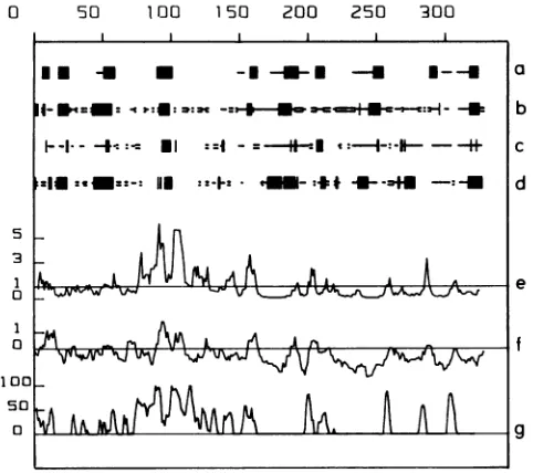
FIG. 6. Computer-assisted prediction of antigenic sites. The DHBV pre-S protein sequence as predicted from cloned viral DHBV 16DNAstartingatnucleotide 801 and endingatnucleotide 1785 (nomenclature as in reference 36) was used. The secondary structures werepredictedaccordingtoChouand Fasman(4)(line a), Gamieretal.(20)(line b), Del6ageand Roux(8) (line c),andLevin etal. (33)(line d). Thegiven conformational states area-helix(-), p-sheet (-) and turn (=). The blank regions indicate aperiodic states. Lines e, f, and g are profiles generated with different computer programs(13,25,47)predicting potential antigenic sites of proteins.Peaksabove thexaxis indicate themostsolventaccessible (line e), hydrophilic(line f),andantigenic(lineg)regions. Foreach graph the scaleisgivenonthe yaxis.
ventional codon of the pre-S region; it could represent a proteolytic processing product, or it could derive from a splicedmRNAwhich has not been detected so far. A protein of30 kDareactingwithSD20 ispresentinvariable amounts insome infected liversand viruspellets and may be initiated atthe AUG codon at nucleotide957,which wouldpredict a 30-kDa pre-S protein. The more complex pre-S protein pattern seen in serumthan that in infected liversis intrigu-ing. More efficientproteolytic degradation ofpre-Sproteins in serum than inliver could be a trivialexplanation.
A hydrophilicity profile indicates that the DHBV pre-S region is ratherhydrophilic, whereasthe S region contains two major hydrophobic peaks (Fig. 6). As speculated for mammalianhepadnaviruses,thehydrophobicdomains ofthe S protein may be involved in its anchoring to the cell membrane,which isreminiscent oftransmembrane proteins ofother viruses (11). The main antigenic site of DHBV as predicted by computer algorithms is located in the pre-S region between amino acids 75 and 110(Fig. 6). Consistent with the computer prediction, the epitope recognized by SD20 is localizedbetweenamino acids 77 and 100(Fig. 5). The sequence divergence of the DHBV and HHBV pre-S proteins is rather high(51%) and has been speculatedtobe at least partly responsible for the different host ranges ofthe two viruses (58). However, the predicted hydrophilicity profile and the antigenic profile were almost indistinguish-able (data not shown), suggestingthat conservation of cer-tainhydrophilic domains is of functionalimportance. SD20 binds to a region which exhibits a highly conserved 6- to 8-amino-acidhelix(aminoacids 92to100)aspredicted byall
A
on November 10, 2019 by guest
http://jvi.asm.org/
[image:6.612.319.560.75.289.2] [image:6.612.82.280.75.362.2]algorithms used. A similar but longer helix(aminoacids 86to 103) is alsopresent in thecorrespondingHHBV pre-S region (datanot shown).
The availability of neutralizing monoclonal antibodies provides a useful tool forinvestigating mechanisms ofvirus neutralization. Despiteextensive studies with synthetic pep-tides and anti-peptideantibodieswhich allowedthe mapping of putative virus recognition sites for cell receptors within the pre-Sregion ofHBV, elucidation of the mechanisms of virus neutralization remained elusive. Itappears from these studies that the S protein has no receptor recognition site and the pre-S2 region has only an auxiliary role in HBV binding to hepatocytes(43, 46). However, it is known that S and pre-S2 proteins elicit protective antibodies in humans andchimpanzees(15,21, 27,46, 51). Thus,neutralization of HBV appears complex and can also be brought about by mechanisms otherthandirect interference with the receptor-binding site, as is known for other viruses (9, 37). For hepadnaviruses,DHBVisthe only convenient model which can beused to decipher the mechanismsofvirus neutraliza-tion both in vivo and in vitro.
ACKNOWLEDGMENTS
We greatly appreciate the help of L. Vitvitski-Trepo in performing immunofluorescence studies and thank R. Schneider, F. Schodel, and T. Weimer for critically reading the manuscript.
Part of this work has been supported by grants from the Deutsche Forschungsgemeinschaft. V.L. is the recipient of afellowshipfrom theAssociation pour laRecherchecontre le Cancer.
LITERATURECITED
1. Budkowska, A., M. M. Riottot, P. Dubreuil, Y. Lazizi, C. Brechot, E. Sobezak, M. A.Petit, and J. Pillot. 1986. Monoclo-nal antibody recognizing pre-S 2 epitope of hepatitis B virus: characterizationofpre-S 2 epitope andanti-pre-S2antibody.J. Med. Virol. 20:111-125.
2. Buscher, M., W. Reiser, H. Will, and H. Schaller. 1985. Tran-scripts and the putative RNA pregenome of duck hepatitis B virus: implications for reversetranscription. Cell40:717-724. 3. Cheung, R. C., W. S. Robinson, P. L. Marion, and H. B.
Greenberg. 1989. Epitope mapping ofneutralizing monoclonal antibodies against duck hepatitis B virus. J. Virol. 63:2445-2451.
4. Chou, G. Y., and G. D. Fasman. 1978. Predictionof secondary structureofproteins from amino acid sequence. Adv. Enzymol. Relat. Areas Mol. Biol. 47:145-148.
5. Cova, L., 0. Hantz, M. Arliaud-Gassin, A. Chevalier, P.
Berthillon, L. Boulay, C. Jacquet, B. Chomel, L. Vitvitski, and C. Trepo. 1985. Comparative study ofDHBV DNA levels and endogenous DNA polymerase activity in naturally infected ducklings in France. J. Virol. Methods 10:251-260.
6. Cova, L., V. Lambert, A. Chevalier, 0. Hantz, I. Fourel, C. Jacquet, C.Pichoud, J. Boulay, B. Chomel,L.Vitvitski, and C. Trepo. 1986.Evidenceforthe presence of duckhepatitisBvirus in wild migrating ducks. J. Gen. Virol. 67:537-547.
7. Deleage, G.,F. F. Clerc, B. Roux,and D. C. Gautheron. 1988. Antheprot: a package for protein sequence analysis using a microcomputer. Comput. Appl. Biosci. 4:351-356.
8. Deleage,G.,and B. Roux. 1987. Aalgorithm for protein second-ary structurepredictionbased on class prediction. Protein Eng. 1:289-294.
9. Dimmock, N. J. 1984. Mechanism of virus neutralization. J. Gen. Virol. 65:1015-1022.
10. Dreesman, G. R., Y. Sanchez, I.Ionescu-Matiu, J. T. Sparrow, H. R. Six, D. L. Peterson, F. B. Hollinger, andJ. L. Melnick. 1982. Antibody to hepatitis B surface antigen after a single inoculation of uncoupled synthetic HBsAg peptides. Nature (London) 295:158-160.
11. Eble, B. E., V. R. Lingappa,and D. Ganem. 1986. Hepatitis B surface antigen: an unusual secreted protein initially
synthe-sized as a transmembrane polypeptide. Mol. Cell. Biol. 6: 1454-1463.
12. Eble, B. E., D. R. MacRae, V. R. Lingappa, and D. Ganem. 1987. Multiple topogenic sequences determine the
transmem-braneorientation ofhepatitisBsurfaceantigen.Mol.Cell.Biol. 7:3591-3601.
13. Emini, E. A., J. V. Hughes, D. S. Perlow, and J. Boger. 1985. Induction of hepatitis A virus-neutralizing antibody bya virus-specific syntheticpeptide. J.Virol. 55:836-839.
14. Emini, E. A., S. Y. Kao, A. J. Lewis, R. Crainic, and E. Wimmer. 1983. Functionalbasis of polio neutralization deter-mined with monospecificneutralizing antibodies. J. Virol. 46: 466-474.
15. Emini, E. A., V.Larson, J. Eichberg, P. Conard,V.M.Garsky, D. R. Lee, R. W. Ellis, W. J. Miller, C. A. Anderson, and R.J. Gerety. 1989.Protectiveeffect ofasynthetic peptide comprising the complete pre-S 2 region ofthe hepatitis B virus surface protein. J. Med. Virol. 28:7-12.
16. Engvall, E., and P. Perlman. 1972. Enzyme-linked immunosor-bent assay(ELISA).III.Quantification ofspecific antibodiesby enzyme-labelled anti-immunoglobulin in antigencoated tubes. J. Immunol. 109:129-135.
17. Feitelson, M. A., P. L. Marion, and W.S. Robinson. 1983. The nature of polypeptides larger in size than the major surface antigen components of hepatitis B and like viruses in ground squirrels, woodchucks, andducks. Virology 130:76-90. 18. Fourel, I., P. Gripon, 0. Hantz, L. Cova, V. Lambert, C.
Jacquet, K. Watanabe, J. Fox, C.Guillouzo, and C.Trepo. 1989. Prolonged duck hepatitis B virus replication in duck hepato-cytes cocultivated with ratepithelial cells: a useful system for antiviraltesting. Hepatology 10:186-191.
19. Fourel, I., 0. Hantz, L. Cova, H. S. Allaudeen, and C. Trepo. 1987. Main propertiesof duck hepatitis B virus DNA polymer-ase: comparison with the human and woodchuck hepatitis B virus DNApolymerases. Antiviral Res.8:189-199.
20. Garnier,J.,D. J. Osguthorpe,andB. Robson. 1978.Analysisof the accuracyandimplications of simple methods for predicting the secondary structure of globular proteins. J. Mol. Biol. 120:97-120.
21. Gerin, J. L., H. Alexander, J. W.-K. Shih, R. H. Purcell, G. Dapolito, R. Engle, N. Green, J. G. Sutcliffe, T. M. Shinnick, and R. A. Lerner. 1983. Chemically synthesized peptides of hepatitis B surface antigen duplicate the d/y specificities and induce subtype-'specific antibodies inchimpanzees. Proc. Natl. Acad. Sci. USA 80:2365-2369.
22. Guldner, H. H., H.J. Netter, C. Szostecki,H. J. Lakomek, and H. Will. 1988. Epitope mapping with a recombinant human 68 kDa (Ul) ribonucleoprotein antigen reveals heterogeneous au-toantibody profiles in human autoimmune sera. J. Immunol. 141:469-475.
23. Gust,I. D., A. G. Coulepsis, W. S. Robinson, and A. J. Zuck-erman. 1986. Taxonomic classification of human hepatitis B virus. Intervirology 25:14-29.
24. Heermann, K.-H., F. Kruse, M. Seifer, and W. H. Gerlich. 1987. Immunogenicity of the gene S and pre-S domains inhepatitis B virions and HBsAg filaments. Intervirology28:14-25.
25. Hopp, T. P., and K. R. Woods. 1981. Prediction of protein antigenic determinants from amino acid sequences. Proc. Natl. Acad. Sci. USA 78:3824-3828.
26. lorio,R. M., and M. A. Bratt. 1985.Neutralization of Newcastle disease virus by monoclonal antibodies to the hemagglutinin-neuraminidase glycoprotein: requirement for antibodies tofour sitesfor complete neutralization. J. Virol. 51:445-451. 27. Itoh, Y., E. Takai, H. Ohnuma, K. Kitayima, F. Tsuda, A.
Machida, S. Mishiro, T. Nakamura, Y. Miyakawa, and M.
Mayumi. 1986. A synthetic peptide vaccineinvolvingthe prod-uct ofthe pre-S 2 region ofhepatitis Bvirus: protectiveefficacy in chimpanzees. Proc. Natl. Acad. Sci. USA83:9174-9178. 28. Klinkert, M. Q., L.Theilmann, E.Pfaff,and H. Schaller. 1986.
Pre-S 1 antigens and antibodies early in the course of acute
hepatitis B virus infection. J. Virol. 58:522-525.
29. Kohler, G., and C. Milstein. 1976. Derivation ofspecific anti-body-producing tissue culture and tumor lines by cell fusion.
on November 10, 2019 by guest
http://jvi.asm.org/
Eur.J. Immunol. 6:511-519.
30. Kyhse-Andersen, J.1984.Electroblottingofmultiple gels usinga
simple apparatus without buffer tank for rapid transfer of protein from polyacrylamide to nitrocellulose. J. Biochem. Biophys.Methods 10:203-209.
31. Laemmli,U. K.1970.Cleavageof structuralproteins duringthe assembly ofthe head ofbacteriophage T4. Nature (London) 227:680-685.
32. Lambert, V.,L.Cova,W. R.Hansen,B.Chomel,andC.Trepo. 1988. DuckhepatitisBvirus in wild waterfowl from the United States,p.523-525.In A. J.Zuckerman(ed.),Viralhepatitisand liverdisease. AlanR.Liss, Inc.,NewYork.
33. Levin, J. M.,B.Robson,andJ. Garnier.1986. Analgorithmfor secondary structure determination in proteins based on
se-quencesimilarity. FEBSLett. 205:303-308.
34. Li, J.S.,L.Cova,R.Buckland,V.Lambert,G.Deleage,andC. Trepo. 1989. Duck hepatitis B virus can tolerate insertion, deletion and partial frameshift mutation in the distal pre-S region.J. Virol. 63:4965-4968.
35. Machida, A., S. Kishimoto, H. Ohnuma, K. Baba, Y. Ito, Miyamoto, G. Funatsu, K. Oda, S. Usuda, S. Togami, T. Nakamura,Y.Miyakawa,and M.Mayumi. 1984.Apolypeptide containing55amino acidresidues codedbythepre-S regionof hepatitis B virus deoxyribonucleicacid bears the receptorfor po!ymerizedhumanaswellaschimpanzeealbumin. Gastroen-terology86:910-918.
36. Mandart, E., A. Kay, and F. Galibert. 1984. Nucleotide
se-quenceofaclonedduckhepatitisBvirusgenome:comparison with woodchuck and human hepatitis B virus sequences. J. Virol. 49:782-792.
37. Mandel, B.1984. Mechanism ofvirusneutralization,p. 32-38. In A. L.NotkingandM. B. A.Oldstone(ed.),Conceptsin viral pathogenesis. Springer-Verlag, NewYork.
38. Maniatis, T.,E. F.Fritsch,andJ.Sambrook. 1982. Molecular cloning: alaboratorymanual. ColdSpring HarborLaboratory, Cold Spring Harbor, N.Y.
39. Marion,P.L.,S. S.Knight,M. A.Feitelson,L.S. Oshiro,and W. S. Robinson. 1983. Major polypeptide ofduck hepatitis B surface antigen particles.J. Virol. 48:534-541.
40. Marion,P.L.,L.S.Oshiro,D.C.Regenery,G. H.Scullard,and W.S. Robinson. 1980.Avirus inBeechey ground squirrelsthat is related to hepatitis B virus of man. Proc. Natl. Acad. Sci. USA 77:2941-2945.
41. Mason,W.S.,G.Seal,andJ.Summers.1980. Avirus ofPekin ducks with structural and biological relatedness to human hepatitis Bvirus.J.Virol. 36:829-836.
42. Milich,D. R.1987.Geneticandmolecular basis forT- andB-cell recognition of hepatitis B viral antigens. Immunol. Rev. 99: 71-103.
43. Neurath, A. R., andS. B. H. Kent. 1989. Thepre-S region of hepadnavirus envelope proteins. Adv. Viral oncol.34:64-142. 44. Neurath, A. R., S. B. H. Kent, K. Parker, A. M. Prince, N.
Strick, B. Brotman, and P. Sproul. 1986. Antibodies to a
synthetic peptidefromthepre-S120-145regionofthehepatitis B virusenvelopearevirusneutralizing. Vaccine4:35-37. 45. Neurath,A.R.,S. B. H.Kent,N.Strick,and K. Parker. 1986.
Identification and chemical synthesis of a host cell receptor bindingsiteonhepatitisBvirus. Cell46:429-436.
46. Neurath,A.R.,N.Strick,S. B. H.Kent,K.Parker,C. S.Kim, M.Girard,H. E.Ralph,andJ. Valinsky. 1989.Biologicalroleof pre-S sequences of the hepatitis B virus (HBV) envelope protein, p. 143-158. In J. P. Tam and E. T. Kaiser (ed.), Synthetic peptides:approachestobiologicalproblems.Alan R. Liss, Inc., NewYork.
47. Parker, J. M. R., D. Guo, and R. S. Hodges. 1986. New hydrophilicityscale derivedfromhigh performance liquid chro-matography peptide retention data: correlation of predicted surfaceresidues withantigenicityandX-ray-derived accessible sites. Biochemistry25:5425-5432.
48. Persing,D.H.,H. E.Varmus,and D.Ganem.1987. Thepre-S1 protein of hepatitis B is acylated at its amino terminus with myristicacid. J. Virol. 61:1672-1677.
49. Pontisso, P., M.-A. Petit, M. J. Bankowski, and M. E. Peeples. 1989. Human liverplasmamembranes containreceptors for the hepatitis B virus pre-Sl region and, via polymerized human serumalbumin, for thepre-S2region. J. Virol. 63:1981-1988. 50. Pugh, J. C., J. J. Sninsky, J. W. Summers, and E. Schaeffer.
1987. Characterization of apre-S polypeptide on the surfaces of infectious avianhepadnavirus particles. J. Virol. 61:1384-1390. 51. Purcell, R.H., andJ.L.Gerin. 1985.Prospectsfor a secondand thirdgeneration of hepatitis B vaccines. Hepatology 5:159-163. 52. Rigby, P. W. J., M.Dieckmann,C.Rhodes,and P. Berg.1977. Labeling deoxyribonucleicacid tohigh specific activityinvitro by nick translation with DNA polymerase I. J. Mol. Biol. 113:237-251.
53. Schaeffer, E., and J. J. Sninsky. 1984. Predicted secondary structure similarity in the absence of primary amino acid se-quence: hepatitis B virus open reading frames. Proc. Natl. Acad. Sci. USA 81:2902-2906.
54. Schlicht, H. J., C. Kuhn, B. Guhr, R.J. Mattaliano, and H. Schaller.1987. Biochemical andimmunologicalcharacterization of the duck hepatitis B virus envelope proteins. J. Virol. 61:2280-2285.
55. Schodel, F.,R.Sprengel,T.Weimer,D.Fernholz,R.Schneider, andH. Will.1989. Animal hepatitis B viruses.Adv. Viral Oncol. 8:73-102.
56. Schulman, M., C. D.Wilde,andG.Kohler. 1978. A better cell line for making hybridomas secreting specific antibodies. Na-ture(London)276:269-270.
57. Southern, E. M. 1975. Detection ofspecific sequences among DNAfragments separated by gel electrophoresis. J. Mol. Biol. 98:503-517.
58. Sprengel, R., E. F. Kaleta, and H. Will. 1988. Isolation and characterization ofahepatitis B virus endemic in herons. J. Virol. 62:3832-3839.
59. Sprengel, R., C. Kuhn, C. Manso, and H. Will. 1984. Cloned duck hepatitis B virus DNA is infectious in Pekin ducks. J. Virol. 52:932-937.
60. Sprengel, R.,C.Kuhn,H.Will,andH. Schaller. 1985. Compar-ative sequence analysis of duck and human hepatitis B virus genomes. J. Med. Virol. 15:323-333.
61. Sprengel, R., and H. Will. 1987. Duck hepatitis B virus, p. 363-386. In G. Darai (ed.), Virus disease in laboratory and captive animals. Martinus Nijhoff Publishing, Boston. 62. Strebel, K., E. Beck, K. Strohmeyer, and H. Schaller. 1986.
Characterization of foot-and-mouth disease virusgeneproducts with antiseraagainst bacterially synthesized fusion proteins.J. Virol.57:983-990.
63. Summers, J. W., J. M. Smolec,and R. Snyder. 1978. A virus similartohepatitis Bvirus associated withhepatitis and
hepa-tomainwoodchucks.Proc.Natl.Acad.Sci. USA75:4533-4537. 64. Tuttleman, J.S., J.C.Pugh,andJ.W.Summers. 1986. In vitro experimental infection of primary duck hepatocyte cultureswith duckhepatitisB virus. J. Virol. 58:17-25.
65. Vitvitski, L., M. L. Meyers, J. J. Sninsky, P. Berthillon, P. Chevalier,M. A.Sells,G.Acs,andC.Tr6po.1988. Expression of the Xproduct of hepatitisBvirusand WHV ininfected livers and transfected 3T3 cells: evidence for cross reactivity and correlation with core/e gene expression, p. 341-344. In A.J. Zuckerman(ed.), Viral hepatitisandliverdisease. Alan R.Liss, Inc.,NewYork.
66. Voller, A.,D.Bidwell,and A. Barlett.1976.Microplateenzyme immunoassay for immunodiagnosis ofvirusinfections,p. 506-512. In N. R. Rose andH. Friedman(ed.), Manualof clinical immunology,1sted. AmericanSociety for Microbiology, Wash-ington,D.C.
67. Wahl,G.M., M.Stern,andG. R. Stark.1979. Efficient transfer oflarge DNA fragmentsfrom agarose gels to diazobenzylox-methyl-paperandrapid hybridization by usingdextran sulfate. Proc. Natl.Acad.Sci. USA76:3683-3687.
68. Yokosuka, O., M. Omata, and Y. Ito. 1988. Expression of pre-Sl, pre-S2, and C proteins in duckhepatitisB virus infec-tion. Virology167:82-86.
69. Zhou,Y. Z.1980. Aviruspossiblyassociated withhepatitisand hepatomain ducks. ShanghaiMed. J. 3:641-644.
![FIG. 4.TosequencetranslationSD20[60]) Pre-S/S protein sequence alignment of several avian hepadnaviruses](https://thumb-us.123doks.com/thumbv2/123dok_us/1322049.85923/5.612.85.537.75.300/fig-tosequencetranslationsd-pre-protein-sequence-alignment-avian-hepadnaviruses.webp)