0022-538X/90/094051-08$02.00/0
Copyright © 1990, American Society forMicrobiology
Characterization of Hepatitis
Delta Antigen: Specific
Binding
to
Hepatitis Delta Virus
RNA
JU-HUNG LIN,"2'3 MING-FUCHANG,"2t SUSAN C. BAKER,"12SUGANTHAGOVINDARAJAN 14 AND MICHAEL M. C. LAI'2*
Howard Hughes MedicalInstitute' and Departments ofMicrobiology2 andPathology,4 Universityof Southern California,
School of Medicine, LosAngeles, California 90033-1054, and Institute of Molecular Biology, Academia Sinica,
Nankang, Taiwan 115293
Received 9 August 1989/Accepted 26May 1990
Ithas previously been shown thathuman hepatitis virus delta antigenhasanRNA-bindingactivity(Chang
etal., J. Virol. 62:2403-2410, 1988). In thepresentstudy, thespecificityof suchanRNA-proteininteraction
was demonstrated by expressing various domains of the delta antigen in Escherichia coli as TrpE fusion
proteins and testing theirRNA-binding activities inaNorthwestern protein-RNA immunoblotassayandRNA
gel mobility shiftassay.Hepatitis delta virus (HDV) RNA bound specificallytothedelta antigen in thepresence
ofanexcessamountof unrelatedRNAs andarelatively high salt concentration. Bothgenome-and
antigenome-sense HDV RNAs and at least two different regions of HDV genomic RNA bound to the delta antigen. Surprisingly, these two different regions of HDV genomic RNA couldcompetewith each other fordelta antigen binding, although they do not have common nucleotide sequences. In contrast, this binding could not be competed withbyother viralorcellular RNA. Since both the genomic and antigenomic HDV RNAshadstrong
intramolecular complementarysequences,these resultssuggest thatthebinding ofdelta antigen is probably specific fora secondarystructure uniquetothe HDVRNA. By expressing differentsubdomains ofthedelta antigen, wefound that the middle one-third of delta antigenwasresponsiblefor binding HDV RNA. Neither
theN-terminalnortheC-terminal domain bound HDV RNA. Binding between the delta antigen and HDV RNA
wasalsodemonstrated within the HDV particles isolated from the plasma ofahuman delta hepatitis patient.
This invivobinding resistedtreatmentwith 0.1% sodium dodecyl sulfate and 0.5% Nonidet P-40. In addition,
weshowed that the antiserum fromahuman patient with delta hepatitis reacted with all three subdomains of
the deltaantigen,indicating that all of the domainsareimmunogenic in vivo. These studies demonstrated the
specific interaction between delta antigen and HDVRNA.
Hepatitis delta virus(HDV) isadefective virus associated with severe hepatitis in humans (1, 13, 19). It requires hepatitis B virusas ahelper virus(32). The HDV particle is
a 36-nm enveloped virus containing the hepatitis B virus surface antigenonthe virion surface. Inside the envelope is the HDV-specific delta antigen (HDAg) (32). The HDAg is also detected in the nucleiof infectedhepatocytes (31). Delta antigen is the only HDV-specific structural protein in the virion.The viralgenomeisacircularsingle-stranded RNA of
approximately 1.7 kilobases (kb) (20, 26, 41) which has extensive intramolecular complementary sequences. Thus, its RNA structure resembles that ofviroid and some plant virusoid RNAs. HDV RNA has a unique autocatalytic
cleavageandligation activity (24, 34, 35, 43, 44). Italso has five open reading frames which can potentially code for proteins of more than 100 amino acids each (26). One of these open reading frames, in the antigenomic sense,
en-codes delta antigen (8, 23, 26, 41). The remaining open
reading frames have notbeen demonstrated to encode any
proteins.
The deltaantigens inthe liver orserumof deltahepatitis patients are composed of two protein species of approxi-mately 27 and 24 kilodaltons (kDa) (2, 4, 29, 42, 45). The
same two proteins are detected in the liver and serum of
chimpanzees experimentally infected with HDV (2, 45). Recently, it hasbeen demonstrated that these twoproteins
*Correspondingauthor.
tPresentaddress:DepartmentofBiochemistry,National Taiwan
University, CollegeofMedicine, Taipei,Taiwan.
were derived from two different HDV RNA species with differentcoding capacities (Y.-P. Xia, M.-F. Chang, D. Wei, S.Govindarajan,and M. M. C.Lai,Virology, in press). The HDAg is phosphorylated and is localized in the nuclei both in infected animals (14) and in transfected cells (8). It also hasanRNA-binding activity (8). However, the specificity of
thisRNA-protein interaction hasnotyetbeendemonstrated. Since HDAg appears to be required for the replication of HDV RNA (22), the interaction between HDAg and HDV RNAprobably plays an important role in HDV RNA
syn-thesis. Prevailing evidence suggests that HDV RNA repli-cates through a rolling-circle mechanism. This evidence includesthedetection, in HDV-infected hepatocytes (9, 27), ofHDV-specific RNA of longer than genomic length, which representsintermediatesof RNAreplication,andtheability ofHDV RNAtocleave andligateitselfautocatalytically (24, 34, 35, 43, 44). Conceivably, the interaction ofHDAgwith HDV RNA might regulate these activitiesorRNA replica-tion.
To furthercharacterize the interaction between theHDAg and HDVRNA, wehave constructed TrpEfusionproteins containingvarious domains ofHDAgand tested their RNA-binding activities. In this report, we demonstrate that this interaction is very specific and occurs between HDV RNA
and themiddle one-third ofHDAg. Furthermore, this inter-actioncan bedemonstrated within the HDV particle.
MATERIALS ANDMETHODS
Construction of plasmids expressing TrpE-HDAg fusion proteins.The 0.9-kbPstI-SstIIinsert ofplasmidpECE-d-E, 4051
on November 10, 2019 by guest
http://jvi.asm.org/
Pst I,Sst II digestion Sma I digestion
T4DNApolymerase IdBAP
Sma I/Sst II PstI/SmaI Junction -ATCCCCGGG---[TGA--- GGG
GAT-sequences -TAGGGG| ---ACT----GGG]CCC
CTA-FIG. 1. Schematicdiagramoftheconstruction ofaTrpE-HDAg
plasmidvectorfor expression of HDAg. pATH-Dwas designedso
that it expresses, in Escherichia coli, a fusion protein of
TrpE-HDAg containing amino acids 11 to 214 of HDAg. Boxes with
shaded areas represent TrpE sequences. Solid boxes indicate the
HDAg codingregion. The HDAgstopcodon is represented byan
asterisk. The solid circle represents the simian virus 40 origin of replication. The sequences at thejunctions of the pATH-2vector
and HDV insertareshown. BAP, Bacterialalkaline phosphatase.
whichcontainsa1.3-kb EcoRI fragment ofHDVcDNA (26),
was blunt-ended by using T4 DNA polymerase and sub-cloned, in frame,into theSmaI siteof pATH-2 (11) DNAto
generate pATH-D (Fig. 1). pATH-D expresses a
TrpE-HDAg fusion protein containing amino acids 11 to 214 of HDAg (26). The 0.7-kb StuI-XbaI fragment was removed
fromthepATH-D, and theremaining DNAwasfilled in, by
using the Klenow fragment of DNA polymerase I, and ligated with DNA ligase to generate pATH-N, which
con-tains sequences representing the N-terminus of the HDAg (amino acids 11 to 78). The purified 0.7-kb StuI-XbaI frag-ment ofpATH-D was subcloned, in frame, into the
SmaI-XbaIsite ofapATH-2vector to generatepATH-MC, which
contains sequences representing the middle portion and carboxyl terminus of the HDAg (amino acids79to 212). A 0.4-kb SmaI-XbaIfragment was removed from the plasmid
MC, and the remaining DNA was filled in by using the
KlenowfragmentofDNApolymerase I before self-ligation
to generate pATH-M, which contains sequences
represent-ing onlythe middle portionofthe HDAg(amino acids 79to
163). A 0.3-kb SalI insertwasremoved from pATH-D, and
the remaining DNA was filled in by using the Klenow
fragment ofDNA polymerase I and religated to generate
pATH-X. A 0.46-kb SmaI fragment was further removed
from the pATH-X plasmid, and the remaining DNA was
religated to generate pATH-C, which represents the
C-terminusof the HDAg(amino acids 164to212).
Constructionofother plasmids. The plasmid pHD-P, which
containsthe protein-condingdomain of HDVRNA (7),was
constructedby excisinganEcoRI fragment (positions965to
487 through 1683/0) from plasmid
pS29,
which contains the full-length cDNA of HDV RNA (Sall-Sall, 1,683 basepairs
[bp]). The fragment was cloned into the
EcoRI
site of pTZ-18U (U.S. Biochemicals). The plasmid pHD-V, which con-tains the viroidlike domain (7) of HDV RNA, was con-structed by inserting aSalI-EcoRI
fragment(positions487to 965), derived frompS29,
into the polylinker of pTZ-18U between theEcoRI
andSall sites. Transcription byT7RNA polymerase of pHD-P, linearized by Hindlll,yielded
an RNA of 1.2 kb, corresponding tothe protein-coding domain (7) of the genomic-sense HDV RNA. Transcriptionof pHD-V, linearized by Sall, yielded a genomic-sense HDV RNA subfragment corresponding to the viroidlike domain (7).Preparation of TrpE-HDAg fusion proteins and HDAg-specific antiserum. The preparation of TrpE-HDAg fusion proteins and immunization schedule followed theprocedures described by Hardy and Strauss (17) with minor modifica-tions. Briefly, a culture of
MC1061
cellscontaining plasmids encoding the hybrid TrpE-HDAg fusion proteins was in-duced with 3-indoleacrylic acid, bacterial lysates were pre-pared, and the TrpE-HDAg fusion proteins were concen-trated in the insoluble fraction. For the purpose of immunization, the insoluble fraction of TrpE-HDAg fusion protein D (approximately 500 p.g) was purified by electro-phoresis on a 7.5% polyacrylamide gel containing 0.1% sodium dodecyl sulfate (SDS). The fusion protein was iden-tified by staining with Coomassie blue and excised from the gel. The gel slice was homogenized in 1 ml of phosphate-buffered saline and then emulsified with an equal volume of Freund complete adjuvant. This mixture was injected sub-cutaneously along the back of New Zealand White rabbits weighing 3 to 4 kg. The rabbits were boosted every 2 weeks by injection in the hindleg with 200 to 400,ug
of gel-purified fusion protein D in incomplete Freund adjuvant. Rabbits were bled from the ear vein 1 week following boost injec-tions. Serum was separated, and antibody titer was deter-mined by enzyme-linked immunosorbent assay.RNA-protein binding assays by the Northwestern blot pro-cedure. The Northwestern protein-RNA blot analysis was carried out as described previously (8). Briefly, TrpE-HDAg fusion proteins which contained different portions of the HDAg were prepared as described above. The fusion pro-teins were separated by SDS-polyacrylamide gel electropho-resis (PAGE) (7.5% polyacrylamide) and then electrotrans-ferred to a nitrocellulose membrane at 200 mA overnight at 4°C.The membrane was then incubated in standard binding buffer (SBB; 10 mM Tris hydrochloride
[Tris-HCl,
pH 7.0], 1 mM disodium EDTA, 50 mM NaCl, 0.02% bovine serum albumin, 0.02% Ficoll, and 0.02% polyvinylpyrrolidone). The binding buffer also contained a32P-labeled
RNA tran-scribed in vitro(105
cpm/ml per 1.7 kb of RNA; specific activity, 2 x10⁷
cpm/,Lg)
and 100 pLg of nonspecific total cellular RNA isolated from DBT cells, a murine astrocytoma cell line, per ml as a nonspecific competitor (36). The probes used in this study included the full-length HDV genomic RNA, viroidlike domain(EcoRI-SalI
fragment in the region between nucleotides 487 and 965) (26), a protein-coding domain (SalI-PstI fragment in the region between nucleo-tides 965 and 654 through 1683/0), the full-length antigenomic RNA of HDV, and mouse hepatitis virus (MHV) nucleocap-sid RNA (37). The T7 polymerase transcription procedures were done as described previously (44).32P-labeled
RNA bound to the fusion proteins on the nitrocellulose membrane was visualized by autoradiography, and the amount of radioactivity bound to the membrane was determined byon November 10, 2019 by guest
http://jvi.asm.org/
[image:2.612.59.298.60.331.2]scintillation counting of specific bands excised from the nitrocellulose filter.
RNA-protein binding studies by the RNA mobility shift assay. The RNA mobility shift assay was carried out by a modified procedure as described previously (18, 30, 38). Briefly, 20 ,ug of partially purified and heat-denatured TrpE-HDAg fusion protein was incubated in 10 ,ul of buffer containing 2.5 mM Tris-HCl (pH 7.4), 100 mM NaCl, 2 mM MgCl2, 1 mM dithiothreitol, and 20% glycerol with 40 U of RNasin, 10 ,ug of tRNA, and 10p.g of total cellular RNA at 4°C for 5 min. If required, unlabeled RNA competitors (usually 500 ng in 2 ,ul) were added, and the reaction mixes were incubated for an additional 10 min at 4°C. Subse-quently, 1 ,ul (10 ng) of32P-labeled RNA (approx. 20,000 cpm) was added, and the reaction mixture was incubated for an additional 30 min at 4°C. Then, electrophoresis was carried out in 1, 0.8, or0.6% low-melting-point agarose gels with 10 mM sodium phosphate (pH 7.0) as the running buffer,maintaining the voltage at about 20 to 25 V at 4°C, for 15 h. The gels were then dried and visualized by autoradi-ography.
Slot-blot RNA analysis. HDV genomic RNA from the plasma of an HDV-infected patient was prepared by disrupt-ing hepatitis delta virions purified from the patient's serum with the lysis buffer containing 80 mM Tris-HCl (pH 6.8), 0.1 M2-,-mercaptoethanol, and0.1% (wt/vol) SDS, followed by immunoprecipitation with HDAg-specific antiserum pre-pared against TrpE-HDAg fusion protein. The final pellet containingHDV RNA was suspendedin buffer (10 mM Tris-HCl [pH 7.4], 60 mM NaCl, 1 mM EDTA, and 1% SDS), followed by extraction with phenol-chloroform (1:1) and ethanol precipitation. The ethanol-precipitated RNA was denatured inlOx SSC (1x SSC contains 0.15 M NaCl and 15 mM sodium citrate)-13% formaldehyde at 65°C for 15 min andboundto anitrocellulose membrane by filtration with a slot-blot apparatus (Schleicher & Schuell). The nitrocellu-lose membrane was then baked under vacuum for 1.5 h at 80°C and prehybridized in sealed plasticbags in50% form-amide-5x SSC-50 mM sodium phosphate (pH 7.0)-1% SDS-5x Denhardt solution(lx Denhardt solution contains 0.02% each bovine serum albumin, polyvinylpyrrolidone, andFicoll),and 250p.g of denaturedsalmon sperm DNA per ml for 4 h at 42°C. Filters were then hybridized at 42°C overnight inthesamesolutionwith32P-labeledDNAprobes of HDV monomer RNA, which were prepared by using random hexamersasprimers(12).Afterhybridization, filters
werewashed in 0.1x SSC-0.1%SDSat50°C and exposedto X-rayfilm.
RESULTS
Construction ofplasmidDNAencoding TrpE-HDAgfusion protein andgeneration of antiserum againstdeltaantigen. To facilitate studies of the protein structure and RNA-binding capacity of HDAg,
plasmid
DNAcapable
ofexpressing
a TrpE-HDAg fusion protein was constructed. We used a bacterial expression vector containing the TrpE promoter (11) and insertedanHDVcDNAfragmentencoding
the delta antigen (Fig. 1). Thisplasmidencodesaproteinconsisting
of theN-terminus oftheTrpEprotein
andallofHDAg
except for the N-terminal 10 amino acids. Bacteria transformedby
this vector wereinducedby
3-indoleacrylic acid,
andTrpE-HDAg fusionproteinwaspartially
purified
from the insolu-bleproteinfraction. PAGEanalysisof theinsoluble fraction showed a predominant protein of 60 kDa(Fig. 2A).
Thisproteinwasisolated from thegeland used for immunization
A.
M I tkd) :* :7
97-It
B.
kkd)
1 2 3
c68-s
29- _ _
-18x
43-i
8-29-%b.
14-FIG. 2. Expression ofthe TrpE-HDAg fusion protein D and specificity oftheantiserumprepared against this fusion protein. (A) TrpE-HDAgfusion protein D containing a nearly full-length HDAg sequence wasexpressed as described in Materials and Methods and analyzed by SDS-PAGE (lane 1). The polyacrylamide gel was stained with Coomassie blue anddried. Molecular mass markers (laneM) were run in parallel. The arrow indicates the TrpE-HDAg fusion protein of60kDa.(B)Antiserum prepared against the TrpE-HDAg fusion protein was used to immunoprecipitate in vitro-translated HDAg. The [35S]methionine-labeled, invitro-translated HDAg was prepared as described previously (8). The immunopre-cipitated proteins wereseparatedbySDS-PAGEand thenexposed to an X-ray film. Lane 1, In vitro-translated HDAg; lane 2, in vitro-translatedHDAgimmunoprecipitatedwithpreimmune serum; lane3, invitro-translatedHDAgimmunoprecipitatedwithantiserum prepared againstTrpE-HDAgfusion protein. The arrowhead indi-cates the immunoprecipitated product of HDAg. Molecular mass markers aregivenontheleft (inkilodaltons).
ofrabbits. The antiserum obtained specifically precipitated the HDAgwhichwastranslated in vitro from the HDV RNA (Fig.2B),confirmingthatthis TrpEfusion proteincontained the deltaantigen.
Construction and characterization of plasmids encoding different domains ofHDAgasTrpE fusion proteins. We have previously shown that the delta antigen hasanRNA-binding activity in vitro (8). However, the specificity of this
RNA-proteininteraction hasnotyetbeenstudied. Analysisof the predicted amino acid sequence ofHDAg showed that this
protein, particularly its N-terminaltwo-thirds, had ahighly basic nature, with 31.5% ofthe amino acid residues being arginine or lysine (Fig. 3). Furthermore, the protein also containedtwostretchesof sequencesresemblingthe leucine
zipper motif (16, 21, 28), which has been found in many
1 MSBSEDB GCGBDILEW VSG (EEL ELDLBEEE IImENP
51 LIIUGIIGB DGEPP AZ14Et4E IDAP PL BGGFTDEE
101 D1REBLBaJ MQISSG-S SLS. EL&ITF ZEBBAGSVG
151 GVNPLEGGSB GAPGCaVS M2GVPESPFA BTG DIBG SQEPtWDILF
201 PADPPFSPQS CBE
FIG. 3. Predicted amino acid sequence of HDAg. The amino acid sequence ofHDAgis predictedfrom the publishedsequence (26). Thebasic amino acidsarginine (R)andlysine (K)are under-lined. Leucine residues ofleucine zipperlike motifs (3, 10, 25) are
indicatedbyasterisks.
on November 10, 2019 by guest
http://jvi.asm.org/
[image:3.612.368.516.78.225.2] [image:3.612.325.558.579.676.2]A. A, B. HDAg 2142 aa
TrpE
M _ 5n
C |Y~4 49 aa
D 4aa a
B C.
Fusiorn Protein (KDna) 60 46
46 43
N M C MC
"A.
Ir
v
N M C MC
52
C. D.
[image:4.612.62.299.76.380.2]N M C MC
FIG. 4. Structure and expression of the TrpE-HDAg fusion proteins D, N, M, C, and MC. (A) Schematic diagram of the
structuresof theTrpE-HDAgfusionproteins.Thenumber of amino acids(aa) and predicted molecularmassfor eachmutantproteinare
indicated.(B) TrpE-HDAgfusionproteinswereinduced in E. coli, partially purified as describedinMaterialsandMethods, and
ana-lyzed by SDS-PAGE. Coomassie blue stainingof thegelis shown. Molecular massmarkers (in kilodaltons) were run inparallel. (C) Western blot analysis ofTrpE-HDAg fusion proteins was carried
outwith antiserumfromahuman deltahepatitis patientanddetected
by '25I-labeled protein Aasdescribed before(8).Theautoradiogram
is shown.
nucleicacid-binding proteins.This basicdomainandleucine zippersequence mayaccountfortheability of HDAgtobind RNA. Wethereforeattemptedtoidentifydomains ofHDAg whichareresponsible forthe RNA-binding activity.
For thispurpose, HDAg was divided into three separate domains. Thesedomainswerefused with thebacterialTrpE
protein (see Materials and Methods and Fig. 4A). These fusion proteins were induced by 3-indoleacrylic acid and
shown to be the predominant proteins in the insoluble fraction of the bacteria (Fig. 4B). The electrophoretic
mo-bilityof theN-terminal fusion protein variedslightly depend-ing on the electrophoretic conditions. These proteins were
transferred to nitrocellulose membrane and detected by Western procedures with antiserum from a human delta
hepatitis patient. The results in Fig. 4C show that the antibody bound to all three proteins, suggesting that all of themcontain epitopes of the deltaantigen expressed during infection in humans. Furthermore, since all three fusion proteins were detected by the serumfrom adeltahepatitis
patient, this result indicates that all three protein domains
areimmunogenic during in vivo infection.
Determinationby Northwestern procedure ofRNA-binding
FIG. 5. NorthwesternblotanalysisofHDAgwith variousRNA
species. The differentTrpE-HDAgfusion proteins were separated bySDS-PAGE, electrotransferredtonitrocellulosemembranes,and
then incubated with various 32P-labeled RNAs as described in Materialsand Methods. The 32P-labeled RNAs usedwere(A)HDV
genomic RNA, (B) MHV RNA; (C)HDVantigenomic RNA, and
(D) the viroidlike domain ofthe HDV RNA. N, M, C, and MC
represent the TrpE-HDAg fusion proteins containing the
N-ter-minus, middle portion, C-terN-ter-minus, and middle portion plus
C-terminusofHDAg, respectively,asshown inFig.3.
domain on HDAg. The three plasmids expressing different TrpE-HDAg fusion proteins were used to determine the specificity of the interactions between HDAg and HDV RNA. The various fusionproteins were separated by SDS-PAGE and transferredto anitrocellulosemembrane, which was then incubated with 32P-labeled RNA from various
sources. When 32P-labeled full-length HDV genomic RNA wasused,the RNA bound specificallytothemiddle domain (M)anddidnotbindtotheN-orC-terminus ofHDAg(Fig. 5A). Consistently, the combined middle and C-terminal domains also bound RNA. In contrast, when a murine coronavirus (MHV) RNA of comparable size and of the
same specific activity was tested, practically no [32P]RNA boundtoanyof these fusionproteins (Fig. 5B). Since all of thesebindingstudiesweredone in thepresenceofan excess amount(100 ,ig/ml)of total cytoplasmicRNAisolated from
a mouse cell line as a nonspecific competitor RNA, this result suggests that HDV RNA binds specifically to the middle domain of the HDAg. The binding was equally
effective when the binding study was performed in the
presence of150 mM NaCl, indicatingthe specificityof this interaction(data not shown).
To determine the possible binding sites of the protein on
the HDVRNA,wetested theantigenomic-senseHDVRNA and both the viroidlike domain(7) (HDV nucleotides487 to 965), which includes all of the HDVsequences requiredfor thecleavage ofgenomic-senseandanti-genomic-senseHDV RNA, andtheprotein-codingdomainofHDV RNA (nucle-otides 965to487) inthisbindingassay. Theresults showed thatall three RNAsboundtothemiddledomain of the TrpE-HDAgfusionprotein (Fig. 5Cand D, and datanot shown). The relativebindingefficiencies ofdifferent RNAs testedare
N M C
*-68
N M C
on November 10, 2019 by guest
http://jvi.asm.org/
[image:4.612.346.532.77.299.2]TABLE 1. Relativebinding of different domains of HDAg to various RNA species"
Relative binding (% ofcontrol)
Expt RNA or sequence
N M C MC
1 Genomic RNA 3.5 76.0 0.3 100.0
MHVRNA 0.8 4.8 0.1 0.1
2 AntigenomicRNA 3.2 100.0 0.1 b
Viroidlike domain 3.4 62.9 0.5
Protein-coding domain 3.5 45.2 0.1
a RNA-proteinbindingassayswere performed by theNorthwesternblot procedure as shown in Fig. 5. The TrpE fusionproteinsN, M, C, and MC represent
fusionsof bacterial TrpEwiththe N-terminus, middle portion, C-terminus, and middle portion plus C-terminus, respectively, oftheHDAg, as shown in Fig. 4.
The32P-labeledRNA bound to each protein was identified by autoradiography, cut from the gel, and quantitated by scintillation counting. In experiment 1, the
relativebinding wascalculatedas the percentage of totalcountsof genomic RNA bound to the fusion protein containing themiddleand C-terminal domains. In
experiment2, total counts of antigenomic RNA bound to the fusion protein containing themiddle portion was used as the normalization standard. MHV RNA
istheMHV RNA encoding the nucleocapsid protein (37).
b-,Notdetermined.
summarized in Table 1. Since the HDV RNA had a very high percentage of intramolecular complementarity, the antige-nomic RNA had a primary sequence and secondary struc-ture very similar to those of the HDV genomic RNA. This result further indicates that the middle domain of HDAg binds specifically to the HDV RNA. The finding that both the viroidlike and the protein-coding domains bound to the same extent to HDV RNA suggests that there are at least twodifferent binding sites or that the two domains have a commonsecondary structure which is unique to HDV RNA. Determination of specificity of HDAg-HDV RNA interac-tions by RNA mobility shift assay. Tofurther determine the specificity ofthe HDAg-RNAinteractions, we used an RNA mobility shift assay to study the RNA-protein interaction. The32P-labeled HDV RNA was incubated with the various TrpE-HDAgfusion proteins in the presence of excess cellu-lar RNA and then analyzed by agarose gel electrophoresis under nondenaturing conditions. Figure 6A shows that the HDV RNA and the middle domain of HDAg formed a slow-moving RNA-protein complex. So did the middle- plus
A 1 2 3 4 5 6 BI 2 3 4 5
carboxyl-terminal domains and the total HDAg. In contrast, the N- and C-terminalportions did not form such an RNA-protein complex. This result confirmed the result obtained by the Northwestern procedure. The specificity of such an
RNA-proteininteraction was further studied by competition assays. Figure 6B shows that the RNA-protein complex formed between thegenomic RNA and the HDAg could be competed with by an excess of the cold genomic RNA. Furthermore, the antigenomic RNA could form a complex with the HDAg (lanes4and 5),while the coronavirus (mouse hepatitis virus) RNAdidnot.
The nature of binding sites on the genomic RNA was studied further. Figure7shows that the viroidlike domain of the HDV RNA could form an RNA-protein complex with HDAg, confirmingtheresultsobtained fromthe Northwest-ern procedure. This complex formation could becompeted with by an excess of the unlabeledviroidlike RNA itselfor by total HDV RNA. In contrast, it could not be competed with by either MHV RNA or the RNA representing the plasmidvector(pT7). Mostsurprisingly,thiscomplexcould becompetedwithbytheprotein-codingdomain(nucleotides 965 to 487) of HDV RNA, which did not overlap the viroidlike domain. Since these two domains do not share
1 2 3 4 5 6 7
I,
L*4.
FIG. 6. RNA mobility shift assay. 32P-labeled HDV genomic
RNAwascomplexedwith differentTrpE-HDAgfusionproteinsand
thenseparated by electrophoresis on0.6% low-melting-point
agar-ose gels. After electrophoresis, the gel was dried and
autoradio-graphed. (A)Lanes: 1,32P-labeled RNAonly; 2,N-terminaldomain
ofHDAg; 3,middledomain; 4,C-terminaldomain; 5,middle-plus
C-terminaldomains; 6, full-length HDAg. (B)Lanes:1, 32P-labeled
genomic RNA only; 2, 32P-labeled genomic RNA and full-length HDAg; 3, 32P-labeled genomic RNA plus 50-fold excess of cold
genomicRNA and HDAg; 4,32P-labeledantigenomicRNAonly; 5,
32P-labeledantigenomic RNA andHDAg.
E1I
FIG. 7. CompetitivebindingofHDAgwith the viroidlikedomain ofHDV RNA.32P-labeled viroidlike domain ofHDVgenomicRNA transcribed frompHD-Vwascomplexedwith thefull-lengthHDAg and various cold RNAspecies (50-foldexcess). TheRNA-protein
complexeswere separated byelectrophoresis on 1% low-melting-point agarose gels. Lanes: 1,
[32P]RNA
only; 2, [32P]RNA and HDAg; 3,[32P]RNA,
HDAg, andexcesscoldviroidlikedomain of HDVRNA; 4, [32P]RNA, HDAg,andexcesscoldfull-lengthHDVRNA; 5, [32P]RNA,HDAg,andexcesscoldprotein-codingdomain ofHDVRNA; 6,
[32P]RNA,
HDAg, andexcess cold MHV (coro-navirus) RNA; 7, [32P]RNA, HDAg,andexcesscoldplasmidRNAtranscribedfrompT7vectoritself. k
,.kA -A
on November 10, 2019 by guest
http://jvi.asm.org/
[image:5.612.100.273.479.625.2] [image:5.612.398.486.503.616.2]1 2 3 4
-.r
A (kb)
1 2 B
_I
-C-1
-2
_10 -3
w:
FIG. 8. Lack ofbinding ofsingle-stranded anddouble-stranded MHV RNA. 32P-labeled plus-strand mRNA 7 ofMHV was
tran-scribed invitro from theplasmidconstructcontaining the3'end1.7 kb of theMHVgenomic sequence(C. K. Shieh, unpublished)and hybridized with the antisenseconstructof thesameMHV sequence. Afterhybridization, the double-strandedRNA wasused forbinding with the near-full-length TrpE-HDAg fusion protein. Lanes: 1, positive-strand RNAonly; 2, positive-strand RNAand HDAg; 3, double-strandedRNAonly; 4, double-stranded RNAand HDAg.
nucleotide sequence(26), this result suggests that the bind-ing oftheHDAg to HDV RNA is probably specificnotfor HDV primary sequence but for the secondary structure of the HDV RNA.
Since the entire HDV RNAcouldformadouble-stranded rod structure because of strong intramolecular hydrogen bonding (20),weexaminedwhether thebinding of HDAgto HDV RNAwas the result ofthedouble-strandedness of the RNA. Wesynthesizedboth plus- andminus-strandRNAof MHV mRNA 7(1.7kb) and thenhybridizedtheminvitroto form adouble-stranded RNA. This RNAwasusedto inter-act with HDAg. Asshown inFig. 8, double-strandedRNA migrated more slowly than the single-stranded RNA, but neither ofthembound to HDAg. This result suggests that the HDAgdidnotbind to the double-stranded RNA but rather to asecondary structure unique to HDV RNA.
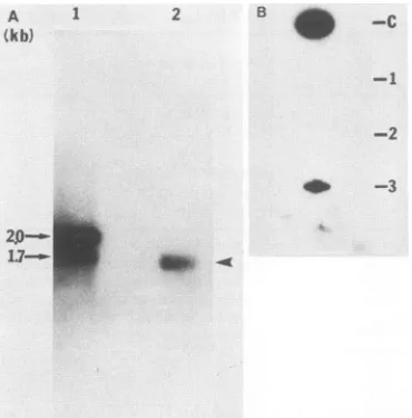
[image:6.612.142.214.75.238.2]Binding of HDAg to HDV RNA in vivo. To determine whether the specific interaction between HDAg and HDV RNAobserved in vitro also occurred in the hepatitis delta virion, we examined the status of HDV RNA in HDV virions isolated from the plasma of a patient with acute delta hepatitis. Electrophoreticanalysis of the RNA showed that only one HDV-specific RNA species was detectable (Fig. 9A). ThisRNAmigratedslightly faster than themonomeric RNAof 1.7 kb inaglyoxal-agarose gel, consistent with the interpretation that this RNA is a circular RNA (20, 23, 26, 41).Todeterminewhether this RNA was bound to the delta antigen within the virus particle, the purified virus particle wasdisrupted with 0.1% SDS and then immunoprecipitated with the rabbit antiserum prepared against the TrpE-HDAg fusion protein. Immunoprecipitation was performed in the presenceof0.5% Nonidet P-40. The immunoprecipitate was extracted with phenol-chloroform (1:1), transferred to a nitrocellulose membrane, and probed with an HDV-specific DNA probe. The HDV-specific RNA was precipitated with this antiserumbut not by the preimmune serum (Fig. 9B). A controlinvitro-transcribedHDV RNAwithout HDAg could notbe precipitated (Fig. 9B). This result indicates that the HDVRNAwasbound to the HDAg inside the hepatitis delta
FIG. 9. Analysis of HDV RNA from the plasma of a delta hepatitis patient. (A) Northern (RNA) blot analysis of the HDV RNA.HDV RNA(lane2)wasprepared from thepatient's plasmaas described in Materials and Methods. The probe usedwas a 3p_ labeledmonomercDNAof HDV RNA(26).RNAsize markers of 2.0and 1.7 kb(lane 1)weremadeby in vitrotranscription with T7 RNA polymerase of the pT7 plasmids containing HDV cDNA insertsof respective sizes andruninparallel. (B) Slot-blot analysis of HDV RNA that was immunoprecipitated with HDAg-specific antiserum. Thepatient's plasma wastreated with 0.1% SDS and thenprecipitated with various antisera. Theimmunoprecipitatewas extracted withphenol-chloroform, transferredonto anitrocellulose membraneby filtration, andprobed with 32P-labeled HDV cDNA. Slot1 wasHDV RNAtranscribed in vitro andimmunoprecipitated byHDAg-specific antiserum. Slots 2 and3 represent HDV RNA from theplasma ofadeltahepatitispatient whichwas immunopre-cipitated with either preimmune serum (slot 2) or HDAg-specific antiserumprepared against fusion protein D(slot 3). C, In vitro-transcribed HDVRNAwithoutimmunoprecipitation (positive con-trol).
virion by a bond whichwasstableaftertreatmentwith0.1% SDS and 0.5% Nonidet P-40.
DISCUSSION
Previous studieshavesuggested that HDAg, which is the
only protein encodedby HDV, has anRNA-binding ability (8). The currentstudy demonstrated the specificity ofsuch anRNA-proteininteraction. HDV RNA bindsspecificallyto HDAg. Unrelated viral (MHV) RNA does not bind to HDAg.Althoughwehavenotexhaustively testedevery kind ofRNA,thefactthat allof thebinding studies performedin this report were done in the presence ofa large excess of nonspecific competitorRNAisolatedfrom uninfected mam-maliancells suggests that thisbinding is very specificto the HDV RNA. Thisspecific bindingwasdemonstratedbytwo
independentmethods:aNorthwestern blot procedure and an RNA mobility shift assay. The binding occurred at a rela-tively high salt concentration. Furthermore, binding could be competed with by homologous RNAs but not byother RNAs. These results suggest that there is aspecific interac-tion between HDVRNAandHDAg. Anadditional interest-ingfindingwasthat thisspecific bindingwasnotthe result of a specific nucleotide sequence but rather ofa specificRNA conformation unique to HDV RNA. This conclusion was deducedfrom the
finding
that the binding ofthe viroidlike domain ofHDVRNAtoHDAg could becompetedwithby2p0--
on November 10, 2019 by guest
http://jvi.asm.org/
[image:6.612.345.530.76.264.2]the protein-coding domain, which does not share sequence with the viroidlike domain. It is not clear what this RNA conformation is. The most logical explanation is that both domains form a similar rod structure because of intramolec-ular complementarity (20, 41); however, this study has ruled out the simple double-stranded structure as the basis of RNA-protein binding. Previous analysis has suggested that HDV RNA is capable of forming a more complex structure (7, 43). Thus, a tertiary structure or an alternative RNA conformation may be responsible for the specificity of this RNA-protein interaction.
The function of the HDAg-HDV RNA interaction is not clear. This specific binding is consistent with the previous finding that HDAg is localized in the nucleus, where HDV RNA replicates in infected hepatocytes (8, 15, 39). Further-more, it has been demonstrated, by HDV cDNA transfec-tion, that HDAg is required for the replication of HDV RNA (22). Thus, the specific binding of HDAg to HDV RNA may play an important role in HDV RNA replication.
The binding domain on the HDAg protein is also specific; only the middle domain of the protein binds HDV RNA. It is interesting that this domain is highly hydrophilic and con-tains many basic amino acid residues (Fig. 3). However, the basic nature of the middle domain of the HDAg does not seem to be sufficient for its specific binding to HDV RNA, since the N-terminal domain of the protein is equally basic and yet it does not bind to the RNA. Therefore, the specific interaction between HDAg and HDV RNA probably has specific sequence requirements. Significantly, the HDV RNA sequence encoding the middle domain of the HDAg has recently been shown to be conserved among clinical isolates of HDV (Y.-C. Chao, M.-F. Chang, I. Gust, and M. M. C. Lai, Virology, in press). Thus, the binding of HDAg to HDV RNA is an important function for HDV RNA replication. Both the N-terminal and middle domains of HDAg contain a stretch of leucine zipperlike sequences (Fig. 3). This type of sequence has been shown to induce the dimerization of the proteins (10, 33) and, as a result, is responsible for the binding of proteins to DNA (16, 21, 28). Indeed, both the N-terminal and middle domains can form dimers (unpublished observation), and yet only the middle domain binds HDV RNA. Thus, dimer formation is not sufficient for the specific interaction between HDAg and HDV RNA. In contrast, the C-terminus is relatively free of charged amino acids. No nucleic acid-binding properties were detected with this domain.
HDAg has been shown to be an internal viral structural protein of HDV, i.e., it can only be detected when theviral envelope is disrupted with detergents (5, 6, 32). However, HDAg does not form a classical nucleocapsid structure. The data presented in this report show that at least some of the HDAg indeed binds to the circular HDV RNA via a bond which is resistant to treatment with 0.1% SDS and 0.5% Nonidet P-40. Whether this interaction between HDAg and HDV RNA is the same as that observed in vitro is not yet clear.
One additional piece ofinformation on the properties of the HDAg was revealedfrom thisstudy. Serumfrom adelta hepatitis patient detected all threedomains of HDAg in an immunoblotting procedure. Thus, all three domains appear to be immunogenic in humans.Thisfinding hasrecently been demonstrated independently (40). These bacterial TrpE-HDAg fusion proteins may be suitable as adiagnosticprobe in the future.
ACKNOWLEDGMENTS
ThisworkwassupportedbyPublic HealthServicegrantAI-26741 (to M.M.C.L.)fromthe National Institutes of Health andagrant from the National Science Council of the Republic of China. M.M.C.L. is anInvestigator ofthe HowardHughes Medical Insti-tute. M.-F.C.ispartially supportedbyaSmootFellowship awarded byNorris Cancer Centerof the University of Southern California. S.C.B. is apostdoctoral fellow of the Arthritis Foundation.
LITERATURE CITED
1. Arico,S., M.Aragona,M.Rizzetto,F.Caredda,A.Zanetti,G. Marinucci, S. Diana, P. Farci, M. Arnone, N. Caporaso, A. Ascione, P. Dentico, G. Pastore, G. Raimondo, and A. Craxi. 1985.Clinical significance ofantibodytothehepatitisdeltavirus insymptomless HBsAgcarriers.Lancetii:356-358.
2. Bergmann, K. F., and J. L. Gerin. 1986. Antigens ofhepatitis delta virus inthe liver and serum ofhumans and animals. J. Infect.Dis. 154:702-705.
3. Biedenkapp,H., U.Borgmeyer,A. E.Sippel,and K.-H. Klemp-nauer. 1988. Viralmyb oncogene encodesa sequence-specific DNA-binding activity.Nature(London) 335:835-837.
4. Bonino,F., K. H. Heermann,M. Rizzetto, and W. H. Gerlich. 1986.Hepatitisdeltavirus:proteincomposition of deltaantigen and itshepatitisBvirus-derivedenvelope.J.Virol.58:945-950. 5. Bonino,F., B.Hoyer,E.Ford, J. W.-K.Shih,R. H.Purcell,and J. L. Gerin. 1981. The deltaagent: HBsAgparticleswith delta antigenand RNA in the serumofanHBVcarrier. Hepatology
1:127-131.
6. Bonino,F., B.Hoyer,J. W.-K.Shih,M.Rizzetto,R. H.Purcell, and J. L. Gerin. 1984. Delta hepatitis agent: structural and antigenic properties of the delta-associated particle. Infect. Immun. 43:1000-1005.
7. Branch, A. D., B. J. Benenfield, B. M. Baroudy, F. V. Wells, J. L. Gerin,and H. Robertson. 1989. A UV-sensitive structural element in a viroid-like domain of the hepatitis delta virus. Science 243:649-652.
8. Chang, M.-F.,S.C.Baker, L. H.Soe,T.Kamahora,J.G.Keck, S. Makino, S.Govindarajan, and M. M. C. Lai. 1988. Human hepatitis delta antigen is a nuclear phosphoprotein with RNA-binding activity. J. Virol. 62:2403-2410.
9. Chen, P.-J., G. Kalpana, J. Goldberg, W. Mason, B. Werner, J. L.Gerin,andJ. Taylor. 1986.Structureandreplication ofthe genome ofthehepatitis deltavirus. Proc. Natl.Acad. Sci. USA 83:8774-9778.
10. Dang, C. V., M.Mcguire,M.Buckmire,and W.M. F. Lee. 1989. Involvement ofthe 'leucinezipper' regionintheoligomerisation and transforming activity of human c-myc protein. Nature (London) 337:664-666.
11. Dieckmann, C. D., and A. Tzagoloff. 1984.
Assembly
of the mitochondrial membranesystem. J.Biol. Chem. 260:1513-1520. 12. Feinberg, A. P., and B. Vogelstein. 1983. Atechnique
for radiolabellingDNArestrictionendonuclease fragments tohigh
specific activity. Anal. Biochem. 132:6-13.
13. Govindarajan, S., K. P.Chin,A.G.Redeker, and R. L.Peters. 1984. FulminantBviralhepatitis: role of deltaagent. Gastroen-terology86:1416-1420.
14. Govindarajan, S., B. Lim, andR. L. Peters. 1984. Immunohis-tochemical localization of the delta antigen associated with hepatitis Bvirusinliverbiopsysections embedded inAraldite. Histopathology 8:63-67.
15. Gowans, E. J., B. M. Baroudy, F. Negro, A. Ponzetto, R. H. Purcell, and J. L. Gerin. 1988. Evidence for
replication
of hepatitis delta virus RNA in hepatocyte nuclei after in vivo infection. Virology 167:274-278.16. Halazonetis, T. D., K. Georgopoulos, M. E. Greenberg, and P. Leder. 1988.c-jun dimerizeswith itselfand with
c-fos,
forming complexesofdifferent DNAbindingaffinities. Cell 55:917-924. 17. Hardy, W. R., andJ.H.Strauss. 1988.Processing
thenonstruc-turalpolyproteins ofSindbis virus: studyofthe kinetics invivo byusingmonospecific antibodies. J. Virol. 62:998-1007. 18. Heaphy, S., C. Dingwall,I. Ernberg, M.J. Gait, S. M. Green,
J. K. A. D. Lowe,M. Singh, and M. A.Skinner. 1990. HIV-1 regulatorof virion expression (Rev) protein binds to an RNA
on November 10, 2019 by guest
http://jvi.asm.org/
stem-loop structure located within the Rev response element region. Cell60:685-693.
19. Jacobson, I. M., J. L. Dienstag, B. C. Werner,D. B. Brettler, P. H. Levine, and I. K. Mushahwar. 1985. Epidemiology and clinical impact of hepatitis D virus infection. Hepatology 5: 188-191.
20. Kos, A., R.Dikema,A. C.Arnberg, P. H. van der Merde,and H. Schelekens. 1986. The HDV possesses a circular RNA. Nature(London) 323:558-560.
21. Kouzarides, T., and E. Ziff. 1988.Therole oftheleucinezipper in thefos-jun interaction. Nature(London) 336:646-651. 22. Kuo, M. Y.-P., M. Chao, and J. Taylor. 1989. Initiation of
replication of the human hepatitis delta virus genome from cloned DNA: role of deltaantigen.J. Virol. 63:1945-1950. 23. Kuo, M. Y.-P., J. Goldberg, L. Coates,W.Mason, J. Gerin,and
J. Taylor. 1988. Molecularcloning ofhepatitis delta virusfrom aninfected woodchuck liver: sequence, structure,and applica-tions. J. Virol. 62:1855-1861.
24. Kuo,M.Y.-P., L.Sharmeen, G. Dinter-Gottlieb, and J. Taylor. 1988. Characterization ofself-cleavingRNAsequences onthe genome andantigenome of humanhepatitis delta virus. J. Virol. 62:4439-4444.
25. Landschulz, W. H., P. F. Johnson, and S. L. McKnight. 1988. Theleucine zipper: a hypothetical structure common to a new class ofDNA-bindingproteins. Science 240:1759-1764. 26. Makino, S., M.-F. Chang, C.-K. Shieh, T. Kamahora, D. M.
Vannier, S.Govindarajan,and M. M. C. Lai. 1987. Molecular cloning and sequencing of a human hepatitis delta virus RNA. Nature(London) 329:343-346.
27. Makino, S., M.-F. Chang, C.-K. Shieh, T. Kamahora, D. M. Vannier, S.Govindarajan,and M. M. C. Lai. 1987. Molecular biology of a human hepatitis delta virus RNA, p. 549-564. In W. Robinson, K. Koike, and H. Will (ed.), Hepadna viruses. Alan R. Liss, Inc., New York.
28. Nakabeppu, Y., K. Ryder, and D. Nathans. 1988. DNA-binding activities of three murine jun proteins: stimulation by fos. Cell 55:907-915.
29. Pohl, C., B. M. Baroudy, K. F. Bergmann, P. J. Cote, R. H. Purcell, J. Hoofnagle, and J.L. Gerin. 1987. A human monoclo-nal antibody that recognizes viral polypeptides and in vitro translation products of the genome of the hepatitis D virus. J. Infect. Dis. 156:622-629.
30. Query, C. C., R. C.Bentley, and J. D. Keene. 1989. A specific 31-nucleotide domain ofUl RNA directly interacts with the 70K small nuclear ribonucleoprotein component. Mol. Cell. Biol. 9:4872-4881.
31. Rizzetto, M., M. G. Canese, S. Arico, 0. Crivelli, F. Bonino, C. G.Trepo, andG. Verme. 1977. Immunofluorescence detec-tion ofanewantigen-antibody system (delta/anti-delta) associ-ated to the hepatitis B virus in the liver and in the serum of HBsAg carriers. Gut18:997-1003.
32. Rizzetto, M., B. Hoyer, M. G. Canese, J. W.-K. Shih, R. H.
Purcell, and J. L. Gerin. 1980. Deltaagent:association of delta antigen with hepatitis B surface antigen andRNA in serum of delta-infected chimpanzees. Proc. Natl. Acad. Sci. USA 77: 6124-6128.
33. Schuermann, M., M.Neuberg, J. B. Hunter, T.Jenuwein, R.-P. Ryseck, R. Bravo, and R. Muller.1989. Theleucine repeat motif inFoxprotein mediates complex formation with Jun/AP-1 and is required for transformation. Cell 56:507-516.
34. Sharmeen,L., M. Y.-P.IKuo,G.Dinter-Gottlieb,andJ. Taylor. 1988. Antigenomic RNA of human hepatitis delta virus can undergoself-cleavage. J. Virol. 62:2674-2679.
35. Sharmeen, L., M. Y.-P.Kuo, andJ. Taylor 1989. Self-ligating RNA sequences on the antigenome of human hepatitis delta virus.J. Virol. 63:1428-1430.
36. Stohlman, S.A., R.S. Baric, G. N.Nelson, L. H. Soe, L. M. Welter, and R. J. Deans. 1988. Specific interaction between coronavirus leader RNA and nucleocapsid protein. J. Virol. 62:4288-4295.
37. Stohlman,S. A.,and M. M. C. Lai. 1979. Phosphoproteinsof murinehepatitis viruses.J.Virol. 32:672-675.
38. Surowy, C.S.,V. L. VanSanten,S. M.Scheib-Wixted,and R.A. Spritz. 1989. Direct, sequence-specific binding of the human U1-70kribonucleoprotein antigen proteintoloop1 ofUl small nuclear RNA. Mol. Cell. Biol.9:4179-4186.
39. Taylor, J., W.Mason,J.Summers,J.Goldberg, C.Aldrich,L. Coates,J. L.Gerin,and E. Gowans. 1987.Replicationofhuman hepatitisdelta virus in primary culture of woodchuck hepato-cytes.J. Virol. 61:2891-2895.
40. Wang, J.-G., R. W. Jansen, E. A. Brown, and S. M.Lemon. 1990. Immunogenic domains ofhepatitis delta virus antigen: peptide mappingofepitopes recognized by human and wood-chuck antibodies. J. Virol. 64:1108-1116.
41. Wang, K.-S., O.-L. Choo, A. J. Weiner, J.-H. Ou, R. C. Najarian, R. M. Thayer, G. T. Mullenbach, K. J. Denniston, J. L. Gerin, and M. Houghton. 1986. Structure, sequenceand expression of thehepatitis delta viral genome. Nature(London) 323:508-513.
42. Weiner,A.J., Q.-L. Choo, K.-S. Wang, S.Govindarajan,A. G. Redeker, J.L.Gerin,andM. Houghton. 1988. A single antige-nomic open reading frame of the hepatitis delta virus encodes theepitope(s) of bothhepatitis delta antigen polypeptides p248 andp278.J. Virol. 62:594-599.
43. Wu, H.-N., Y.-J.Lin,F.-P.Lin,S.Makino, M.-F.Chang,and M. M.C.Lai. 1989.Humanhepatitis delta virus RNA subfrag-mentscontainanautocleavage activity. Proc. Natl. Acad. Sci. USA 86:1831-1835.
44. Wu, H.-N.,and M. M. C. Lai. 1989. Reversible cleavage and ligation of hepatitis delta virusRNA.Science 243:652-654. 45. Zyzik, E.,A.Ponzetto, B.Forzani, C. Hele, K.Heermann, and
W. H. Gerlich. 1987. Proteins of hepatitis virus in serum and liver, p.565-577. In W.Robinson, K. Kolike, and H. Will (ed.), Hepadnaviruses. Alan R. Liss, Inc., New York.