0022-538X/91/063284-09$02.00/0
Copyright ©1991,AmericanSocietyforMicrobiology
Herpes
Simplex
Virus
Origin-Binding
Protein
(UL9) Loops
and
Distorts the Viral Replication Origin
ANDREW KOFF, JOHNF. SCHWEDES, ANDPETERTEGTMEYER*
DepartmentofMicrobiology, State University ofNew YorkatStony Brook, Stony Brook, New York11794-8621
Received 30 November1990/Accepted 17 March 1991
To investigate therole ofthe herpes simplex virus origin-binding protein (UL9) in the initiation of DNA replication,wehaveexamined the effect of UL9bindingonthestructureof the viralorigin of replication. UL9 loops and alters the DNA helix of the origin regardless of the phasing of the bindingsites. DNase I and micrococcal nucleasefootprinting show that UL9 binds two sites in the originand loops the AT-rich DNA between them independent of the topology of the DNA. KMnO4 and dimethyl sulfatefootprintingfurthershow that UL9alters the DNA helix in the ATregion. Incontrast totheloopingreaction,however,helicaldistortion requires the freeenergy of supercoiled DNA. UL9 also loops and distorts the origin DNA ofa
replication-defective mutant witha6-bp insertion in the AT region. Because the helical distortion ofthis mutant DNAis different from that of functional origins, weconclude thatanimperfecttertiary structureof themutantDNA
maycontributetoitsloss ofreplication function.
Herpes simplex virus (HSV)isanexcellent modelsystem for the study ofprotein-protein and protein-DNA interac-tions at a eukaryotic origin of replication. Genetic and
biochemicalanalyses aresimplified, since mostof the com-ponentsnecessaryforviral DNAmetabolismareencodedby
the virus.HSVmaysharesomeof theinitiationeventsofthe well-characterized Cairn's type DNA replication origins
eventhough studiesof thereplicativeintermediates of HSV suggestthat viral DNAreplicationoccursbyarolling circle
mechanism (2, 16, 22-24,49).
HSVcontainstwotypesofreplication origins. One origin, oriL, is inthe long uniquesegment ofthevirus (15, 18, 27, 51), and theother,
oris,
is in therepeat region flanking the shortunique segment(15, 41, 49). These origins are highlyhomologous, and both are functional in vivo. The optimal
originsequenceshavebeen defined for
oris
(10, 28, 43). This origin contains a large 45-bp palindrome centered on analternatingATmotif andanadditional 30 bpleftward of the
palindrome (28).Two UL9-binding sites overlap the ends of each arm of the palindrome (13, 35). A third, weaker, UL9-binding site has recently been identified (12, 50). Inser-tions ofincreasing numbers of AT dinucleotides into the centerof thepalindrome have anoscillating effectonorigin
function (28). Substitution of the AT region in the centerof thepalindrome with GC-rich sequences impairs replication
(28, 43). Therefore, the ATregion has both a spacing role,
presumably between the UL9 bindingsites, andanunknown
functional role inreplication.
HSV encodes proteins both directly and indirectly in-volved in viral DNA replication. Seven viral genes are
sufficientforreplication of the virus(32,52). Theseencodea
DNApolymerase, polymerase accessory protein, helicase,
primase, a single-stranded DNA-binding protein, the UL8
protein of unknown function, and an origin recognition
protein, UL9 (reviewed in reference 7). Genetic and bio-chemical studies indicate that some of these proteins can formcomplexes withmultiple functional activities(6, 8, 20, 34). It isUL9 thatimparts specificitytothe HSV replication complex (19, 39). UL9appearstobindas adimertoapairof
*Corresponding author.
overlapping inverted pentanucleotiderepeats (25)atsites I, II, andIII in the origin (13, 25, 35). Site Ionthe left side of
the palindrome is astrongerbinding site than siteII onthe
right side (12, 13). Site III is the weakest binding site and binds UL9 only in the presence of sites I and 11 (12).
Differences in binding affinity probably reflect base varia-tions in the recognition elements of thebinding sites.
In vitro replication systems for simian virus 40 (SV40)
(40), Escherichiacoli(1, 33), andbacteriophage A (33) have identified common events in the initiation ofreplication of Cairns-type origins. In thefirst stage ofinitiation, an origin
recognition protein specificallybindstomultiple recognition sites andunwindstheDNA strands atthe originof replica-tion. The second stage starts with the assembly of the replication machinery at the partially unwound origin. A helicase contained in this replication complex extends the partially unwound origin into a replicationbubble. Finally,
primase and DNA polymerases initiate the polymerization reaction inthereplication bubble. The E. coli and bacterio-phage A origin recognition and helicase activities are on
separate proteins,whereas the SV40 origin-binding protein, T antigen, has both activities. However, the origin-binding proteinin allcaseshas thecapacitytocatalyzethe firstevent in the initiation ofreplication, theunwinding of theduplex origin DNA. We wanted to determine whether UL9 could alter thestructure of the HSVorigin of replication.
Inthisstudy,weshow thatUL9alters thestructureof the originofreplicationintwoways. Neither of these structural changes requires theaddition ofexogenous ATP. Nuclease footprinting demonstratesthat UL9loopstheAT-rich DNA between UL9-binding sites I and II. Chemicalfootprinting
shows that the helix of theloopedDNAcanbe distortedby
UL9 when free energy is provided by supercoiling. Foot-printing of mutant origins with insertions in the AT-rich DNAindicates thatUL9 alsoloops anddistorts themutant origins. The helical distortion of a replication-defective origin, however, is differentfrom that of functionalorigins.
MATERIALS AND METHODS
Cells and virus.Sf9 cells and baculovirusweremaintained
as described by Summers and Smith (45). Recombinant
3284
on November 10, 2019 by guest
http://jvi.asm.org/
baculovirus expressing the HSV type 1 (HSV-1) origin-binding protein UL9 (Autographa californica nuclear poly-hedrosis virus [AcNPV]-UL9) was a generous gift of M.
Challberg (36).
Cloning of HSV origins. pS201, the wild-type HSV-2 origin, and two AT insertion mutants, pS201AT3 andpS201 AT23, were kindly provided by D. Galloway (28). Origin-containing ApaI restriction fragments were released from these plasmids and purified by low-melting-point agarose gel electrophoresis as described by Maniatis et al. (29). These restriction fragments were subcloned into an ApaI site inserted between the polylinker and the forward primer-binding site of pUC19. The transformants were sequenced, and clones with identical orientations were selected. We defined the clone containing the wild-type origin derived from pS201 as WT. The replication-competent, 46-bp inser-tion mutant was redefined as [AT]23, and the replication-defective, 6-bp insertion mutant was redefined as [AT]3. DNA was isolated from these clones by the alkaline lysis method (3) and purified in a CsCl gradient as described by Maniatis et al. (29).
Purification of recombinant UL9. Sf9 cells were infected with AcNPV-UL9 at a multiplicity of 10 to 20 viruses per cell and incubated at 27°C for 68 to 72 h. AU purification procedures were carried out at 4°C. Infected cells were gently shaken off flasks and collected by centrifugation at 1,000 x g. The cell pellet was washed with 8.33 ml of phosphate-buffered saline per g of wet weight of cells and collected by centrifugation at 1,000 x g. Hypotonic lysis and high salt nuclear protein extraction were carried out as described previously by Elias et al. (14). The clarified extracts were adjusted to 0.5 M NaCl by dilution with 2.4 volumes of buffer B (20 mM N-2-hydroxyethylpiperazine-N'-2-ethanesulfonic acid [HEPES]-NaOH [pH 7.6], 0.5 mM dithiothreitol (DTT), 0.5 mM phenylmethylsulfonyl fluoride, 0.5 mM Na2EDTA [pH 8.0],10%glycerol) containing 2 ,ug of leupeptin per ml. The adjusted extract was loaded onto a DEAE-Sephadex column (12 ml/h, 2.5 cm by 3.5 cm) equil-ibrated in buffer B with 0.5 M NaCl. The flow-through fraction was collected, adjusted to 0.2 M NaCl with 1.5 volumes of buffer B, and applied to a Whatman P11 column (5 ml/h, 1 cm by 7 cm) equilibrated in buffer B with 0.2 M NaCl. The column was washed with 10 volumes of buffer B and with 0.3 M NaCI in buffer B until the optical density at 280 nm was zero. UL9 was eluted in 10 volumes of 0.4 M NaCl in buffer B and collected inhalf-column-volume frac-tions. Fractions containing UL9 were identified by sodium dodecyl sulfate-polyacrylamide gel electrophoresis and Coomassie staining. The UL9 fractions were dialyzed against buffer C (20 mM HEPES-NaOH [pH 7.6], 0.5 mM DTT, 0.5 mM phenylmethylsulfonyl fluoride, 0.5 mM Na2EDTA [pH 8.0], 0.3 MNaCl, 50% glycerol) and stored at -20°C. UL9 protein purified by this scheme was approxi-mately 80% pure by Coomassie staining and had stable DNA-binding properties for at least 2 months.
Footprinting with DNAse I. Plasmid DNA was cut with EaeI and 3' end labeled with the Klenow fragment of DNA polymerase as described previously (25). The end-labeled DNA wasextracted withphenol-chloroform-isoamyl alcohol (FCHISAM) and purified on a Bio-Gel P6 (100 to 200 mesh) spin columnequilibrated with H20. The eluate was adjusted to conditions recommended by New England BioLabs for digestion by EcoRI and PvuII. After digestion, the origin-containing restriction fragments were isolated on a nonde-naturing 5% acrylamide (29:1) gel and electroeluted. DNA wasprecipitated with ethanol and resuspended at 0.1 ng/ul.
Variable amounts of protein, as indicated in the
figure
legends, wereincubatedwith 0.1 ngofprobein the presence of 5.5 ,ug ofpoly(dI-dC) poly(dI-dC-25%glycerol-0.5 mM DTT-0.2 M NaCl-0.5 mM Na2EDTA-4 mM ATP-5 mMMgCl2inatotal volume of 60pil.To
accurately
reflect in vivo temperature, the binding reactions were incubated at37°C
for 15 min. Then 0.025 U of DNase I was added in atotal volume of 1 ,ulof nuclease dilutionbuffer {20mM[pipera-zine-N,N'-bis(2-ethanesulfonic
acid)]
(PIPES)-NaOH
(pH
7.0), 0.1 M MgCl2, 0.05 M CaCl2, 1 mMNaCl,
0.1 mM Na2EDTA (pH8.0)}, and the reaction wascontinued for 1 min. Reactionswerestoppedbytheaddition of6 ,u of0.5 M Na2EDTA (pH 8.0) and an equal volume ofbCHISAM.
DNA was isolated from the aqueousphase
by
elution throughaP6spincolumnequilibrated
withH20.
Theeluate waslyophilized andresuspendedin 10 ,ul of95% formamidesamplebuffer andelectrophoresedona10%
acrylamide-8.3
M urea sequencing gel. Gels were dried andexposed
to X-rayfilm at-70°C withanintensifier.Footprinting with MNase. The
procedures
and buffers wereidenticaltothose used forDNaseIfootprinting
except that 0.4 U of micrococcal nuclease(MNase)
was used insteadofDNase I.Footprinting withKMnO4.Theprocedureused is identical to that described by Parsons et al. (37).
Briefly,
0.2 ,ug of supercoiled plasmid DNAcontaining
the HSVorigin
of interest was incubated with UL9 under the conditions de-scribed above except thatpoly(dI-dC)
poly(dI-dC)
wasnot included. KMnO4 was added to 30 mM, and incubationcontinuedfor4min. Reactionswere
stopped
by
theaddition of 1/10 volume of3-mercaptoethanol,
and DNA wasex-tracted by DCHISAM and recovered from the aqueous phase by using a Sephadex G50
(fine)
spin
column equili-brated withH20.
DNA was linearized with NdeI and pre-cipitated with ethanol. Linearized DNA was annealed toeither a 5' end-labeled M13 forward or reverse
primer
(as
described in the figurelegends)
and extended.Electropho-resiswascarriedout on8%
acrylamide-7
Mureasequencing
gels. Gels were dried andexposed
toX-ray
film at-70°C
with an intensifier.Footprintingwith DMS.The
procedures
andbufferswereidenticaltothose used forKMnO4
footprinting
exceptthat4IlI
ofdimethyl sulfate(DMS)
diluted 1:20 inH20
was used instead ofKMnO4. Incubation with DMSwascarriedoutfor 3 minbefore the modification reaction wasstopped
with 14 ,ulof1.5% sodiumdodecyl
sulfate-0.1 MNa2EDTA-7.15
M,-mercaptoethanol.
RESULTS
UL9loops the DNA between
origin-binding
sites. DNase I and MNasefootprinting
analyses
wereusedtodetermine the effectsofUL9binding
tovariousreplication
origins.
DNA is resistant to DNase I cleavage ifprotein
binding
blocks accessofDNase Itotheminorgrooveoraltersthegeometryof the minor groove (17). MNase cuts the
phosphate
back-bone of distorted DNA but is inhibitedby
guanine
andcytosineresidues around the site of
cleavage
(11,
53).
Both functional (WT and[AT]23)
and nonfunctional([AT]3)
repli-cation origins
(Fig.
1) were used to determine whether thereplication defect of
[AT]3
was causedby
changes
in UL9binding to this
origin.
Increasing
amounts of recombinant UL9were incubated withsingly
end-labeled,
origin-contain-ing restriction fragments. Protein-DNA
complexes
wereallowed toformat
37°C
and were treated witheitherDNaseon November 10, 2019 by guest
http://jvi.asm.org/
[AT]23 REPLICATION COMPETENT
ATATATATATATATATATATATATATATATATATATATATATATAT
[AT]3REPLICATION DEFECTIVE
ATATAT
WILD TYF
E
-0
In<
V) 1-x
Ji
I o
10 20 30 40 1,0 60 70
*TTTTCT¢tWOR GCTT oCCCCJ.L...
GCAGGATTATCATATAIATAAT
cATCCGTTCACCETCGG.... .... ... .. ... .
.G. .G.G.A.A..T. .T.G.T..C.T.T.T.A.T.G.4.A.C.G.
TA&AAA lAl;xCici CGAA =C. CGTCCTATAGTATATATATTATTAGGGCAUCSCdCC C
NUCLEOTIDEPOSITON
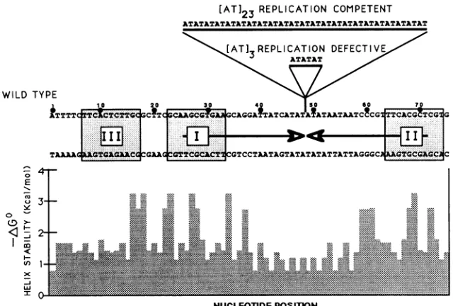
FIG. 1. Sequences ofthefunctional andnonfunctional HSV-2 replication origins. Both DNA strands of the minimal origin sequence defined byLockshornandGalloway (28)areshown,andnucleotide positionsarenumbered abovethe topstrand. Thepositionof thelarge centralpalindromeis indicated by arrows between the DNA strands. The tworecognitionelementsforUL9bindingandathirdhomologous region(12, 25, 50) are indicated bystippledboxes. The sequences ofinsertion mutations[AT]3and[AT]23betweenwild-type nucleotides 48 and 49 are shown above thewild-typesequence. Aplotof helixstabilitybased on nearestneighborpairs (5)is shownbelow the bottomstrand ofthewild-type DNA.
I or MNase. After the reaction was stopped, the DNA was isolated and analyzed on sequencing gels.
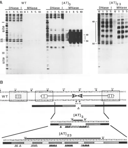
Extractsfromuninfected cells orcellsinfected with non-recombinant baculovirus were purified in the same manner asUL9 and had noevidentorigin-bindingactivity(data not shown). The binding of highly purified UL9 strongly pro-tected sites I and II in the WT and mutant origins from DNase I(Fig. 2). Binding to site III, above siteIin thegel, was not detected. Weaker DNase I protection over both endsof the AT-rich region in the WT and mutant replication
originswasalso observed. In the absenceof protein, DNase I cut the inserted AT-rich DNA segments at the expected
intervals of 2 bp (31). UL9 binding to the [AT]23 mutant
origin caused a strikingly periodic pattern of DNase
sensi-tivity throughout the region corresponding to the [AT]23 dinucleotide insertion. Regionsofsensitivity alternated with regions of nuclease resistance with a periodicity of approx-imately 10 to 11 bp. The periodicity was not evident in the shorterwild-type or [AT]3origins.
The structure of the originDNA wasprobed further with MNase(Fig. 2). In the absenceof UL9, MNase failed to cut mostregions of WT and mutant DNAs but did cut a single site in the AT-rich DNA in the WT origin and at regular 2-bp intervals throughout the [AT]3and [AT]23insertions. In the presenceof UL9, MNase sensitivity in the AT-rich region of
[AT]23became periodic with aninterval of 10 to 11 bp. The areasof MNase periodicity overlapped the areas of DNase I periodicity (Fig. 2). UL9 binding induced MNase hypersen-sitivity throughout the AT region of [AT]3. Shorter expo-suresof the autoradiogram in Fig. 2 show that the regions of DNA maximally cleaved by MNase were separated by approximately one helical turn. Thus, the most prominent MNase cleavage sites in both mutant origins matched the helical repeat of AT DNA. In the WT origin only a single, weakly sensitive MNase site was identified. The paucity of
nuclease sites in the shorter ATregion of theWTorigin may reflect the sequencecontextof theregionorsteric hindrance
by UL9blocking accessofMNase.
Both DNase I and MNase cut the AT regions in the insertion mutants with a periodicity of 10 bp, but the two patterns were out of register. It is unlikely that these distinctive patterns reflect UL9bindingtotheATregionand directnucleaseprotectionbecause UL9 would have to bind to the DNA at intervals of less than 10 bp. Rather, these
nuclease-sensitivity patternsarecharacteristic of DNA that is looped (21). We conclude that the AT regions of the mutant origins are looped between the UL9-binding sites
through protein-protein interactions. Thedistance between the centers ofUL9-binding sites I and II in the wild-type
origin is41bp. BecausealternatingAT sequencesareeasily deformed(31,44), theATregionof thewild-typeoriginmay facilitatelooping. Furthermore,UL9itself may have consid-erable flexibility that would allow the formation of small DNA loops. Lee and Schleif (26) have presented evidence that theAraCproteincanformasmallrepression loopwith a spanof 33bp.
UL9 distorts the origin of replication. To characterize further the structure formed in the looped region ofDNA,
we used KMnO4 footprinting analysis (Fig. 3). KMnO4
oxidizesthymineandcytosineresidues induplexDNA that is structurally distorted (4). KMnO4-sensitive changes in-clude DNA bending, DNA unwinding with a loss of base pairing, anduntwistingof theDNAhelix. KMnO4 modifica-tionscanbedetected because the Klenowfragmentof DNA polymerase I pauses at or nearmodified nucleotides. UL9 was incubated with supercoiled plasmids containing the various origins described in the legend to Fig. 1. DNA-protein complexes were allowed to form, and KMnO4was added to the binding reaction. After the reaction was stopped, DNA was isolated and linearized. Primers were )E
W
on November 10, 2019 by guest
http://jvi.asm.org/
[image:3.612.150.470.71.287.2]A WT [AT]3
DNase I MNase DNase I MNase
A 1 S 1f01A S n 1 3 5 10 0 1 3 5 10
* 1s_
IS i
50
-[AT]2 3
DNase I MNase
0:
__w_
1 3 5 0 1 3 5-j48 **w P
-50
so- 4=S
asa
s.) _
Un
a
B
1 10 20 30 40 50 60 70
ATTTTC[TTCACTCTTGCGCTTC
GCAAGCGTGAJ1GCAGGATTATCATATATATAATAATCCCGTITCACGCTCGTGACC
WT
11111
LTAAAAGLGGGJLCCGAAqCTCCC CGTCCTAATAGTATATATATTATTAGGGCA
0JC
4A.GTGC2AGC~~~~~~~~~ TGGCG2fTGG4 8 50
[AT]
3r 4j~~~4 50 5
GTCCTAATAGTATATATATATATATTATTAGGGCAA
A A
[AT]23
48
GTCCTAATAGTATATATATATATATATA
Adl
50
LTATATATATATATATATATATATATATATATATATATTATTAGGGCAA
AAA
AnA
tAAA
FIG. 2. Nucleasefootprintingofthe WT and mutantreplication origins. (A)DNase Iand MNasefootprints ofthe bottom strandoflinear originDNA.Increasingamountsof UL9wereincubated with0.1 ngofsinglyend-labeledprobes fromWTand mutantoriginsat37°C.The numbers above each lanerepresent the relative amountsofUL9. Thenumber1abovethegel lanesindicates 0.3 ,ugofUL9,and 10indicates 3
jig
ofUL9.Protein-DNA complexesweretreatedwitheitherDNase I or MNase asindicatedaboveeachsetof lanes. Nucleotidepositions 48and50 areindicated foreachorigin.The vertical black lines between these nucleotides represent the inserted AT DNA.SitesIandIIare indicatedtotheleft ofthe WTfootprint. SiteIIremainsat afixedpositionatthebottomoftheorigins,whereassiteIismovedtowardthe topofthepanels in the mutant origins. (B)Summary ofthe nucleasefootprintingdata on the bottomstrand. Nucleotides 48and 50are identifiedtoorient the sequences with the dataabove.Bars represent DNase Iprotection causedbyUL9binding; the blackregionswere strongly protected, the stippled regionswerepartially protected, andopen regionswerenotprotected. Trianglesrepresentthenucleotides sensitiveto MNasecleavage in the presence of UL9. Filledtriangles were moresensitive than open triangles.The bottom strand of the inserted ATregion andflankingsequences of the mutants are shown below the WTorigin.Nucleotidepositions48 and 50areshown,and the linesabove the sequenceindicate the insertednucleotides.annealedtotheDNAandextendedby Klenow polymerase. Figure 3A shows the experimental data for the bottom
strand; in Fig. 3B, we illustrate the data for both strands
schematically.
UL9bindinginduced dramaticKMnO4
sensitivity
in both functional and nonfunctional origins. However, both quan-titative andqualitativedifferences in the patterns ofKMnO4
sensitivitywereobserved whenfunctional andnonfunctional
m ommmmm ::..3
A
on November 10, 2019 by guest
http://jvi.asm.org/
[image:4.612.105.514.72.545.2]WT
0 1 3 5
[AT]3
0 1 3 5
19 =
..O
.0
;.. ?!-.
53/54 t R,I" l
56/57
IMa
a
40_
_:
inser4
is3
1 0 20 30 40 50 60 70
la a m a a a m
6 CACT&fTTGcPCTTCTCAAGCGTGAI CAGGATTAkTCATA±AAAAATAATCCCGTVTCACGCTCjG
aa TCCTAAT TAT Aa ATTATT
[AT] X ; 3 U
40 50 60 70
U.TCAAGCaGGATTATCATA ATATATATATATATATATATATATATATATATATATATATATATA TATAATAATCCCGT TC
T
13 H
1111
[image:5.612.94.515.67.508.2]a{TTC WcAGTCCTAATAGTAT ATATATATATATATATATATATATATATATATATATATATATAT ATATTATTA A A
FIG. 3. KMnO4 footprinting of the various replication origins. (A) KMnO4 footprints of the bottom strand of circularorigin DNA. IncreasingamountsofUL9wereincubated withsupercoiledplasmid DNA containing the origins of replication indicated above each panel. The numbersaboveeach lanerepresenttherelative increase in UL9asdescribed in thelegendtoFig.2.TheATregions of the various origins areshowntotheleft of the footprints. Nucleotidesmostreactivetomodification by KMnO4areindicatedby black boxes, and theirlocations
areidentifiedby nucleotidepositionontheleft ofeachpanel. Theopenrectangle indicatesthelocation ofaKMnO4-sensitive positionoutside of theATregioninthe WTorigin. (B) Schematicrepresentation of KMnO4 footprints. Both strands ofthevariousoriginsareshownand numberedabovethetopstrand. UL9-binding sitesareshownasstippled boxes. The central palindrome is shown between the strandsof DNA in theWTorigin.UL9-binding siteII,in all theorigins, isalignedontheright side of the figure, and ATinsertsareenclosedinopenboxes. KMnO4 signalsarerepresentedbyvertical bars between the DNA strands. The solid barsrepresentthemostreactive nucleotides, andthe stippled barsrepresentlessreactivenucleotides. Theopenrectangles indicate the location ofaKMnO4-sensitive position outside oftheAT region in the WT origin.
origins werecompared (Fig. 3). In the functional wild-type
and [AT]23 origins, UL9 binding increased the KMnO4 sensitivitytothegreatest extent at nucleotides 53to 54and 56and57ofthebottom strand. In contrast,protein binding to the nonfunctional [AT]3 origin induced thegreatest
sen-sitivityto KMnO4 intheinserted DNA. Similar differences betweenfunctionalandnonfunctional originswereobserved
in thetoporigin strand;inthepresenceof UL9,nucleotides
40 and 41 werethemost reactivepositionsinthefunctional origins, whereas the insert region was most reactive in the defectiveorigin (Fig. 3B;datanotshown).Weconcludethat UL9 distorts the DNA helix in the ATregionregardlessof its length. Nevertheless,differences in thephasingof the flank-ing UL9-bindflank-ing sites lead to different patterns of helical distortion in the AT DNA.
Most UL9-induced KMnO4 modifications were present
A
[AT]2
30 1 3 5
e e
___
_ma
B
30
[AT]2
UI
53J54
EtW
56/57_on November 10, 2019 by guest
http://jvi.asm.org/
DMS
_ +
-_
-KMnO4
- + UL9
U,w
21 20
38
3
60AtvN A AAA AA *
GATTATCATATATATAATAATCC
11 1111111 1
FIG. 4. KMnO4 and DMSfootprintingof the top strand of the WTorigin. Protein (1.0 ,g) wasincubated with supercoiled DNA containing the WT origin of replication andtreated with the reagents indicated above each panel. The presence or absence of UL9 is shownabove each lane.After modificationthe DNA wasrecovered, linearized, and annealed to a sequencing primer. Stops in the extension of thisprimer identified modificationson the top strand of the DNA. Thecytosine residue at nucleotide 44 is shown on the right of the DMS footprint.On theleft ofthefigure, we indicate the positions of nucleotides20and 21. Aschematicrepresentation of the data is illustrated below the gel. The top strand of DNA from nucleotides38 to60is shown.Barsrepresentthe KMnO4stops,and caratsrepresent theDMSstops.
DNA I
ATP +
UL9 - +
KMnO4
I III
+
_ + - +
19-
---I C
.*
"'
42A
I
-.
4 11111 liii 60
AATAGTATATATATTATTAGG
FIG. 5. Energy requirements of the looping and distortion of originDNA. UL9(1.0j,g)wasincubatedwithWTorigin DNA,and theprotein-DNAcomplexes weretreated with KMnO4. The topo-logicalforms ofthe DNAsubstratesareindicated above eachpair of lanes. Form I represents covalently closed supercoiled DNA, and form III is DNAlinearizedat anNdeI site before incubation with UL9.Thepresence orabsenceofATPandof UL9 isindicated. The ATregionandnucleotide19 arealigned and indicated in the various footprints. The KMnO4 footprinting dataare illustrated schemati-cally below the gel. The bottom strand of DNA is shown from nucleotides40 to60,andthe stopscausedby KMnO4modification arerepresentedby bars.
between UL9-bindingsites I and II (Fig. 3 and 4). Many of thesesiteswerelocated at the samepositionsrelativetosites I or II in both WT and mutant origins. For examples, see
modifiednucleotides 40 and 41 on the top strand and 53 to 54 and 56 and 57 on the bottom strand. These observations suggest that most structural changes are induced by the
binding ofUL9 to sites I and II. Surprisingly, UL9 altered theKMnO4modificationof severalnucleotides to the left of
siteIin the WTorigin. UL9induced modification of
nucle-otide 19 in the bottom strand of the origin and reduced
modification of nucleotides 20and 21 in the top strand (Fig.
4).DNAin thisregionmayhave anatypical helical structure that is changed by UL9 binding. Interestingly, these
posi-tions arelocated betweenUL9-binding sites Iand III in the WTorigin. Thesefindings suggest that UL9 may bind to site III, in agreement with the findings ofWeir et al. (50) and Eliasetal. (12), even though our DNase footprints did not detect stable protein binding at this location. We do not
know why the UL9-induced modification at nucleotide 19 appearstomaintainafixed positionrelative to site II in WT and mutantorigins.
To complement the KMnO4 footprinting, we used DMS
footprinting analysis(Fig. 4). DMS modifies both theNi and N3positions of adenine and the
Ni
position of cytosine insingle-stranded DNA, and it modifies the N3 and N7
posi-tions of adenine andguanine,respectively, in either double-stranded orsingle-stranded DNA(30). Klenow polymerase pausesattheDMS-modified nucleotides, and thus the
mod-ifiedbasecanbedetectedbyprimerextensionanalysis(37). The HSV-2 wild-type and mutant origins contain a single
cytosine residue atposition44 embedded in the alternating
AT-rich DNA. This nucleotide would be resistanttoDMSin
double-strandedordistortedDNAbut would besensitive in unwound DNA.
Although UL9protectedsites I and II from DMS modifi-cations only to a limited extent, it induced sensitivity to
KMnO4 and hypersensitivity to DMS throughout the AT
region ofthe WTorigin (Fig.4). Similarfindingsweremade in thecaseof bothinsertionmutants(datanotshown).These
changes confirmthat the DNA helix is altered in thisregion.
We cannot,however,determinewhetherpausing ofKlenow atthecytosinewascausedby methylation of this residuein a region ofsingle-stranded DNA or by methylation of the
adjacent adenine. In preliminary experiments using DMS modification in conjunction with single-strand specific
nu-cleases, we have not been able to detect single-stranded
DNA in the ATregion (datanotshown).
Loopingand helixdistortionareseparateactivities of UL9. Intheexperimentsdescribedabove,weperformednuclease assays for loopingon linear DNA and chemical
sensitivity
assaysfor helixdistortion on supercoiledDNA. To investi-gate the energy requirementsforUL9-induced helixdistor-tion, we compared the KMnO4 modification of linear and
supercoiledDNAin the presenceorabsence of ATP
(Fig. 5).
UL9efficiently distorted the ATregion
ofsupercoiled
WT DNA.Linearization of thesameplasmid, however,rendered the origin completely resistant toKMnO4 modification. An increase in theamountofUL9proteinin thebinding
reactionon November 10, 2019 by guest
http://jvi.asm.org/
[image:6.612.110.248.79.287.2] [image:6.612.366.509.83.311.2]withlinearized substrates didnotinduce the KMnO4-sensi-tive structure (data not shown). ATP had no effect on the
UL9-induced modifications of the originDNA. These results indicate that UL9 requires the free energy available in
supercoiled DNAtodistort the helix of HSV originDNA in
anATP-independent fashion. UL9 binding leadstoDNase I protectionofsites I andII of WT origins at similar protein
concentrations, using either linear or supercoiled DNA
substrates (datanotshown). Thus, UL9 does notbindmore
stronglytosupercoiledthantolinear DNA templates, and it is notbetter UL9 binding thatleadsto helix distortion. We conclude that looping and helix distortion are separate activitiesof UL9.
DISCUSSION
UL9, the origin recognition protein ofHSV, binds to at least two sites in the HSV origin in a cooperative manner
(12). The AT-rich sequences between UL9-binding sites I
andIIhaveafunctional roleaswellas aspacing role (28, 42).
In thepresentstudy, weinvestigated the function of UL9at the viral replication origin by analyzing theDNA structures formed in both functional and nonfunctional origins in the
presenceofUL9. Weshowthat binding of UL9totheorigin loopsanddistortstheDNAbetweenthebinding sites. These
events can be separated by their requirements for free
energy.
Our studies also offer insight into the effects of insertion mutations on origin function. We compared structural changes induced by UL9 bindingtoWTandmutantorigins, using nucleasefootprinting. The mutantorigins, constructed by Lockshorn and Galloway (28), have insertions that
ex-tend the AT region betweenUL9-bindingsites I and II. The shorter insertion mutant, [AT]3, has a 6-bp insertion that
changes the phasing ofUL9-binding sites by halfaturn and blocksreplication. The longer insertionmutant, [AT]23, has
a 46-bp insertion that enhances replication. We found that
UL9 boundtositesIandIIofthe WT originandbothmutant
origins at similar protein concentrations. UL9 binding to [AT]23 and to [AT]3 resulted in nuclease cuttingof the AT regions in oscillating patterns with aperiod of 10 to 11 bp. Both DNaseIandMNasecutthe AT regions of the insertion mutants in this periodic pattern. These nuclease-sensitivity patternsarecharacteristic of DNA thatisloopedorwrapped
around protein (21).Weconclude that theATregions of both mutants are loopedbetween the UL9-binding sites through
protein-proteininteractions.
Aperiodicnuclease-cuttingpatternwasnotevident in the WTorigin in which thespanofnuclease-exposed sites in the
AT region is not sufficientto exhibit periodicity. Neverthe-less, we assume thattheATregion of the WTorigin is also
looped by the same protein-protein interactions that are so
evident inthecaseofbothmutants. Thus,UL9bindstosites I andIIandloopsthe ATregion regardless ofthephasing of
the UL9-binding sites. Presumably, the flexibility of the protein and DNAaresufficientto compensatefor the spatial and phasing differences in the three origins. Nevertheless, the wild-type and mutant origins would have different
ter-tiary structures in the region oflooped DNA. The altered tertiary structure ofthe [AT]3 origin might contributeto its loss ofreplication function.
Todetermine whetherUL9binding alterstheDNAduplex in the functional and nonfunctional replication origins, we usedchemical footprinting techniques. Figure 1 showsafree
energy plot of base-pair stability in the HSV origin. The region of origin DNA in which the least amount of free
energy would be necessary to distort the DNA
duplex
correspondstothe ATregionbetween thetwo
UL9-binding
sites. We found that UL9 distorts the DNA in the ATregion
ofall three
origins. Interestingly,
thepattern of helix distor-tion in the AT segmentwas differentin the functional and nonfunctionalorigins.
Thisfinding
is consistent with the idea that the looped DNA of the [AT]3 origin has an alteredtertiarystructurethatmayblock theinitiationof
replication.
Incontrasttolooping,helixdistortionwasabsolutely depen-dent on the origin being supercoiled. These findingsimply
thatdifferences in thepreexisting
structures of relaxed andsupercoiled DNA determine the ultimate effects of UL9
binding and DNAloopingonthehelical structure of the AT
region in vitro. Although circularization of viral DNA is necessaryforDNAreplicationinvivo(38), thesuperhelicity ofviral DNA
during lytic
infection hasnotbeen established. Recently Weir et al. (50) and Elias et al. (12) haveinvestigated thefunction ofUL9-binding site IIIatthe left endoftheorigin. Theyhave shown that deletionormutation of this segment reduces DNAreplicationandUL9bindingto theremainder of theorigin severalfold. Ourdataare consis-tent with the idea that UL9interacts with site III. We have found that UL9 alters the KMnO4 modification of 3 nucleo-tides between sites I and III even though our DNase foot-prints did not detect stable protein binding at site III. Perhaps, UL9 binds transiently to siteIII and interacts with UL9bound to sites I orII toalter DNA structure.
Replication origins from prokaryotic and eukaryotic
sources allcontain sequences rich in AT DNA. In Saccha-romyces cerevisiae, the origin unwinds transiently even in the absence of protein (46). Protein-induced unwinding of the yeast originis not a highly sequence-specific event (47)
as it is in the SV40 origin (9). An elegant series of
experi-ments using yeastorigins has demonstrated that the energet-icsof DNA distortion may control replication initiation (48)
and that therole of origin-binding proteins may be simply to lower the thermodynamic activation barrier of DNA distor-tion. The AT motif in the HSV origin is thermodynamically
unstable (5) and highly reactive to both chemical and enzy-matic probes (31, 44). However, the free energy associated with UL9 binding to the HSV origin is not sufficient to distort linear DNA. KMnO4 modification of the HSVorigin also requires the free energy available in supercoiled DNA. ATP or ATP hydrolysis cannot substitute as an energy source.
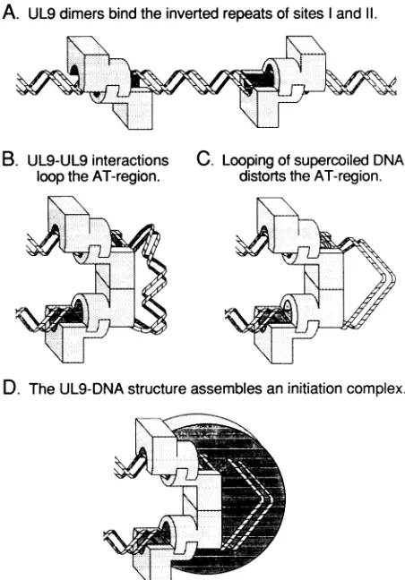
Figure 6 shows a model for formation of a functional UL9-DNA complex at the wild-type origin of replication. Step A, two dimers of UL9 bind to inverted repeats in sites I and II of the HSV origin. The AT-rich DNA between these sites consists of approximately four turns of the double helix. Step B, direct interactions between the dimers of UL9 bend theintervening DNAinto a partial loop. The loop in the WT origin is probably wrapped tightly around the protein core, as illustrated, whereas the inserted DNA of the mu-tants [AT]3 and [AT]23 would be free from the protein and thus partly susceptible to nucleases. Step C, DNA looping, in conjunction with the free energy of supercoiled DNA, distorts the DNA helix in the AT region. The alteredhelical structureof DNA may serve as part of a recognition element for other proteins involved in the initiation of replication. Step D, assembly of a complete initiation complex would require interactions with both UL9 and looped DNA. We suggest that this complex would be utilized in the next step of replication. Using this model, the replication defect of the [AT]3origin may be explained in the following way. Altered phasing ofthe UL9-binding sites results in improper
on November 10, 2019 by guest
http://jvi.asm.org/
C. Loopingof supercoiled DNA distorts theAT-region.
D. TheUL9-DNAstructureassemblesaninitiationcomplex.
FIG. 6. Model for structure ofUL9-origin complexes. Steps A through Daredescribed in the text.
actions betweenUL9moleculesbound toeachsite, creating
an inappropriately looped and untwisted structure.
Differ-ences in MNase and KMnO4sensitivities of looped DNA in
the functional and nonfunctional origins support this idea. Interactions ofadditional replication factors with the UL9-origin complex may be blocked because the tertiary struc-tureof the complex is incorrect. Furtherfootprinting analy-ses,in thepresenceofadditionalreplicationproteins,willbe important in determining subsequent events in the initiation ofHSVDNA replication.
ACKNOWLEDGMENTS
This workwas supported by Public Health Service grants CA-18808, CA-28146, and CA-09176 awarded by the National Cancer Institute.
WethankJim Borowiec, Kris Mann, PaulaEnrietto, and Joseph Lipsick for helpful comments on the manuscript. Judy Stenger contributed Fig. 6.
REFERENCES
1. Baker, T. A., L. L. Bertsch, D.Bramhili,K.Sekimizu, E.Wahle, B. Yung, and A. Kornberg. 1988. Enzymatic mechanism of initiation ofreplication from the origin ofthe Escherichia coli chromosome. CancerCells 6:19-24.
2. Ben-Porat, T., and S. A. Tokazewski. 1977. Replication of herpesvirus DNA. II. Sedimentation characteristics of newly
synthesized DNA. Virology 79:292-301.
3. Birnboim,H. C.,andJ. Doly.1979. Arapid alkaline extraction
procedure for screening recombinant plasmid DNA. Nucleic
AcidsRes. 7:1513-1523.
4. Borowiec, J. A., L. Zhang, S. Sasse-Dwight, and J. D. Gralla. 1987. DNA supercoiling promotes formation of a bent repres-sion loop in lac DNA. J. Mol. Biol. 196:101-111.
5. Breslauer, K. J., R. Frank, H. Blocker, and L. A. Marky. 1986. Predicting DNAduplex stability from the basesequence. Proc. Natl. Acad. Sci. USA 83:3746-3750.
6. Calder, J. M., and N. D. Stow. 1990. Herpes simplex virus helicase-primase: the UL8 protein is not required for DNA-dependent ATPase and DNAhelicase activities. Nucleic Acids Res. 18:3573-3578.
7. Challberg, M. D., and T. J. Kelly. 1989. Animal virus DNA replication. Annu. Rev. Biochem. 58:671-718.
8. Crute, J. J., T. Tsurumi, L. Zhu, S. K. Weller, P. D. Olivo, M. D. Challberg, E. S. Mocarski, and I. R. Lehman. 1989. Herpes simplex virus 1 helicase-primase: a complex of three herpes-encoded gene products. Proc. Natl. Acad. Sci. USA 86:2186-2189.
9. Dean, F. B., J. A. Borowlec, Y. Ishimi, S. Deb, P. Tegtmeyer, and J. Hurwitz. 1987. Simian virus 40 large tumor antigen requires three corereplication origindomains for DNA unwind-ing and replication in vitro. Proc. Natl. Acad. Sci. USA 84: 8267-8271.
10. Deb, S., and M. Doelberg. 1988. A 67-base-pair segment from theOri-S region of herpes simplex virus type 1 encodesorigin function. J. Virol. 62:2516-2519.
11. Dingwall, C., G. P. Lomonossoff, and R. A. Laskey. 1981. High sequence specificity of micrococcal nuclease. Nucleic Acids Res. 9:2659-2673.
12. Elias, P., C. M. Gustafsson, and 0. Hammarsten. 1990. The origin binding protein ofherpes simplexvirus 1 binds coopera-tively to the viral origin of replication oris. J. Biol. Chem. 265:17167-17173.
13. Elias, P., and I. R. Lehman. 1988. Interaction of originbinding protein with an origin of replication of herpes simplex virus 1. Proc. Natl. Acad. Sci. USA85:2959-2963.
14. Elias, P., M. E. O'Donnell, E. S. Mocarski, and I. R. Lehman. 1986. A DNA binding proteinspecific for anorigin ofreplication of herpes simplex virus type 1. Proc. Natl. Acad. Sci. USA 83:6322-6326.
15. Frenkel, N., H. Locker, and D. A. Vlazny. 1980. Studies of defective herpes simplex viruses. Ann. N.Y. Acad. Sci. 354: 347-370.
16. Friedmann, A., J. Shlomal, and Y. Becker. 1977. Electron microscopy of herpes simplex virus DNA molecules isolated from infected cells bycentrifugation in CsCl density gradients. J. Gen. Virol. 34:507-522.
17. Galas, D. J., and A. Schmitz. 1978. DNAase footprinting: a simple method for thedetection of protein-DNA binding spec-ificity. Nucleic Acids. Res. 5:3157-3170.
18. Gray, C. P., and H. C.Kaerner. 1984. Sequence oftheputative origin ofreplication in the UL region of herpes simplex virus type 1 ANG DNA. J. Gen. Virol. 65:2109-2119.
19. Heilbronn, R., and H. zur Hausen. 1989. A subset ofherpes simplex virus replication genes induces DNA amplification within the host cell genome. J. Virol. 63:3683-3692.
20. Hernandez, T. R., and I. R. Lehman. 1990. Functional interac-tionbetween the herpes simplex-1 DNA polymeraseand UL42 protein. J. Biol. Chem. 265:11227-11232.
21. Hochschild, A., and M. Ptashne. 1986. Cooperative bindingofk repressor to sites separated byintegralturns ofthe DNAhelix. Cell 44:681-687.
22. Jacob, R. J., L. S. Morse, and B. Roizman. 1979. Anatomy of herpes simplex virus DNA. XII. Accumulation ofhead-to-tail concatemers in nuclei of infected cells and their role in the generation of the fourisomeric arrangements of viral DNA. J. Virol. 29:448-457.
23. Jacob, R. J., and B. Roizman. 1977. Anatomy ofherpessimplex virus DNA. VIII. Properties of the replicating DNA. J. Virol. 23:394-411.
24. Jongeneel, C. V., and S. L. Bachenheimer. 1981. Structure of replicating herpes simplex virus DNA. J. Virol. 39:656-660. 25. Koff, A., and P. Tegtmeyer. 1988. Characterization of major
A. UL9 dimers bind the inverted repeats of sites I and 11.
B. UL9-UL9 interactions loop the AT-region.
on November 10, 2019 by guest
http://jvi.asm.org/
[image:8.612.64.290.73.397.2]recognition sequences for a herpes simplex virus type 1 origin-binding protein. J. Virol. 62:4096-4103.
26. Lee, D.-H., and R. F. Schleif. 1989. In vivo DNA loops in araCBAD:size limits and helical repeat. Proc. Natl. Acad. Sci. USA 86:476-480.
27. Lockshorn, D., and D. A. Galloway. 1986. Cloning and charac-terizationof oriL2, a largepalindromicDNAreplication origin of herpes simplex virus type 2. J. Virol. 58:513-521.
28. Lockshorn, D., and D. A. Galloway. 1988. Sequence and struc-tural requirements of a herpes simplex viral DNA replication origin. Mol. Cell. Biol. 8:4018-4027.
29. Maniatis, T., E. F. Fritsch, and J. Sambrook. 1980. Molecular cloning: alaboratorymanual.Cold SpringHarborLaboratory, Cold Spring Harbor, N.Y.
30. Maxam, A. M., and W. Gilbert. 1977. A new method for sequencing DNA. Proc. Natl. Acad. Sci. USA 74:560-564. 31. McClellan, J. A., E. Palecek, and D. M. J. Lilley. 1986. (A-T)n
tracts embedded in random sequence DNA-formation of a structure which is chemically reactive andtorsionally deform-able. Nucleic AcidsRes. 14:9291-9309.
32. McGeoch, D. J., M. A. Dalrymple, A. Dolan, D. McNab, L. J. Perry, P. Taylor, and M. D. Challberg. 1988. Structures of herpes simplex virus type 1 genes required for replication of virus DNA. J. Virol. 62:444-453.
33. Nossal, N. G. 1983. Prokaryotic DNA replication systems. Annu. Rev. Biochem.52:581-616.
34. O'Donnell,M. E., P.Elias, B. E. Funnell, and I. R. Lehman. 1987. Interaction between the DNA polymerase and single-stranded DNAbinding protein (infected cellprotein8)of herpes simplexvirus 1. J. Biol. Chem.262:4260-4266.
35. Olivo,P.D., N.J. Nelson, and M.D.Challberg. 1988. Herpes simplex virusDNAreplication: the UL9 gene encodes anorigin bindingprotein. Proc. Natl. Acad. Sci. USA 85:5414-5418. 36. Olivo, P. D., N. J. Nelson, and M. D. Challberg. 1989. Herpes
simplex virustype 1 geneproducts required forDNA replica-tion: identificationand overexpression. J. Virol.63:196-204. 37. Parsons, R., M. E. Anderson, and P. Tegtmeyer. 1990. Three
domains in the simian virus 40 core origin orchestrate the binding, melting, and DNA helicase activitiesof T antigen. J. Virol. 64:509-518.
38. Poffenberger, K. L., and B. Roizman. 1985. A noninverting genomeofaviable herpessimplexvirus 1: presence of head-to-tail linkages in packagedgenomes andrequirements for circu-larization afterinfection.J. Virol. 53:587-595.
39. Rabkin, S. D., and B. Hanlon. 1990. Herpessimplexvirus DNA synthesis at a preformed replication fork in vitro. J. Virol.
64:4957-4967.
40. Stillman, B. 1989. Initiation ofeukaryotic DNAreplication in vitro. Annu. Rev. Cell Biol. 5:197-245.
41. Stow,N.D. 1982. Localization ofanoriginofDNAreplication withintheTRs/IRsrepeated regionof theherpes simplexvirus type1 genome. EMBO J. 1:863-867.
42. Stow,N. D.1985.Mutagenesis ofaherpes simplex virusorigin ofDNAreplicationand its effectonviral interference. J. Gen. Virol. 66:31-42.
43. Stow, N. D., and E. C. McMonagle. 1983. Characterization of theTRs/lRsoriginof DNAreplicationofherpes simplexvirus type1. Virology 130:427-438.
44. Suggs, J. W.,and R.W.Wagner.1986.Nucleaserecognition of an alternating structure in a d(AT)14 plasmid insert. Nucleic Acids Res. 14:3703-3716.
45. Summers, M. D., and G. E. Smith. 1987. A manual of methods for baculovirus vectors and insect cell culture procedures. Appendix. Tex. Agric. Exp. Stn. Bull. 1555:1-48.
46. Umek, R. M., and D. Kowalski. 1987. Yeast regulatory se-quences preferentially adopt a non-B conformation in super-coiled DNA. Nucleic Acids Res. 15:4467-4480.
47. Umek, R. M., and D. Kowalski. 1988. The ease of DNA unwinding as a determinant of initiation at yeast replication origins.Cell52:559-567.
48. Umek, R. M., and D. Kowalski. 1990. Thermal energy sup-pressesmutational defects in DNA unwinding at a yeast repli-cationorigin. Proc. Natl. Acad. Sci. USA87:2486-2490. 49. Vlazny, D. A., and N. Frenkel. 1981. Replication of herpes
simplex virus DNA: localization of replication recognition sig-nals within defective virus genomes. Proc. Natl. Acad. Sci. USA78:742-746.
50. Weir, H. M., J. M. Calder, and N. D. Stow. 1989. Binding of the herpes simplex virus type 1 UL9 gene productto an origin of viralDNAreplication. Nucleic Acids Res. 17:1409-1425. 51. Weller, S. K., A. Spadaro, J. E. Schaffer, A. W. Murray, A. M.
Maxam, and P. A. Schaffer. 1985. Cloning, sequencing, and functionalanalysisoforiL, aherpes simplexvirus type 1 origin ofDNAsynthesis. Mol. Cell. Biol. 5:930-942.
52. Wu, C. A., N. J.Nelson, D. J.McGeoch, and M. D. Challberg. 1988. Identification of herpes simplex virus type 1 genes re-quired fororigin-dependent DNA synthesis. J. Virol. 62:435-443.
53. Zhang, L., and J. D. Gralla. 1989. Micrococcal nucleaseas a probe for bound and distorted DNA in lac transcription and repressioncomplexes. Nucleic Acids Res. 17:5017-5028.