0022-538X/91/115961-07$02.00/0
Copyright C) 1991, American Society for Microbiology
Specific Binding of
Host
Cell
Proteins
to
the 3'-Terminal Stem-Loop
Structure of Rubella Virus
Negative-Strand
RNA
HIRA L. NAKHASI,l* XI-QING CAO,' TRACEY A. ROUAULT,2 AND TEH-YUNG LIU1
Division of Biochemistry and Biophysics, CBER, Foodand Drug Administration,' and Cell Biology and Metabolism
Branch, NationalInstituteof Child Health andHuman Development,2Bethesda, Maryland20892 Received14 June 1991/Accepted 13 August 1991
At the 5' endof the rubella virus genomic RNA, there are sequences that can form a potentially stable
stem-loop (SL) structure. The complementary negative-strand equivalent of the 5'-end SL structure of
positive-strand rubella virusRNA[5' (+)SL structure] is thoughttoserve asa promoterfor the initiation of positive-strand synthesis. We screened the negative-strand equivalent of the 5' (+) SL structure (64
nucleotides) and the adjacent region of the negative-strandRNAfor their abilitytobindtohostcell proteins.
Specific binding to the 64-nucleotide-long potential SL structure of three cytosolic proteins with relative
molecular masses of 97, 79, and 56 kDa was observed by UV-induced covalent cross-linking. There was a significant increasein the binding of the 97-kDaproteinfromcellsuponinfectionwithrubella virus.Altering theSLstructurebydeletingsequencesin eitheroneof thetwopotential loops abolishedthebindinginteraction. The 56-kDa proteinalso appearedtobind specificallytoanSL derived fromthe3'end ofpositive-strandRNA. The 3'-terminalstructureofrubellavirus negative-strand RNA shared thesameprotein-bindingactivity with
similarstructuresin alphaviruses, suchasSindbis virus andeasternequineencephalitis virus. A possible role
forthe host proteinsin thereplication ofrubella virusand alphaviruses is discussed.
Rubellavirusconsists of a single-stranded polyadenylated genomic RNA of positive polarity, encapsidated by a capsid protein and contained within a lipid bilayer envelope in which the two virus-specific glycoproteins, El and E2, are
embedded (9, 20, 27). Ininfected cells, a subgenomic RNA
derived from the 3'end ofthegenomic RNA is synthesized (20). Thecomplete nucleotide sequence of the rubella virus
genomicRNAhas beenrecently determined, and it is 9,757
nucleotides long (4). The genomic RNA contains two long open reading frames, a 5'-proximal open reading frame of 6,615 nucleotides which encodes the nonstructural proteins
and a 3'-proximal openreading frame of3,189 nucleotides
which encodes the structuralproteins (4). The sequence of the3'-terminalregion of boththewild-typeand the
vaccine-typevirusgenomicRNAhas beenpreviously reported (3, 5,
6, 15, 17, 24, 26, 28).
Little is known about the replication of rubella virus. However, theorganization ofthe genomeof rubella virus is
similartothat ofalphavirusesand thusimpliesasimilarityin
thereplication strategy (4, 16, 20). Sequence comparison of
rubella virus genomic RNA with the alphavirus genomic
RNA does not reveal significant homology (4). However,
sequences similartothreeregionsofalphavirusesarefound in the rubella virus genomic RNA (4). The three
regions,
which are highly conserved among alphaviruses, include sequences at the 5' end of the genome, a 51-nucleotide conserved sequence near the 5' end ofthe
genomic RNA,
and a 20-nucleotide conserved sequence at the
junction
of thegenomic andsubgenomic
RNA (4). Functionalanalysis
of the three conserved sequences in Sindbis virus RNA
by
Strauss and coworkers (10, 18, 19, 23) showed that these sequence elements are
important
for virusreplication.
Pre-sumably, eachperforms adifferentfunctionin virus
replica-tion (10,18, 19).
Previously, Schlesinger
andcolleagues
(11,12, 25) had arrived at a similar conclusion about the
func-* Correspondingauthor.
tions of the conserved sequences while studying Sindbis virus defective interfering RNA replication.
While the conserved sequences of Sindbis virus RNA were being analyzed, it became evident that they can fold intostem-loop (SL)structures (22). Becauseofthe possible
role ofSL structures as protein recognition sites, we were
intrigued bythe observation made by Frey and coworkers (4) that the sequence at the 5' end of the rubella virus genome can form a potential stable SL structure. Primer
extensionanalysisof the 5' endof the rubella virus genomic
RNArevealed that there are strong stop bands bothat the
beginningof thepotential rubella virus SL structure andat
the 5' endofgenome RNA(4). Potential for formation of SL
structures also has been observed at the 3' end of rubella virus RNA (6, 17, 26, 28).
Understandingthemechanism of RNA virusreplicationis ofprime significancebecause it is centraltothe
pathogenic-ity ofalarge group of viruses. Despite itsimportance, very
little isknownaboutthedetailsofthisreplicationprocessin
highereukaryoticcells. Often,hostfactorsarealsorequired
for RNAreplication. The host factors may playavariety of roles in this process, including enzymatic, structural, and
regulatory functions. In mostcases,thehost factors are not well characterized, nor are their functions well defined.
Specific interaction of cellular
proteins
with RNArecogni-tion elements might be one of the crucial steps involved in RNA replication. Such
binding
sites may be definedby
alinear sequence or a
particular
sequence structure in theRNA. SL structures of RNA have been
implicated
to be important fortranslation,transcription,
andreplication.
Spe-cifically,the SLstructure atthe 5' end of the
poliovirus
RNA hasbeen showntoplayarole inorganizing
viral and cellularproteins involved in
positive-strand
production (1).
Simi-larly, it has been proposed that a cellularRNA-binding
protein playsarole in
mediating
Tat-dependent long
terminal repeatactivation ofhumanimmunodeficiency
virus(7).
Previously, we have shown that host
proteins
interactspecifically
withapotential
SL structurefound in the distal 5961on November 10, 2019 by guest
http://jvi.asm.org/
3' end of the genomic RNA of rubella virus and that the increase in their binding activity after infection coincided temporally with the appearance of the negative-strand RNA synthesis (16). In this study,weexaminedthe3'-terminalSL
structure of the negative-strand RNA [3' (-) SL] for the
abilitytobindtoprotein(s) presentin host cells.
MATERIALS AND METHODS
Viral infection of cells. Vero 76 cells were grown in
T75
tissue culture flasksand infected withaplaque-purifiedstock
ofthe wild-type rubella virus, M33 strain (5 PFU per cell). The viral titration and the period of infection were deter-mined as described earlier (16).
Preparation of cell lysates. Cell lysates from both unin-fected and inunin-fected cells were prepared according to the
protocol described previously (16). Protein concentration was determined by the bicinchoninicacid assay (Pierce).
In vitro synthesis of RNAtranscripts. Synthesis of
oligo-nucleotidetemplatealong withthe 17-base T7promoter and
its purificationwasperformed aspreviously described (16). T7polymerase wasobtainedfrom David Haile(CellBiology andMetabolism Branch, National Institute of Child Health and Human Development). Both labeled and unlabeled
transcriptsweregeneratedaspreviously described(13).The
oligonucleotide sequence for the template of the rubella virus 3' (-) SL RNA structure was 5'-ACCTCGCTTAGG ACTCCCATTCCCATGGAGAAACTCCTAGATGAGGT
CCTATAGTGAGTCGTATTA-3';
for Sindbis virusthetem-plate was 5'-GATTGGCGGCGTAGTACACACTATTGAA TCAAAACAACCGACCAATTGCACTACCCTATAGTGA
GTCGTA1TA-3';
and foreasternequine
encephalitis virusthe template was 5'-GATAGGGTATGGTGTAGAGGCAG CCACCCGACCTATCCTATCCTATAGTGAGTCGTAT
TA-3'. The RNA for the 3'-end SL of the rubella virus
positive-strand RNA [3' (+) SL] was synthesized as
de-scribed previously (16). The iron response element (IRE) probe was synthesized as described in reference 21.
Poly(I C) waspurchased fromBoehringerMannheim
Bio-chemicals, Inc.
Gel retardation assays. Gel retardation assays were
per-formedby using 500 to 700pmolof
32P-labeled
RNAprobe(approximately 20,000 cpm) and 20 ,ug of protein in 20-,ul
volumes containing cytolysis buffer and RNase inhibitor
(Inhibit-ACE; 5 Prime-3 Prime, Inc., Boulder, Colo.) (8).
Bindingwasperformed at room temperature for 20 min. The
complexeswereresolved on a 6% polyacrylamide
nondena-turinggel for 1.5 hwithTris-borate-EDTA buffer as
previ-ously described (21). After electrophoresis, the gels were
fixed anddried.
InvitroUV-induced cross-linking andSDS-PAGEanalysis ofcross-linkedproteins. Celllysates (20 ptg)from uninfected andinfected cells were incubated with 700 pmol of a
high-specific-activitySL RNA probe (70,000cpm/ng)for 30min
at 4°C. RNA-protein complexes werecross-linked at room temperature in a water bath with a UV Stratalinker 2400
(StratageneCo.) for30 min at 1,200 ,uJ x 100. Samples were then treated with RNase T1 (1 U) for 10 min at room
temperature. Samples were boiled in Laemmli sample buffer and analyzed by 10% sodium dodecyl
sulfate-polyacryl-amidegelelectrophoresis (SDS-PAGE) (17).
RESULTS
Identification of cellular proteinsthatbindto SL structure at3' end of rubella virus negative-strand RNA. To look for
A 1000bp
5'(+)SL
z1r
3'(+)SL
lB
46NT(+)
2"
-1_-~~~~~ ~ ~ ~~~~~~~~~~~.
PolyA3' (+)
PnivI IAV W
46NT(-)
3'(-)SL
B GGUACCU U LoopB
C~~~~~~ G Loop A C -G U-A A-U A -U C A-U'
I A
G
I/ G-C
A-U
G-C
G-C
3'-GUUACCUUCGAUAGCCU A-5'
3'(-) SLRNARUBELLA VIRUS
C
\LoopB
Loop A C-G
G -C 'U C-G' C-G A-U A-U
3'-CU-AACGUGAUG-5'
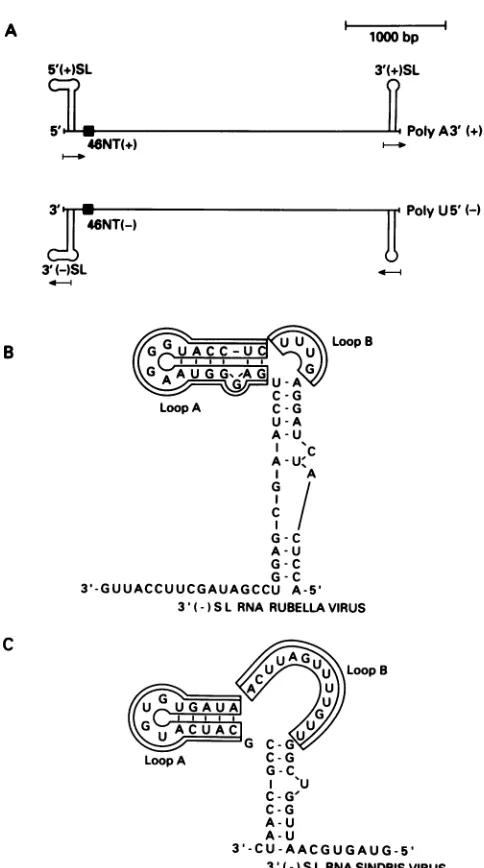
3'(- ) SLRNA SINDBIS VIRUS FIG. 1. (A) Schematic representation of rubella virus positive (+)- andnegative (-)-strand RNAs; 5' (+) SL, 5'-end SLstructure
ofthepositivestrand; 3' (+)SL,3'-endSL structure of thepositive strand; 3' (-) SL, 3'-end SL structure of the negative strand; 46NT(+) or (-), 46-nucleotide conserved sequence of either the positive-orthenegative-strand rubella virusRNA.(B)ProposedSL
structure ofthe 3'(-) SL of rubella virusRNA. (C) Proposed SL
structureofthe3'(-) SL of Sindbis virusRNA.
theproteins whichmight interact with the 3' SL of negative-strandRNA, wesynthesized an RNA molecule encompass-ing the 64 bases of the possible SL structure (Fig. 1A). The radiolabeled RNA was used as aprobe to search for RNA-binding proteins by the use of the gel mobility shift assay and
UV cross-linking technique from both uninfected and in-fected cytosols (Fig. 2). Using lysates from infected and uninfected cells with several RNA probes as competitors, we showed that the two band-shift complexes (I and II) observed arespecificforthe 3' (-) SL RNA(Fig. 2A). When the proteins from the lysates were UV cross-linked in the presence ofthe RNA probe and resolved by SDS-PAGE, two protein bands of 56 and 79 kDa were observed from
. .-1I 11
ruiyu0 1-1
on November 10, 2019 by guest
http://jvi.asm.org/
[image:2.612.328.570.69.503.2]3'(-) RNA SL
Uninf. Inf.
f 1r I
Cl( ~ Cl).
T
i
.
T
B.
ProbeLysate
C-)
3'(-) RNA SL
I . I
Uninf. Inf.
11
--J cl)
Competitor Mr kDa
200-u cn
0..w
LCa X. - -c
VW 0 f
97--Complex I
* -Complex II
68-.*.-p56
[image:3.612.70.530.78.337.2]
43-1 2 3 4
5 6
78 9
1
2
3
45
6
78
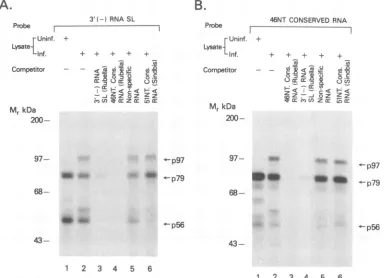
FIG. 2. (A) Gel retardationassayofthe rubella virus 3' (-) SL RNA. Lysates (20,ug)from uninfected(Uninf.) andinfected (Inf.) cells were incubatedwith 700 pmol ofRNAprobe aloneorinthepresence ofa500-foldexcessof specific[3' (-)SL] ornonspecific [IREor
poly(I* C)]RNA.(B) SDS-PAGE analysis oftheRNA-proteincomplexesformedby UVcross-linking with the3' (-)SL probe. Theamount ofcelllysateand the quantity ofthe probe is thesame asin panel A. The RNA samples used for thecompetitionareshown. Arrowspoint tocomplexesatthe indicated molecularmasses.
uninfected lysates (Fig. 2B, lane 1), whereas anadditional
protein band of 97 kDawas observed from infected lysates
(Fig. 2B, lane 5). The weak binding seen at97 kDa in the uninfected extracts was not greater than the nonspecific background (Fig. 2B, lanes 1 to 4). An excess unlabeled
RNA ofidentical sequence and polarity could specifically block thebinding activity (Fig. 2B, lanes 2 and 6), whereas unrelatedRNAs, suchas anIREorpoly(I * C), didnotblock the activity (Fig. 2B, lanes 3, 4, 7, and 8). The slight competition of p56 between the labeled rubella virus probe and the IRE probe is not significant as was evidenced by quantitating the bands by scanning densitometry. Further-more,whenmolar ratios of specific and nonspecific
compet-itorstotheprobe weredecreased, the specificitywasreadily
apparent and the IRE no longer showed any effect. The specificity of the binding activity was confirmed by the
absence of competition in the presence ofan RNA probe
upstreamof the SL structure (see Fig. 7A andB,lanes 5). To address the specificity of the interactions, we made
several variants of the SL RNA ofrubella virus (Fig. 1B).
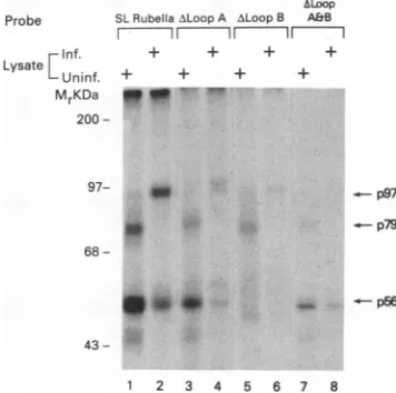
The results ofcross-linking experimentsareshowninFig.3.
Computer-based modeling (29) predictsthat the3' SL of the
negative-strand RNA canforma long stem with twoloops designated A and B (Fig. 1B). When sequences whichcan
potentially form either one of the two loops were deleted,
there was drastic reduction in the protein-binding activity from both uninfectedand infected celllysates (Fig. 3, lanes
3to8).The weakbindingof cellularproteinstothe truncated RNA probes was not greater than the nonspecific
back-ground (Fig. 3, lanes 3 to 8). However, altering the
se-quencesin the possible stemregion sothat the base-paired structure was nolongerpresent didnotchange thebinding activity (datanotshown). Theintensityof theprotein-RNA
complexes observed both in the gel shift assay and in
UV-inducedcross-linking varied fromexperimentto
exper-iment.
Similarities inbinding activities between 3' SLstructuresof
negative-strand and positive-strand rubella virus RNA. We
Probe
ALoop
SL Rubella -LoopA ALoopB A&B
fI I II 7
Inf. + +
Lysate Uninf. + +
MrKDa ^
200
-97- 4
- p97 p79
68
-_wlw -1-p56
4
43 .~
1 2 3 4 5 6 7 8
FIG. 3. Effectof mutations in the 3'(-)SLRNA of rubella virus
on the RNA-protein interaction. SDS-PAGE analysis of RNA-protein complexesfromuninfected(Uninf.)andinfected(Inf.)cell lysateswhichwereincubated withequalamountsof RNAprobes.
SLRubella,3'(-)SLRNA;Aloop A,deletion of nucleotides32 to
48; A loop B,deletion of nucleotides 49 to 52; A loops A and B,
deletion ofnucleotides 32to52. Arrowspointtocomplexesat the indicatedmolecularmasses.
A.
ProbeLysate
Competitor
C-0
_~
-~ ~'nzk ~ ~ *--9p97
*.
p79
on November 10, 2019 by guest
http://jvi.asm.org/
[image:3.612.344.522.483.660.2]A.
Probe 3'(+) RNASL
I
I~~~~~~~~~~~~~~~~~~~~
Lysate Uninf.
J
-1 -j
+
Competitor -- :q,, >:
....w_.
Inf.
-j
- t' 2n
B. Probe Lysate
Competitor
3'(-_ RNA SL Uninf. Inf.
-l r ---~ I
T +
.w....
w.
Mr kDa
*:1
_
997 -.
-68
--4
1 2 3 4 5 6 1 2 3 4 5 6
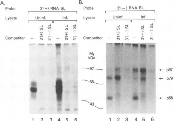
FIG. 4. Similarities in the protein-binding activitybetween the 3' (-) SL and the 3' (+) SL RNA structures. SDS-PAGEanalysis of RNA-protein complexes fromuninfected(Uninf.)and infected(Inf.)celllysateswhichwereincubatedwithequalamounts of either the3'(-) SLorthe 3' (+) SLRNAprobe.A100-foldexcessof coldRNAof the3'(-)SL RNAorthe 3'(+)SL RNAwasused in thecompetition
assay.Arrows pointtowardcomplexesattheindicatedmolecular masses.
determined whether the binding activity of the 3' SL of
negative-strand RNA [3' (-) SL] has any common compo-nent(s) with the binding activity of the 3' SL of
positive-strand RNA [3' (+) SL]thatwehavepreviouslystudied and
characterized (16). As shown in Fig. 4A,lanes 3 and 6,the
binding activity of the 3' (+) SL RNA from uninfected and
infected celllysateswas blocked byan excess of 3' (-) SL RNA.However, thereverse wasnot true.Whencold 3'(+)
SL RNAwasusedforcompetition, onlythebinding activity
of 56-kDaproteinwasblocked(Fig. 4B,lanes 2 and5).p79
andp97 bindingtothe 3'(-) SLprobewasenhanced when
3' (+)SLRNAwas addedto the reaction(Fig. 4B, lanes2 and 5). A nonspecific RNA probe did not function as a
competitor for 3' (-) RNASLcomplexformation (Fig. 2A,
lanes4, 5, 8, and 9).
3'(-)SLRNA-binding activityof rubella virus is sharedby
otheralphaviruses. Comparison ofthepossible3' SL struc-tureofnegative-strandRNAof rubella virus with the poten-tial 3' SL ofnegative-strand RNAs of alphaviruses suchas
Sindbis virus showed striking similarities in the overall secondary structure eventhough there is little homology in
thenucleotidesequence(Fig. 1C). We turnedtoUV-induced cross-linking of cellular proteins with 3' (-) SL RNAs to
examine whether SLRNAs from alphaviruses have binding activity similar to that observed with rubella virus. Using
celllysates from uninfected and infected cells, weobserved
proteins cross-linked with the 3' (-) SL RNA of Sindbis
virus RNA that were similar in molecular weight to the
proteins observedcross-linked with the rubellavirus 3' (-)
SLRNA,with the exception of the 56-kDaprotein (Fig. 5, compare lanes 9 and 13 with lanes 1 and 5). The
RNA-protein complexes withrubella virusand Sindbis virus 3' (-)
SL RNAwere blocked by their homologous and heterolo-gous3'(-) SL RNAs (Fig.5,lanes2, 3, 6, 7, 10, 11, 14, and
15). Similar results were observed when the 3' (-) SL of
eastern equine encephalitis virus RNA was used in the
competition assay (Fig. 5, lanes 4, 8, 12, and 16). The specificity of the RNA-protein interactionwith the Sindbis
virus and eastern equine encephalitis virus 3' (-) SLRNA
wasdemonstrated bylack ofcompetitionin thepresenceof
nonspecificRNAs(datanotshown). To further confirmthat similar protein-RNA complexes are recognized by both
rubella virus and Sindbis virus SL structures, we made
variants ofSindbis virus 3' (-)SL RNA. The resultsof the cross-linking experiment showed that deletion ofsequences which can potentially form loop B resulted in significant
reduction of thebinding activity of 97- and 79-kDa proteins
(Fig. 6,lanes3 and4).Theeffectwasnotsoprominentwith p56 binding; possibly,thereisalowerrateofbindinginthis
experiment. We observed variability in binding ofp56 in many otherexperimentsaswell.
Interaction of 46-nucleotide conservedsequencewith
bind-ing activityof 3' (-)SL RNA. Itwasobserved(4)thatthere
is a stretch of 46 nucleotides located 224 nucleotides from
the 5' end of the rubella virus genome (Fig. 1A)which has
50% overall homology with the 51-nucleotide conserved sequence of alphaviruses. The 51-nucleotide sequence of Sindbis virus has been implicated in viral replication (19).
We therefore asked whether the 46-nucleotide regioninthe
rubella virus RNAplaysarole in viralreplicationsimilarto
that of the3'(-)SL RNAviaRNA-protein interaction. The
results (Fig. 7) showed that the negative strand of the 46-nucleotide conserved sequence of rubella virus blocked thebinding activity of the 3'(-) SL RNA (Fig. 7A, lane4)
and that the same was true when the 3' (-) SL RNA was
used as a competitorin the binding assay for the negative
strand of the 46-nucleotide conserved sequence (Fig. 7B, lane4). However, the51-nucleotide conserved sequenceof negative-strand RNAofSindbis virus didnotcompete with
thisinteractionexceptforthe 56-kDa protein (Fig. 7Aand B,
lanes6). Quantitationofthep56bandby scanning densitom-etry revealed that the competition by the 51-nucleotide sequence from Sindbis virus is significant. To prove the specificityof this interaction, wefoundthat RNA from the
adjoining region of the46-nucleotide sequencedid not com-petewith theactivity (Fig. 7Aand B, lanes5).
4- p97
4-. p79
- p56
on November 10, 2019 by guest
http://jvi.asm.org/
[image:4.612.170.472.84.294.2]3' -) SLRNARubella Virus
rUninf. + + + +
LysateLl Inf.
3'(-)SL RNA Sindbis Virus
++ + + ++ + + +±+
Competitor - =Zz 2 = X
2c
- X: 2 zC=' U ) a : fu'
Mr kDa
200-97
-
68-a a
a
-- p97
-- p79
p56a
p56b 43
1 2 3 4 5 6 7 8 9 10 11 12 13 14 15 16
FIG. 5. Similarities in theprotein-binding activity betweenthe 3' (-)SLRNAsof rubella virus andalphaviruses(Sindbisvirus andeastern
equineencephalitis virus[EEV]).SDS-PAGEanalysis of RNA-protein complexes formed inthepresence orabsence of unlabeled 3' (-) SL
RNAs of rubella virus, Sindbisvirus, andeastern equine encephalitis virus with equal amounts of probes. The molecularmasses of the
complexesareshown. Uninf., uninfected; Inf.,infected.
DISCUSSION
The rubella virus genomic RNA is predicted to form
potentially stable SLstructuresatboth the5'andthe 3'ends
(4, 6, 28).Thesesecondarystructuresin theRNAhave been implicated in viral replication (4). Similar structures inthe Sindbisvirus RNA havebeen showntobe important for the replication of Sindbis virus and its defective interfering particles (10, 12, 18, 19).
Previously,wehaveshown that host cellproteins interact
specificallywiththe possible 3'(+) SL of the genomic RNA
of rubella virus and that the increase in protein-binding
3'l(-SSL
Probe 3l(--)SL hLoop B
RNA RNA
Irif. t +
Lysate ''
Uninf. +
Mr kDa 200
97 _ -p97
68
----p56
43
1 2 3 4
FIG. 6. Effect ofmutationsin the3'(-)SL RNA of Sindbis virus
on the protein-RNA interaction. SDS-PAGE analysis of RNA-protein complexes from uninfected (Uninf.)andinfected(Inf.)cell lysates.Aloop B, nucleotides25to37weredeletedinthe 3'(-)SL RNA.Arrowsdepict the molecularmassesof thecomplexes.
activity coincided with the appearance of negative-strand
RNA synthesis (16). In this study, we demonstrated the
existenceof anothergroupofcellular proteins which interact
specifically with the potential 3'-end SL structure of the negative-strand rubella virus RNA [3' (-)SLI. The interac-tionofthese proteins is specificasdemonstrated by the lack
ofinhibitionby unrelated RNA. Mutationalanalysis of the 3' (-)SLstructureshowedthatthesequenceswhichcanform
two potential loop structures are involved in the protein-binding activity (Fig. 3).Thecellular protein interaction with
the 3' (-) SL RNA suggests an involvement of these
proteinsin the replication process. Recently, we wereable to demonstrate the initiation ofnegative-strand RNA syn-thesis fromachloramphenicol acetyltransferase RNAwhich
hadrubella virus 5'- and 3'-endpossible SL structures(14).
The initiation of negative-strand RNA synthesis was
achievedonlyafterviral infection(14).Mutations in the first 44nucleotidesatthe5' end of theSindbis virusRNA,which
are capable offorming an SL structure, showed that this regionhasaroletoplayinviralreplication sincesomeofthe
mutationswereeitherlethalorresulted inpoorviralgrowth
in different cell types (19). This led these investigators to
suggestthat there is an interaction of host factors with the
Sindbis virus SL structures.
While analyzing the interaction of the cellular proteins
with thepossible3'(-)SLstructure,werealized thatoneof theproteins, i.e.,the 56-kDaprotein,hasamolecularmass
(61'kDa) approximately similar to that of the 3' (+) SL RNA'-binding protein from uninfected cells(16). We there-fore reasoned that the two proteins may be the same.
Competition analysisofthebindingactivitybetweenthe two SLstructuresshowedthat indeedthetwoproteins recognize
similarstructures.Recognitionof the 56-kDaprotein byboth
the positive-strand and negative-strand SL RNAs suggests
that thisproteinisinvolvedin twoprocesses,onetoinitiate
the negative-strand synthesis and the other to initiate the
positive-strand synthesis. Occurrence ofmultiple
nonidenti-cal RNA-binding domains in a single protein has been
Probe
on November 10, 2019 by guest
http://jvi.asm.org/
[image:5.612.155.455.76.296.2] [image:5.612.108.240.495.686.2]A.
B.
3'(-) RNASL
+ + + + +
Z&8n c3Z o CC S- et C< : <S -_
9Z
zoz Zz4-p97
_ _ 79
68-p56
43-Probe 46NTCONSERVEDRNA FUninf. +
Lysate-Inf. + + + + +
Competitor - - ui
cc
n: cXuza: Urcr:
Mr kDa
200-97- .8,
* p97
aim_la
4a
,p79
68--p56
43-1 2 3 4 5 6
1 2 3 4 5 6
FIG. 7. Involvement ofthe 46-nucleotide conservedsequence(46NT. Cons.)in the 3'(-)SLRNA-proteininteraction. (A)SDS-PAGE analysis ofRNA-protein complexesfrom uninfected(Uninf.)and infected(Inf.)celllysateswith the 3'(-)SL RNA. Thecompetitionassay
wasdone in thepresenceofa500-foldexcessofspecificandnonspecific RNAs.(B) SDS-PAGEanalysisofRNA-protein complexesfrom uninfected and infected celllysateswith thenegativestrand of the 46-nucleotide conservedsequence. Thedesignationof eachlane isindicated atthetop.Arrowspoint toward the molecularmassesof thecomplexes. 51NT Cons., 51-nucleotideconserved sequence.
observed with several RNA-binding proteins, and these
distinct domains have been postulated to have several
dif-ferent functions (2). The discrepancy in the molecular
masses of the 3' (-) SL RNA-binding protein (56 kDa)
reported in thepresentstudy and the 3' (+) SLRNA-binding protein (61 kDa) reported earlier (16) will bethe subject of furtherstudy.
Basedonthe observation (4) that the 5' (+) SLs ofrubella
virus andSindbis virus have similar stable RNA secondary
structures, wereasoned that the 3' (-) SL RNA of Sindbis
virusbinds to proteins similarto those which interact with the3' (-) SL of rubella virus RNA. Indeed, thetwopotential negative-strand SL structures compete with each other for thesame cellular binding proteins, and the similarity in the
binding activity is also extended to another member of the alphavirusfamily,easternequine encephalitis virus. Reduc-tion in the binding activities with the mutantforms of both
theSindbis virus and rubella virus 3' (-) SL RNAstructures
furthersuggeststhat the binding interaction between thetwo
viruses involves similar structures. However, it remains to
be seen whether exchange of the 5' (+) SL structures
between the Sindbis virus and the rubella virus RNAs can
allow initiation of replication from either of the viral
ge-nomes.
Intherubellavirusgenomic RNA, within 224 nucleotides fromthe 5'end, there isastretch of 46 nucleotides whichhas
50% overall similarity with the Sindbis virus 51-nucleotide conserved sequence (4). This region of the Sindbis virus, which is conserved among all alphaviruses, is located 155
nucleotides from the 5' end and is capable of forming double-hairpin structures in a computer-based model (22).
The function of this element is not clear, but it has been
implicated in viral replication (19). Secondary structure
analysisof the first 250 bases of Sindbis virus RNAbythese
investigators (19) predictedthat the 51-nucleotide conserved
regionis inclose proximity tothe 5'-terminal SL structure.
We reasoned that the 46-nucleotide conservedregion inthe rubella virusRNA,which hassimilaritytothe 51-nucleotide conservedsequenceof Sindbisvirus,mayhaveasimilar role
to play in rubella virus replication. We showed that the
proteins which interact with the 3' (-) SL RNA are also
recognized by the complementary strand ofthe 46-nucleo-tide element and vice versa (Fig. 7A and B), thereby
implying that common proteins are interacting with two elements of the rubella virus RNA. This interaction is
specific for the rubella virus RNA and is different from Sindbis virus RNA (Fig. 7). The consequences of this and other RNA-protein interactions in viral replication are not
clearatpresentandneed furtherstudying.
Inconclusion, we showedthatthepossible SLstructures at the 5' and 3' ends of rubella virus RNA, which are
necessary for viral replication, bind specifically tocellular proteins. There is a common protein which interacts with both elements.Similarityinthebinding activity ofthe3'-end
elements of the negative-strand RNA of rubella virus and
alphaviruses implies that common factors are involved in
theirrespective replication processes. Probe
rUninf. Lysate
CInt. Compelitor
Mr kDa
200-
on November 10, 2019 by guest
http://jvi.asm.org/
[image:6.612.129.514.85.363.2]ACKNOWLEDGMENTS
We thank Charles Rice(Washington University), Neil Goldman,
andYuan DeVries for criticalreading of the original manuscript. We are most grateful to Rick Klausner for the stimulating discussions
during this study. We also thank John Ewell for synthesizing the
oligonucleotidesand David Haile for the generous gift of T7 RNA
polymeraseand help in scanning densitometry. REFERENCES
1. Andino, R., G. E. Rieckhof, and D. Baltimore. 1990. A
func-tional ribonucleoprotein complex forms around the 5' end of
poliovirusRNA. Cell 63:369-380.
2. Bandziuilis, R. J., M. S. Swanson, and G. Dreyfuss. 1989. RNA-binding proteins as developmental regulators. Genes Dev. 3:431-437.
3. Clark, D. M., T. W. Loo,I.Hui, P. Chong, and S. Gillam. 1987. Nucleotide sequence and in vitro expression of rubella virus 24S
subgenomic messenger RNA encoding the structural proteins
El,E2 andC. Nucleic Acids Res. 15:3041-3057.
4. Dominguez, G., C.-Y. Wang, and T. K. Frey. 1990. Sequence of
the genome RNA of rubella virus: evidence for genetic rear-rangementduring togavirusevolution. Virology 177:225-238. 5. Frey, T. K., and L. D. Marr. 1988. Sequence of the region
coding for virion protein C andE2and thecarboxy terminusof the nonstructural proteins of rubella virus: comparison with alphaviruses. Gene62:85-99.
6. Frey, T. K., L. D. Marr, M. L. Hemphill, and G. Dominguez.
1986. Molecular cloning and sequence of the region of the
rubella virus genome coding for glycoprotein El. Virology
154:228-232.
7. Gatignol,A., A. Buckler-White, B. Berkhout, and K.-T. Jeang.
1991. Characterization ofahuman TAR RNA binding protein
thatactivates the HIV-1LTR.Science 251:1597-1660. 8. Hentze, M. W., T. A. Rouault, J. B. Harford, and R. D.
Klausner. 1989.Oxidation-reductionas amolecularmechanism ofaregulatoryRNA-protein interaction. Science244:357-359. 9. Ho-Terry, L., and A. Cohen. 1982.Rubellavirionpolypeptides: characterizationbypolyacrylamide gel electrophoresis, isoelec-tric focusing andpeptide mapping.Arch. Virol.72:47-54.
10. Kuhn, R. J., Z. Hong, and J. H. Strauss. 1990. Mutagenesis of
the 3' nontranslated region of Sindbis virus RNA. J. Virol.
64:1465-1476.
11. Levis, R., H. Haung, and S. Schlesinger. 1987. Engineered defective interfering RNAs of Sindbis virus express bacterial chloramphenicol acetyltransferase in avian cells. Proc. Natl. Acad. Sci. USA 84:4811-4815.
12. Levis, R., B. G. Weiss, M. Tsiang, H. V. Haung, and S.
Schlesinger. 1986. Deletionmapping of Sindbis virus DI RNAs
derived fromcDNAs defines thesequencesessential for
repli-cation andpackaging. Cell44:137-145.
13. Milligan, J. F., D. R. Groebe, G. W. Witherell, and 0. C. Uhlenbeck. 1987. Oligoribonucleotide synthesis usingT7RNA
polymerase andsyntheticDNAtemplates. Nucleic Acids Res.
15:8783-8798.
14. Nakhasi, H. L., X.-Q. Cao, T. A. Rouault, and R. D.Klausner. Unpublished data.
15. Nakhasi, H. L., B. C. Meyer, and T.-Y. Liu. 1986. Rubella virus cDNA: sequence and expression of El envelope protein. J. Biol. Chem. 261:16616-16621.
16. Nakhasi, H. L., T. A.Rouault, D. J. Haile, T.-Y. Liu, and R. D. Klausner. 1990. Specific high-affinity binding of host cell pro-teins to the 3' region of rubellavirus RNA. New Biol. 2:255-264.
17. Nakhasi, H. L., D. Thomas, D. Zheng, and T.-Y. Liu. 1989. Nucleotide sequence of capsid, E2 and El protein genes of
rubella virus vaccine strain RA27/3. Nucleic Acids Res. 17: 4393-4394.
18. Niesters, H. G. M., and J. H.Strauss. 1990. Defined mutation in the 5' end nontranslated sequence of Sindbis virus RNA. J. Virol. 64:4162-4168.
19. Niesters, H. G. M., and J. H.Strauss. 1990.Mutagenesis of the
conserved 51-nucleotide region of Sindbis virus. J. Virol. 64: 1639-1647.
20. Oker-Blom, C., I. Ulmanen, L.Kaariainen, and R. F. Pettersson. 1984. Rubella virus 40S genomicRNA specifies a24S subge-nomic mRNA thatcodes foraprecursor to structuralproteins.
J. Virol.49:403-408.
21. Rouault, T. A., M. W. Hentze, S. W. Caughman, J. B.Harford, and R. D. Klausner.1988. Binding ofacytosolicproteintothe
iron-responsive element of human ferritin messenger RNA.
Science241:1207-1210.
22. Strauss, E. G., and J. H. Strauss. 1986.Structure andreplication ofthealphavirusgenome, p.35-90. InS.Schlesingerand M. J.
Schlesinger (ed.), The togaviridae and flaviviridae. Plenum
PublishingCorp.,New York.
23. Strauss, J. H., and E. G. Strauss. 1988. Evolution of RNA viruses. Annu. Rev.Microbiol.42:657-683.
24. Takkinen,K., G.Vidgren, J.E.Ulf-Hellman, N. Kalkkinen,C. Wrenstedt, and R. F. Pettersson. 1988. Nucleotide sequenceof
therubellaviruscapsidproteingene revealsanunusuallyhigh
G/Ccontent.J.Gen. Virol. 69:603-612.
25. Tsiang, M., B. G.Weiss, and S.Schlesinger. 1988. Effects of5'
terminal modification on the biological activity of defective interferingRNAsof Sindbis virus. J.Virol. 62:47-53.
26. Vidgren, G., K. Takkinen, N. Kalkkinen, L. Kaarianen, and R. F. Pettersson. 1987.Nucleotidesequenceofthe genescoding
forthemembraneglycoproteinsEl and E2 of rubella virus. J.
Gen. Virol. 68:2347-2357.
27. Waxham, M. N., andJ. S. Wolinsky. 1983. Immunochemical
identification of rubellavirushemagglutinin. Virology 126:194-203.
28. Zheng, D., L. Dickens, T.-Y. Liu, and H. L. Nakhasi. 1989. Nucleotide sequenceofthe24Ssubgenomicmessenger RNA of avaccine strain (HPV77) ofrubellavirus: comparison with a wild typestrain(M33). Gene82:343-349.
29. Zuker, M.,and P. Stiegler. 1981. Optimal computerfoldingof large RNA sequences using thermodynamics and auxiliary
information. Nucleic Acids Res.9:133-148.
![FIG.5.complexesRNAsequine Similarities in the protein-binding activity between the 3' (-) SL RNAs of rubella virus and alphaviruses (Sindbis virus and eastern encephalitis virus [EEV])](https://thumb-us.123doks.com/thumbv2/123dok_us/1312389.84599/5.612.108.240.495.686/complexesrnasequine-similarities-protein-binding-activity-alphaviruses-sindbis-encephalitis.webp)