Monoclonal Antibody 2424 with Neutralizing Activity against HIV-1
JRFL
Rajnish Kumar,a,bRuimin Pan,cChitra Upadhyay,a,bLuzia Mayr,a,b*Sandra Cohen,a,bXiao-Hong Wang,a,b
Preetha Balasubramanian,a,bArthur Nádas,dMichael S. Seaman,eSusan Zolla-Pazner,a,bMiroslaw K. Gorny,a,bXiang-Peng Kong,c Catarina E. Hioea,b
Veterans Affairs New York Harbor Healthcare System, New York, New York, USAa; Departments of Pathology,bBiochemistry and Molecular Pharmacology,cand
Environment Medicine,dNYU School of Medicine, New York, New York, USA; Division of Viral Pathogenesis, Beth Israel Deaconess Medical Center, Harvard Medical School, Boston, Massachusetts, USAe
ABSTRACT
The V3 region of HIV-1 gp120 is important for virus-coreceptor interaction and highly immunogenic. Although most anti-V3 antibod-ies neutralize only the sensitive tier 1 viruses, anti-V3 antibodantibod-ies effective against the more resistant viruses exist, and a better under-standing of these antibodies and their epitopes would be beneficial for the development of novel vaccine immunogens against HIV. The HIV-1 isolate JRFL with its cryptic V3 is resistant to most V3-specific monoclonal antibodies (MAbs). However, the V3 MAb 2424 achieves 100% neutralization against JRFL. 2424 is encoded by IGHV3-53 and IGLV2-28 genes, a pairing rarely used by the other V3 MAbs. 2424 also has distinct binding and neutralization profiles. Studies of 2424-mediated neutralization of JRFL produced with a mannosidase inhibitor further revealed that its neutralizing activity is unaffected by the glycan composition of the virus envelope. To understand the distinct activity of 2424, we determined the crystal structure of 2424 Fab in complex with a JRFL V3 peptide and showed that the 2424 epitope is located at the tip of the V3 crown (307IHIGPGRAFYT319), dominated by interactions with HisP308,
ProP313, and ArgP315. The binding mode of 2424 is similar to that of the well-characterized MAb 447-52D, although 2424 is more side
chain dependent. The 2424 epitope is focused on the very apex of V3, away from nearby glycans, facilitating antibody access. This fea-ture distinguishes the 2424 epitope from the other V3 crown epitopes and indicates that the tip of V3 is a potential site to target and incorporate into HIV vaccine immunogens.
IMPORTANCE
HIV/AIDS vaccines are crucial for controlling the HIV epidemics that continue to afflict millions of people worldwide. However, HIV vaccine development has been hampered by significant scientific challenges, one of which is the inability of HIV vaccine candidates evaluated thus far to elicit production of potent and broadly neutralizing antibodies. The V3 loop is one of the few immunogenic targets on the virus envelope glycoprotein that can induce neutralizing antibodies, but in many viruses, parts of V3 are inaccessible for antibody recognition. This study examined a V3-specific monoclonal antibody that can completely neu-tralize HIV-1 JRFL, a virus isolate resistant to most V3 antibodies. Our data reveal that this antibody recognizes the most distal tip of V3, which is not as occluded as other parts of V3. Hence, the epitope of 2424 is in one of the vulnerable sites on the virus that may be exploited in designing HIV vaccine immunogens.
T
he HIV-1 envelope glycoprotein (Env) is the only virus-en-coded protein expressed on the surface of the virus and is the sole target for virus-neutralizing antibodies (Abs). On the virion surface, the HIV Env spike is a compact heterodimeric trimer made up of gp120 and gp41 subunits (1–3). The surface gp120 subunit is responsible for interacting with the host cell through binding to CD4 and the coreceptor, the chemokine receptor CCR5 or CXCR4 (4–7). On the basis of primary amino acid se-quences, gp120 is divided into five conserved regions (C1 to C5), which are interspersed with five variable regions (V1 to V5) (8). The CD4-binding site and the chemokine receptor-binding site are both highly conformational and discontinuous. The chemo-kine receptor binding site in particular is composed of the invari-ant2 and3 strands of the V1V2 stem region,20 and21 strands in the conserved C4 region, and the third variable (V3) region of gp120 (3,9). Vulnerable sites on the HIV Env have been identified based on their recognition by broadly neutralizing hu-man monoclonal antibodies (MAbs). On gp120, these epitope sites include the CD4-binding site (10, 11), a cluster of glycansReceived15 May 2015Accepted6 June 2015
Accepted manuscript posted online24 June 2015
CitationKumar R, Pan R, Upadhyay C, Mayr L, Cohen S, Wang X-H,
Balasubramanian P, Nádas A, Seaman MS, Zolla-Pazner S, Gorny MK, Kong X-P, Hioe CE. 2015. Functional and structural characterization of human V3-specific monoclonal antibody 2424 with neutralizing activity against HIV-1 JRFL. J Virol 89:9090 –9102.doi:10.1128/JVI.01280-15.
Editor:F. Kirchhoff
Address correspondence to Xiang-Peng Kong, [email protected], or Catarina E. Hioe, [email protected].
*Present address: Luzia Mayr, INSERM U1109, Université de Strasbourg, Strasbourg, France.
R.K. and R.P. contributed equally to this work.
Copyright © 2015, American Society for Microbiology. All Rights Reserved.
doi:10.1128/JVI.01280-15
on November 7, 2019 by guest
http://jvi.asm.org/
recognized by MAb 2G12 (12–14), and the glycan-bearing regions in V1V2 and V3 (15–17). These Abs recognize structurally com-plex epitopes and display an unusual VH domain exchange or an extreme level of somatic hypermutations and/or CDR3 lengths; thus, inducing such Abs by vaccination is not a simple feat.
The crown of the V3 loop, on the other hand, is highly immu-nogenic; antibodies to the V3 crown are induced in the vast ma-jority of human subjects following HIV infection or after vaccina-tion with HIV gp120 vaccines (18–23). The importance of V3 as a vaccine immunogen is further established by the fact that V3 is essential for HIV-1 infectivity (24,25) and that antibodies binding to V3 can block the virus infection (26–31) Most V3-specific MAbs isolated from HIV-1-infected individuals are also highly cross-reactive, recognizing gp120 proteins from viruses of differ-ent HIV-1 subtypes. However, these V3-specific MAbs neutralize mainly the relatively sensitive tier 1 viruses and are ineffective against tier 2 and tier 3 isolates (2,26,32). The failure of anti-V3 MAbs to neutralize tier 2 and tier 3 viruses in the face of recogni-tion of their corresponding soluble gp120 proteins indicates that V3 epitopes are present but inaccessible on the functional Env spikes on the virions (33, 34). Nonetheless, there are distinct epitopes on the V3 loop (17,35). Many are occluded, but some may be more exposed. Immunogenic V3 epitopes that are acces-sible on the virus Env would be valuable new targets for HIV vaccine development.
In this study, we present the crystal structure of a V3 epitope recognized by human MAb 2424, which is distinct from the other V3 MAbs in its capacity to neutralize JRFL, a relatively resistant 1 isolate. MAb 2424 was isolated from a chronically HIV-infected subject living in New York City by a cellular method in which peripheral blood mononuclear cells (PBMCs) were trans-formed by Epstein-Barr virus, fused with heteromyeloma cells (36,37), and selected based on enzyme-linked immunosorbent assay (ELISA) reactivity with the V3 (consensus B)-MLV gp70 fusion protein. Unlike the other V3 MAbs, 2424 preferentially binds to V3 of subtype B viruses. Moreover, the 2424 gene usage is unusual among human anti-V3 MAbs; 2424 is IgG1 with a kappa light chain and is encoded by IGHV3-53 and IGLV2-28 genes (38). In agreement with functional studies, the crystallographic structure reveals a distinct epitope at the tip of the V3 crown that is not as influenced by glycans as the other V3 epitopes. Although it is unknown if potent 2424-like Abs can be generated by vacci-nation, this V3 epitope represents another important site that may be incorporated into the immunogens for vaccines designed to elicit HIV-1-neutralizing antibodies.
MATERIALS AND METHODS
Human monoclonal antibodies (MAbs) and HIV-1 Env proteins. An-ti-V3 human MAbs 2424, 447-52D, 2219, 2557, 3074, and 3869 were obtained from an existing panel of HIV-specific MAbs. MAbs 2424, 447-52D, and 2219 were derived from the cells of HIV-1-infected subjects from the United States, while MAbs 2257, 3074, and 3869 were produced from Cameroonian individuals infected with CRF02_AG or other non-B
subtypes (22,38–41). All of these MAbs were generated by the cellular
method as described previously (36,37). The irrelevant human
anti-par-vovirus B19 MAb 1418 was used as a negative control (42). MAb b12
(provided by Dennis Burton and Carlos Barbas) and MAb 2G12 (pro-vided by Hermann Katinger) were obtained through the NIH AIDS Re-agent Program, Division of AIDS, NIAID, NIH, while MAb PG9 was provided by Wayne Koff (International AIDS Vaccine Initiative [IAVI]’s
Neutralizing Antibody Consortium) or purchased from Polymun Scien-tific.
Recombinant gp120 proteins of various subtypes (produced in 293T cells) were purchased from Immune Technology Corp., except for
gp120JRFL(produced in CHO cells by Progenics, Inc.) which was obtained
from Vaccine Research and Development Branch of Division of AIDS, NIAID, NIH. Stabilized trimeric BG505-SOSIP.664 gp140 was a generous gift from John P. Moore (Weill Cornell Medical College).
Antibody binding assay.The binding of human V3 MAbs to recom-binant gp120s was determined by ELISA. Briefly, 96-well ELISA plates
were coated with gp120 (1.0g/ml in phosphate-buffered saline [PBS]) at
4°C overnight, blocked with 3% bovine serum albumin (BSA) in PBS, and reacted for 2 h at 37°C with MAbs serially diluted in PBS with 1% BSA. Bound MAbs were detected with alkaline phosphatase-conjugated goat
anti-human IgG andp-nitrophenyl phosphate substrate.
Cell lines, plasmids, and viruses.Cells of the TZM.bl line were ob-tained through the NIH AIDS Research and Reference Reagent Program (contributed by J. Kappes and X. Wu). 293T/17 cell line was purchased from the American Type Culture Collection (ATCC).
HIV-1 Env-expressing plasmids for generating pseudoviruses were obtained from the NIH AIDS Research and Reference Reagent Program, Division of AIDS, NIAID, NIH. Pseudoviruses were produced by
cotrans-fecting 293T cells withenv,rev, and pNL4-3.Luc.R-E- or pSG3 using a
ProFection kit (Promega, Madison, WI) or polyethylenimine (PEI)
MAX40,000 (Polysciences, Warrington, PA). Glycan-modified HIV-1JRFL
was generated as described above in the presence of 25M kifunensine or
20M swainsonine (Sigma, St. Louis, MO). Supernatants were harvested
after 48 h and clarified by centrifugation and 0.22-m filtration.
Single-use aliquots were stored at⫺80°C until use.
Point mutations in JRFL Env were introduced in pCAGGS JRFL.JB gp160 using the QuikChange II XL site-directed mutagenesis kit (Strat-agene, La Jolla, CA, USA), according to the manufacturer’s instructions. All mutant constructs were sequenced to confirm the correct amino acid changes. Plasmid pCAGGS JRFL.JB gp160 was kindly supplied by the NIH Vaccine Research Center.
Neutralization assay.Virus neutralization was measured as described
previously (43). Briefly, serially diluted MAbs were incubated with the
virus for 1 h at 37°C. TZM.bl cells were then added to virus-MAb mixtures in Dulbecco’s modified Eagle medium (DMEM) containing 10% fetal bovine serum and DEAE-dextran (Sigma, St. Louis, MO). After 48 h, virus infection was determined using the Bright-Glo luciferase assay system (Promega, Madison, WI). For neutralization assays with soluble CD4 (sCD4), the virus was preincubated with recombinant human sCD4 (Pro-genics Pharmaceuticals, Tarrytown, NY) for 30 min at 37°C, before addi-tion of serially diluted MAbs. Virus infecaddi-tion was measured in TZM.bl
cells using a-galactosidase-based assay (Promega, Madison, WI).
Fab production and purification.The Fab fragment of MAb 2424 was
prepared by papain digestion as described previously (35,44). Briefly, the
IgG molecule was mixed with papain (Worthington, Lakewood, NJ) at a 20:1 molar ratio in 100 mM Tris (pH 6.8) with 1 mM cysteine hydrochlo-ride and 4 mM EDTA. The mixture was incubated for 1 h at 37°C, and the reaction was stopped with 10 mM iodoacetamide. The Fab fragment was separated from the Fc fragment and the undigested IgG by a protein A column and further purified by size exclusion chromatography. The Fab fragment was then concentrated to about 10 mg/ml for crystallization.
Crystallization, data collection, structure determination, and re-finement.The V3JRFLpeptide was synthesized by Biomatik (Wilmington, DE), dissolved in water, and mixed with Fab 2424 at a 10:1 molar ratio. Crystallization conditions were screened and optimized using the vapor diffusion hanging-drop method. Well-diffracted crystals of Fab alone were obtained with a well solution of 20% polyethylene glycol 8000, 0.1 M Tris (pH 8.5), 36% glycol, whereas those of the Fab/epitope complex were obtained in a well solution of 28% polyethylene glycol 4000, 0.17 M
Li2SO4, 0.085 M Tris (pH 8.5), 15% glycerol. X-ray diffraction data sets
were collected at the synchrotron beamline GM/CA-CAT of the Advanced Unique HIV-Neutralizing V3 MAb
on November 7, 2019 by guest
http://jvi.asm.org/
Photon Source (APS), Argonne National Laboratory. All data sets were
processed using the HKL 2000 package (45) and XDS (46), and structures
were determined by molecular replacement using a homologous Fab
structure (PDB ID3KDM) as the initial model. Cycles of refinement for
each model were carried out in COOT and Phenix (47,48). The NCS
constraints were imposed for the three complexes in the refinement for the 3.18-Å structure of the Fab 2424/V3 complex. Final structural analyses were carried out using ICM, and figures were generated using PyMOL
(Schrödinger, LLC) and ICM (49).
Protein structure accession numbers.Coordinates and structure fac-tors of Fab 2424/V3 complex and Fab 2424 have been deposited in the Protein Data Bank under accession numbers 4XMK and 4XML, respec-tively.
Nucleotide sequence accession numbers.The IGHV and IGKV se-quences of 2424 have been deposited in GenBank with accession numbers
EU794426and KP050787, respectively.
RESULTS
V3-specific MAb 2424 potently neutralizes HIV-1 JRFL isolate. Most V3-specific MAbs isolated from HIV-infected subjects or vaccine recipients are capable of neutralizing tier 1 HIV-1 isolates but not the more resistant tier 2 or tier 3 viruses, due to the cryptic nature of many V3 epitopes on the Env spikes of tier 2 and tier 3 viruses. However, one V3-specific MAb, 2424, shows a potent neutralizing activity against a relatively resistant HIV-1 isolate, JRFL, indicating the presence of an Ab-accessible neutralizing V3
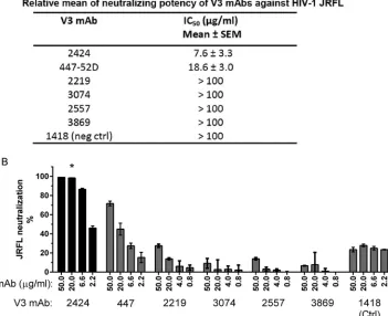
epitope on this virus. As shown inFig. 1A, among six V3 MAbs tested, 2424 was the most effective, with a 50% inhibitory concen-tration (IC50) of 7.6g/ml, whereas the remaining five had IC50s
of 18.7g/ml or⬎100g/ml. The IC50s of other V3 MAbs against
JRFL were also shown previously to range from 11 to⬎50g/ml (26). More recently, when a panel of 48 V3 MAbs was tested, 2424 was also found to be the most potent against JRFL (50). The greater potency of 2424 was further shown by its ability to achieve a neutralization plateau of 100%, a level unattainable by 447-52D and the other V3 MAbs tested (Fig. 1B; also, seeFig. 3to5).
2424 is also unlike the other V3 MAbs in terms of its Ig gene usage. This MAb has VH and VL gene pairing not used by the other V3 MAbs (50). The 2424 VH and VL genes are somatically mutated 14.4% and 5.1% from the germ line IGHV3-53 and IGLV2-28 genes, respectively. Heavy-chain complementarity-determining region 3 (CDR H3) is composed of 17 amino acids, similar in length to normal human Igs (51). Moreover, while the majority of V3 MAbs uselight chains, 2424 uses alight chain. Only 17 out of 70 human V3 MAbs with known VH and VL genes uselight chains (23,38,50,52). Together, these data suggest that 2424 is a unique MAb targeting an epitope in the V3 loop that may be more accessible or occluded dif-ferently compared to the other V3 epitopes.
2424 displays subtype B-specific binding and neutralizing activities.To better characterize MAb 2424 and its epitope, we FIG 1Neutralization potency of 2424 versus the other V3-specific MAbs against JRFL. (A) Relative neutralizing potency of 2424 and five other V3 MAbs against
JRFL. These V3 MAbs were isolated from HIV-infected individuals (22,38–40). Means and standard errors of IC50s from four independent experiments are
shown. (B) Titration curves of the six V3 MAbs against JRFL. Neutralization assay was performed with JRFL pseudovirus and TZM.bl target cells as previously
reported (43). MAbs were titrated and incubated with virus at 37°C for 1 h. TZM.bl cells were then added to the virus-MAb mixtures in DMEM containing 10%
fetal bovine serum and DEAE-dextran. Neutralization activity was assessed 48 h later using the luciferase detection system. The parvovirus-specific MAb 1418 was included as a negative control. Means and standard deviations were calculated from duplicate wells. Representative data from multiple repeat experiments are
shown. Statistical analysis was performed by two-way analysis of variance (ANOVA). *,P⬍0.001 compared to MAbs 447-52D, 2219, 3074, 2557, 3869, and 1418.
on November 7, 2019 by guest
http://jvi.asm.org/
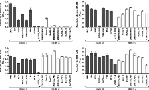
[image:3.585.116.467.78.364.2]assessed the cross-reactivity of 2424 in ELISA using recombinant gp120 proteins from HIV-1 subtype B and C (Fig. 2). Three V3 MAbs with weaker JRFL neutralization activities, 447-52D, 3074, and 2219, were also tested for comparison. 2424 displayed a gp120-binding profile distinct from those of the other three MAbs. 2424 was reactive mainly with subtype B gp120s, whereas 447-52D, 3074, and 2219 recognized most of the subtype B and sub-type C gp120s tested. 2424 also poorly reacted with recombinant gp140 proteins of subtype C (CN54), subtype A (UG37), and subtype A (BG505-SOSIP.664) (optical density at 405 nm [OD405]⫽0.28,
0.15, and 0.313, respectively; OD405of irrelevant MAb 1418⫽0.15),
although it was reactive with few V3 peptides of subtype A viruses, such as KE-Ken29 (OD405⫽1.3) and D687 (OD405⫽0.5).
The neutralizing breadth and potency of MAb 2424 were eval-uated using TZM.bl target cells and pseudoviruses expressing 15 HIV-1 Envs from tier 1 to 3 subtype B and non-B viruses (2,32) (Table 1). 2424 neutralized six viruses (BX08.16, BaL26, SS1196.1, REJO4541, JRCSF, and JRFL.JB), and three of these viruses (JRFL, JRCSF, and REJO4541) are relatively resistant to other V3 MAbs (26,50). This neutralization pattern paralleled the 2424 binding to V3 peptides of the corresponding viruses. All six viruses belong to subtype B, confirming the subtype B specificity of this MAb. In contrast, the other V3 MAbs studied here (447-52D, 2219, 2557, 3074, and 3869) neutralized viruses from both B and non-B sub-types (26, 50). These data indicate that MAb 2424 is relatively potent against JRFL, JRCSF, and REJO4541, but its binding and neutralizing breadth are more restricted than those of the other V3 MAbs.
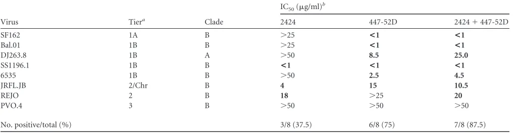
We next determined how the addition of 2424 affected virus neutralization of the other V3 MAbs. Neutralizing activities of 2424 and 447-52D were tested individually or in a 1:1 combina-tion against eight HIV-1 pseudoviruses (Table 2). The combina-tion of 2424 and 447-52D was able to neutralize seven of the vi-ruses, while on its own, 447-52D neutralized six viruses. However, the IC50s of the combined MAbs were generally higher than those
of the individual MAbs, indicative of potential competition and steric hindrance between the two MAbs.
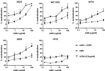
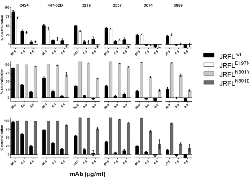
Neutralizing activity of 2424 is affected by soluble CD4 and removal of the N-glycan at position 301 but not by changes in N-glycan composition of the virus Env.To further characterize the 2424 epitope, we tested whether 2424 neutralizing activity was affected by virus pretreatment with soluble CD4. CD4 binding to the virus Env causes conformational changes that induce exposure of masked epitopes on V3 (53). A fixed concentration of soluble CD4, which on its own yielded⬃20% neutralization, was incu-bated with JRFL, prior to addition of titrated amounts of 2424. Three other V3 MAbs (447-52D, 3074, and 3869) and an irrele-vant parvovirus-specific MAb (1418) were tested for comparison. CD4 treatment augmented virus neutralization by 2424, similar to that observed with 447-52D, 3074, and 3869 (Fig. 3). The neutral-ization levels attained were significantly higher than the calculated sums of percent neutralization attained by each MAb alone and CD4. The other V3 MAbs, 2219 and 2557, were similarly affected by CD4 (54). These data indicate that CD4-induced conforma-tional changes modulate MAb accessibility of the 2424 epitope, similar to the other neutralizing V3 epitopes.
FIG 2Binding of MAb 2424 versus the other V3 MAbs to gp120 proteins of clade B and clade C HIV-1 isolates. The binding of MAbs (1g/ml) to gp120 used
at 1g/ml to coat the well surface was detected by ELISA. An irrelevant control MAb, MAb 1418, was also tested in parallel and displayed no binding to any gp120
in the panel (OD405ⱕ0.2; data not shown). Data from one representative experiment are shown; the experiments were repeated twice.
Unique HIV-Neutralizing V3 MAb
on November 7, 2019 by guest
http://jvi.asm.org/
[image:4.585.43.543.65.369.2]Next we examined the contribution of N-glycan composition in influencing the accessibility of 2424 epitope vis-a-vis the other V3 epitopes. Our previous studies demonstrated that V3 epitopes on JRFL were better exposed when the virus was produced in the presence of a mannosidase inhibitor, kifunensine or swainsonine, which enriches the viral Env glycoproteins with high mannose-type N-linked glycans (27). Hence, we evaluated neutralization of JRFL produced with or without kifunensine by 2424 and five other V3 MAbs (Fig. 4A). JRFL neutralization by 2424 was unaltered whether the virus was produced with or without kifunensine. Ki-funensine treatment also had minimal effect on 3074, but this MAb did not neutralize JRFL. In contrast, 447-52D, 2219, 2557, and 3869 were more effective against JRFL produced in the pres-ence of kifunensine than untreated JRFL. Swainsonine treatment also had no effect on JRFL neutralization by 2424, although it enhanced virus sensitivity to 447-52D (Fig. 4B). MAb 2G12, which recognizes a cluster of N-glycans bearing the high-mannose type (55), and the CD4-binding site-specific MAb b12 were tested for
comparison (Fig. 4A). 2G12 displayed higher neutralization against JRFLKifthan untreated JRFL, whereas neutralization by
b12 was changed slightly, consistent with previously published data (56). The irrelevant control MAb 1418 had no neutralizing activity against treated or untreated virus.
[image:5.585.40.552.81.274.2]Although various levels of neutralization were observed with the different V3 MAbs against untreated JRFL, the six V3 MAbs had similar activity of binding to the JRFL gp120 protein (Fig. 4C). These data demonstrated that the epitopes recognized by the six V3 MAbs are present on the monomeric JRFL Env but are masked to different degrees when expressed on the virus. Neutralization data of JRFL produced with or without a mannosidase inhibitor further indicate that, while the exposure of many V3 epitopes was modulated by the N-glycan composition of the virus Env, the 2424 epitope was not affected. Two N-glycans previously implicated as controlling V3 exposure include those at position 197 at the C-ter-minal base of V1V2 and position 301 at the N-terC-ter-minal base of V3 (57–59), and both glycans were assigned to be the complex type TABLE 1Neutralization activity of MAb 2424 against HIV-1 pseudoviruses
Virus Tiera Clade V3 sequenceb
MAb 2424
IC50
(g/ml)c
Peptide
bindingd
Signature
motife
Bx08.16 1B B CTRPNNNTRKSIHIGPGRAFYTTGDIIGDIRQAHC <0.4 ⴙⴙⴙ ⴙ
Bal.26 1B B CTRPNNNTRKSIHIGPGRAFYTTGEIIGDIRQAHC <0.4 ⴙⴙⴙ ⴙ
SS1196.1 1B B CTRPNNNTRKSIHIGPGRAFYATGGVIGDIRQAHC <0.4 ⴙⴙⴙ ⴙ
JRFL.JB 2/Chr B CTRPNQNTRKSIHIGPGRAFYTTGEIIGDIRQAHC 5.4 ⴙⴙⴙ ⴙ
JRCSF 2/Chr B CTRPSNNTRKSIHIGPGRAFYTTGEIIGDIRQAHC 6.6 ⴙⴙⴙ ⴙ
REJO4541 2 B CTRPNNNTRKSIHIAPGRAFYATGEIIGDIRKAYC 11 ⴙⴙⴙ ⴙ
Bal.01 1B B CTRPNNNTRKSINIGPGRAFYTTGEIIGDIRQAHC ⬎50 NT ⫺
SF162.LS 1A B CTRPNNNTRKSITIGPGRAFYATGDIIGDIRQAHC ⬎50 ⫾ ⫺
6535.3 1B B CTRPNNNTRKSINLGPGRAFYATGDIIGDIRQAHC ⬎50 ⫹ ⫺
YU2 2 B CTRPNNNTRKSINIGPGRALYTTGEIIGDIRQAHC ⬎50 ⫺ ⫺
MW965.26 1A C CTRPNNNTRKSVRIGPGQTFYATGAIIGDIRQAHC ⬎50 ⫺ ⫺
HO31.7 2 B CTRPSNNTRKSITIGPGRAFYTTGDIIGDIRRAHC ⬎50 NT ⫺
DJ263.8 1B A CTRPNNNTRRSVRIGPGQTFYATGDIIGDIRQAHC ⬎50 ⫺ ⫺
HO61.14 3 B CTRPNNNTRKSIPIGPGRAFYTTGDIIGDIRQAHC ⬎50 NT ⫺
ZM109F 1B C CIRPGNNTRKSIRLGPGQTFYATGDVIGDIRKAYC ⬎50 ⫺ ⫺
aChr, chronic. b
Boldface indicates the 2424 epitope signature motif.
cMAb concentration needed to reach 50% inhibition in the TZM.bl neutralization assay. IC
50s of⬍50g/ml are in bold.
d
ELISA reactivity against biotinylated V3 peptides. Reactivity with scrambled peptide was an OD405ofⱕ0.5. Average OD405s are classified as follows:⫺, 0.2 to 0.5;⫾, 0.5 to 0.75;
⫹, 0.75 to 1.5;⫹⫹, 1.5 to 2.0;⫹⫹⫹, 2.0 to 3.5. NT, not tested.
e
His308 , Pro313
, and Arg315
constitute a signature motif for the 2424 epitope.
TABLE 2Virus neutralization by 2424 and 447-52D individually or in combination
Virus Tiera Clade
IC50(g/ml)b
2424 447-52D 2424⫹447-52D
SF162 1A B ⬎25 <1 <1
Bal.01 1B B ⬎25 <1 <1
DJ263.8 1B A ⬎50 8.5 25.0
SS1196.1 1B B <1 <1 <1
6535 1B B ⬎50 2.5 4.5
JRFL.JB 2/Chr B 4 15 10.5
REJO 2 B 18 ⬎25 20
PVO.4 3 B ⬎50 ⬎50 ⬎50
No. positive/total (%) 3/8 (37.5) 6/8 (75) 7/8 (87.5)
a
Chr, chronic.
bTotal MAb concentration needed to reach 50% neutralization in the TZM.bl neutralization assay. IC
50s ofⱕ25g/ml are in bold.
on November 7, 2019 by guest
http://jvi.asm.org/
[image:5.585.40.553.575.707.2](60). However, for JRFL, there is a glycosylation site at position 301 but not at position 197. To evaluate the importance of N-gly-can at these positions, mutations were introduced into JRFL Env at position 301 (N to D or N to Y) to remove the specific glycan or at position 197 (D to N) to add a glycosylation site. The muta-tions at position 301 rendered the virus sensitive to all V3 MAbs tested, including 2424 (Fig. 5). Nonetheless, mutations that re-move the glycan at residue 301 also enhanced sensitivity of differ-ent HIV-1 isolates to antibodies against the CD4-binding site and the CD4-induced epitopes, although the MPER epitopes in the gp41 subunit were unaffected (57,59,61,62), indicating that mu-tations at this position induce global structural alterations which affect not only V3 but also other distant sites. In contrast, the D197N mutation did not affect virus neutralization by any of the six V3 MAbs tested. The contribution of residue 197 in shielding V3 epitopes is not associated with N-glycan; rather, this residue is involved in stabilizing the interprotomer interactions between V1V2 and V3 that allow V1V2 to shield V3 in the Env trimeric spike. Only specific mutations that disrupt these interactions (e.g., D197H and D197Q) are able to release V3 from the V1V2 masking (S. Zolla-Pazner, unpublished data).
The crystal structure of MAb 2424 and its epitope reveals that 2424 targets a vulnerable region at the apex of the V3 crown.To define the epitope of MAb 2424, we determined the structure of the 2424 antigen-binding fragment (Fab) cocrystallized with a 23-mer V3JRFLpeptide, NNTRKSIHIGPGRAFYTTGEIIG (residues
301 to 325 in the HxB2 numbering system [63]). The structure of
the 2424 Fab-epitope complex was solved by molecular replace-ment and refined to 3.18-Å resolution with anRwork/Rfreevalue of
22.5%/28.3% (Fig. 6AandTable 3). The crystals grew in an ortho-rhombic space group with three Fab-epitope complexes in the asymmetric unit. Since the noncrystallographic symmetry con-straints (NCS) were imposed in the refinement, only one complex is described in detail here. The residues of the light and heavy chains are numbered following the convention of Kabat and Wu (64), preceded by “L” and “H,” respectively, and the residues of the V3 peptide are preceded by a “P.” Although a 23-mer peptide was used in the crystallization, only 11 residues, with the sequence IHIGPGRAFYT (residues 307 to 319), were observed in the elec-tron densities and thus built into the final model. We also deter-mined the structure of Fab 2424 alone and refined it to a 2.68-Å resolution (Rwork/Rfree⫽20.0%/28.5%) (Table 3). The Fab
struc-ture is very similar to that in the Fab-epitope complex, with only one residue, PheH58, in the antigen-binding site of the Fab alone having a different side chain orientation.
The Fab/V3 epitope complex revealed that 2424 binds the very apex of the V3 crown (Fig. 6and7), forming direct contacts with 9 residues, P308HIGPGRAFYP318. However, IleP309 and PheP317 have only very little backbone contact (⬃6.5 Å2and 4.3 Å2,
respec-tively) with the antibody, and their side chains point away from the antigen-binding surface of 2424. The antibody binds the epitope using the ladle mode, one of the two binding modes typ-ically used by anti-V3 crown MAbs (65). In this binding mode, the arch of the V3 crown (four residues at the apex, typically GPGR for FIG 3Effects of CD4 engagement on virus neutralization by 2424 versus the other V3 MAbs. Neutralization of JRFL pseudovirus by V3 MAbs 2424, 447-52D,
3074, and 3869 was evaluated after treatment with or without sCD4. Virus was pretreated with 2.5g/ml of sCD4 for 30 min at 37°C or left untreated and then
incubated with serially diluted MAb for 1 h at 37°C. TZM.bl cells were added to virus-MAb mixtures in DMEM containing 10% fetal bovine serum and
DEAE-dextran. After 48 h, virus infection was measured based on-galactosidase activity. Means and standard errors from two independent experiments are
shown. *,P⬍0.01 based on the two-way ANOVA to show a synergistic difference above the calculated sum of percent neutralization attained by MAb and sCD4
on their own.
Unique HIV-Neutralizing V3 MAb
on November 7, 2019 by guest
http://jvi.asm.org/
[image:6.585.86.496.65.340.2]subtype B or GPGQ for subtype C) points directly toward the antigen-binding site (Fig. 6A). This is different from the other binding mode, called the cradle mode, in which the V3 crown lies sideways in the long antigen-binding groove of the antibody (35, 66). Like other anti-V3 crown MAbs, the antigen-binding site is negatively charged, so that it can accommodate the positively charged V3 crown. Interestingly, residue P315-P317 forms a small 310helical turn, which is rarely observed in V3 crown structures
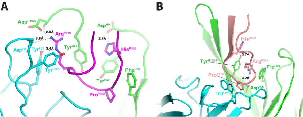
despite the early prediction from a sequence analysis that the C-terminal part of V3 has a propensity to be helical (67). All CDR loops participate in the antigen-antibody interactions, and they form a relatively small but deep binding site basin. There are three specific binding pockets that interact with the side chains of HisP308, ProP313, and ArgP315, respectively (Fig. 7A). The contact
areas of these three residues (240 Å2) account for over 60% of the contact areas of all the epitope residues with 2424 (383 Å2). In
agreement with these structural data, the presence or absence of these three specific residues dictates whether 2424 binds strongly or poorly to V3 from diverse HIV-1 isolates (Table 1). Thus, the side chains of these three residues dominate the antigen-antibody interaction for 2424, forming the signature motif for 2424 recog-nition. This is consistent with the 2424 specificity for subtype B viruses, which often have HisP308and ArgP315.
Comparison of the antigen-antibody interactions of 2424 with 447-52D.We compared structurally the antigen-antibody in-teraction of 2424 with that of 447-52D, a well-characterized anti-V3 crown MAb. The most obvious similarity between these two MAbs is their mode of antigen binding: both use the ladle mode, but 447-52D has a 22-amino-acid CDR H3 that serves as the handle of the ladle (Fig. 6B). They also have some similarities in the details of their anti-gen binding. For example, the side chain ArgP315of the epitope in
2424 forms a salt bridge with a heavy-chain aspartic acid, and it is also surrounded by several Tyr residues from both heavy and light chains (Fig. 7A). Similarly, the side chain ArgP315in 447-52D also forms a salt bridge with a heavy-chain aspartic acid, and it is also sandwiched between the side chains of two aromatic amino acids of the antibody (Fig. 7B). In addition, ProP313of V3 in both MAbs is placed in a
hydrophobic environment with its side chain stacked against the side chain of either a Phe (2424) or a Trp (447-52D). However, the two MAbs also have several differences in their antigen-antibody interac-tions. First, the epitope of 2424 is two residues shorter in the N ter-minus than that of 447-52D; this focuses 2424 binding to the very apex of the V3 crown. Second, 2424 is highly side chain specific, bury-ing the side chains of HisP308and ArgP315in deep pockets in the anti-gen-binding site, while 447-52D utilizes a beta-sheet machain in-teraction with the N terminus of the V3 crown, rendering 447-52D FIG 4Neutralizing activities of MAb 2424 versus the other V3 MAbs against JRFL produced in the presence or absence of a mannosidase inhibitor. (A) HIV-1
JRFL pseudoviruses were produced in transfected 293T cells in the absence or presence of 25M kifunensine (JRFLKif) and tested for neutralization by V3 MAbs
(2424, 447-52D, 2219, 2557, 3869, and 3074), MAb 2G12, CD4bs MAb b12, or an irrelevant MAb, MAb 1418. (B) HIV-1 JRFL pseudoviruses were produced in
293T cells with or without 20M swainsonine (JRFLSwain) and tested for neutralization by V3 MAbs 2424 and 447-52D. Serially diluted MAbs were incubated
with virus at 37°C for 1 h. TZM.bl cells were then added to the virus-MAb mixtures in complete DMEM containing DEAE-dextran. Neutralization activity was assessed after 48 h. Results with 2424 and 447-52D were averages from duplicate wells, and data from one representative experiment are shown. Results with other
MAbs are shown as averages from two to three experiments.Pvalues were determined by the two-way ANOVA. (C) V3 MAb reactivity with gp120 JRFL was
assessed in ELISA. Recombinant gp120 protein (1g/ml) was coated on 96-well plates and reacted with serially diluted V3 MAbs. Data from a representative
experiment with averages and standard deviations from duplicate wells are shown.
on November 7, 2019 by guest
http://jvi.asm.org/
[image:7.585.47.538.68.361.2]more broadly reactive. The limited contacts between 2424 and the V3 crown and the 2424 epitope being farther away from nearby glycans may explain how its binding is less affected by the glycan composition of gp120 (Fig. 4). However, its side chain specificity is likely to limit the neutralization breadth of 2424.
DISCUSSION
This study presents the crystallographic structure of MAb 2424 in complex with its V3 epitope, elucidating the distinctive capacity of this V3 MAb to neutralize HIV-1 JRFL, a virus isolate resistant to many other V3 MAbs (26,52). The presence of such a MAb indi-cates that, although epitopes on the V3 crown region of tier 2 or tier 3 viruses are often occluded, some are accessible to Abs. The broadly neutralizing MAb PGT 128 that is highly potent against tier 2 and tier 3 viruses also binds to a complex epitope consisting in part of a short beta-strand segment of the V3 loop and N-linked glycans at the V3 base (17). 2424 uses a different approach to access its epitope in that it targets the very tip of the V3 crown, away from the many N-glycans shrouding the HIV Env surface. In support of this idea, we demonstrate that treating the virus with a mannosidase inhibitor, which changes the sugar composition of N-glycans on the virus Env, did not affect virus neutralization by
2424. On the other hand, most other V3 MAbs were more potent against viruses bearing Env with homogenously high mannose-type glycans, suggesting the important contribution of complex-type N-glycans in masking the epitopes of these V3 MAbs, either by direct steric hindrance or by facilitating formation of more compact Env trimers. All together, the data presented herein in-dicate that, unlike other V3 epitopes, the 2424 epitope is devoid of and is minimally masked by N-glycans; thus, it constitutes a dis-tinct target which would complement the other neutralizing epitopes on the virus Env.
Another distinctive feature of MAb 2424 is its ability to mediate 100% neutralization against JRFL. Although the mechanistic ex-planations for this activity remain unknown, the result suggests the possibility that MAb 2424 can attain binding saturation of the virus Env due to the relative distance of its epitope from the sur-rounding glycans. Nonetheless, the neutralizing activity of MAb 2424 was further enhanced after CD4 binding and also affected by the removal of N-glycan from residue 301 at the base of V3, indi-cating that the 2424 epitope is not completely exposed on the prefusion Env spikes. The binding affinity of MAb 2424 is not noticeably different from that of the other V3 MAbs when tested with soluble gp120 monomer. On the Env trimers, the V3 distal tip FIG 5Neutralization activities of 2424 versus the other V3 MAbs against HIV-1 pseudoviruses expressing wild-type or mutant JRFL Envs. HIV-1 JRFL pseudoviruses bearing wild-type or mutated Envs were produced in transfected 293T cells and incubated by titrated V3 MAbs (2424, 447-52D, 2219, 2557, 3074, and 3869) for 1 h at 37°C. TZM.bl cells were then added to the virus-MAb mixtures in complete DMEM containing DEAE-dextran. Neutralization activity was assessed after 48 h. Results are means and standard errors from two experiments.
Unique HIV-Neutralizing V3 MAb
on November 7, 2019 by guest
http://jvi.asm.org/
[image:8.585.40.541.61.417.2]may be constrained to preferentially adopt the conformation rec-ognized by 2424 and/or may be better oriented for binding by 2424 compared to the other V3 crown MAbs.
Together with the current 2424-V3 structure, many crystal
structures of human V3 MAbs in complex with their cognate V3 epitopes have been resolved (35,68–70). They reveal three major modes of Ab-epitope interactions, designated ladle, cradle, and Janus. The ladle mode is exemplified by 447-52D, which grasps V3 by the long CDR H3 handle of the ladle. 2424 belongs to this same category but is distinct from 447-52D in that it interacts only with the tip of the V3 crown with the bowl portion of the ladle. The potency of 2424 to neutralize relatively resistant viruses such as JRFL and REJO and its indifference to mannosidase inhibitors imply that the very tip of V3 targeted by 2424 is minimally shielded by glycans in these viruses. In contrast, the cradle mode is represented by MAbs 2219 and 2557, whose antigen-binding sites are shaped like a cradle, and the V3 crown lies in it. MAbs in this category are effective only when the virus Env is enriched with the high-mannose-type N-glycans, indicating the critical contribu-tion of N-glycans in shielding these MAbs’ epitopes. In addicontribu-tion, there is a third binding mode, the Janus mode, which is character-ized by MAbs 3074 and 268; they approach V3 from two opposing sides to contact, respectively, the conserved hydrophobic core and the strain-specific hydrophilic face, in the middle segment of the V3 crown (35). MAb 3074 neutralizes JRFL poorly whether the virus is produced with or without a mannosidase inhibitor, indi-cating that this MAb cannot reach into the hydrophobic core of V3 when presented on the JRFL Env spike and that this conserved site is concealed by factors other than N-glycans.
The distinct epitope and neutralization profile of MAb 2424 correspond to its Ig constant and variable gene usage. MAb 2424 has a kappa light chain and is encoded by IGHV3-53 and IGLV2-28 genes. Among 51 human anti-V3 MAbs evaluated by Gorny et al. (38), the majority (76.5%) use lambda light chains. Only 12 (23.5%) utilize the IGHV3 gene family, and these IGHV3-encoded V3 MAbs are paired preferentially with kappa light chains. However, although V3 Abs are skewed away from using FIG 6Structural comparison of MAbs 2424 and 447-52D. (A) Ribbon
repre-sentation of the Fab 2424/V3JRFLcomplex. The epitope and the light and heavy
chains are magenta, cyan, and green, respectively. For simplicity, only the Fv
region is shown. (B) Fab 447-52D/V3MNcomplex. The epitope is shown in
[image:9.585.43.280.67.299.2]salmon. (C) Superimposition of the two Fabs. Both light and heavy chains of 447-52D are in gray. (D) Superimposition of the V3 peptides bound to 2424 and 447-52D. The N terminus of 2424 epitope is shorter than that of 447-52D.
TABLE 3Crystallographic data collection and refinement statisticsa
Parameter Fab 2424/V3JRFL Fab 2424
Data collection
Space group P22121 P21
Cell dimensions
a,b,c(Å) 95.71, 121.62, 139.84 69.59, 45.04, 74.78
␣,,␥ 90, 90, 90 90, 96.75, 90
Resolution (Å) 3.18 (3.37–3.18) 2.68 (2.84–2.68)
CC (1/2) 99.2 (76.5) 99.3 (70.4)
⌱/⌱ 11.6 (2.0) 11.5 (2.0)
Completeness (%) 99.9 (99.9) 99.1 (98.6)
Refinement
Resolution (Å) 48.28–3.18 47.86–2.68
Unique reflections 53,260 13,118
Rwork/Rfree 22.5/28.3 20.0/28.5
No. of atoms
Protein 10,085 3,287
Solvent 0 33
B factors (Å2)
Protein 69.7 59.7
Solvent 47.3
RMS deviations
Bond length (Å) 0.009 0.009
Bond angle (°) 1.183 1.225
aNumbers in parentheses refer to the outer resolution shell. RMS, root mean square.
on November 7, 2019 by guest
http://jvi.asm.org/
[image:9.585.40.548.470.715.2]IGHV3, this gene family is common among Abs from healthy uninfected individuals. As shown by Tiller et al. (51), 54.8% of 183 recombinant Abs isolated from uninfected individuals belong to the IGHV3 gene family. It remains to be determined if such V3 Abs can be generated in these individuals by vaccination. 2424-like Abs were not the dominant response elicited in mice upon immunization with gp120 JRFL alone or in complex with MAb, as indicated by the inability of serum Ab pools from the immunized animals to neutralize 2424-sensitive viruses such as JRFL or REJO (27), although the composition of dominant and nondominant Ab responses were not yet delineated and the gp120/MAb com-plex vaccines tested were not selected and optimized for present-ing the 2424 epitope. Nonetheless, many epitopes in the V3 crown has been shown to be highly immunogenic in humans and in animal models. MAb 2424 also is not as highly mutated, with somatic hypermutation rates of about 14% and 5% in its VH and VL genes and a CDR H3 length of 17 amino acids. This level of affinity maturation is lower than those required to generate the extremely potent and broadly neutralizing MAbs targeting other Env sites, such as PG6 and PG19 (15), PGT 141-145, PGT121, PGT127, PGT128, and PGT135 (16), VRC01 (66), NIH45-46 (71), 10E8 (72), and the most recently reported 35O22 (73). Rather, it is in the range observed with influenza virus-specific Abs after repeated administration of influenza vaccines (74), although it remains above the mutation rates achieved after immunization with gp120 protein alone or with prime-boost vaccines of ALVAC and gp120 protein (58).
The crystallographic structures of 2424 and its V3 epitope re-veal the important contribution of HisP308and ArgP315, both of which are characteristic of subtype B V3, and provide an explana-tion for the 2424 specificity for subtype B Env. In contrast, the consensus Env sequences of subtype A and subtype C viruses, which encompass the vast majority of HIV-1 isolates circulating in the areas of epidemicity in Africa and Asia, commonly contain an Arg at position 308 and a Gln at position 315. Induction of Abs
directed to the 2424 epitope site at the apex of V3 with the subtype B and non-B sequences would be desirable. No MAb has been identified thus far that targets the non-B counterpart of the 2424 epitope to determine whether this site is also accessible on non-B HIV-1 isolates. Nonetheless, targeting this site would constitute an additional approach to the other V3-targeting vaccine strate-gies that focus on the complex PGT128-like peptidoglycan epitopes or the V3 epitopes recognized via the cradle or Janus mode. Abs against disparate targets on V3 and other sites on the HIV Env are needed to prevent the transmission of a diverse array of HIV-1 isolates and block escape variants that would readily emerge under pressure from only one Ab class.
ACKNOWLEDGMENTS
We thank Constance Williams, Vincenza Itri, and Timothy M. O’Neal for preparing the monoclonal antibodies used in the study, Barbara Volsky for sharing technical expertise in pseudovirus generation and neutraliza-tion assays, and Michael Tuen for general lab support.
This study was supported in part by NIH grants AI102740, AI100151, and AI082274 and by research funds from the Henry M. Jackson Foun-dation and the Department of Veterans Affairs (VA), Veterans Health Administration, Office of Research and Development. C.E.H. is the recip-ient of the VA Research Career Screcip-ientist and VA Merit Review Awards. GM/CA-CAT at APS, Argonne National Laboratory, was funded in whole or in part with federal funds from the National Cancer Institute (Y1-CO-1020) and the National Institute of General Medical Sciences (Y1-GM-1104). Use of the Advanced Photon Source was supported by the U.S. Department of Energy, Basic Energy Sciences, Office of Science, under contract no. DE-AC02-06CH11357.
The content is solely the responsibility of the authors and does not necessarily represent the official views of the National Institutes of Health or the Department of Veterans Affairs.
REFERENCES
1.Gaschen B, Taylor J, Yusim K, Foley B, Gao F, Lang D, Novitsky V, Haynes B, Hahn BH, Bhattacharya T, Korber B.2002. Diversity con-FIG 7Details of the antigen-antibody interactions in the 2424 and 447-52D Fab/V3 peptide complexes. The color scheme is the same as forFigs. 6AandB. The
key residues involved in the antigen-antibody interactions are shown as sticks. (A) Fab 2424/epitope complex. Note that three residues, HisP308, ProP313, and
ArgP315, of V3 play key roles in the antigen-antibody interaction: (i) the side chain HisP308is stacked with that of TyrH52and forms a potential hydrogen bond with
AspH54, (ii) the side chain of ProP313is stacked with that of PheH58, and (iii) the side chain of ArgP315is surrounded by three Tyrs and forms a salt bridge with
AspH100B. (B) 447-52D Fab/epitope complex. Note again that three residues, HisP308, ProP313, and ArgP315, of V3 play key roles in the antigen-antibody
interaction.
Unique HIV-Neutralizing V3 MAb
on November 7, 2019 by guest
http://jvi.asm.org/
[image:10.585.41.535.65.256.2]siderations in HIV-1 vaccine selection. Science296:2354 –2360.http://dx .doi.org/10.1126/science.1070441.
2.Seaman MS, Janes H, Hawkins N, Grandpre LE, Devoy C, Giri A, Coffey RT, Harris L, Wood B, Daniels MG, Bhattacharya T, Lapedes A, Polonis VR, McCutchan FE, Gilbert PB, Self SG, Korber BT, Montefiori DC, Mascola JR.2010. Tiered categorization of a diverse panel of HIV-1
Env pseudoviruses for assessment of neutralizing antibodies. J Virol84:
1439 –1452.http://dx.doi.org/10.1128/JVI.02108-09.
3.Kwong PD, Wyatt R, Robinson J, Sweet RW, Sodroski J, Hendrickson
WA.1998. Structure of an HIV gp120 envelope glycoprotein in complex
with the CD4 receptor and a neutralizing human antibody. Nature393:
648 – 659.http://dx.doi.org/10.1038/31405.
4.Lifson JD, Feinberg MB, Reyes GR, Rabin L, Banapour B, Chakrabarti S, Moss B, Wong-Staal F, Steimer KS, Engleman EG.1986. Induction of CD4-dependent cell fusion by the HTLV-III/LAV envelope glycoprotein.
Nature323:725–728.http://dx.doi.org/10.1038/323725a0.
5.Feng Y, Broder CC, Kennedy PE, Berger EA.1996. HIV-1 entry cofactor: functional cDNA cloning of a seven-transmembrane, G protein-coupled
receptor. Science272:872– 877.http://dx.doi.org/10.1126/science.272.5263
.872.
6.Deng H, Liu R, Ellmeier W, Choe S, Unutmaz D, Burkhart M, Di Marzio P, Marmon S, Sutton RE, Hill CM, Davis CB, Peiper SC, Schall TJ, Littman DR, Landau NR.1996. Identification of a major co-receptor
for primary isolates of HIV-1. Nature381:661– 666.http://dx.doi.org/10
.1038/381661a0.
7.Dragic T, Litwin V, Allaway GP, Martin SR, Huang Y, Nagashima KA, Cayanan C, Maddon PJ, Koup RA, Moore JP, Paxton WA.1996. HIV-1
entry into CD4⫹cells is mediated by the chemokine receptor CC-CKR-5.
Nature381:667– 673.http://dx.doi.org/10.1038/381667a0.
8.Modrow S, Hahn BH, Shaw GM, Gallo RC, Wong-Staal F, Wolf H. 1987. Computer-assisted analysis of envelope protein sequences of seven human immunodeficiency virus isolates: prediction of antigenic epitopes
in conserved and variable regions. J Virol61:570 –578.
9.Rizzuto CD, Wyatt R, Hernandez-Ramos N, Sun Y, Kwong PD, Hen-drickson WA, Sodroski J.1998. A conserved HIV gp120 glycoprotein
structure involved in chemokine receptor binding. Science280:1949 –
1953.http://dx.doi.org/10.1126/science.280.5371.1949.
10. Wu X, Yang ZY, Li Y, Hogerkorp CM, Schief WR, Seaman MS, Zhou T, Schmidt SD, Wu L, Xu L, Longo NS, McKee K, O’Dell S, Louder MK, Wycuff DL, Feng Y, Nason M, Doria-Rose N, Connors M, Kwong PD, Roederer M, Wyatt RT, Nabel GJ, Mascola JR.2010. Rational design of envelope identifies broadly neutralizing human monoclonal antibodies to
HIV-1. Science329:856 – 861.http://dx.doi.org/10.1126/science.1187659.
11. Klein F, Gaebler C, Mouquet H, Sather DN, Lehmann C, Scheid JF, Kraft Z, Liu Y, Pietzsch J, Hurley A, Poignard P, Feizi T, Morris L, Walker BD, Fatkenheuer G, Seaman MS, Stamatatos L, Nussenzweig
MC.2012. Broad neutralization by a combination of antibodies
recogniz-ing the CD4 bindrecogniz-ing site and a new conformational epitope on the HIV-1
envelope protein. J Exp Med209:1469 –1479.http://dx.doi.org/10.1084
/jem.20120423.
12. Trkola A, Purtscher M, Muster T, Ballaun C, Buchacher A, Sullivan N, Srinivasan K, Sodroski J, Moore JP, Katinger H.1996. Human mono-clonal antibody 2G12 defines a distinctive neutralization epitope on the
gp120 glycoprotein of human immunodeficiency virus type 1. J Virol70:
1100 –1108.
13. Scanlan CN, Pantophlet R, Wormald MR, Ollmann Saphire E, Stanfield R, Wilson IA, Katinger H, Dwek RA, Rudd PM, Burton DR.2002. The broadly neutralizing anti-human immunodeficiency virus type 1 antibody 2G12 recognizes a cluster of alpha1¡2 mannose residues on the outer face
of gp120. J Virol76:7306 –7321.http://dx.doi.org/10.1128/JVI.76.14.7306
-7321.2002.
14. Murin CD, Julien JP, Sok D, Stanfield RL, Khayat R, Cupo A, Moore JP, Burton DR, Wilson IA, Ward AB. 2014. Structure of 2G12 Fab2 in complex with soluble and fully glycosylated HIV-1 Env by negative-stain
single-particle electron microscopy. J Virol88:10177–10188.http://dx.doi
.org/10.1128/JVI.01229-14.
15. Walker LM, Phogat SK, Chan-Hui PY, Wagner D, Phung P, Goss JL, Wrin T, Simek MD, Fling S, Mitcham JL, Lehrman JK, Priddy FH, Olsen OA, Frey SM, Hammond PW, Kaminsky S, Zamb T, Moyle M, Koff WC, Poignard P, Burton DR.2009. Broad and potent neutralizing antibodies from an African donor reveal a new HIV-1 vaccine target. Sci-ence326:285–289.http://dx.doi.org/10.1126/science.1178746.
16. Walker LM, Huber M, Doores KJ, Falkowska E, Pejchal R, Julien JP,
Wang SK, Ramos A, Chan-Hui PY, Moyle M, Mitcham JL, Hammond PW, Olsen OA, Phung P, Fling S, Wong CH, Phogat S, Wrin T, Simek MD, Koff WC, Wilson IA, Burton DR, Poignard P.2011. Broad neu-tralization coverage of HIV by multiple highly potent antibodies. Nature 477:466 – 470.http://dx.doi.org/10.1038/nature10373.
17. Pejchal R, Doores KJ, Walker LM, Khayat R, Huang PS, Wang SK, Stanfield RL, Julien JP, Ramos A, Crispin M, Depetris R, Katpally U, Marozsan A, Cupo A, Maloveste S, Liu Y, McBride R, Ito Y, Sanders RW, Ogohara C, Paulson JC, Feizi T, Scanlan CN, Wong CH, Moore JP, Olson WC, Ward AB, Poignard P, Schief WR, Burton DR, Wilson
IA.2011. A potent and broad neutralizing antibody recognizes and
pen-etrates the HIV glycan shield. Science334:1097–1103.http://dx.doi.org
/10.1126/science.1213256.
18. Carrow EW, Vujcic LK, Glass WL, Seamon KB, Rastogi SC, Hendry RM, Boulos R, Nzila N, Quinnan GV, Jr. 1991. High prevalence of antibodies to the gp120 V3 region principal neutralizing determinant of HIV-1MN in sera from Africa and the Americas. AIDS Res Hum
Retrovi-ruses7:831– 838.http://dx.doi.org/10.1089/aid.1991.7.831.
19. Vaine M, Wang S, Liu Q, Arthos J, Montefiori D, Goepfert P, McElrath MJ, Lu S.2010. Profiles of human serum antibody responses elicited by three leading HIV vaccines focusing on the induction of Env-specific
antibodies. PLoS One5:e13916.http://dx.doi.org/10.1371/journal.pone
.0013916.
20. Gottardo R, Bailer RT, Korber BT, Gnanakaran S, Phillips J, Shen X, Tomaras GD, Turk E, Imholte G, Eckler L, Wenschuh H, Zerweck J, Greene K, Gao H, Berman PW, Francis D, Sinangil F, Lee C, Nitay-aphan S, Rerks-Ngarm S, Kaewkungwal J, Pitisuttithum P, Tartaglia J, Robb ML, Michael NL, Kim JH, Zolla-Pazner S, Haynes BF, Mascola JR, Self S, Gilbert P, Montefiori DC.2013. Plasma IgG to linear epitopes in the V2 and V3 regions of HIV-1 gp120 correlate with a reduced risk of
infection in the RV144 vaccine efficacy trial. PLoS One8:e75665.http://dx
.doi.org/10.1371/journal.pone.0075665.
21. Zolla-Pazner S, Edlefsen PT, Rolland M, Kong XP, deCamp A, Gottardo R, Williams C, Tovanabutra S, Sharpe-Cohen S, Mullins JI, deSouza MS, Karasavvas N, Nitayaphan S, Rerks-Ngarm S, Pitisuttihum P, Kaewkung-wal J, O’Connell RJ, Robb ML, Michael NL, Kim JH, Gilbert P.2014. Vaccine-induced human antibodies specific for the third variable region of HIV-1 gp120 impose immune pressure on infecting viruses. EBioMedicine 1:37– 45.http://dx.doi.org/10.1016/j.ebiom.2014.10.022.
22. Gorny MK, Williams C, Volsky B, Revesz K, Wang XH, Burda S, Kimura T, Konings FA, Nadas A, Anyangwe CA, Nyambi P, Krachma-rov C, Pinter A, Zolla-Pazner S.2006. Cross-clade neutralizing activity of human anti-V3 monoclonal antibodies derived from the cells of individ-uals infected with non-B clades of human immunodeficiency virus type 1.
J Virol80:6865– 6872.http://dx.doi.org/10.1128/JVI.02202-05.
23. Scheid JF, Mouquet H, Feldhahn N, Seaman MS, Velinzon K, Pietzsch J, Ott RG, Anthony RM, Zebroski H, Hurley A, Phogat A, Chakrabarti B, Li Y, Connors M, Pereyra F, Walker BD, Wardemann H, Ho D, Wyatt RT, Mascola JR, Ravetch JV, Nussenzweig MC.2009. Broad diversity of neu-tralizing antibodies isolated from memory B cells in HIV-infected individuals.
Nature458:636 – 640.http://dx.doi.org/10.1038/nature07930.
24. Cao J, Sullivan N, Desjardin E, Parolin C, Robinson J, Wyatt R, Sodroski J.1997. Replication and neutralization of human immunodefi-ciency virus type 1 lacking the V1 and V2 variable loops of the gp120
envelope glycoprotein. J Virol71:9808 –9812.
25. Saunders CJ, McCaffrey RA, Zharkikh I, Kraft Z, Malenbaum SE, Burke B, Cheng-Mayer C, Stamatatos L.2005. The V1, V2, and V3 regions of the human immunodeficiency virus type 1 envelope differentially affect
the viral phenotype in an isolate-dependent manner. J Virol79:9069 –
9080.http://dx.doi.org/10.1128/JVI.79.14.9069-9080.2005.
26. Hioe CE, Wrin T, Seaman MS, Yu X, Wood B, Self S, Williams C, Gorny MK, Zolla-Pazner S.2010. Anti-V3 monoclonal antibodies dis-play broad neutralizing activities against multiple HIV-1 subtypes. PLoS One5:e10254.http://dx.doi.org/10.1371/journal.pone.0010254. 27. Kumar R, Tuen M, Liu J, Nadas A, Pan R, Kong X, Hioe CE.2013.
Elicitation of broadly reactive antibodies against glycan-modulated neu-tralizing V3 epitopes of HIV-1 by immune complex vaccines. Vaccine 31:5413–5421.http://dx.doi.org/10.1016/j.vaccine.2013.09.010. 28. Zolla-Pazner S, Cohen S, Pinter A, Krachmarov C, Wrin T, Wang S, Lu
S.2009. Cross-clade neutralizing antibodies against HIV-1 induced in
rabbits by focusing the immune response on a neutralizing epitope. Virol-ogy392:82–93.http://dx.doi.org/10.1016/j.virol.2009.05.039.
29. Montefiori DC, Graham BS, Zhou J, Zhou J, Bucco RA, Schwartz DH,
on November 7, 2019 by guest
http://jvi.asm.org/
Cavacini LA, Posner MR.1993. V3-specific neutralizing antibodies in sera from HIV-1 gp160-immunized volunteers block virus fusion and act synergistically with human monoclonal antibody to the conformation-dependent CD4 binding site of gp120. NIH-NIAID AIDS Vaccine Clinical
Trials Network. J Clin Invest92:840 – 847.
30. Spear GT, Takefman DM, Sharpe S, Ghassemi M, Zolla-Pazner S.1994. Antibodies to the HIV-1 V3 loop in serum from infected persons contrib-ute a major proportion of immune effector functions including
comple-ment activation, antibody binding, and neutralization. Virology204:609 –
615.http://dx.doi.org/10.1006/viro.1994.1575.
31. Emini EA, Schleif WA, Nunberg JH, Conley AJ, Eda Y, Tokiyoshi S, Putney SD, Matsushita S, Cobb KE, Jett CM, et al.1992. Prevention of HIV-1 infection in chimpanzees by gp120 V3 domain-specific
monoclo-nal antibody. Nature355:728 –730.http://dx.doi.org/10.1038/355728a0.
32. Li M, Gao F, Mascola JR, Stamatatos L, Polonis VR, Koutsoukos M, Voss G, Goepfert P, Gilbert P, Greene KM, Bilska M, Kothe DL, Salazar-Gonzalez JF, Wei X, Decker JM, Hahn BH, Montefiori DC. 2005. Human immunodeficiency virus type 1 env clones from acute and early subtype B infections for standardized assessments of vaccine-elicited
neutralizing antibodies. J Virol 79:10108 –10125. http://dx.doi.org/10
.1128/JVI.79.16.10108-10125.2005.
33. Agarwal A, Hioe CE, Swetnam J, Zolla-Pazner S, Cardozo T. 2011. Quantitative assessment of masking of neutralization epitopes in HIV-1.
Vaccine29:6736 – 6741.http://dx.doi.org/10.1016/j.vaccine.2010.12.052.
34. Krachmarov CP, Honnen WJ, Kayman SC, Gorny MK, Zolla-Pazner S, Pinter A.2006. Factors determining the breadth and potency of neutraliza-tion by V3-specific human monoclonal antibodies derived from subjects in-fected with clade A or clade B strains of human immunodeficiency virus type
1. J Virol80:7127–7135.http://dx.doi.org/10.1128/JVI.02619-05.
35. Jiang X, Burke V, Totrov M, Williams C, Cardozo T, Gorny MK, Zolla-Pazner S, Kong XP.2010. Conserved structural elements in the V3
crown of HIV-1 gp120. Nat Struct Mol Biol17:955–961.http://dx.doi.org
/10.1038/nsmb.1861.
36. Gorny MK, Xu J-Y, Gianakakos V, Karwowska S, Williams C, Sheppard HW, Hanson CV, Zolla-Pazner S. 1991. Production of site-selected neutralizing human monoclonal antibodies against the third variable
do-main of the HIV-1 envelope glycoprotein. Proc Natl Acad Sci U S A88:
3238 –3242.http://dx.doi.org/10.1073/pnas.88.8.3238.
37. Gorny MK.1994. Production of human monoclonal antibodies via fusion of Epstein-Barr virus-transformed lymphocytes with heteromyeloma, p
276 –281.InCelis JE (ed), Cell biology: a laboratory handbook, vol 2.
Academic Press, New York, NY.
38. Gorny MK, Wang XH, Williams C, Volsky B, Revesz K, Witover B, Burda S, Urbanski M, Nyambi P, Krachmarov C, Pinter A, Zolla-Pazner S, Nadas A.2009. Preferential use of the VH5-51 gene segment by the human immune response to code for antibodies against the V3
do-main of HIV-1. Mol Immunol46:917–926.http://dx.doi.org/10.1016/j
.molimm.2008.09.005.
39. Gorny MK, Revesz K, Williams C, Volsky B, Louder MK, Anyangwe CA, Krachmarov C, Kayman SC, Pinter A, Nadas A, Nyambi PN, Mascola JR, Zolla-Pazner S.2004. The v3 loop is accessible on the surface of most human immunodeficiency virus type 1 primary isolates and serves
as a neutralization epitope. J Virol78:2394 –2404.http://dx.doi.org/10
.1128/JVI.78.5.2394-2404.2004.
40. Gorny MK, Williams C, Volsky B, Revesz K, Cohen S, Polonis VR, Honnen WJ, Kayman SC, Krachmarov C, Pinter A, Zolla-Pazner S. 2002. Human monoclonal antibodies specific for conformation-sensitive epitopes of V3 neutralize human immunodeficiency virus type 1 primary
isolates from various clades. J Virol76:9035–9045.http://dx.doi.org/10
.1128/JVI.76.18.9035-9045.2002.
41. Gorny MK, Xu JY, Karwowska S, Buchbinder A, Zolla-Pazner S.1993. Repertoire of neutralizing human monoclonal antibodies specific for the
V3 domain of HIV-1 gp120. J Immunol150:635– 643.
42. Gigler A, Dorsch S, Hemauer A, Williams C, Kim S, Young NS, Zolla-Pazner S, Wolf H, Gorny MK, Modrow S.1999. Generation of neutralizing human monoclonal antibodies against parvovirus B19
pro-teins. J Virol73:1974 –1979.
43. Montefiori DC.2005. Evaluating neutralizing antibodies against HIV, SIV, and SHIV in luciferase reporter gene assays. Curr Protoc Immunol Chapter
12:Unit 12.11.http://dx.doi.org/10.1002/0471142735.im1211s64.
44. Burke B, Gomez-Roman VR, Lian Y, Sun Y, Kan E, Ulmer J, Srivastava IK, Barnett SW.2009. Neutralizing antibody responses to subtype B and
C adjuvanted HIV envelope protein vaccination in rabbits. Virology387:
147–156.http://dx.doi.org/10.1016/j.virol.2009.02.005.
45. Otwinowski Z, Minor W.1997. Processing of X-ray diffraction data
collected in oscillation mode, p 307–326.InCarter CW, Sweet R (ed),
Macromolecular crystallography, part A, vol 276. Academic Press, New York, NY.
46. Kabsch WX.2010. XDS. Acta Crystallogr D Biol Crystallogr66:125–132.
http://dx.doi.org/10.1107/S0907444909047337.
47. Emsley P, Cowtan K.2004. Coot: model-building tools for molecular
graphics. Acta Crystallogr D Biol Crystallogr60:2126 –2132.http://dx.doi
.org/10.1107/S0907444904019158.
48. Adams PD, Afonine PV, Bunkoczi G, Chen VB, Davis IW, Echols N, Headd JJ, Hung LW, Kapral GJ, Grosse-Kunstleve RW, McCoy AJ, Mo-riarty NW, Oeffner R, Read RJ, Richardson DC, Richardson JS, Terwilliger TC, Zwart PH.2010. PHENIX: a comprehensive Python-based system for
macromolecular structure solution. Acta Crystallogr D Biol Crystallogr66:
213–221.http://dx.doi.org/10.1107/S0907444909052925.
49. Abagyan RA, Totrov M, Kuznetsov D.1994. ICM—a new method for protein modeling and design: applications to docking and structure
pre-diction from the distorted native conformation. J Comput Chem15:488 –
506.http://dx.doi.org/10.1002/jcc.540150503.
50. Li L, Wang XH, Williams C, Volsky B, Steczko O, Seaman MS, Luthra K, Nyambi P, Nadas A, Giudicelli V, Lefranc MP, Zolla-Pazner S, Gorny MK.2015. A broad range of mutations in HIV-1 neutralizing human monoclonal antibodies specific for V2, V3, and the CD4
bind-ing site. Mol Immunol66:364 –374.http://dx.doi.org/10.1016/j.molimm
.2015.04.011.
51. Tiller T, Tsuiji M, Yurasov S, Velinzon K, Nussenzweig MC, Warde-mann H.2007. Autoreactivity in human IgG⫹memory B cells. Immunity 26:205–213.http://dx.doi.org/10.1016/j.immuni.2007.01.009.
52. Li L, Wang XH, Banerjee S, Volsky B, Williams C, Moody MA, Zolla-Pazner S, Gorny MK.2012. Clonal analysis of human anti-V3
monoclonal antibodies selected by a V3 tetramer. Hum Antibodies21:65–
73.http://dx.doi.org/10.3233/HAB-130264.
53. Sattentau QJ, Moore JP.1991. Conformational changes induced in the human immunodeficiency virus envelope glycoprotein by soluble CD4
binding. J Exp Med174:407– 415.http://dx.doi.org/10.1084/jem.174.2
.407.
54. Upadhyay C, Mayr LM, Zhang J, Kumar R, Gorny MK, Nadas A, Zolla-Pazner S, Hioe CE.2014. Distinct mechanisms regulate exposure of neutralizing epitopes in the V2 and V3 loops of HIV-1 envelope. J Virol 88:12853–12865.http://dx.doi.org/10.1128/JVI.02125-14.
55. Doores KJ, Fulton Z, Huber M, Wilson IA, Burton DR.2010. Antibody 2G12 recognizes di-mannose equivalently in domain- and nondomain-exchanged forms but only binds the HIV-1 glycan shield if domain
ex-changed. J Virol84:10690 –10699.http://dx.doi.org/10.1128/JVI.01110-10.
56. Doores KJ, Burton DR.2010. Variable loop glycan dependency of the broad and potent HIV-1-neutralizing antibodies PG9 and PG16. J Virol 84:10510 –10521.http://dx.doi.org/10.1128/JVI.00552-10.
57. Li Y, Cleveland B, Klots I, Travis B, Richardson BA, Anderson D, Montefiori D, Polacino P, Hu SL.2008. Removal of a single N-linked glycan in human immunodeficiency virus type 1 gp120 results in an
en-hanced ability to induce neutralizing antibody responses. J Virol82:638 –
651.http://dx.doi.org/10.1128/JVI.01691-07.
58. Haynes BF, Moody MA, Alam M, Bonsignori M, Verkoczy L, Ferrari G, Gao F, Tomaras GD, Liao HX, Kelsoe G. 2014. Progress in HIV-1
vaccine development. J Allergy Clin Immunol134:3–10; quiz 11.http://dx
.doi.org/10.1016/j.jaci.2014.04.025.
59. Binley JM, Ban YE, Crooks ET, Eggink D, Osawa K, Schief WR, Sanders
RW.2010. Role of complex carbohydrates in human immunodeficiency
virus type 1 infection and resistance to antibody neutralization. J Virol 84:5637–5655.http://dx.doi.org/10.1128/JVI.00105-10.
60. Leonard CK, Spellman MW, Riddle L, Harris RJ, Thomas JN, Gregory
TJ.1990. Assignment of intrachain disulfide bonds and characterization
of potential glycosylation sites of the type 1 recombinant human immu-nodeficiency virus envelope glycoprotein (gp120) expressed in Chinese
hamster ovary cells. J Biol Chem265:10373–10382.
61. Wang W, Nie J, Prochnow C, Truong C, Jia Z, Wang S, Chen XS, Wang
Y.2013. A systematic study of the N-glycosylation sites of HIV-1 envelope
protein on infectivity and antibody-mediated neutralization. Retrovirol-ogy10:14.http://dx.doi.org/10.1186/1742-4690-10-14.
62. Gach JS, Quendler H, Tong T, Narayan KM, Du SX, Whalen RG, Binley JM, Forthal DN, Poignard P, Zwick MB.2013. A human antibody to the Unique HIV-Neutralizing V3 MAb
on November 7, 2019 by guest
http://jvi.asm.org/
CD4 binding site of gp120 capable of highly potent but sporadic cross
clade neutralization of primary HIV-1. PLoS One8:e72054.http://dx.doi
.org/10.1371/journal.pone.0072054.
63. Ratner L, Fisher A, Jagodzinski LL, Mitsuya H, Liou RS, Gallo RC, Wong-Staal F.1987. Complete nucleotide sequences of functional clones
of the AIDS virus. AIDS Res Hum Retroviruses3:57– 69.http://dx.doi.org
/10.1089/aid.1987.3.57.
64. Kabat EA, Wu TT.1991. Identical V region amino acid sequences and segments of sequences in antibodies of different specificities. Relative con-tributions of VH and VL genes, minigenes, and complementarity-determining regions to binding of antibody-combining sites. J Immunol 147:1709 –1719.
65. Burke V, Williams C, Sukumaran M, Kim S-S, Li H, Wang X-H, Gorny MK, Zolla-Pazner S, Kong X-P. 2009. Structural basis of the cross-reactivity of genetically related human anti-HIV-1 monoclonal
antibod-ies: implications for design of V3-based immunogens. Structure17:1538 –
1546.http://dx.doi.org/10.1016/j.str.2009.09.012.
66. Sok D, Laserson U, Laserson J, Liu Y, Vigneault F, Julien JP, Briney B, Ramos A, Saye KF, Le K, Mahan A, Wang S, Kardar M, Yaari G, Walker LM, Simen BB, St John EP, Chan-Hui PY, Swiderek K, Kleinstein SH, Alter G, Seaman MS, Chakraborty AK, Koller D, Wilson IA, Church GM, Burton DR, Poignard P.2013. The effects of somatic hypermutation on neutralization and binding in the PGT121 family of broadly
neutraliz-ing HIV antibodies. PLoS Pathog9:e1003754.http://dx.doi.org/10.1371
/journal.ppat.1003754.
67. LaRosa GJ, Davide JP, Weinhold K, Waterbury JA, Profy AT, Lewis JA, Langlois AJ, Dreesman GR, Boswell RN, Shadduck P.1990. Conserved sequence and structural elements in the HIV-1 principal neutralizing
deter-minant. Science249:932–935.http://dx.doi.org/10.1126/science.2392685.
68. Stanfield RL, Gorny MK, Williams C, Zolla-Pazner S, Wilson IA.2004. Structural rationale for the broad neutralization of HIV-1 by human
monoclonal antibody 447-52D. Structure12:193–204.
69. Gorny MK, Sampson J, Li H, Jiang X, Totrov M, Wang XH, Williams
C, O’Neal T, Volsky B, Li L, Cardozo T, Nyambi P, Zolla-Pazner S, Kong XP.2011. Human anti-V3 HIV-1 monoclonal antibodies encoded by the VH5-51/VL lambda genes define a conserved antigenic structure.
PLoS One6:e27780.http://dx.doi.org/10.1371/journal.pone.0027780.
70. Bell CH, Pantophlet R, Schiefner A, Cavacini LA, Stanfield RL, Burton DR, Wilson IA.2008. Structure of antibody F425-B4e8 in complex with a V3 peptide reveals a new binding mode for HIV-1 neutralization. J Mol Biol375:969 –978.http://dx.doi.org/10.1016/j.jmb.2007.11.013. 71. Diskin R, Scheid JF, Marcovecchio PM, West AP, Jr, Klein F, Gao H,
Gnanapragasam PN, Abadir A, Seaman MS, Nussenzweig MC, Bjork-man PJ.2011. Increasing the potency and breadth of an HIV antibody by
using structure-based rational design. Science334:1289 –1293.http://dx
.doi.org/10.1126/science.1213782.
72. Huang J, Ofek G, Laub L, Louder MK, Doria-Rose NA, Longo NS, Imamichi H, Bailer RT, Chakrabarti B, Sharma SK, Alam SM, Wang T, Yang Y, Zhang B, Migueles SA, Wyatt R, Haynes BF, Kwong PD, Mascola JR, Connors M.2012. Broad and potent neutralization of HIV-1
by a gp41-specific human antibody. Nature491:406 – 412.http://dx.doi
.org/10.1038/nature11544.
73. Huang J, Kang BH, Pancera M, Lee JH, Tong T, Feng Y, Georgiev IS, Chuang GY, Druz A, Doria-Rose NA, Laub L, Sliepen K, van Gils MJ, de la Pena AT, Derking R, Klasse PJ, Migueles SA, Bailer RT, Alam M, Pugach P, Haynes BF, Wyatt RT, Sanders RW, Binley JM, Ward AB, Mascola JR, Kwong PD, Connors M.2014. Broad and potent HIV-1 neutralization by a human antibody that binds the gp41-gp120 interface.
Naturehttp://dx.doi.org/10.1038/nature13601.
74. Moody MA, Yates NL, Amos JD, Drinker MS, Eudailey JA, Gurley TC, Marshall DJ, Whitesides JF, Chen X, Foulger A, Yu JS, Zhang R, Meyerhoff RR, Parks R, Scull JC, Wang L, Vandergrift NA, Pickeral J, Pollara J, Kelsoe G, Alam SM, Ferrari G, Montefiori DC, Voss G, Liao HX, Tomaras GD, Haynes BF.2012. HIV-1 gp120 vaccine induces af-finity maturation in both new and persistent antibody clonal lineages. J
Virol86:7496 –7507.http://dx.doi.org/10.1128/JVI.00426-12.