Tulane Virus Recognizes the A Type 3 and B Histo-Blood Group
Antigens
Dongsheng Zhang,aPengwei Huang,aLu Zou,cTodd L. Lowary,cMing Tan,a,bXi Jianga,b
Division of Infectious Diseases, Cincinnati Children’s Hospital Medical Center,aand University of Cincinnati College of Medicine,bCincinnati, Ohio, USA; Department of Chemistry and Alberta Glycomics Centre, University of Alberta, Edmonton, Alberta, Canadac
ABSTRACT
Tulane virus (TV), the prototype of theRecovirusgenus in the calicivirus family, was isolated from the stools of rhesus monkeys and can be cultivatedin vitroin monkey kidney cells. TV is genetically closely related to the genusNorovirusand recognizes the histo-blood group antigens (HBGAs), similarly to human noroviruses (NoVs), making it a valuable surrogate for human NoVs. However, the precise structures of HBGAs recognized by TV remain elusive. In this study, we performed binding and blocking experiments on TV with extended HBGA types and showed that, while TV binds all four types (types 1 to 4) of the B antigens, it recognizes only the A type 3 antigen among four types of A antigens tested. The requirements for HBGAs in TV replication were demon-strated by blocking of TV replication in cell culture using the A type 3/4 and B saliva samples. Similar results were also observed in oligosaccha-ride-based blocking assays. Importantly, the previously reported, unexplained increase in TV replication by oligosaccharide in cell-based blocking assays has been clarified, which will facilitate the application of TV as a surrogate for human NoVs.
IMPORTANCE
Our understanding of the role of HBGAs in NoV infection has been significantly advanced in the past decade, but direct evidence for HBGAs as receptors for human NoVs remains lacking due to a lack of a cell culture method. TV recognizes HBGAs and can replicatein vitro, providing a valuable surrogate for human NoVs. However, TV binds to some but not all saliva samples from A-positive individuals, and an unexplained observation of synthetic oligosaccharide blocking of TV binding has been reported. These issues have been resolved in this study.
N
oroviruses (NoVs), an important cause of nonbacterial acute gastroenteritis in humans, constitute the genus No-rovirusof the calicivirus (CV) family. Other genera include Sapo-virus, which also causes acute gastroenteritis in humans, Lagovi-rus, Vesivirus, Nebovirus, and the recently proposed genera RecovirusandValovirus, which cause a wide variety of diseases in different animal species (1–3). NoVs are further divided into over 30 genotypes classified into five genogroups; two of these (GI and GII) cause major epidemics of acute gastroenteritis in humans, while the remaining three (GIII to GV) mainly infect animals. Both GI and GII human NoVs recognize the human histo-blood group antigens (HBGAs) in a strain-specific manner as potential receptors or attachment factors (4–7), and a number of patterns of binding to different ABO, secretor, and Lewis antigens have been described (8). Human NoVs are difficult to study due to the lack of anin vitroculture method and animal models (9).Tulane virus (TV) is the prototype of the genusRecovirusand was originally isolated from the stools of rhesus monkeys (10). The TV can be cultivated in monkey kidney-derived cell lines, including LLC MK-2, Vero, and MA104. Recoviruses are geneti-cally closest to the NoVs among the known genera in the calicivi-rus family (10). A cryo-electron microscopy (cryo-EM) study showed that the TV capsid exhibits a typical icosahedral symmetry with unique conformational flexibility among C/C dimers of the protruding (P) domain compared with the A/B dimers of the TV capsid (11). A challenge of rhesus monkeys within vitro-cultivated TV resulted in intestinal infection with mild gastroenteritis, indi-cating that TV is an enteric pathogen (12). Because the TV also recognizes the HBGAs like NoVs (13), it has been used as a surro-gate for human NoVs (14–18).
HBGAs are complex carbohydrates distributed abundantly on mucosal epithelia of the respiratory, genitourinary, and digestive tracts and on the surfaces of red blood cells. These antigens are synthesized from various disaccharide precursors through se-quential addition of monosaccharides catalyzed by specific glyco-syltransferases that are encoded by three major HBGA gene fam-ilies, including the ABO, secretor (H), and Lewis families.
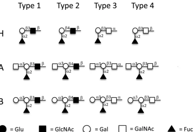
Depending on the oligosaccharide residue and their link-ages of the disaccharide precursors, HBGAs can be divided into six types (19). For example, the A, B, and H antigens can be synthesized on type 1 [-Gal(1¡3)-GlcNAc], type 2 [ -Gal(1¡4)-GlcNAc], type 3 [-Gal(1¡3)␣-GalNAc], type 4 [-Gal(1¡3)-GalNAc], type 5 [-Gal(1¡3)-Gal], and type 6 [-Gal(1¡4)-Glc] chains, differing in the linkages or resi-dues of the disaccharide precursors (19). The structures of type 1 to 4 antigens that were studied here are shown inFig. 1. The H antigens are produced by the addition of a fucose in an
␣(1¡2) linkage to the -galactose of the six different core chains. Both the A and B antigens are produced from the six
Received9 September 2014Accepted8 November 2014
Accepted manuscript posted online12 November 2014
CitationZhang D, Huang P, Zou L, Lowary TL, Tan M, Jiang X. 2015. Tulane virus recognizes the A type 3 and B histo-blood group antigens. J Virol 89:1419 –1427.
doi:10.1128/JVI.02595-14. Editor:S. López
Address correspondence to Xi Jiang, jason.jiang@cchmc.org.
Copyright © 2015, American Society for Microbiology. All Rights Reserved.
doi:10.1128/JVI.02595-14
on November 7, 2019 by guest
http://jvi.asm.org/
types of H antigens by the addition of an N -acetylgalac-tosamine (A) or a galactose (B) in an␣(1¡3) linkage to the terminal -galactose. All six types of ABH antigens can be found on erythrocyte and tissue surfaces; of these six, types 1 to 4 are considered the most important (19), and procedures to synthesize these glycansin vitroare available (20,21).
The binding of HBGAs by TV has been demonstrated by vari-ous binding and blocking assays; however, the precise structures of HBGAs responsible for TV binding remain unclear. A recent study showed that TV recognizes both type A and B saliva with variations among saliva samples in each type (13).We also ob-served differences of TV binding to type B versus type A saliva, in which all type B saliva samples but only a subset of type A saliva samples were reactive to TV. These results indicate that further study to define the binding patterns of TV to human HBGAs is necessary. In addition, unexplainable results regarding the role of HBGAs in TV replication were reported: while significant reduc-tion of TV replicareduc-tion in cell cultures was observed following treatments of the TV with type A and B saliva samples, treatments with bovine serum albumin (BSA)-conjugated A and B oligosac-charides increased TV titers (13). Thus, our understanding of the roles of HBGAs in TV replication and host range remains limited. In this study, we performed further binding and blocking ex-periments using reagents specific to different types of the ABH antigens to clarify these discrepancies. We demonstrated that the TV bound all four tested types (types 1 to 4) of the B antigens, consistent with the results of TV binding of all types B and AB HBGAs in the saliva-binding assays. However, TV bound only the A type 3 antigen among four types (types 1 to 4) of A oligosaccha-ride conjugates tested, suggesting that the lack of binding or weak binding of type A saliva samples to TV may be due to a lack of expression or low expression of the A type 3 antigens in these saliva. Accordingly, we showed that the blocking activities of saliva samples on TV replication in cell culture correlated with the
bind-ing signals by a monoclonal antibody (MAb) recognizbind-ing the A type 3/4 antigens. Finally, we found the reason for the previously described increase of TV replication by BSA-conjugated HBGA oligosaccharides and demonstrated the expected inhibition by these oligosaccharides. Together, our new data support the notion that TV recognizes HBGAs as attachment factors and that such recognition is required for TV infection and replication.
MATERIALS AND METHODS
Virus, MAbs, and synthetic oligosaccharides.Plaque-purified, low-pas-sage-number TV stock was used for the plaque assay; the H type 3/4-specific MAb MBr1 (to globo-H) was purchased from Enzo Life Sciences, Inc. (Farmingdale, NY), and A type 3/4-specific MAb HE-10 was pur-chased from Sigma-Aldrich (St. Louis, MO). Polyacrylamide (PAA)-con-jugated type A and B trisaccharides of approximately 30 kDa containing 20 mol% carbohydrate were purchased from GlycoTech Corporation, Rockville, MD; bovine serum albumin (BSA)-conjugated type A and B trisaccharides with 20 to 25 carbohydrate chains attached per BSA mole-cule were purchased from Glycorex AB (Lund Sweden). The H type 1 to 4, A type 1 to 4, and B type 1 to 4 BSA conjugates were prepared as described previously (22), using antigens synthesized as reported previously (20,
21). Briefly, the antigens were synthesized with an alkene at the reducing end that was converted to the corresponding amine salt using a photo-chemically induced thiol-ene reaction with cysteamine. The amine was then coupled with di-p-nitrophenyl adipate to generate an activated ester derivative (23). These activated derivatives were reacted (22) with bovine serum albumin to give the neoglycoconjugates used the analyses. The structures of these different antigens are shown inFig. 1; antigen loadings on BSA ranged from 11 to 16, as determined by matrix-assisted laser desorption ionization (MALDI) mass spectrometry.
Purification of TV and preparation of TV specific antibodies.Rhesus monkey kidney LLC-MK2 cells (ATCC, Manassas, VA) were cultured in rolling bottles with medium 199 (Mediatech, Manassas, VA) supple-mented with 10% fetal bovine serum (FBS), 200 U/ml penicillin, and 200 g/ml streptomycin. Cells with 80 to 90% confluence were inoculated with 10 ml (multiplicity of infection [MOI]⫽0.4)) of TV inoculum for 1 h.
FIG 1Structures of H type 1 to 4, A type 1 to 4, and B type 1 to 4 antigens conjugated to BSA. Synthetic H type 1 to 4, A type 1 to 4, and B type 1 to 4 antigens were coupled to BSA through the reducing end. Gal, galactose; GalNAc,N-acetylgalactosamine; GlcNAc,N-acetylglucosamine; Fuc, fucose. Symbolic nomen-clature used is that developed by the Consortium for Functional Glycomics.
on November 7, 2019 by guest
http://jvi.asm.org/
[image:2.585.129.460.64.290.2]Then the inoculum was removed, and 50 ml medium without FBS was added and cultured for 72 h. A total of 600 ml of TV-infected LLC-MK2 cell culture medium was concentrated in a dialysis tube against polyeth-ylene glycol (PEG) 8000. Then the sample was adjusted for density (re-fractive index of 1.365) with cesium chloride and centrifuged at 288,000⫻ g(41,000 rpm, SW41Ti rotor; Beckman) for 40 h. The gradient fractions were collected by bottom puncture. TV virions in the fractions were iden-tified and quaniden-tified by transmission electron microscopy (TEM), SDS-PAGE, Western blotting, and reverse transcription-PCR (RT-PCR). The CsCl-purified TVs were used for generation of antibody against TV.
For generation of an anti-TV antibody, female BALB/c mice (n⫽4; Harlan-Sprague-Dawley, Indianapolis, IN) at 3 to 4 weeks of age were immunized with purified TV three times through intraperitoneal injec-tion with adjuvant in 2-week intervals. Blood was collected by retro-or-bital capillary plexus puncture before each immunization and 4 weeks after the final immunization. Sera were processed from blood via a stan-dard protocol. The antisera to TV were evaluated by enzyme-linked
im-munosorbent assay (ELISA) and Western blotting analyses. The antibody specifically recognized the intact TV and its VP-1 protein. The antibody could also block TV binding to HBGAs and neutralized TV replication in LLC-MK-2 cells.
HA and HAI.Type A1, A2, B, and O human red blood cells (RBCs) from Immucor, Inc. (Norcross, GA, USA) were used for hemagglutina-tion (HA) and hemagglutinahemagglutina-tion inhibihemagglutina-tion (HAI) for TV. In addihemagglutina-tion, RBCs from 24 type A-positive individuals from the Memorial Blood Cen-ter (St. Paul, MN, USA) were tested for the role of the A type 3 in causing HA of RBCs by TV. The RBCs were packed by diluting the cells in 0.01 M phosphate-buffered saline (PBS) (without Ca2⫹or Mg2⫹; pH 7.2)
(Invit-rogen, Carlsbad, CA) and centrifuging for 15 min at 500⫻g. The HA of TV to the type B RBCs was performed in 96-well V-bottom plates. Fifty microliters of 0.5% human RBCs in 0.01 M PBS (pH 7.2) and 8 hemag-glutination units (HAUs) of TV (50l of TV containing 5⫻102PFU in PBS) were mixed and incubated for 60 to 100 min at 4 to 8°C. Due to the weak interaction of TV with A type antigen, a method reported by
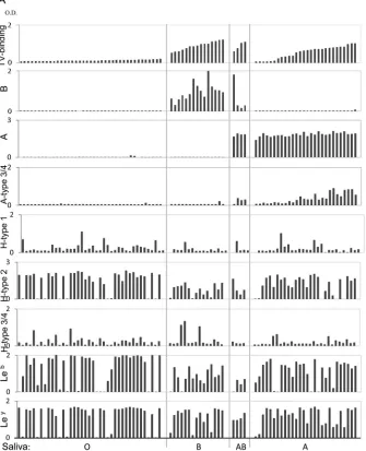
Moro-FIG 2Binding of TV to saliva detected by ELISA. (A) The 86 saliva samples were sorted according to their binding signals to TV within each of the four blood types: O, B, AB, and A. The levels of B type, A type, A type 3/4, H type 1, H type 2, H type 3/4, Leb, and Leyantigens in the saliva samples were determined by ELISA
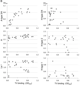
with corresponding specific MAbs. (B) Correlation analysis of binding activity of TV with HBGA levels in the saliva samples was performed. A significant positive correlation (r⫽0.85;P⬍0.05) between TV binding and the A type 3/4 antigen in the type A saliva was found.
Tulane Virus Recognizes A3 and B Antigens
on November 7, 2019 by guest
http://jvi.asm.org/
[image:3.585.126.461.73.486.2]kutti et al. was modified (24). The HA of TV to the type A RBCs was performed in 96-well flat-bottom plates. Eight HAUs (2l of TV contain-ing 2⫻103PFU in PBS) of TV was added to 20l of 0.5% human type A RBCs in 0.01 M PBS (pH 7.2) and mixed. After incubation for 60 to 100 min at 4 to 8°C, agglutination was examined by microscopy. HA inhibi-tion of TV with the type B RBCs by synthetic HBGAs was performed by the same procedures with an additional step of preincubation of TV with serial dilutions of the inhibitors.
Saliva binding assay of TV.A panel of 86 human saliva samples rep-resenting different HBGA types collected in our previous studies (8) was used. The saliva samples were boiled for 10 min to denature potential antibodies that might influence the assay. The levels of the A, B, H type 1, H type 2, and Lewis antigens in the samples were determined previously (8) using MAbs BG-4 (anti-H type 1), BG-6 (anti-Leb), and BG-8 (anti-Ley) (Signet Laboratories Inc. Dedham, MA) and MAbs BCR 9031 (anti-H type 2), BCR 9010 (anti-A), and BCRM 11007 (anti-B) (Accurate Chem-ical & Scientific Corporation, Westbury, NY). The content of the H type 3/4 and A type 3/4 antigens was determined in this study by ELISA using the MAbs MBr1 (Enzo Life Sciences, Inc., Farmingdale, NY) and HE-10 (Sigma-Aldrich, St. Louis, MO), respectively. To test saliva binding by TV, saliva samples at 1:1,000 were used to coat plates in PBS at 4°C overnight and then incubated with TV at 1⫻104PFU/ml. The bound TVs were detected by mouse TV serum (1:3,500) and then by a goat anti-mouse IgG conjugated to horseradish peroxidase (HRP) (1:5,000). Color signals were developed with TMB (3,3=,4,4=-tetramethylbenzidine) and read at 450 nm.
Binding of TV with synthetic ABH tri- and tetrasaccharides.The synthetic ABH type 1 to 4 oligosaccharide-BSA conjugates in PBS were
used to coat plates at 4°C overnight, which were blocked with 5% milk at room temperature for 1 h and then rinsed with 200l of PBS. Next, 100l (1⫻103PFU) of purified TV was added, and plates were incubated at 4°C overnight and rinsed with PBS three times. The bound TVs were detected by mouse anti-TV serum as described above.
Inhibition of TV replication by A type 3/4- and B-positive saliva and oligosaccharide conjugates in cell culture.Inhibition of TV replication was assessed by plaque reduction assay of TV in LLC-MK2 cells in 6-well plates. Type A saliva samples with different levels of the A type 3/4 antigen were tested for their ability to block TV replication, while type B saliva was used as a positive control. Saliva samples were boiled and clarified by centrifugation at 10,000⫻gfor 10 min. The TV inoculum was incubated with saliva starting at a dilution of 1:300 in Dulbecco’s modified Eagle medium (DMEM) with 6-fold serial dilutions. The tubes used were pre-coated with 5% BSA. After 1 h incubation at 37°C, the TV-saliva mixture was inoculated into LLC-MK2 cells and incubated at 37°C for 1.5 h. The plates were overlaid with 0.8% agarose. After a 2-day incubation, the plates were stained with crystal violet, and plaques were counted. The blocking rate (%) of the saliva was calculated by the reduction in plaque numbers in the wells treated with saliva relative to the number in un-treated control wells. Blocking rate (expressed as a percentage) was calcu-lated as follows: (1⫺PFU of test well/PFU of control well)⫻100.
Procedures similar to those used for the plaque reduction assay were also used to determine the roles of the A type 3 and B oligosaccharide conjugates in blocking TV infection in cell cultures. Synthetic oligosac-charide (20g/well) was preincubated with TV inoculum and then trans-ferred to 6-well plates. The test tubes used for incubation of TVs with
FIG 2continued
on November 7, 2019 by guest
http://jvi.asm.org/
[image:4.585.134.451.68.408.2]oligosaccharides were precoated with a BSA solution to avoid potential nonspecific binding of TVs to the test tubes.
RESULTS
Binding of TV to type A saliva correlated with the A type 3/4 antigens in saliva.To test type specificity of human HBGAs rec-ognized by TV, we first performed a saliva binding assay on a panel of 86 saliva samples representing different ABH and Lewis types (8). TV bound strongly to all 15 type B and four type AB saliva samples, but the binding signals among the 28 type A saliva sam-ples varied: 17 bound strongly (optical density [OD]⬎0.5), five bound weakly to moderately (OD 0.2 to 0.5), and the remaining six did not bind or bound poorly (OD⬍0.2) (Fig. 2A). The TV binding signals correlated with neither the A antigens nor other HBGAs, except for the A type 3/4 antigens (r⫽0.85,P⬍0.05) (Fig. 2B), suggesting that A type 3 and/or A type 4 antigens could be responsible for the observed TV binding signals. Subsequent oligosaccharide binding assays further showed that TV binds the A type 3 but not the A type 4 antigen (see below). None of the 39 type O saliva samples revealed binding signals with TV, indicating that TV does not interact with any of the H type and Lewis antigens in human saliva.
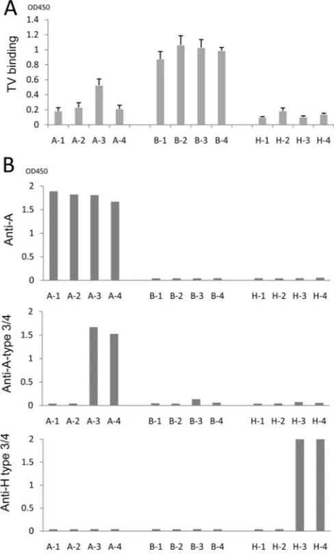
Specificity of binding of TV to the A type 3 and B oligosaccha-rides.To further determine the binding targets of TV, we per-formed oligosaccharide-based binding assays using BSA-conju-gated oligosaccharides representing four types (types 1 to 4) of the A, B, and H antigens). The results indicated that TV strongly bound all four types of the B antigen but only A type 3 among the four A type antigens (Fig. 3A). The binding signals of TV to the A type 3 oligosaccharide were weaker than those to the B antigens (types 1 to 4). TV did not bind to the H antigens (Fig. 3A), con-sistent with the lack of TV binding to any type O saliva (Fig. 2A). The antigenic specificities of these oligosaccharides were also val-idated by corresponding MAbs (Fig. 3B). The A type 3/4 MAb (HE-10) recognized the A type 3 and 4 but not the A type 1 or 2 antigens. This result indicates that MAb HE-10 recognizes an epitope of the A antigens differing from that recognized by TV, in which the former is shared by A types 3 and 4, while the latter is distinct between the two types. Similarly, the H type 3/4 MAb (MBrL) also recognized H type 4 antigens. Finally, MAb (BG2) bound all four types of the A antigens, indicating that a further distinct epitope on the A antigens is recognized by this MAb (Fig. 3A).
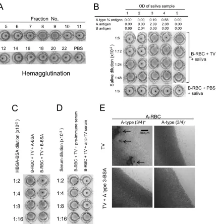
Roles of the A type 3/4 antigens in hemagglutination (HA) and HA inhibition (HAI) with TV.HA and HAI on TV were performed to further prove the specific interaction of TV with human HBGAs. TVs from different fractions of CsCl gradient centrifugation were analyzed by HA and HAI, revealing that the fractions (fractions 7 and 8) containing intact TVs hemaggluti-nated RBCs specifically (Fig. 4A). In our initial study, V-shaped plates were used, which resulted in clear HA for type B RBCs but only marginal HA for the A type 3/4 RBCs (Fig. 4B). To increase the sensitivity of the HA assay, flat-bottom plates were used, and the HA results were read under a microscope (Fig. 4E). The pre-sentation of A type 3/4, H type 3/4, and Lewis y antigens on the surface of the RBCs was determined by HA using MAbs to these antigens. Among RBCs from 24 A-positive individuals, 15 re-sulted in HA with A type 3/4 MAb and 13 (87%) of the 15 RBCs revealed HA with TV (Table 1), while no HA by TV was observed in the other 9 A type 3/4-negative RBCs. Although most A-positive
individuals are Lewis y antigen positive, no significant difference was seen between Lewis y-positive and -negative individuals (52% versus 67%).
The involvement of the B antigens in TV HA was confirmed by blocking of the HA by type B saliva (Fig. 4B), synthetic B type 1 to 4 oligosaccharides (Fig. 4C) and anti-TV serum (Fig. 4D). Both A type 3 and 4 BSA conjugates were also tested in the HAI assay, revealing blocking only by A type 3 BSA conjugate on A type 3/4 RBC HA of TV (Fig. 4E). Interestingly, in the TV HA assay using A type 3/4-positive RBCs, competitive inhibition between the type B antigens and the A type 3/4 antigens occurred, in which the type B saliva completely blocked TV HA of A type 3/4 RBCs (Fig. 4B). This result suggests that the TV most likely uses the same binding site for interaction with both the B and the A type 3 antigens.
Inhibition of TV replication by A type 3/4 and B saliva.To further determine the biological significance of the A type 3/4 and
FIG 3Binding of TV to synthetic HBGA oligosaccharides. (A) Binding of TV to four subtypes of the A, B, and H oligosaccharides. (B) Validation of synthetic ABH oligosaccharides by MAb-based binding assays. Three MAbs, anti-A type 3 (HE-10), anti-globo-H (H type 3/4) (MBr1), and anti-A (BG2), were used. A-1 to A-4, B-1 to B-4, and H-1 to H-4 are A type 1 to 4, B type 1 to 4, and H type 1 to 4 BSA conjugates, respectively.
Tulane Virus Recognizes A3 and B Antigens
on November 7, 2019 by guest
http://jvi.asm.org/
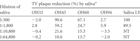
[image:5.585.299.543.69.473.2]B HBGAs in TV replication, we performed a plaque reduction assay on TVs in cell culture using human saliva as a blocking reagent. Nearly complete inhibition (⬃100%) of TV replication by the type B saliva at 1:300 was observed (Table 2), while variable blocking results were obtained when the type A saliva samples were tested (data not shown). To determine the involvement of the A type 3 antigens in the blocking activities, saliva samples from two individuals with strong (OH45 and OH60) and two individ-uals with weak (OH32 and OH94) signals for A type 3/4 were tested. High inhibition of TV replication (⬎20 to 50%) was ob-served after TVs were incubated with saliva OH45 and OH60
(with strong A type 3/4 signals) at a dilution of 1:1,800. As ex-pected, no blocking or weak blocking (⬍10%) was seen with saliva OH32 and OH94 (with weak A type 3/4 signals) even when a higher concentration of saliva (1:300) was used (Table 2). All four saliva samples had similar levels of total type A antigens, indicating that the A type 3/4antigen in the saliva is responsible for the inhi-bition.
Synthetic type B-BSA conjugates reduce TV replication in cell culture.We first repeated the blocking experiments, described in a previous study, that showed that synthetic HBGA-BSA con-jugates resulted in an increase in TV infection (13). Using the
FIG 4Hemagglutination of human red blood cells (RBCs) by TV. (A) Hemagglutination of type B human RBCs by TVs from CsCl gradient fractions. Only fractions 8 and 9 contained intact TVs, which was determined by electron microscopy followed by ELISA and detection by PCR. (B) Inhibition of TV-induced hemagglutination by saliva containing A type 3/4 and B antigens. Strong inhibition by the type B saliva (saliva samples 1 and 2) which correlated with the salivary B antigens was observed. Inhibition was also observed for the A type 3/4-positive saliva (saliva samples 3 and 4), but inhibition was not seen for the type O saliva (saliva sample 5). (C and D) Inhibition of TV induced hemagglutination by type B oligosaccharide and by antibody to TV. (E) Inhibition of TV induced hemagglutination by A type 3 oligosaccharide using flat-bottom plates followed by reading of the results under a microscope. Weak agglutination of A type 3/4-positive RBCs developed, and the hemagglutination could be blocked by a synthetic A type 3 oligosaccharide conjugate. Arrows indicate typical agglutina-tions of RBCs.
on November 7, 2019 by guest
http://jvi.asm.org/
[image:6.585.81.511.62.506.2]same conditions, we observed an increase of plaque formation after incubation of TVs with the BSA-conjugated type A and B oligosaccharides compared with the control group without BSA conjugated oligosaccharides. However, it was noted that a similar increase was also seen when BSA alone without type A or B oligo-saccharides was used (data not shown), indicating that such an increase of viral titers was nonspecific. After stepwise trouble-shooting, we found that the observed “increase” in TV titers was due to nonspecific binding of TV to the incubation tubes, which could be prevented by BSA or the BSA conjugates. Because no additional proteins were included in the control groups, less TV can be removed from the tube to inoculate the cell cultures. As a result, all treatment groups showed more TV plaques after culti-vation. This false result can be avoided by precoating of the tubes with BSA.
After resolving this problem, we repeated the oligosaccharide-blocking assay and observed specific inhibition of TV replication by the type B-BSA conjugate, but the blocking efficiency was sig-nificantly lower than that of saliva (Table 3). Although no block-ing effects on TV replication by other oligosaccharide conjugates were observed, including B-PAA, A-BSA, A type 2-BSA, and A type 3-BSA conjugates, no increase in TV plaques occurred (Table 3). We speculate that the lower blocking efficiency by the oligo-saccharide conjugates could be due to their lower valence com-pared with the multivalent nature of the HBGAs of saliva.
Collec-tively, our data support the notion that HBGAs play an important role in TV replication.
DISCUSSION
In this study, we verified that the TV recognizes all four types of the B antigens but only the A type 3 of the A antigens. The speci-ficity of TV in recognizing the A type 3 antigens has been demon-strated by binding of TV with A type 3/4-positive saliva, detected with an anti-A type 3/4 MAb. We also obtained direct evidence of TV binding to the A type 3 and B antigens by oligosaccharide-based binding and blocking assays and by HA and HAI with A type 3/4- and B-positive human RBCs and saliva. The biological signif-icance of HBGA as a host factor in TV replication has been dem-onstrated by specific blocking of TV replication in cell cultures using type B-specific HBGAs and saliva characterized as contain-ing A type 3/4 antigens. Finally, our study clarified an unexplained result reported in the literature regarding the roles of human HBGAs in TV replication. Thus, we conclude that the recognition by TV of the A type 3 and B HBGAs is highly specific and biolog-ically relevant.
The finding that TV recognizes both the A type 3 and B anti-gens further broadens our understanding of the nature of virus-host interaction of caliciviruses with HBGAs. First, according to the cross-blocking results of TV between the A type 3 and B anti-gens, we predict that the TV may use a common receptor-binding interface to interact with both the A type 3 and B antigens. This is similar to what was observed in human NoVs that use a common HBGA binding interface to bind different ABH and Lewis anti-gens. Thus, we further predict that TV may rely on the shared
-galactose in the disaccharide precursors (core chains) and the fucose linked to the-galactose in the A and B antigens (Fig. 1) as the major binding saccharide for TV to interact with HBGAs. However, since TV does not bind to any types of O saliva, the fucose that linked to the-galactose of the secretor saliva may be dispensable for the interactions.
[image:7.585.40.286.87.222.2]In this study, a MAb that recognizes A type 3/4 antigens was used for HBGA typing of human saliva, and a correlation of saliva binding by this MAb and by TV was observed, raising the question of whether the A type 4 antigen in human saliva is also involved in interaction with TV. According to the oligosaccharide-based binding assays, however, TV recognized A type 3 but not the other three types (1, 2, and 4) of the A antigens, indicating that the TV
TABLE 1Hemagglutination of type A RBCs by TV is correlated to the A type 3/4 antigen on individuals’ RBCs
Antigen
No. of individuals (n⫽24) with RBC typea
No. (%) of RBC samples with induced HA by TVb
A type 3/4
⫹ 15 13 (87)
⫺ 9 0
H type 3/4
⫹ 8 3 (38)
⫺ 16 10 (63)
Lewis y
⫹ 21 11 (52)
⫺ 3 2 (67)
a
RBCs of 24 type A-positive individuals were tested. The presence (⫹) or absence (⫺) of the A type 3, H type 3, and Lewis y antigens in each individual’s RBCs was determined by HA with specific MAbs.
bThe HA assays were performed in 96-well, flat-bottom plates, and HA was determined
by reading the plates under a microscope. A typical TV HA result for A type 3/4 RBCs is shown inFig. 4E.
TABLE 2Inhibition of TV replication in cell culture by A type 3/4 and type B saliva samples
Dilution of saliva
TV plaque reduction (%) by salivaa
OH32 OH45 OH60 OH94 Saliva LY
1:300 ⫺2.0 90.6 67.1 2.7 100
1:1,800 2.8 59.2 24.7 5.9 89.5
1:10,800 ⫺0.4 21.6 15.3 ⫺3.5 NT 1:64,800 ⫺0.2 10.6 13.7 ⫺2.0 NT
[image:7.585.299.545.91.185.2]aFour type A-positive saliva samples with high (OH45 and OH60, OD⬎0.5) or low (OH32 and OH94, OD⬍0.2) levels of A type 3/4 antigen were tested, and a type B saliva sample (saliva LY) was also included. NT, not tested.
TABLE 3Inhibition of TV replication by a plaque reduction assay with synthetic type A and B oligosaccharide conjugates as inhibitors
Oligosaccharide
conjugatea PFU (avg) 95% CIb Plaque reduction (%)c
BSA 21.5 17.4–25.6 ⫺2.4
B-BSA 14.5 13.2–15.8 31.0
B-PAA 20.3 16.4–24.1 3.6
A3-BSA 19.3 15.6–22.9 8.3 A2-BSA 17.5 13.2–21.8 16.7 A-BSA 17.8 13.7–21.8 15.5 Control (TV only) 21.0
a
A- and B-BSA, conjugated A and B trisaccharides; A2- and A3-BSA, BSA-conjugated A type 2 and 3 tetrasaccharides; B-PAA, polyacrylamide-BSA-conjugated B trisaccharide. The control wells were TV not mixed with any oligosaccharides. b95% CI, confidence interval at the 95% confidence level.
c
Plaque reduction was calculation based on plaque numbers in the test wells compared with that of the control wells.
Tulane Virus Recognizes A3 and B Antigens
on November 7, 2019 by guest
http://jvi.asm.org/
[image:7.585.41.285.625.697.2]recognizes an epitope on the A antigens differ from that recog-nized by the MAb. The A antigens contain multiple sugar residues that may form different antigenic epitopes recognized by the MAb and TV, respectively. The recognition of distinct epitopes of man HBGAs in human milk has also been observed between hu-man NoVs and HBGA-specific MAbs (25). Thus, we conclude that TV recognizes the A type 3 antigens specifically, and the cross-reactivity of the MAb to both A types 3 and 4 should not affect our conclusion.
The finding that TV recognizes the A type 3 antigen also helped our understanding of the type specificity of HBGAs recognized by different human NoVs. For example, the prototype Norwalk virus (GI.1) recognizes mainly the type 1 chain of the A and H antigens, while the major epidemic GII.4 NoVs recognize mainly the type 3 chains of the A, B, and H antigens. This type specificity may have a role in clinical infection and prevalence of individual strains of NoVs in different human populations. Future studies to deter-mine the distribution of individual types of the ABH and Lewis antigens in the general population and to explore their roles in the prevalence of individual genotypes of human NoVs are necessary. The demonstration of the role of human HBGAs in TV repli-cation by the cell culture-based blocking experiments described in our study is highly significant, because such a study is impossible to carry out with human NoVs due to the lack of a cell culture method and an animal model. Similar to results of a previous study (13), significant reduction of TV replication in LLC-MK2 cells was seen after treatment of TVs with the A type 3 and B antigens. As an analogy, we predict that human HBGAs or a car-bohydrate receptor also plays an important role in the replication of human NoVs and related caliciviruses (26–29). To look into the report of increased TV infectivity by type A and B oligosaccharides in an earlier study (13), we carefully evaluated the experimental procedures described in the study and identified a potential prob-lem that could cause a false result. We found that TV tends to bind to the test tubes in the preincubation step and that such nonspe-cific binding can be prevented by precoating of the tube with a protein solution, such as BSA. Our conclusion was strongly sup-ported by a demonstration of an increase in TV titer by preincu-bation of TV with BSA and by avoiding such false results by pre-coating the test tubes with a BSA solution. The observed lower blocking efficacy of the oligosaccharide conjugates than human saliva could be due to a lower valence of the oligosaccharide con-jugates than of saliva glycans.
ACKNOWLEDGMENTS
The research described in this article was supported by the National In-stitutes of Health of the United States (R01 AI 089634), the Agriculture and Food Research Initiative Competitive Grants Program of the USDA National Institute of Food and Agriculture (NIFA award 2011-68003-30005), the Alberta Glycomics Centre, and the Canadian Institutes of Health Research (RMF 92091).
REFERENCES
1.Green KY, Ando T, Balayan MS, Berke T, Clarke IN, Estes MK, Matson DO, Nakata S, Neill JD, Studdert MJ, Thiel HJ.2000. Taxonomy of the caliciviruses. J Infect Dis181(Suppl 2):S322–S330.http://dx.doi.org/10 .1086/315591.
2.L’Homme Y, Sansregret R, Plante-Fortier E, Lamontagne AM, Ouar-dani M, Lacroix G, Simard C.2009. Genomic characterization of swine caliciviruses representing a new genus of Caliciviridae. Virus Genes39:
66 –75.http://dx.doi.org/10.1007/s11262-009-0360-3.
3.Thomas C, Jung K, Han MG, Hoet A, Scheuer K, Wang Q, Saif LJ.2014.
Retrospective serosurveillance of bovine norovirus (GIII.2) and nebovirus in cattle from selected feedlots and a veal calf farm in 1999 to 2001 in the United States. Arch Virol159:83–90.http://dx.doi.org/10.1007/s00705 -013-1795-3.
4.Tan M, Jiang X.2014. Histo-blood group antigens: a common niche for norovirus and rotavirus. Expert Rev Mol Med16:e5.http://dx.doi.org/10 .1017/erm.2014.2.
5.Tan M, Jiang X.2011. Norovirus-host interaction: multi-selections by human histo-blood group antigens. Trends Microbiol19:382–388.http: //dx.doi.org/10.1016/j.tim.2011.05.007.
6.Tan M, Jiang X. 2005. Norovirus and its histo-blood group antigen receptors: an answer to a historical puzzle. Trends Microbiol13:285–293.
http://dx.doi.org/10.1016/j.tim.2005.04.004.
7.Tan M, Jiang X.2010. Norovirus gastroenteritis, carbohydrate receptors, and animal models. PLoS Pathog6:e1000983.http://dx.doi.org/10.1371 /journal.ppat.1000983.
8.Huang P, Farkas T, Zhong W, Tan M, Thornton S, Morrow AL, Jiang X.2005. Norovirus and histo-blood group antigens: demonstration of a wide spectrum of strain specificities and classification of two major bind-ing groups among multiple bindbind-ing patterns. J Virol79:6714 – 6722.http: //dx.doi.org/10.1128/JVI.79.11.6714-6722.2005.
9.Papafragkou E, Hewitt J, Park GW, Greening G, Vinje J.2013. Chal-lenges of culturing human norovirus in three-dimensional organoid in-testinal cell culture models. PLoS One8:e63485.http://dx.doi.org/10.1371 /journal.pone.0063485.
10. Farkas T, Sestak K, Wei C, Jiang X.2008. Characterization of a rhesus monkey calicivirus representing a new genus of Caliciviridae. J Virol82:
5408 –5416.http://dx.doi.org/10.1128/JVI.00070-08.
11. Yu G, Zhang D, Guo F, Tan M, Jiang X, Jiang W. 2013. Cryo-EM structure of a novel calicivirus, Tulane virus. PLoS One8:e59817.http://dx .doi.org/10.1371/journal.pone.0059817.
12. Sestak K, Feely S, Fey B, Dufour J, Hargitt E, Alvarez X, Pahar B, Gregoricus N, Vinje J, Farkas T. 2012. Experimental inoculation of juvenile rhesus macaques with primate enteric caliciviruses. PLoS One
7:e37973.http://dx.doi.org/10.1371/journal.pone.0037973.
13. Farkas T, Cross RW, Hargitt E, III, Lerche NW, Morrow AL, Sestak K.
2010. Genetic diversity and histo-blood group antigen interactions of rhe-sus enteric caliciviruses. J Virol84:8617– 8625.http://dx.doi.org/10.1128 /JVI.00630-10.
14. Hirneisen KA, Kniel KE.2013. Comparing human norovirus surrogates: murine norovirus and Tulane virus. J Food Prot76:139 –143.http://dx.doi .org/10.4315/0362-028X.JFP-12-216.
15. Li X, Ye M, Neetoo H, Golovan S, Chen H.2013. Pressure inactivation of Tulane virus, a candidate surrogate for human norovirus and its poten-tial application in food industry. Int J Food Microbiol162:37– 42.http: //dx.doi.org/10.1016/j.ijfoodmicro.2012.12.016.
16. Shearer AE, Hoover DG, Kniel KE.2014. Effect of bacterial cell-free supernatants on infectivity of norovirus surrogates. J Food Prot77:145– 149.http://dx.doi.org/10.4315/0362-028X.JFP-13-204.
17. Tian P, Yang D, Quigley C, Chou M, Jiang X.2013. Inactivation of the Tulane virus, a novel surrogate for the human norovirus. J Food Prot
76:712–718.http://dx.doi.org/10.4315/0362-028X.JFP-12-361. 18. Wang Q, Hirneisen KA, Markland SM, Kniel KE.2013. Survival of
murine norovirus, Tulane virus, and hepatitis A virus on alfalfa seeds and sprouts during storage and germination. Appl Environ Microbiol79:
7021–7027.http://dx.doi.org/10.1128/AEM.01704-13.
19. Mollicone R, Gibaud A, Francois A, Ratcliffe M, Oriol R.1990. Acceptor specificity and tissue distribution of three human alpha-3-fucosyltransferases. Eur J Biochem 191:169 –176. http://dx.doi.org/10 .1111/j.1432-1033.1990.tb19107.x.
20. Meloncelli PJ, Lowary TL.2010. Synthesis of ABO histo-blood group type I and II antigens. Carbohydr Res345:2305–2322.http://dx.doi.org/10 .1016/j.carres.2010.08.012.
21. Meloncelli PJ, West LJ, Lowary TL.2011. Synthesis and NMR studies on the ABO histo-blood group antigens: synthesis of type III and IV struc-tures and NMR characterization of type I-VI antigens. Carbohydr Res
346:1406 –1426.http://dx.doi.org/10.1016/j.carres.2011.03.008. 22. Wu X, Ling C-C, Bundle DR.2004. A new homobifunctional p-nitro
phenyl ester coupling reagent for the preparation of neoglycoproteins. Org Lett6:4407– 4410.http://dx.doi.org/10.1021/ol048614m.
23. Dondoni A, Massi A, Nanni P, Roda A.2009. A new ligation strategy for peptide and protein glycosylation: photoinduced thiol-ene coupling. Chemistry15:11444 –11449.http://dx.doi.org/10.1002/chem.200901746.
on November 7, 2019 by guest
http://jvi.asm.org/
24. Morokutti A, Redlberger-Fritz M, Nakowitsch S, Krenn BM, Wressnigg N, Jungbauer A, Romanova J, Muster T, Popow-Kraupp T, Ferko B.
2013. Validation of the modified hemagglutination inhibition assay (mHAI), a robust and sensitive serological test for analysis of influenza virus-specific immune response. J Clin Virol56:323–330.http://dx.doi .org/10.1016/j.jcv.2012.12.002.
25. Huang P, Morrow AL, Jiang X.2009. The carbohydrate moiety and high molecular weight carrier of histo-blood group antigens are both required for norovirus-receptor recognition. Glycoconj J26:1085–1096.http://dx .doi.org/10.1007/s10719-009-9229-x.
26. Lindesmith L, Moe C, Marionneau S, Ruvoen N, Jiang X, Lindblad L, Stewart P, LePendu J, Baric R.2003. Human susceptibility and resistance to Norwalk virus infection. Nat Med9:548 –553.http://dx.doi.org/10 .1038/nm860.
27. Nystrom K, Le Gall-Recule G, Grassi P, Abrantes J, Ruvoen-Clouet N, Le Moullac-Vaidye B, Lopes AM, Esteves PJ, Strive T, Marchandeau S, Dell A, Haslam SM, Le Pendu J.2011. Histo-blood group antigens act as attachment factors of rabbit hemorrhagic disease virus infection in a virus strain-dependent manner. PLoS Pathog7:e1002188.http://dx.doi.org/10 .1371/journal.ppat.1002188.
28. Stuart AD, Brown TD.2007. Alpha2,6-linked sialic acid acts as a receptor for feline calicivirus. J Gen Virol88:177–186.http://dx.doi.org/10.1099 /vir.0.82158-0.
29. Zakhour M, Maalouf H, Di Bartolo I, Haugarreau L, Le Guyader FS, Ruvoen-Clouet N, Le Saux J C, Ruggeri FM, Pommepuy M, Le Pendu J.2010. Bovine norovirus: carbohydrate ligand, environmental contami-nation, and potential cross-species transmission via oysters. Appl Environ Microbiol76:6404 – 6411.http://dx.doi.org/10.1128/AEM.00671-10.
Tulane Virus Recognizes A3 and B Antigens