Enhances Hepatitis C Virus RNA Replication by Promoting Linkage
between 5
=
and 3
=
Untranslated Regions
Anuj Kumar, Upasana Ray,* Saumitra Das
Department of Microbiology and Cell Biology, Indian Institute of Science, Bangalore, India
Human La protein has been implicated in facilitating the internal initiation of translation as well as replication of hepatitis C virus (HCV) RNA. Previously, we demonstrated that La interacts with the HCV internal ribosome entry site (IRES) around the GCAC motif near the initiator AUG within stem-loop IV by its RNA recognition motif (RRM) (residues 112 to 184) and influ-ences HCV translation. In this study, we have deciphered the role of this interaction in HCV replication in a hepatocellular carci-noma cell culture system. We incorporated mutation of the GCAC motif in an HCV monocistronic subgenomic replicon and a pJFH1 construct which altered the binding of La and checked HCV RNA replication by reverse transcriptase PCR (RT-PCR). The mutation drastically affected HCV replication. Furthermore, to address whether the decrease in replication is a consequence of translation inhibition or not, we incorporated the same mutation into a bicistronic replicon and observed a substantial decrease in HCV RNA levels. Interestingly, La overexpression rescued this inhibition of replication. More importantly, we observed that the mutation reduced the association between La and NS5B. The effect of the GCAC mutation on the translation-to-replication switch, which is regulated by the interplay between NS3 and La, was further investigated. Additionally, our analyses of point mu-tations in the GCAC motif revealed distinct roles of each nucleotide in HCV replication and translation. Finally, we showed that a specific interaction of the GCAC motif with human La protein is crucial for linking 5=and 3=ends of the HCV genome. Taken together, our results demonstrate the mechanism of regulation of HCV replication by interaction of thecis-acting element GCAC within the HCV IRES with human La protein.
H
epatitis C virus (HCV), a major etiologic agent of posttrans-fusion and sporadic non-A, non-B hepatitis (NANBH), be-longs to theHepacivirusgenus of theFlaviviridaefamily (1,2). The HCV genome consists of a 9.6-kb single-stranded positive-sense RNA containing an open reading frame (ORF) encoding an ⬃3,000-residue polyprotein precursor flanked at both ends by highly structured and conserved untranslated regions (UTRs) (1–4). HCV translation is mediated by an internal ribosome entry site (IRES) located mostly within the 341-nucleotide (nt) 5=UTR and extending to 30 to 40 nucleotides downstream of the initiator AUG (iAUG) codon. Sequence elements that are directly involved in HCV replication are located mostly in the 3=UTR (1,4–6). The HCV 3=UTR varies between 200 and 235 nt in length, including a short variable region, a poly(U/UC) tract (with an average length of 80 nt), and an invariant 98-nt X-tail region (7–9). Initiation of HCV replication takes place by formation of a ribonucleoprotein (RNP) complex at the 3=UTR of the viral genome. The newly synthesized negative-strand RNA then serves as a template for the production of the plus-strand copies of viral RNA (10,11).
Some reports have revealed the presence ofcis-acting elements in the HCV 5=UTR that affect viral replication. For example, the sequence upstream of the IRES is essential for viral RNA replica-tion, IRES sequences are required for high-level RNA replicareplica-tion, and stem-loop domain II of the IRES is crucial for replication (12). Several studies have also revealed that the 5=UTR is capable of binding to a liver-specific microRNA, mir-122, resulting in en-hanced HCV replication (13,14).
Apart fromcis-acting elements present in HCV RNA, HCV nonstructural proteins (except NS2) are required for efficient HCV replication. Intracellular changes resulting from HCV rep-lication are not very well understood. Development of a selectable
and efficiently replicating HCV subgenomic replicon in the hu-man hepatoma cell line Huh7 has allowed investigation of viral persistence and pathogenesis mechanisms in the context of HCV replication in cell culture (10,11,15). A significant number of studies have focused on the cellular and viral proteins that directly form RNP complexes with the 5=and 3=UTRs (7,9,11,16).
Sev-eraltrans-acting proteins, such as polypyrimidine tract binding
protein (PTB) (17) and poly(rC) binding protein 2 (PCBP-2) (18), interact with the 5=and/or 3=UTR. These interactions may be important for HCV replication, although the precise roles are not yet known.
Human La protein is a multifunctional phosphoprotein origi-nally identified as an autoantigen in patients with autoimmune disorders. It is an RNA binding protein of the RNA recognition motif (RRM) family. La protein is located predominantly in the nucleus and functions in the maturation of RNA polymerase III transcripts and the unwinding of double-stranded RNA (19,20). La protein associates with U1 RNA (21), telomerase RNA (22), adenovirus VA RNA (23), and the HIVtrans-activation response (TAR) element (24). Other targets of La include the 5=UTRs of poliovirus (25), hepatitis C virus (26,27), encephalomyocarditis
Received27 February 2013 Accepted27 March 2013
Published ahead of print3 April 2013
Address correspondence to Saumitra Das, sdas@mcbl.iisc.ernet.in.
* Present address: Upasana Ray, National Cancer Institute, National Institutes of Health, Bethesda, Maryland, USA.
Copyright © 2013, American Society for Microbiology. All Rights Reserved.
doi:10.1128/JVI.00525-13
on November 7, 2019 by guest
http://jvi.asm.org/
the primary sequence while retaining the overall secondary struc-ture, has a drastic effect on HCV replication in the context of a monocistronic subgenomic replicon and an infectious JFH1 RNA. To further address whether reduced La protein interactions with the GCAC motif affect HCV replication as a consequence of trans-lation inhibition, we mutated the bicistronic replicon pSGR-JFH1/Luc. Interestingly, we found that HCV RNA levels decreased in the pSGR-JFH1/Luc mutant as well. Furthermore, we found that this inhibition of replication can be rescued by La overexpres-sion. We also found that the mutation reduced the interaction between La and NS5B. Additionally, we demonstrated that this mutation affected the translation-to-replication switch regulated by the interplay between NS3 protease and human La protein. Furthermore, we showed that specific interaction of La with the GCAC motif promotes linkage between both ends of the HCV genome, indicating a plausible mechanism for regulating HCV replication by long-range RNA-RNA interactions. To our knowl-edge, this study constitutes the first report demonstrating the im-portance of thecis-acting element GCAC located within the HCV IRES in HCV replication.
MATERIALS AND METHODS
Cell culture.Huh7 and Huh7.5 cells were cultured in Dulbecco’s modi-fied Eagle’s medium (DMEM) supplemented with 10% fetal bovine se-rum (FBS), 100 U of penicillin/ml, and 100g of streptomycin sulfate/ml. Cells were maintained at 37°C in 5% CO2.
Materials.Hemin was purchased from Merck Millipore. Puromycin and cycloheximide were purchased from Sigma.
Plasmids. The HCV monocistronic replicon construct pFKi383 hygubiNS3-3=5.1 (33), the full-length pJFH1 construct (34), and the bicistronic replicon construct pSGR-JFH1/Luc (35) used in this study were generous gifts from Ralf Bartenschlager and Takaji Wakita.
Site-directed mutagenesis.Mutated replicons were generated by us-ing a QuikChange Lightnus-ing multisite-directed mutagenesis kit (Agilent Technologies) according to the manufacturer’s protocol.
Transient transfection.Huh7 cells (80% confluent) were transiently transfected with HCV mono- or bicistronic replicon RNA (2g) by using Lipofectamine 2000 reagent (Invitrogen) in antibiotic-free medium. Cells were harvested by using Tri reagent (Sigma) for RNA isolation, followed by reverse transcriptase PCR (RT-PCR). Cell lysates were prepared by using passive lysis buffer (Promega) to measure luciferase activities, using luciferase assay reagent (Promega).
Semiquantitative RT-PCR.Total RNA was extracted by using Tri re-agent (Sigma). One microgram of total RNA was reverse transcribed by using RT reaction mix (containing 1⫻ reverse transcription buffer [Thermo Scientific], 1 mM deoxynucleoside triphosphate [dNTP] mix [Thermo Scientific], 40 U of RNase inhibitor [Promega], and 40 U of RevertAid Moloney murine leukemia virus [M-MuLV] RT [Thermo
Sci-Quantitative RT-PCR.Total RNA was isolated by using Tri reagent (Sigma) and reverse transcribed by avian myoblastosis virus (AMV) RT (Promega), using an HCV 5=primer (forward) or an HCV 3=primer (reverse) to detect the negative or positive RNA strand, respectively. cDNA (diluted 1:10) was used for PCR amplification by using a real-time assay mixture (Finnzymes) according to the manufacturer’s instructions. The data were analyzed by using an ABI Prism real-time PCR machine, using the comparative⌬⌬CTmethod (36). GAPDH was used as an
inter-nal control.
Tagged cDNA RT-PCR.Total RNA was isolated by using Tri reagent (Sigma) and reverse transcribed with AMV RT (Promega), using a tagged HCV 5=primer. Two rounds of PCR were done by using Hot StartTaq
DNA polymerase (Genei). The first round of PCR was performed on cDNA using the tag-only primer and the HCV 3=primer. The resulting PCR product was diluted 1:100 and used for the second round of PCR using a set of internal primers. The PCR product was run on a 2% agarose gel. GAPDH was used as an internal control.
Immunoprecipitation.Huh7 cells (80% confluent) were transiently transfected with HCV monocistronic replicon RNA by using Lipo-fectamine 2000 reagent (Invitrogen) in antibiotic-free medium. After 24 h, cells were lysed by using buffer containing 100 mM KCl, 5 mM MgCl2, 10 mM HEPES (pH 7.0), 0.05% Nonidet P-40, 1 mM dithiothreitol (DTT), 100 U/ml RNase inhibitor, and 25l/ml protease inhibitor cock-tail. Supernatants were incubated with anti-La antibody. Immunocom-plexes were immobilized on protein G-Sepharose beads, followed by pro-teinase K and DNase treatments. Finally, phenol-chloroform-extracted and alcohol-precipitated RNA was used for RT-PCR. The same protocol was used for experiments using the bicistronic replicon RNA by using anti-NS5B antibody. Antiactin was used as a nonspecific control.
In vitrotranscription.mRNAs were transcribedin vitrofrom linear-ized plasmid constructs under T7 promoters in runoff transcription tions using T7 RNA polymerase (Promega). Standard transcription reac-tions were performed according to the manufacturer’s instrucreac-tions.
In vitrotranslation.In vitro-transcribed mRNAs were translatedin vitro by using micrococcal nuclease-treated rabbit reticulocyte lysate (RRL), amino acid mix, and an RNase inhibitor (Promega), according to the manufacturer’s instructions. Luciferase activity was measured by us-ing luciferase assay reagent (Promega).
Preparation of cell extracts for Western blotting.Huh7 cells were harvested and then lysed in radioimmunoprecipitation assay (RIPA) buf-fer (50 mM Tris [pH 7.4], 150 mM NaCl, 0.25% sodium deoxycholate, 1% Nonidet P-40, 1 mM EDTA, 1 mM phenylmethylsulfonyl fluoride [PMSF], 1 mM sodium orthovanadate, 1 mM sodium fluoride, and 12 l/ml protease inhibitor cocktail) for 30 min at 4°C, followed by centrif-ugation at 12,000⫻gfor 10 min to pellet cellular debris. Supernatants were used for Western blotting.
Western blotting.Protein concentrations in lysates were assayed by a Bradford assay (Bio-Rad), and equal protein amounts in cell extracts were separated by SDS–10% PAGE and transferred onto a nitrocellulose mem-brane (Pall Life Sciences). Samples were then analyzed by
on November 7, 2019 by guest
tion using rabbit anti-La or anti-NS5B antibody followed by a secondary antibody (horseradish peroxidase-conjugated anti-rabbit IgG; Sigma). The blots were developed by using ECL techniques (Amersham Pharma-cia Biotech). Mouse monoclonal antiactin antibody (Sigma) was used to verify equal loading of cell extracts.
Coimmunoprecipitation assay.Transfected Huh7 cells were washed with cold phosphate-buffered saline (PBS) and lysed by using RIPA buffer (50 mM Tris-HCl [pH 8.0], 150 mM NaCl, 12 mM deoxycholate sodium salt, 0.1% sodium dodecyl sulfate, 1% Triton X-100, 0.2 mM sodium vanadate, and 1 mM phenylmethylsulfonyl fluoride). Cell lysates were centrifuged at 12,000⫻gfor 10 min at 4°C, and the supernatants were precleared with protein G-agarose at 4°C for 3 h. In parallel, anti-La (or anti-NS5B) antibody was incubated with protein G-agarose overnight in 1 ml of PBS at 4°C. This was followed by incubation of antibody conjugated with protein G-agarose with the precleared lysates at 4°C for 4 h. Precip-itates were washed three times with RIPA buffer, and bound proteins were fractionated by SDS–10% PAGE and subjected to Western blot analysis using anti-NS5B (or anti-La) antibody.
Purification of La protein. Escherichia coliBL21(DE3) cells were transformed with a bacterial expression vector encoding La (or mutant La). Transformed colonies were inoculated into 100 ml of LB medium containing 80g/ml ampicillin and grown at 37°C in an incubator shaker set at 200 rpm until theA600reached 0.6. The culture was induced with 0.6 mM isopropyl--D-thiogalactopyranoside for 4 h at 37°C. The crude ex-tracts were mixed with a Ni-nitrilotriacetic acid (NTA) agarose slurry (Qiagen) and rocked at 4°C for 2 h. The lysate was loaded onto a column and washed with 50 ml of wash buffer (50 mM Tris [pH 7.4], 300 mM NaCl, and 40 mM imidazole), and the bound proteins were eluted with 500l of elution buffer containing 500 mM imidazole. The eluted pro-teins were dialyzed at 4°C for 6 h in dialysis buffer (50 mM Tris [pH 7.4], 100 mM KCl, 7 mM-mercaptoethanol, and 20% glycerol), aliquoted, and stored at⫺70°C
Two-cycle RNase protection assay.A two-cycle RNase protection as-say was performed as previously described (37). Briefly, total RNA was self-hybridized overnight at 60°C in the absence of radiolabeled probe, followed by RNase treatment for 1 h in digestion mix (500 mM NaCl, 10 mM Tris-HCl [pH 7.5], 5 mM EDTA, 4.5g of RNase A/ml, and 350 U of RNase T1/ml). Samples were subjected to proteinase K treatment fol-lowed by phenol-chloroform extraction and ethanol precipitation with 5 g of tRNA. RNA samples were then suspended in hybridization buffer containing 75 fmol of radiolabeled probe and subjected to a standard RNase protection procedure.
UV cross-linking assay.␣-32P-labeled RNA probes were allowed to form complexes with recombinant La protein at 30°C for 20 min in RNA binding buffer (containing 5 mM HEPES [pH 7.6], 25 mM KCl, 2 mM MgCl2, 3.8% glycerol, 2 mM DTT, and 0.1 mM EDTA) and next irradi-ated with a UV lamp for 15 min. The reaction mixtures were treirradi-ated with 30g of RNase A at 37°C for 30 min, followed by SDS-PAGE analysis and phosphorimaging.
5=-3=coprecipitation assay.A 5=-3=coprecipitation assay was per-formed as previously described (38), with slight modifications. Briefly, 2 g ofin vitro-transcribed biotinylated 5=-UTR RNA was bound to 50l of streptavidin beads for 30 min at room temperature. The32P-labeled 3= -UTR RNA probe was incubated with recombinant La or bovine serum albumin (BSA) in the presence of 10g of tRNA. The RNA-protein com-plex was then incubated with the 5=-UTR RNA-coated streptavidin beads at 4°C for 2 h. The beads were collected and washed. Precipitated RNA was analyzed on a denaturing 5% Tris-borate PAGE gel and autoradio-graphed.
Statistical analysis.All graphs represent means⫾standard deviations (SD). Student’sttest was performed to determine statistical significance, and the criterion for statistical significance was aPvalue of⬍0.05 (ⴱ represents aPvalue of⬍0.05,ⴱⴱrepresents aPvalue of⬍0.005, andⴱⴱⴱ represents aPvalue of⬍0.0005).
RESULTS
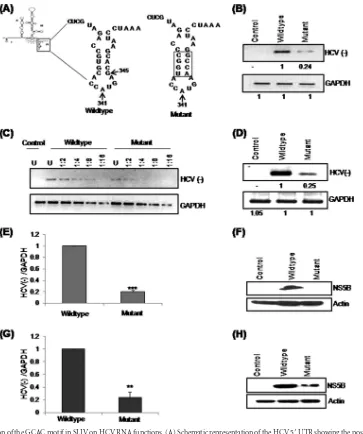
Mutation of the GCAC motif in the SLIV region affects HCV RNA functions.Previously, it was shown that the GCAC sequence motif (which is relatively conserved across HCV genotypes) near the initiator AUG in the SLIV region of the HCV internal ribo-somal entry site (IRES) is important for IRES-mediated transla-tion. Consequently, mutation of the GCAC motif reduces IRES activity drastically (32). However, the role of this GCAC motif in HCV replication is unknown. Thus, to investigate whether the GCAC motif influences HCV replication, we introduced a muta-tion into SLIV in an HCV monocistronic replicon (33) in such a way as to alter the primary nucleotide sequence of the GCAC motif, while a compensatory mutation in the other strand main-tained the secondary structure and GC content (Fig. 1A).
To study the effect of this mutation on HCV replication, Huh7 cells were transiently transfected within vitro-transcribed wild-type or mutated HCV monocistronic replicon RNA. Total RNA was isolated at 24 h posttransfection, and semiquantitative RT-PCR was performed (Fig. 1B). The mutated replicon showed a drastic reduction in HCV negative-strand synthesis compared to the wild-type replicon, suggesting the importance of the GCAC motif in replication.
PCR quantification can be performed only if the PCR product is measured within the exponential phase of the PCR, where the amount of amplified target is directly proportional to the amount of input target. To determine the exponential phase of the PCR empirically, serial dilutions of cDNA samples were made, and PCR amplification was performed over the same number of cy-cles. This assay also suggested that negative-strand RNA levels were low in the case of the mutated replicon (Fig. 1C).
Additionally, to discriminate negative-strand RNAs among a vast excess of positive-strand RNAs, RT-PCR using tagged prim-ers was performed to selectively measure the level of negative-strand synthesis (39). Mutation of the GCAC motif caused an ⬃75% reduction in HCV replication (Fig. 1D). Quantitative RT-PCR further confirmed these observations (Fig. 1E). In parallel, a Western blot analysis was performed where we could not detect NS5B in the case of the mutated replicon (Fig. 1F), which showed a drastic decrease in NS5B production from the mutated RNA.
Similarly, the effect of the mutation on HCV RNA replication in an infectious cell culture system was determined by transient transfection of Huh7.5 cells with JFH1 RNA. HCV negative-strand levels were measured at 24 h posttransfection by quantita-tive RT-PCR (Fig. 1G). The mutated RNA displayed a significant reduction (P⬍0.005) in RNA replication as well as a considerable decrease in the production of NS5B protein, as seen in the Western blot analysis (Fig. 1H).
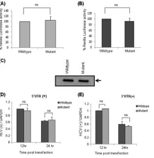
In the above-mentioned experiments, we normalized the transfection efficiency by performing transfection of Huh7 or Huh7.5 cells with the wild-type or mutated viral genome along with aRenillaluciferase reporter construct. At 24 h posttransfec-tion, luciferase assays were performed, and there was no difference in transfection efficiencies (Fig. 2AandB).
To rule out the possibility that low HCV RNA levels might be due to a decrease in the stability/integrity of the mutated viral genome rather than a defect in replication, we checked the integ-rity of wild-type and mutated RNAsin vitrousing formaldehyde agarose gel electrophoresis (Fig. 2C).
Treatment with the replication inhibitor hemin, an iron donor,
on November 7, 2019 by guest
http://jvi.asm.org/
increases intracellular iron levels. Elevated iron levels inactivate NS5B (viral RNA-dependent RNA polymerase [RdRp]) by allow-ing iron to bind specifically and with high affinity to NS5B’s Mg2⫹ binding pocket and thus suppressing new viral RNA synthesis without affecting translation (40,41). We specifically determined
theex vivostability of both the transfected RNAs (positive sense)
by blocking (using hemin) the formation of subsequent HCV RNA strands as a result of replication of input RNA. For this purpose, Huh7 cells were transfected with wild-type or mutated RNA and treated simultaneously with hemin (100M). Total RNA was isolated at two time points (12 and 24 h
posttransfec-tion), and quantitative RT-PCR was performed by using primers corresponding to the 5=and 3=UTRs to determine the levels of positive-strand RNA (Fig. 2D and E). We did not find any change in RNA levels, suggesting that both wild-type and mu-tated RNAs are equally stable, even after transfection. The rel-ative decrease in the RNA level at two subsequent time points might be the consequence of degradation of input RNA in the absence of replication.
Taken together, our results suggest that mutation in the
cis-acting element GCAC in SLIV reduces HCV RNA replication and translation without affecting its stability.
FIG 1Effect of mutation of the GCAC motif in SLIV on HCV RNA functions. (A) Schematic representation of the HCV 5=UTR showing the position of the mutation incorporated into the SLIV region of the HCV monocistronic subgenomic replicon (33). Nucleotide positions of initiator AUG (iAUG) and the GCAC motif are also marked. (B) Huh7 cells were transfected with eitherin vitro-transcribed wild-type or mutated HCV monocistronic subgenomic RNA. The cells were harvested at 24 h posttransfection, and HCV RNA (negative-strand) levels were quantified by using semiquantitative RT-PCR. (C) Serial dilutions of the cDNA samples (1:2, 1:4, 1:8, and 1:16) were made. Amplified PCR products were analyzed by electrophoresis on a 2% agarose gel. GAPDH was used as an internal control. “U” denotes undiluted cDNA sample. (D to F) Tagged cDNA RT-PCR (D), quantitative RT-PCR (E), and Western blot for NS5B using anti-NS5B antibody (F) for the experiment performed in panel B. Actin was used as a control to ensure equal loading. (G) Huh7.5 cells were transfected with wild-type or mutated HCV JFH1 RNA. The cells were harvested at 24 h posttransfection, and HCV RNA (negative-strand) levels were quantified by using quantitative RT-PCR. (H) For the experiment performed in panel G, Western blotting for NS5B was done by using anti-NS5B antibody. Actin was used as a control to ensure equal loading.
on November 7, 2019 by guest
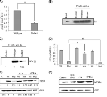
[image:4.585.111.474.63.497.2]Mutation of the GCAC motif reduces La interaction with HCV RNA.Human La protein interaction with the GCAC motif near the initiator AUG within the HCV IRES is critical for internal initiation of translation (31,32). To investigate whether this in-teraction is also important for HCV replication, Huh7 cells were transfected within vitro-transcribed wild-type or mutated repli-con RNA (positive sense) and simultaneously treated with hemin (100M) to block negative-strand synthesis. Cells were harvested at 24 h posttransfection, and ribonucleoprotein (RNP) complexes were immunoprecipitated by using anti-La antibody. The differ-ence in the association of La with input positive-sense wild-type and mutated RNAs was measured by quantitative RT-PCR using RNA isolated from the immunoprecipitated complex. Interest-ingly, we found a significant reduction (P⬍0.005) in HCV posi-tive-sense RNA levels in complex from mutated RNA compared to the wild type (Fig. 3A). Western blotting was performed to show equal precipitation of La by using anti-La antibody (Fig. 3B). This observation suggested that mutation of the GCAC motif re-duced La binding with positive-sense (input) RNA and hence de-creased negative-strand synthesis (as shown inFig. 1). This was confirmed by immunoprecipitation followed by semiquantitative
(data not shown) and tagged cDNA RT-PCR of HCV negative-strand RNA (Fig. 3C).
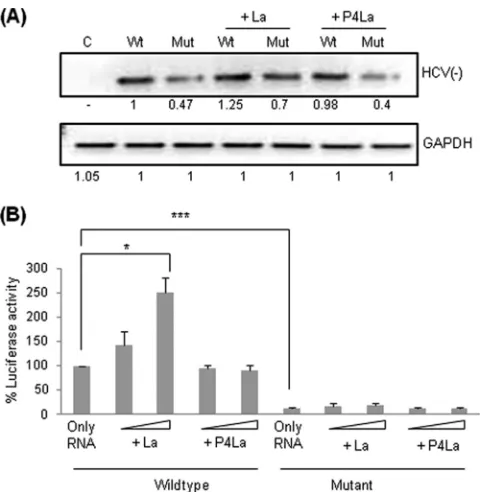
We further investigated whether La overexpression could rescue the observed RNA replication inhibition. Huh7 cells were transiently cotransfected with wild-type or mutated RNA and the plasmid construct expressing La. HCV RNA levels were quantified by using quantitative RT-PCR (Fig. 3D). Interest-ingly, La overexpression rescued the inhibition of replication and increased HCV negative-strand RNA levels significantly (P⬍0.05). P4La, a mutant La protein which interacts ineffi-ciently with the HCV IRES (42), was used as a negative control. P4La overexpression decreased HCV replication of both wild-type and mutated RNAs slightly, perhaps due to competition between endogenous La and P4La supplied by overexpression to bind to the GCAC motif. Similar observations were made at the protein level during Western blot analysis (Fig. 3E). La/ P4La overexpression was verified by semiquantitative RT-PCR (data not shown) and Western blot analysis (Fig. 3F). Taken together, our results suggest that mutation of the GCAC motif decreases La interaction with HCV positive-strand RNA and consequently affects negative-strand synthesis. This effect of FIG 2Effect of mutation of the GCAC motif on HCV RNA stability. (A) Huh7 cells were cotransfected with eitherin vitro-transcribed wild-type or mutated HCV monocistronic subgenomic RNA and aRenillaluciferase reporter construct. At 24 h posttransfection, a luciferase assay was performed, and the results are represented graphically (“ns” represents values that are not significant). (B) Huh7.5 cells were cotransfected with eitherin vitro-transcribed wild-type or mutated HCV JFH1 RNA and aRenillaluciferase reporter construct. At 24 h posttransfection, a luciferase assay was performed, and the results are represented graphically. (C) Integrity ofin vitro-transcribed wild-type and mutated HCV monocistronic RNAs was checked in a 0.8% formaldehyde agarose gel. (D and E) Huh7 cells were transfected with wild-type or mutated HCV monocistronic subgenomic RNA and treated simultaneously with hemin (100M). Total RNA was isolated at 12 h and 24 h posttransfection, and the level of input positive-strand HCV was checked by quantitative RT-PCR using primers corresponding to either the 5=UTR (D) or the 3=UTR (E). GAPDH was used as an internal control for normalization [HCV(⫹) denotes HCV positive strand].
on November 7, 2019 by guest
http://jvi.asm.org/
[image:5.585.136.451.64.388.2]the GCAC mutation can be rescued by supplying exogenous La by overexpression of the La gene.
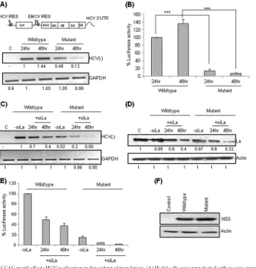
Mutation of the GCAC motif affects HCV replication inde-pendent of translation.The role of the La interaction with the GCAC motif in HCV replication might be dependent on or inde-pendent of translation. To investigate this, an HCV bicistronic replicon, pSGR-JFH1/Luc, where luciferase translation is driven by the HCV IRES and nonstructural HCV proteins are produced by the encephalomyocarditis virus (EMCV) IRES, was used (35). We mutated the GCAC motif in the HCV IRES of this bicistronic replicon without affecting the EMCV IRES, and semiquantitative RT-PCR was performed at 24 h and 48 h posttransfection. At the 24-h time point, we observed an⬃50% reduction in HCV nega-tive-strand synthesis of the GCAC-mutated RNA compared to the wild type (Fig. 4A), providing evidence that the GCAC motif has a distinct role in replication independent of translation. Quantita-tive RT-PCR further corroborated this result (data not shown). At 48 h posttransfection, we found only a marginal increase in HCV RNA levels in cells transfected with wild-type RNA but a
consid-erable decrease in cells transfected with mutated RNA, further suggesting that the mutated RNA does not replicate efficiently and that the GCAC motif is an essentialcis-acting element required for efficient HCV replication.
To investigate the effect of the GCAC motif mutation on HCV translation, in vitro-transcribed wild-type and mutated pSGR-JFH1/Luc RNAs werein vitrotranslated in RRL. A drastic reduc-tion in the internal initiareduc-tion of translareduc-tion of mutated RNA was observed (data not shown). This was further confirmed underex vivoconditions. Huh7 cells were transiently transfected with equal amounts of wild-type or mutated RNA. Cells were lysed at 24 and 48 h posttransfection, and extracts were assayed for luciferase ac-tivity. Consistent with thein vitroresults, translation efficiency was reduced drastically (P⬍0.0005) for the RNA with a mutated GCAC motif (Fig. 4B).
Furthermore, we investigated the effect of La silencing on rep-lication of wild-type and mutated pSGR-JFH1/Luc RNAs. Huh7 cells were transiently cotransfected with equal amounts of wild-type or mutated RNA and small interfering RNA targeting the La FIG 3Effect of mutation of the GCAC motif on La interaction with HCV RNA. (A) Huh7 cells were transfected within vitro-transcribed (positive-sense) wild-type or mutant monocistronic subgenomic RNA and simultaneously treated with hemin (100M). Cells were harvested at 24 h posttransfection, and ribonucleoprotein (RNP) complexes were immunoprecipitated by using anti-La antibody, followed by quantitative RT-PCR of the positive-sense RNA extracted from the immunoprecipitated complex. (B) Western blotting was done to verify equal precipitation of La. (C) Huh7 cells were transfected with wild-type or mutated HCV monocistronic subgenomic RNA. The cells were harvested at 24 h posttransfection, and RNA-protein complexes were precipitated by using anti-La antibody, followed by tagged cDNA RT-PCR for HCV negative-strand RNA. IP, immunoprecipitation. (D) La overexpression can rescue the inhibition of HCV RNA replication. Huh7 cells were cotransfected with wild-type or mutant HCV monocistronic subgenomic RNA along with a plasmid construct expressing either La or P4La (mutant La). The cells were harvested at 24 h posttransfection, and negative-strand HCV RNA levels were quantified by quantitative RT-PCR (“C” denotes control, “Wt” denotes wild-type RNA, “Mut” denotes mutated RNA, and “ns” represents values that are not significant). (E) Western blotting was done by using anti-NS5B antibody to check NS5B production levels. (F) La and P4La overexpression was checked by Western blot analysis using anti-La antibody.
on November 7, 2019 by guest
[image:6.585.115.474.66.390.2]gene (siLa) (50 nM). HCV RNA levels were quantified at 24 h and 48 h posttransfection (Fig. 4C). Although we observed a decrease in the replication of both wild-type and mutated RNAs after La silencing, this decrease was greater for RNA with the mutated GCAC motif. La silencing was verified by Western blot analysis (Fig. 4D). A similar observation was found when translation was tested in a luciferase-based translation assay (Fig. 4E).
To investigate if the GCAC mutation in the HCV IRES has any effect on the EMCV IRES activity of the pSGR-JFH1/Luc bicis-tronic RNA, we performed Western blot analyses to detect the viral proteins NS3 (Fig. 4F) and NS5B (data not shown) in the presence of hemin (100M). As expected, no change in the trans-lation level was observed for both wild-type and mutated RNAs.
Therefore, the results suggest that although viral proteins are being synthesized equally well in wild-type and mutated RNAs, replication is still inhibited, indicating that this replication inhibi-tion might be independent of translainhibi-tion inhibiinhibi-tion.
GCAC facilitates interaction between La and viral RdRp. Host and viral proteins interact with each other directly via pro-tein-protein interactions or indirectly via RNA to form a func-tional replication complex on positive-sense viral genomic RNA. Formation of these complexes is a prerequisite for replication (43,44). To investigate whether the GCAC motif located in the positive strand of the HCV IRES plays any role in mediating the interaction of La with viral RdRp (NS5B), we performed coimmu-noprecipitation assays. Huh7 cells were transfected within vitro -transcribed positive-sense wild-type or GCAC motif-mutated bicistronic subgenomic replicon RNA and treated with hemin (100M). Immunoprecipitation was performed at 24 h post-transfection by using anti-La antibody, and Western blot analysis was performed to detect NS5B. Although equal amounts of La were immunoprecipitated in the cases of both wild-type and mu-tated RNAs, less La-associated NS5B was immunoprecipimu-tated by the mutated RNA than by the wild type (Fig. 5A).
FIG 4Mutation of the GCAC motif affects HCV replication independent of translation. (A) Huh7 cells were transfected within vitro-transcribed wild-type or mutant pSGR-JFH1/Luc RNA. The cells were harvested at 24 h and 48 h posttransfection, and HCV RNA levels were quantified by using semiquantitative RT-PCR. The schematic above the panel represents the pSGR-JFH1/Luc replicon (35). (B) Luciferase levels were also assayed. (C) Effect of La silencing on HCV replication. Huh7 cells were cotransfected with wild-type or mutant pSGR-JFH1/Luc RNA and siLa (50 nM). The cells were harvested at 24 h and 48 h posttransfection, and HCV RNA levels were quantified by using semiquantitative RT-PCR. (D) Western blotting was done for La by using anti-La antibody to check silencing. Actin was used as a control to ensure equal loading. (E) A luciferase assay was also performed, and the results are represented graphically. (F) Western blotting for NS3 was done using by anti-NS3 antibody to check EMCV IRES-mediated translation. Actin was used as a control to ensure equal loading.
on November 7, 2019 by guest
http://jvi.asm.org/
[image:7.585.113.474.65.443.2]In a similar experiment, we performed immunoprecipitation with anti-NS5B antibody, followed by Western blot analysis for La. As in the above-described experiment, we found less La asso-ciated with NS5B when the GCAC motif was mutated (Fig. 5B).
These results suggest that the mutation in the GCAC motif of the HCV positive-strand RNA affects an interaction between La and NS5B, which may be crucial for initiation of negative-strand synthesis.
Exogenous La can rescue HCV replication inhibition but not translation inhibition.Since mutation of the GCAC motif inhib-its replication by affecting interactions between La and viral poly-merase, we were interested in investigating the effect of exogenous La on replication inhibition. To address this, Huh7 cells were tran-siently cotransfected with equal amounts of wild-type or mutated pSGR-JFH1/Luc RNA and a construct encoding La or P4La. HCV RNA levels were quantified at 24 h posttransfection by using semi-quantitative RT-PCR. As described above (Fig. 3D), we found that exogenous La rescued replication of the mutated RNA (Fig. 6A).
Similarly, luciferase assays performedex vivodemonstrated a dose-dependent increase in IRES activity for wild-type RNA but not for mutated RNA (Fig. 6B). This result was also confirmed underin vitroconditions by incubating wild-type and mutated pSGR-JFH1/Luc RNAs in the presence of increasing concentra-tions of purified recombinant La protein in thein vitrotranslation reaction mix. Consistent with theex vivoresults,in vitrodata also revealed a dose-dependent stimulation of HCV IRES-mediated translation for wild-type but not for mutated RNA (data not shown). These observations suggest that exogenous La supple-mentation can rescue replication inhibition but not IRES-medi-ated inhibition caused by GCAC motif mutation.
Mutation of the GCAC motif in the SLIV region affects the translation-to-replication switch. Previously, it was demon-strated that NS3 (viral factor) and human La protein (host factor) share similar binding sites in the SLIV region near the GCAC motif, and interplay between NS3 protease and human La protein regulates the translation-to-replication switch of HCV RNA (45). To investigate the effect of the GCAC motif mutation on the
trans-lation-to-replication switch, wild-type or mutated pSGR-JFH1/ Luc RNA was transiently cotransfected with either an NS3pro
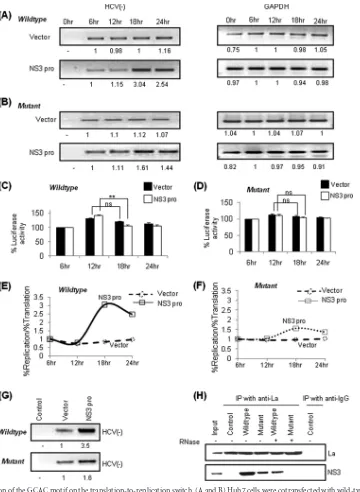
-en-coding construct or vector DNA. At different time points (0, 6, 12, 18, and 24 h posttransfection), HCV negative-strand RNA was measured by semiquantitative RT-PCR (Fig. 7AandB) and tagged cDNA RT-PCR (data not shown). GAPDH was used as an internal control. Overexpression of NS3pro caused a greater increase in HCV negative-strand wild-type RNA levels at the 18-h time than mutated RNA levels. Vector DNA was used as a control.
In parallel, luciferase assays were performed at the same time points (starting at 6 h posttransfection) to assess the rate of IRES-mediated translation (Fig. 7CandD). Ratios of HCV RNA ampli-fied (representing replication) to luciferase synthesis (represent-ing translation) at each time point showed that wild-type HCV negative-strand RNA levels at 18 h posttransfection were⬃ 3.6-fold higher than those of the vector control (Fig. 7E). This ratio was found to be only⬃1.7-fold higher than that of the vector control for mutated RNA (Fig. 7F).
To further confirm our observation, the replication complex was immunoprecipitated by using anti-NS5B antibody at 18 h posttransfection, and HCV negative-strand RNA isolated from the complex was quantified by semiquantitative RT-PCR (Fig. 7G). Our results clearly showed an increase in negative-strand synthesis in the immunoprecipitated complex from the Huh7 cells transfected with the plasmid construct overexpressing NS3pro.
Moreover, we investigated the effect of mutation on the inter-action between La, NS3, and HCV RNA during the translation-to-replication switch by coimmunoprecipitation assays. Less NS3 associated with La was immunoprecipitated by mutated RNA FIG 5Mutation of the GCAC motif affects interactions between La and NS5B.
Huh7 cells were transfected within vitro-transcribed wild-type or mutated pSGR-JFH1/Luc RNA along with treatment with hemin (100M). At 24 h posttransfection, immunoprecipitation was done by using anti-La antibody (A) or anti-NS5B antibody (B), followed by Western blot analysis for La and NS5B. Anti-IgG antibody was used as a negative control for immunoprecipi-tation. “Control” indicates no transfection.
FIG 6Effect of exogenous La on HCV replication and translation. (A) Huh7 cells were cotransfected with wild-type or mutated pSGR-JFH1/Luc RNA and a construct expressing La/P4La (mutant La). The cells were harvested at 24 h posttransfection, and replication was checked by semiquantitative RT-PCR (“C” denotes control, “Wt” denotes wild type, and “Mut” denotes mutant). (B) A luciferase assay was also performed to check HCV IRES-mediated trans-lation.
on November 7, 2019 by guest
[image:8.585.301.541.64.310.2] [image:8.585.91.237.65.224.2]than by the wild type, suggesting the importance of the GCAC motif in mediating the interaction of La and NS3 with HCV RNA (Fig. 7H).
To further validate the above-described observations, we used
translation inhibitors, i.e., puromycin and cycloheximide, which are known to induce dissociation or freezing of ribosomes, respec-tively, and hence have contrasting effects on negative-strand synthesis (46,47). Huh7 cells were transfected with wild-type or FIG 7Effect of mutation of the GCAC motif on the translation-to-replication switch. (A and B) Huh7 cells were cotransfected with wild-type (A) or mutant (B) pSGR-JFH1/Luc RNA and a construct expressing NS3proor the vector. Total RNA was isolated at different time points, and HCV RNA was quantified by using
semiquantitative RT-PCR (left). GAPDH was used as an internal control (right). (C and D) For the experiments detailed in panels A and B, luciferase assays were performed to detect translation levels. The percent luciferase activities were plotted for each reaction at different time points, taking the 6-h time point as 100% (control) (“ns” represents values that are not significant). (E and F) The ratio of percent replication to translation levels in vector-transfected control cells was compared with that of cells expressing NS3proand is graphically represented by plotting the ratio of percent replication to translation on theyaxis and time on
thexaxis. (G) For the experiments performed in panels A and B, the RNA protein complex was immunoprecipitated at 18 h posttransfection by using anti-NS5B antibody followed by semiquantitative RT-PCR for HCV negative-strand RNA. (H) Huh7 cells were cotransfected within vitro-transcribed wild-type or mutated pSGR-JFH1/Luc RNA and a construct expressing NS3. Immunoprecipitation was done at 18 h posttransfection by using anti-La antibody, followed by Western blotting for NS3 by using anti-NS3 antibody. Western blotting for La was done to verify equal precipitation. Anti-IgG antibody was used as a negative control for immunoprecipitation. “Control” represents no transfection.⫺and⫹represent “without RNase A treatment” and “with RNase A treatment,” respectively.
on November 7, 2019 by guest
http://jvi.asm.org/
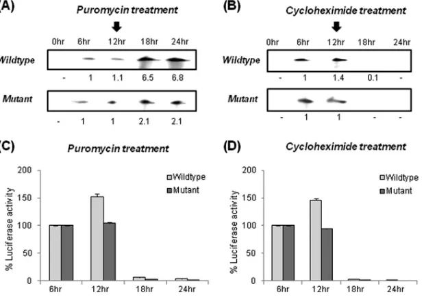
[image:9.585.112.472.64.556.2]mutated pSGR-JFH1/Luc RNA, followed by treatment with puro-mycin or cycloheximide at 12 h posttransfection. HCV negative-strand RNA was detected at different time points by using a two-cycle RNase protection assay (37). Our results clearly showed that puromycin treatment caused a greater increase in HCV negative-strand wild-type RNA synthesis at the 18-h time point than that of mutated RNA (Fig. 8A). In contrast, cycloheximide treatment caused a gradual decrease in both wild-type and mutated HCV negative-strand RNA levels (Fig. 8B). Controls were given no drug treatment (data not shown).
In parallel, we performed luciferase assays at different time points to determine the effect of puromycin and cycloheximide on HCV IRES-mediated translation (Fig. 8CandD). These observa-tions clearly suggested the importance of the GCAC motif in reg-ulating the translation-to-replication switch in the HCV life cycle by “directly” participating in negative-strand synthesis.
Site-directed mutagenesis reveals differential roles of indi-vidual GCAC motif nucleotides in HCV translation and replica-tion.To further investigate the role of the GCAC motif in replica-tion at the single-nucleotide level, different point mutareplica-tions (G345A, A347C, or C348G) were introduced into the pSGR-JFH1/Luc replicon, and HCV RNA levels produced by these mu-tated replicons were checked by semiquantitative RT-PCR. The G345A and C348G mutations caused a considerable reduction in HCV RNA levels, whereas the A347C mutation did not, suggest-ing that these nucleotides are not equally important for RNA rep-lication (Fig. 9A).
We further studied whether the various GCAC point muta-tions affected translation and replication equally or not. To study this, translation of RNAs containing point mutations was checked
in vitro(Fig. 9BandC). Interestingly, it was observed that the
A347C mutation had a greater inhibitory effect on translation than did the G345A and C348G mutations. Previously, it was shown that the A338U mutation renders RNA translationally
in-competent (48), whereas the C324U mutation does not affect translation (32). Therefore, RNAs bearing the A338U and C324U mutations were used as translation controls. Additionally, using a UV cross-linking assay, we investigated the binding of recombi-nant La protein with the wild-type and mutated RNAs. The results of thein vitrobinding studies of point mutants (Fig. 9D) corrob-orate the translation data (Fig. 9BandC), suggesting that it could be because of reduced and differential affinities of La for mutated RNAs. Taken together, our observations suggested differential ef-fects of GCAC motif nucleotides on replication and translation.
La protein promotes interaction of 5=and 3=ends of the HCV genome via the GCAC motif.Previously reported observations have shown that La protein binds both the 5=and 3=UTRs of the HCV genome (26, 49, 50). La also can undergo dimerization through its C terminus (20). It is therefore plausible that La pro-tein could act as a propro-tein bridge to mediate long-range RNA-RNA interactions between the 5=and 3=ends of the HCV genome for efficient RNA replication. To test this possibility, a 5=-3= co-precipitation assay was used, wherein radiolabeled 3=-UTR RNA was precipitated by using the biotin-labeled 5=UTR in the pres-ence of La protein (Fig. 10A). We observed a significant increase (P⬍0.0001) in the 5=-UTR–3=-UTR interaction in the presence of La protein (Fig. 10B). In contrast, in control reaction mixtures where La protein was absent or the unrelated protein BSA was used, very little coprecipitation was observed. Similarly, P4La (mutant La) could not coprecipitate 5=and 3=UTRs (data not shown). Moreover, increasing amounts of the unlabeled 5=- or 3=-UTR RNA could compete away the interaction between 5=and 3=UTRs (Fig. 10C), demonstrating that La linkage to both ends of the HCV RNA is a specific interaction.
Since the GCAC motif plays an important role in HCV repli-cation, it was therefore likely that La promotes this interaction between the 5=and 3=UTRs via the GCAC motif. To test this hypothesis, we performed a 5=-3=coprecipitation assay with mu-FIG 8The GCAC motif participates directly in enhancing HCV replication. (A and B) Huh7 cells were transfected within vitro-transcribed wild-type or mutated pSGR-JFH1/Luc RNA, followed by treatment with puromycin (A) or cycloheximide (B) (12 h posttransfection) and detection of HCV negative-strand RNA at different time points (as indicated) by using a two-cycle RNase protection assay (the arrow above the 12-h time point indicates the time point of addition of puromycin or cycloheximide). (C and D) Luciferase assays were performed to check HCV IRES-mediated translation.
on November 7, 2019 by guest
[image:10.585.136.455.67.284.2]tated RNAs; RNAs bearing an ACCG, G345A, C348G, or A338U mutation formed significantly fewer 5=-3=interactions, whereas the A347C and C324U mutations did not affect these interactions (Fig. 10DandE). Interestingly, coprecipitation assays of GCAC mutations gave results similar to those obtained by RT-PCR assays (Fig. 9A), suggesting that La mediating specific interactions be-tween the GCAC motif (within the 5=UTR) and the 3=UTR is an important mechanism required for efficient HCV RNA replica-tion.
DISCUSSION
Hepatitis C virus translation and replication are facilitated by sev-eral host and viral protein factors. One such host cell protein that facilitates HCV RNA translation as well as replication is the hu-man La protein (11,26,27). Previous publications reported that FIG 9Effect of point mutations of the GCAC motif on HCV replication and
translation. (A) Huh7 cells were transfected within vitro-transcribed wild-type or mutated pSGR-JFH1/Luc RNA. The cells were harvested at 24 h posttrans-fection, and HCV negative-strand RNA levels were quantified by using semi-quantitative RT-PCR. GAPDH was used as an internal control (ACCG indi-cates the same mutant represented inFig. 1A, whereas G345A, A347C, C348G, A338U, and C324U represent point mutants). (B) One microgram of wild-type and various mutated pSGR-JFH1/Luc RNAs was translated in RRL, and percent luciferase activities were plotted (“ns” represents data that are not significant). (C) Wild-type and mutated (G345A, A347C, and C348G) HCV IRES-Luc RNAs (42) were translatedin vitroin RRL, and percent luciferase activities were plotted. (D)In vitrobinding of␣-32P-labeled wild-type and
mutated HCV IRES RNAs with increasing concentrations of recombinant La was performed by using a UV cross-linking assay. No protein was used as a negative control.
FIG 10La protein interaction with the GCAC motif promotes 5=-to-3= link-age. (A) Schematic representation of the 5=-3=coprecipitation assay carried out with a biotinylated 5=UTR and a32P-labeled 3=UTR. The location of the
GCAC motif is also indicated. (B) Autoradiograph of precipitated RNAs in the absence or presence of increasing amounts of La (25, 50, and 100 ng). BSA (100 ng) was used as an unrelated protein control. (C) Increasing amounts of un-labeled (cold) 5=UTR and 3=UTR competed away the binding of the32
P-labeled 3=UTR to the biotinylated 5=UTR (Bio-5=UTR). (D and E) The same assay was performed with wild-type and various indicated mutated 5=-UTR RNAs in the presence of La (50 ng). Nonspecific RNA (unrelated nonviral RNA) that replaced the 5=UTR in the assay mixture was used as a control.
on November 7, 2019 by guest
http://jvi.asm.org/
[image:11.585.287.537.65.538.2] [image:11.585.47.291.70.468.2]and not of decreased replication. Our observations using a repli-cation inhibitor, hemin (40,41), suggested that mutation of the GCAC motif does not affect RNA stability.
Furthermore, we found that an important reason behind the low level of negative-strand synthesis in the case of the mutant was the reduction in La protein association with the positive-sense mutated RNA. Therefore, an overexpression of La allowed an in-crease in the level of replication of the mutant RNA significantly. Moreover, our observations from the experiments performed with the bicistronic replicon clearly indicated the possibility of the existence of an independent role of the La protein interaction with the GCAC motif in replication. Here, we also found that La over-expression could rescue replication inhibition but not translation inhibition. It is possible that, apart from binding the La, the GCAC motif is involved in some other tertiary RNA-RNA interactions in the HCV genome that might be critical for internal initiation of translation (but not replication). It seems that, in the case of the mutated bicistronic replicon RNA, these interactions are not re-stored even after supplying La exogenously, and thus, IRES-me-diated translation could not be rescued, although replication was restored to some extent.
The regulatory functions of IRES elements of HCV and picor-naviruses are mediated through their interactions with many host proteins (18,43,51). For example, PTB interacts with multiple pyrimidine tracts in the HCV 5=UTR and positively regulates HCV IRES translation (16). In the current study, we observed that thecis-acting element GCAC in the HCV IRES regulates the inter-action between the host factor La and the viral polymerase NS5B. While levels of NS5B synthesized by transfected (positive-sense) wild-type and mutated pSGR-JFH1/Luc RNAs were equal, the association of NS5B with La (and vice versa) in an immunopre-cipitated complex was reduced in mutated RNA compared to the wild type. Although we have not deciphered whether the interac-tion of NS5B and La in an RNP complex is a direct protein-protein interaction or is mediated indirectly via HCV RNA, it is still very clear that mutation of the GCAC motif affects this interaction. Therefore, we suggest that the GCAC motif mutation might block the initiation of negative-strand synthesis by affecting the binding of La protein to the GCAC motif and thus reduces the recruitment of NS5B at the replication site.
Recently, it was demonstrated that La protein and NS3 pro-tease share a binding region near the initiator AUG in SLIV. It has been speculated that as soon as viral positive-strand RNA enters a cell, La binds to the GCAC motif and facilitates synthesis of viral proteins by IRES-mediated translation. However, once a thresh-old level of NS3 protein is synthesized, NS3 inhibits the
La-medi-ated enhancement of translation by dislodging La protein from SLIV. This in turn allows HCV RNA to undergo replication (45). It is possible that NS3 does not dislodge La protein completely but instead competes with La until equilibrium is attained, as both proteins have similarKd(dissociation constant) values for binding to the HCV IRES. Therefore, it appears that this interaction of La with the GCAC motif switches its role from translation to replica-tion in the presence of NS3 by initiating the assembly of a func-tional replication complex on the positive strand involving NS5B and 5=- and 3=-UTR RNA elements. This hypothesis was sup-ported by our data where the mutation of the GCAC motif (which affects negative-strand synthesis) reduces La and NS3 interac-tions. Therefore, apart from the role of NS3 in translation inhibi-tion, this study suggests a possible role of NS3 in replication initi-ation, perhaps as a part of the replication complex formed by RNA circularization (as described below and as shown inFig. 11).
It is well known that extensive evolution of HCV quasispecies takes place during persistent infection, which may lead to escape from host immune effector mechanisms through changes in B-and T-cell epitopes (52). Our studies also provide support for the possibility that replication can be regulated by incorporating point mutations in HCV RNA to promote long-term persistence of HCV infection in humans. Moreover, this kind of regulation might be a general phenomenon in the HCV life cycle due to the high level of conservation of the GCAC motif across most geno-types.
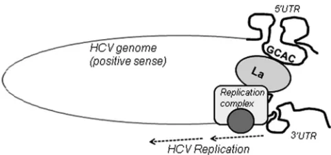
Genome circularization by long-range RNA-RNA interactions between the functional domains of the 5=and 3=ends is a common feature of many positive-stranded RNA viruses. However, specific details of circularization mechanisms likely vary from family to family, e.g., RNA-RNA interactions for flaviviruses (53), RNA-protein-protein-RNA interactions for picornaviruses (51), and cRNA-protein-protein-RNA interactions for coronaviruses and alphaviruses (54). The HCV genome contains well-defined RNA structural elements in its UTRs. Previous studies demonstrated that the protein-independent interaction of domain IIId and short stem-loop 5BSL3.2 is essential for replication (55). Also, previ-ously reported data indicated the participation of different cellular factors in inducing interactions between the HCV 5=and 3=UTRs (38,56). Although many studies have shown the interaction of La protein with both 5=and 3=UTRs (26,49,50), our study is the first to demonstrate that La binds to the 5=and 3=ends via the highly conserved GCAC motif and that this binding may aid in HCV genome circularization. As the GCAC motif plays an essential role
on November 7, 2019 by guest
[image:12.585.300.541.64.177.2]in La and NS5B associations, it is possible that 5=-to-3= cross-communication through La positively regulates HCV replication by providing a suitable platform for the viral polymerase NS5B to localize at the appropriate site to form a functional replication complex (Fig. 11).
Since La has RNA chaperone activity, it is also possible that this activity could be important for viral genome replication in various ways, e.g., by assisting in refolding of local RNA elements into different conformations and/or annealing complementary se-quences in the 5=and 3=UTRs. There may be many other factors involved in HCV RNA replication that may interact along with La, NS5B, and NS3 at the GCAC motif. Future studies should focus on identifying the protein partners of this complex. Furthermore, it would be interesting to investigate whether this RNA-protein complex interacts with any other part of the HCV RNA to enhance the replication process.
Taken together, our study demonstrates that human La pro-tein plays a critical role in regulating HCV replication by interact-ing with the GCAC motif. Our results firmly support the existence of La-dependent 5=-to-3=communication in the HCV genome that involves RNA elements essential for viral translation and rep-lication. Particularly, this study highlights the role of the GCAC motif within the IRES in HCV RNA replication in addition to its role in internal initiation of translation. Therefore, we may also consider this motif as a potent antiviral target. The interaction of La and GCAC was targeted previously to inhibit primarily IRES-mediated translation (42). However, this interaction can also be targeted specifically to block viral RNA replication. Since the La protein is less abundant in the cytoplasm of the host cell than in the nucleus, blocking of this protein might have drastic effects on viral RNA functions. These findings may also have important con-sequences for understanding the HCV replicative cycle and the genetic variability of the virus.
ACKNOWLEDGMENTS
We are grateful to Ralf Bartenschlager and Takaji Wakita for sharing plas-mid constructs. We thank Charles M. Rice for providing Huh7.5 cells. We also thank Rebecca Cerio for critical reading of the manuscript and our laboratory members for their helpful discussions.
This work was supported by a grant from the Department of Biotech-nology, India, to S.D. A.K. and U.R. were supported by predoctoral fel-lowships from the Council of Scientific and Industrial Research, India.
REFERENCES
1.Suzuki T, Aizaki H, Murakami K, Shoji I, Wakita T.2007. Molecular biology of hepatitis C virus. J. Gastroenterol.42:411– 423.
2.Suzuki T, Ishii K, Aizaki H, Wakita T.2007. Hepatitis C viral life cycle. Adv. Drug Deliv. Rev.59:1200 –1212.
3.Brown EA, Zhang H, Ping LH, Lemon SM.1992. Secondary structure of the 5=nontranslated regions of hepatitis C virus and pestivirus genomic RNAs. Nucleic Acids Res.20:5041–5045.
4.Choo QL, Kuo G, Weiner AJ, Overby LR, Bradley DW, Houghton M.
1989. Isolation of a cDNA clone derived from a blood-borne non-A, non-B viral hepatitis genome. Science244:359 –362.
5.Tsukiyama-Kohara K, Iizuka N, Kohara M, Nomoto A.1992. Internal ribosome entry site within hepatitis C virus RNA. J. Virol.66:1476 –1483. 6.Yi M, Lemon SM.2003. 3=nontranslated RNA signals required for
rep-lication of hepatitis C virus RNA. J. Virol.77:3557–3568.
7.Ito T, Lai MM.1999. An internal polypyrimidine-tract-binding protein-binding site in the hepatitis C virus RNA attenuates translation, which is relieved by the 3=-untranslated sequence. Virology254:288 –296. 8.Moradpour D, Penin F, Rice CM.2007. Replication of hepatitis C virus.
Nat. Rev. Microbiol.5:453– 463.
9.Tanaka T, Kato N, Cho MJ, Shimotohno K.1995. A novel sequence
found at the 3=terminus of hepatitis C virus genome. Biochem. Biophys. Res. Commun.215:744 –749.
10. Blight KJ, Kolykhalov AA, Rice CM.2000. Efficient initiation of HCV RNA replication in cell culture. Science290:1972–1974.
11. Domitrovich AM, Diebel KW, Ali N, Sarker S, Siddiqui A.2005. Role of La autoantigen and polypyridimine tract binding protein in HCV replica-tion. Virology335:72– 86.
12. Friebe P, Lohmann V, Krieger N, Bartenschlager R.2001. Sequences in the 5=nontranslated region of hepatitis C virus required for RNA replica-tion. J. Virol.75:12047–12057.
13. Jopling CL, Yi M, Lancaster AM, Lemon SM, Sarnow P.2005. Modu-lation of hepatitis C virus RNA abundance by a liver-specific microRNA. Science309:1577–1581.
14. Shimakami T, Yamane D, Jangra RK, Kempf BJ, Sapniel C, Barton DJ, Lemon SM.2012. Stabilization of hepatitis C virus RNA by an Ago2-miR-122 complex. Proc. Natl. Acad. Sci. U. S. A.109:941–946.
15. Lohmann V, Korner F, Koch J, Herian U, Theilmann L, Bartenschlager R.1999. Replication of subgenomic hepatitis C virus RNAs in a hepatoma cell line. Science285:110 –113.
16. Ali N, Siddiqui A. 1995. Interaction of polypyrimidine tract-binding protein with the 5=noncoding region of the hepatitis C virus RNA genome and its functional requirement in internal initiation of translation. J. Virol.
69:6367– 6375.
17. Chung RT, Kaplan LM.1999. Heterogeneous nuclear ribonucleoprotein I (hnRNP-I/PTB) selectively binds the conserved 3=terminus of hepatitis C viral RNA. Biochem. Biophys. Res. Commun.254:351–362.
18. Fukushi S, Okada M, Kageyama T, Hoshino FB, Nagai K, Katayama K.
2001. Interaction of poly(rC)-binding protein 2 with the 5=-terminal stem loop of the hepatitis C-virus genome. Virus Res.73:67– 69.
19. Tan EM.1989. Antinuclear antibodies: diagnostic markers for autoim-mune diseases and probes for cell biology. Adv. Immunol.44:93–151. 20. Wolin SL, Cedervall T. 2002. The La protein. Annu. Rev. Biochem.
71:375– 403.
21. Madore SJ, Wieban ED, Pederson T.1984. Eukaryotic small ribonucleo-proteins. Anti-La human autoantibodies react with U1 RNA-protein complexes. J. Biol. Chem.259:1923–1933.
22. Ford LP, Shay JW, Wright WE.2001. The La antigen associates with the human telomerase ribonucleoprotein and influences telomere length in vivo. RNA7:1068 –1075.
23. Francoeur AM, Mathews MB.1982. Interaction between VA RNA and the lupus antigen La: formation of a ribonucleoprotein particle in vitro. Proc. Natl. Acad. Sci. U. S. A.79:6772– 6776.
24. Chang YN, Kenan DJ, Keene JD, Gatignol A, Jeang KT.1994. Direct interactions between autoantigen La and human immunodeficiency virus leader RNA. J. Virol.68:7008 –7020.
25. Meerovitch K, Svitkin YV, Lee HS, Lejbkowicz F, Kenan DJ, Chan EK, Agol VI, Keene JD, Sonenberg N.1993. La autoantigen enhances and corrects aberrant translation of poliovirus RNA in reticulocyte lysate. J. Virol.67:3798 –3807.
26. Ali N, Siddiqui A.1997. The La antigen binds 5=noncoding region of the hepatitis C virus RNA in the context of the initiator AUG codon and stimulates internal ribosome entry site-mediated translation. Proc. Natl. Acad. Sci. U. S. A.94:2249 –2254.
27. Ali N, Pruijn GJM, Kenan DJ, Keene JD, Siddiqui A.2000. Human La antigen is required for the hepatitis C virus internal ribosome entry site-mediated translation. J. Biol. Chem.275:27531–27540.
28. Kim YK, Jang SK.1999. La protein is required for efficient translation driven by encephalomyocarditis virus internal ribosomal entry site. J. Gen. Virol.80:3159 –3166.
29. Kim YK, Back SH, Rho J, Lee SH, Jang SK. 2001. La autoantigen enhances translation of BiP mRNA. Nucleic Acids Res.29:5009 –5016. 30. Goodier JL, Fan H, Maraia RJ.1997. A carboxy-terminal basic region
controls RNA polymerase III transcription factor activity of human La protein. Mol. Cell. Biol.17:5823–5832.
31. Pudi R, Abhiman S, Srinivasan N, Das S.2003. Hepatitis C virus internal ribosome entry site-mediated translation is stimulated by specific interac-tion of independent regions of human La autoantigen. J. Biol. Chem.
278:12231–12240.
32. Pudi R, Srinivasan P, Das S.2004. La protein binding at the GCAC site near the initiator AUG facilitates the ribosomal assembly on the hepatitis C virus RNA to influence internal ribosome entry site-mediated transla-tion. J. Biol. Chem.279:29879 –29888.
33. Frese M, Barth K, Kaul A, Lohmann V, Schwarzle V, Bartenschlager R.
on November 7, 2019 by guest
http://jvi.asm.org/
J. Virol.65:3384 –3387.
38. Isken O, Baroth M, Grassmann CW, Weinlich S, Ostareck DH, Os-tareck-Lederer A, Behrens SE. 2007. Nuclear factors are involved in hepatitis C virus RNA replication. RNA13:1675–1692.
39. Craggs JK, Ball JK, Thomson BJ, Irving WL, Grabowska AM.2001. Development of a strand-specific RT-PCR based assay to detect the repli-cative form of hepatitis C virus RNA. J. Virol. Methods94:111–120. 40. Fillebeen C, Rivas-Estilla AM, Bisaillon M, Ponka P, Muckenthaler M,
Hentze MW, Koromilas AE, Pantopoulous K.2005. Iron inactivates the RNA polymerase NS5B and suppresses subgenomic replication of hepati-tis C virus. J. Biol. Chem.280:9049 –9057.
41. Bartolomei G, Cevik RE, Marcello A.2011. Modulation of hepatitis C virus replication by iron and hepcidin in Huh7 hepatocytes. J. Gen. Virol.
92:2072–2081.
42. Mondal T, Ray U, Manna AK, Gupta R, Roy S, Das S.2008. Structural determinant of human La protein critical for internal initiation of trans-lation of hepatitis C virus RNA. J. Virol.82:11927–11938.
43. Isken O, Grassmann CW, Sarisky RT, Kann M, Zhang S, Grosse F, Kao PN, Behrens SE.2003. Members of the NF90/NFAR protein group are involved in the life cycle of a positive-strand RNA virus. EMBO J.22:5655– 5665.
44. Scheller N, Mina LB, Galão RP, Chari A, Giménez-Barcons M, Noueiry A, Fischer U, Meyerhans A, Diez J.2009. Translation and replication of
Virol.2:296 –307.
50. Spångberg K, Wiklund L, Schwartz S.2001. Binding of the La autoanti-gen to the hepatitis C virus 3=untranslated region protects the RNA from rapid degradation in vitro. J. Gen. Virol.82:113–120.
51. Herold J, Andino R.2001. Poliovirus RNA replication requires genome circularization through a protein-protein bridge. Mol. Cell7:581–591. 52. Kato N, Sekiya H, Ootsuyama Y, Nakazawa T, Hijikata M, Ohkoshi S,
Shimotohno K.1993. Humoral immune response to hypervariable region 1 of the putative envelope glycoprotein (gp70) of hepatitis C virus. J. Virol.
67:3923–3930.
53. Hahn CS, Hahn YS, Rice CM, Lee E, Dalgarno L, Strauss EG, Strauss JH.1987. Conserved elements in the 3=untranslated region of flavivirus RNAs and potential cyclization sequences. J. Mol. Biol.198:33– 41. 54. Frolov I, Hardy R, Rice CM.2001.cis-Acting RNA elements at the 5=end
of Sindbis virus genome RNA regulate minus- and plus-strand RNA syn-thesis. RNA7:1638 –1651.
55. Romero-López C, Berzal-Herranz A. 2009. A long-range RNA-RNA interaction between the 5=and 3=ends of the HCV genome. RNA15: 1740 –1752.
56. Wang L, Jeng KS, Lai MM.2011. Poly(C)-binding protein 2 interacts with sequences required for viral replication in the hepatitis C virus (HCV) 5=untranslated region and directs HCV RNA replication through circularizing the viral genome. J. Virol.85:7954 –7964.