In Vivo
Knockdown of the Herpes Simplex Virus 1
Latency-Associated Transcript Reduces Reactivation from Latency
Zachary L. Watson,
aShannan D. Washington,
dDane M. Phelan,
aAlfred S. Lewin,
aSonal S. Tuli,
bGregory S. Schultz,
cDonna M. Neumann,
d,eDavid C. Bloom
aaDepartment of Molecular Genetics and Microbiology, University of Florida College of Medicine, Gainesville,
Florida, USA
bDepartment of Ophthalmology, University of Florida College of Medicine, Gainesville, Florida, USA
cDepartment of Obstetrics and Gynecology, University of Florida College of Medicine, Gainesville, Florida, USA
dDepartment of Pharmacology and Experimental Therapeutics, Louisiana State University Health Sciences
Center, New Orleans, Louisiana, USA
eDepartment of Ophthalmology, Louisiana State University Health Sciences Center, New Orleans, Louisiana,
USA
ABSTRACT
During herpes simplex virus (HSV) latency, most viral genes are
si-lenced, with the exception of one region of the genome encoding the
latency-associated transcript (LAT). This long noncoding RNA was originally described as
having a role in enhancing HSV-1 reactivation. However, subsequent evidence
show-ing that the LAT blocked apoptosis and promoted efficient establishment of latency
suggested that its effects on reactivation were secondary to establishment. Here, we
utilized an adeno-associated virus (AAV) vector to deliver a LAT-targeting
hammer-head ribozyme to HSV-1-infected neurons of rabbits after the establishment of
HSV-1 latency. The rabbits were then induced to reactivate latent HSV-1. Using this
model, we show that decreasing LAT levels in neurons following the establishment
of latency reduced the ability of the virus to reactivate. This demonstrates that the
HSV-1 LAT RNA has a role in reactivation that is independent of its function in
es-tablishment of latency. In addition, these results suggest the potential of AAV
vec-tors expressing LAT-targeting ribozymes as a potential therapy for recurrent HSV
dis-ease such as herpes stromal keratitis, a leading cause of infectious blindness.
IMPORTANCE
Herpes simplex virus (HSV) establishes a lifelong infection and
re-mains dormant (latent) in our nerve cells. Occasionally HSV reactivates to cause
dis-ease, with HSV-1 typically causing cold sores whereas HSV-2 is the most common
cause of genital herpes. The details of how HSV reactivates are largely unknown.
Most of HSV’s genes are silent during latency, with the exception of RNAs made
from the latency-associated transcript (LAT) region. While viruses that make less LAT
do not reactivate efficiently, these viruses also do not establish latency as efficiently.
Here we deliver a ribozyme that can degrade the LAT to the nerve cells of latently
infected rabbits using a gene therapy vector. We show that this treatment blocks
re-activation in the majority of the rabbits. This work shows that the LAT RNA is
impor-tant for reactivation and suggests the potential of this treatment as a therapy for
treating HSV infections.
KEYWORDS
gene therapy, latency, lncRNA, reactivation, ribozymes
H
erpes simplex virus (HSV) establishes a lifelong latent infection within peripheral
nerve ganglia following initial infection of the mucus membranes supplied by the
neurons. During latency, the viral genomes are maintained as circular episomes within
the nucleus of the neuron. Periodically, the virus reactivates from individual neurons,
and the newly produced virions are transported via the nerve fibers to the original site
Received8 May 2018Accepted29 May 2018
Accepted manuscript posted online6 June
2018
CitationWatson ZL, Washington SD, Phelan
DM, Lewin AS, Tuli SS, Schultz GS, Neumann DM, Bloom DC. 2018.In vivoknockdown of the herpes simplex virus 1 latency-associated transcript reduces reactivation from latency. J Virol 92:e00812-18.https://doi.org/10.1128/JVI .00812-18.
EditorRozanne M. Sandri-Goldin, University of
California, Irvine
Copyright© 2018 American Society for
Microbiology.All Rights Reserved. Address correspondence to David C. Bloom, dbloom@ufl.edu.
VIRUS-CELL INTERACTIONS
crossm
on November 6, 2019 by guest
http://jvi.asm.org/
of infection. HSV typically infects the mucosal epithelia of the oral cavity or eyes (HSV-1)
and also the genitalia (HSV-2), where it can cause cold sores or ocular herpes and
genital herpes, respectively. Of special significance, HSV-1 reactivation in the eyes of
some individuals is responsible for herpes stromal keratitis (HSK), which is the leading
cause of infectious blindness in the United States (1). During latency, most viral genes
are silenced, with the exception of one region of the genome encoding the
latency-associated transcript (LAT). The HSV-1 LAT is transcribed from the viral long repeats as
an 8.3- to 8.5-kb polyadenylated primary transcript (Fig. 1). This long noncoding RNA
(lncRNA) is the only viral transcript known to accumulate abundantly during latency
(2–4). The primary transcript is spliced to yield a 2.0-kb intron (5). This intron is highly
stable, with an observed half-life of 24 h in cell culture (6). It is also stable
in vivo
and
is easily detectable by
in situ
hybridization within about 25 to 30% of latently infected
ganglionic neurons (7, 8). Genetic studies involving deletions and mutations within the
LAT promoter and 5
=
exon-coding region have yielded a multitude of phenotypes.
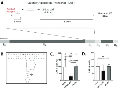
FIG 1Schematic of the HSV-1 genome showing the regions encoding the HSV-1 LAT and design andin vitrotesting results of a ribozyme targeting the LAT. (A) The LAT locus (located in both copies of the RLregion of the genome) is expanded to show the 8.5-kb primary LAT, the 5=and 3=exons, and the stable 2.0-kb intron. (B) Design of a synthetic hammerhead ribozyme targeting the LAT 5=exon. The 5=exon of the HSV-1 LAT was analyzed for hammerhead cleavage sequences. Antisense flanking regions were designed around the conserved catalytic domain to hybridize with the region surrounding the selected triplet. Mfold analysis predicted base pairing in stem II (arrow), but not in the flanking regions, so as to rule out intramolecular base pairing interfering with efficient binding between the ribozyme and the target. (C and D) Quantification of the HSV-1 LAT 5=exon (C) and the 2.0-kb LAT intron (D) in rabbit skin cells following transfection with a LAT-expressing plasmid and either mock treated or cotransfected with LATRz-235 or RhoRz. RNA was isolated, cDNA was transcribed, and LAT levels were analyzed by TaqMan real-time PCR that was normalized to the untargeted cellular control rabbit GAPDH. Normalized data are graphed as percentages of the 5=exon or LAT intron in treated cells relative to untreated cells (None). Each bar represents the average expression of four wells of a 24-well plate (n⫽4). Error bars represent standard deviations.
on November 6, 2019 by guest
http://jvi.asm.org/
[image:2.585.51.536.80.450.2]These pertain to antiapoptosis (9), neuronal tropism (10), reactivation (11–14), leakiness
of lytic gene transcription during latency (15), and histone H3 methylation (16, 17).
Study of the LAT region is further complicated by the fact that there are at least 8
microRNAs as well as other noncoding RNAs (ncRNAs) transcribed from the LAT locus,
whose functions are still being elucidated by mutational analysis (for a review, see
reference 18). While LAT promoter mutants display many phenotypes, the mechanism
of the LAT RNA in these processes is unknown.
LAT promoter mutants exhibit a reduction in genome copy numbers 2- to 3-fold
within sensory ganglia compared to wild-type HSV, presumably due to the LAT’s ability
to protect neurons from apoptosis (9, 19) or cell death (20) during the later stages of
the acute infection, as the virus establishes neuronal latency. Given this observation, it
has been generally accepted that decreased reactivation of LAT mutants is a
conse-quence of fewer latent genomes being available to reactivate (21), though conclusive
evidence is lacking. Therefore, the goal of this study was to determine if the LAT RNA
is directly involved in facilitating HSV-1 reactivation. To examine this specific function
of the LAT RNA, we performed
in vivo
knockdown of the LAT after latency had been
established. Adeno-associated virus (AAV) vectors were used to deliver a ribozyme to
target the HSV-1 LAT being expressed in latently infected sensory neurons. This
approach exploits the ability of some serotypes of AAV to be inoculated on the skin or
in the cornea, subsequently taken up by nerve termini, and ultimately transported by
axonal transport to the neuronal cell bodies, where the HSV-1 latent infection resides
(22). We have previously used this approach to deliver small interfering RNAs (siRNAs)
to latently infected neurons in the rabbit trigeminal ganglia (TG) to knockdown the
cellular insulator protein CTCF (23). Here we demonstrate that using an AAV vector to
deliver a ribozyme targeting the LAT after latency has been established blocks induced
reactivation in
⬎
60% of the rabbit eyes.
RESULTS
A ribozyme targeting the HSV-1 LAT knocks down the LAT RNA
in vitro
.
For
targeting the LAT, we chose a hammerhead ribozyme, a class of catalytic RNA
mole-cules composed of a catalytic loop and three stems that cleave RNAs with high
specificity (24). Hammerhead ribozymes can be engineered to act in
trans
(25, 26) to
specifically target virtually any RNA sequence of interest. Our group has designed
multiple such ribozymes against HSV-1 transcripts and tested their efficacy both
in vitro
and
in vivo
(27). However, to date none of these ribozymes have been directed to the
LAT RNA, nor have they been delivered to neurons
in vivo
. A synthetic hammerhead
ribozyme targeting the HSV-1 LAT 5
=
exon (Fig. 1A) was designed, synthesized, and
cloned into an AAV-green fluorescent protein (GFP) plasmid and was designated
LATRz-235, indicating the target cleavage site at the 235th nucleotide of the LAT 5
=
exon sequence (Fig. 1B and Table 1). The 5
=
exon region of the LAT was targeted initially
because this region corresponds to the regions mapped by deletion mutants that
resulted in decreased reactivation (28). The efficiency of target knockdown was tested
by cotransfecting rabbit skin cells with this plasmid alongside a LAT expression plasmid
and measuring the amount of LAT RNA by reverse transcription followed by TaqMan
real-time PCR (Fig. 1C). RNA levels were normalized to the nontargeted housekeeping
gene transcript for GAPDH (glyceraldehyde-3-phosphate dehydrogenase). Identical
transfections were done using a plasmid encoding an antirhodopsin ribozyme that is
not predicted to have an effect on LAT RNA expression as a control.
LAT 5
=
exon expression was reduced by 71% in cells cotransfected with the plasmid
expressing LATRz-235 compared to LAT levels in cells transfected with the LAT
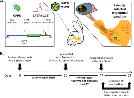
expres-TABLE 1Sequence of LATRz-235 and its target in the LAT 5=exonRibozyme Sequence Target sequence
LATRz-235 GGACACUGAUGAGCGCUUCGGCGCGAAACGAAC GUUCGUCUGUCC
RhodRz397a GUACCAAAGCGCGGCUUCGCGAGUAGUCAAGCC CAUGGUCUUCGG
aDesign and characterization described in reference 26.
In VivoKnockdown of HSV-1 LAT Reduces Reactivation Journal of Virology
on November 6, 2019 by guest
http://jvi.asm.org/
[image:3.585.41.372.82.117.2]sion plasmid only. In contrast, LAT 5
=
exon expression was not affected by
cotransfec-tion of the control non-HSV-specific antirhodopsin ribozyme (Fig. 1C). When levels of
the LAT intron were examined, there was no significant reduction in level, likely due to
its long half-life (Fig. 1D). No amplification was detected in controls lacking reverse
transcriptase (not shown). These results demonstrate that LATRz-235 is effective in
reducing the levels of the primary LAT transcript. We then used LATRz-235 to knock
down LAT
in vivo
using the strategy and overall design depicted in Fig. 2A and B).
Delivery of the AAV vector expressing the LAT ribozyme to the eyes of latently
infected rabbits blocks HSV-1 reactivation.
Since the goal of this study was to
determine whether
in vivo
knockdown of the LAT RNA after latency is established
affects the ability of HSV-1 to reactivate, the rabbit model was used. The advantage of
the rabbit model for the study of HSV-1 reactivation is that the latent virus can be
efficiently and synchronously induced to reactivate following transcorneal
iontopho-resis of

-adrenergic agonists, such as epinephrine (29). In addition, the rabbit model
has many features that parallel HSV-1 recurrent disease in humans, including
reactiva-tion at the original site of infecreactiva-tion. Rabbits are infected in both eyes with HSV-1 by
ocular scarification, and after 30 days, the virus is latent within the trigeminal ganglia,
and no infectious virus can be detected in the TG or in the tears, except for occasional
spontaneous shedding (30, 31). In this study, both eyes of latently infected rabbits were
treated with either of two AAV vectors. One set of rabbits was treated with an AAV
vector expressing a ribozyme targeting human rhodopsin (AAV-RhoRz) as a control,
whereas the other set of rabbits were treated with the HSV-1 LAT-targeting ribozyme
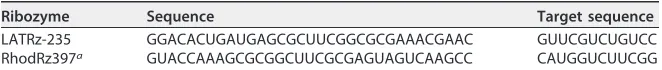
FIG 2Delivery of ribozyme targeting the HSV-1 LAT into latently infected rabbits. (A) The LAT-targeting ribozyme was cloned and packaged in an AAV vector (AAV-LATRz-235) that also contains a humanized green fluorescent protein (GFPh) reporter. This vector can be applied to the corneas of rabbits latently infected with HSV-1, allowing the AAV to be taken up by nerve termini under the cornea, and subsequent transport of the AAV to the trigeminal ganglia (TG) neurons. (B) Time line for testing the effect of the LAT ribozyme on epinephrine-induced reactivation.
on November 6, 2019 by guest
http://jvi.asm.org/
[image:4.585.45.499.79.413.2](AAV-LATRz-235). We have previously demonstrated that AAV applied to the rabbit eye
is efficiently transported to the TG and can efficiently transduce
⬎
70% of the total
neurons (22). Seventeen days after treatment with AAV, the rabbit eyes were
ionto-phoresed with epinephrine and swabbed daily for 12 days to collect tears. These swabs
were then assayed for the presence of infectious virus to measure reactivation. HSV-1
reactivation was detected (at least one positive swab) in only 33% of rabbit eyes
treated with AAV-LATRz-235 compared to 80% of those treated with AAV-RhoRz
(Fig. 3) (
P
⫽
0.04). In addition, the total number of positive swabs was significantly
reduced in the AAV-LATRz-235-treated group (
P
⫽
0.0026). Therefore,
AAV-LATRz-235 vector treatment significantly blocked detectable reactivation in the majority of
the rabbits and eyes compared to controls and reduced the overall shedding of
virus.
After the reactivation study was complete, rabbit TG were dissected, and one TG per
rabbit was processed and analyzed by immunohistochemistry (IHC) to detect GFP
expression from AAV-LATRz-235. These analyses revealed that
⬎
80% of the neurons in
the TG were transduced by AAV and expressed GFP (Fig. 4A and B). The other TG was
subjected to DNA/RNA extraction. The RNA was reverse transcribed, and quantitative
PCR (qPCR) analysis was performed to quantify the HSV-1 LAT (Fig. 4C). Significantly less
LAT was detected in the AAV LATRz-235-treated groups. When the DNA from the same
TG was analyzed by qPCR, there were no differences in HSV-1 genomes between the
two groups (Fig. 4D). Taken together, these results demonstrate that AAV LATRz-235
decreases LAT levels in the latent ganglia and that this reduction results in decreased
reactivation, without destabilizing latent genomes or measurably interfering with their
maintenance. Finally, when the levels of the LAT intron RNA were examined by reverse
transcriptase quantitative PCR (RT-qPCR), there was no significant difference between
the AAV-LATRz-235- and AAV-RhoRz-treated groups (Fig. 4E). This is consistent with the
degree of knockdown of the LAT intron
in vitro
(Fig. 1D) and is likely due to the high
stability of the intron.
DISCUSSION
In this study, we demonstrate that a ribozyme that targets the HSV-1 LAT, when
delivered to the eye of a rabbit that has been latently infected with HSV-1, completely
FIG 3Treatment with AAV LATRz-235 reduces induced reactivationin vivo. Eyes of latently infected rabbits were treated with either AAV-LAT Rz-235 or AAV-RhoRz. Seventeen days after AAV vector treatment, the rabbits were induced to reactivate. The eyes were then swabbed daily for 12 days to detect infectious HSV-1 as evidence of reactivation. Data are grouped by rabbits (L1 to L9, treated with the AAV LATRz-235, and R1 to R11, treated with the AAV-RhoRz) and by right (OD) and left (OS) eyes of each rabbit. A plus sign (⫹) indicates that infectious virus was detected in that eye for that day. Rabbits or eyes that exhibited at least one positive swab during the 12-day period are in red. Red crosshatches through OS for R9 and OD for R10 indicate that these eyes were prevented from being iontophoresed due to corneal scarring and were excluded from the reactivation study. Percentages of rabbits, eyes, or swabs that were positive for the two treatment groups are indicated at the bottom of each column. Exact versions of chi-square analysis for all pairwise comparisons with control (AAV-RhoRz) were performed.
In VivoKnockdown of HSV-1 LAT Reduces Reactivation Journal of Virology
on November 6, 2019 by guest
http://jvi.asm.org/
[image:5.585.45.543.73.257.2]blocks detectable reactivation of the virus in
⬎
60% of the eyes compared to eyes
treated with an AAV vector expressing a ribozyme targeting a heterologous target.
This result supports a direct role of the HSV-1 LAT RNA in facilitating efficient
reactivation from latency, which is separate from its function in enhancing
estab-lishment of latency.
It is important to note that these findings are not in conflict with studies that have
provided clear evidence for a role of the HSV-1 LAT in efficient establishment (19, 32)
and/or maintenance (33, 34) of latent infections. Studies have shown that HSV-1
recombinants with deletions in the LAT promoter and portions of the 5
=
exon coding
region have an increase in apoptotic neurons during the acute infection (9, 20, 35),
resulting in a 2- to 3-fold decrease in total HSV-1 DNA detected in the ganglia during
latency (36). Even though LAT promoter mutants also exhibited a 90% or greater
reduction in their ability to reactivate in the rabbit eye (14, 37), it previously was not
possible to determine the role of qualitative or quantitative differences in
establish-ment of latency on the ability to reactivate. Our ability in the present study to knock
down the LAT RNA after latency is established without altering the integrity of the
genome allowed us to separate the LAT’s effects on reactivation from effects on
establishment. This may open the door for future studies to tease out the specific
aspects of the LAT that mediate this role in reactivation.
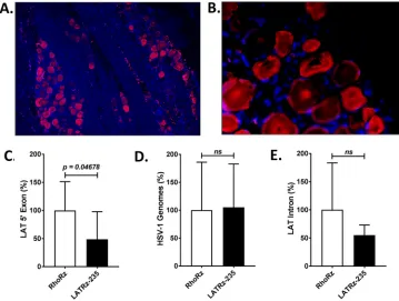
FIG 4Analysis of rabbit TG for AAV transduction, LAT expression, and HSV-1 DNA load. (A and B) Immunofluo-rescence (IF) demonstrating long-term expression of GFP in a high percentage of latently infected rabbit TG neurons. IF with mouse primary antibody to GFP was performed to identify populations of neurons harboring the AAV-LATRz-235 vector, which expressed GFP from the CBA promoter. Alexa Fluor 548 (Invitrogen) was used as the secondary antibody for immunofluorescence visualization using a Leica deconvolution microscope with Slidebook 5.0. Panel A represents an original magnification of⫻10 and panel B the original magnification of⫻40 from the same section. DAPI staining in blue represents nuclei of satellite cells present in the sections. RNA was isolated and analyzed by RT-PCR using primers and probes specific for the LAT 5=exon or LAT 2.0-kb intron regions of the genome. Relative quantities were determined for the LAT and then normalized to the relative quantities of rabbit GAPDH RNA to account for variations in RNA recovery between samples. (C) LAT 5=exon expression significantly decreased following treatment with AAV-LATRz-235 expressing the anti-LAT ribozyme. The average values for each virus are presented along with standard deviations (P⬍0.05). (D) HSV-1 genome load was determined by qPCR using primers for the HSV-1 DNA polymerase gene (see Materials and Methods and Table 2). (E) LAT stable 2.0-kb intron RNA levels do not show significant decrease following treatment with AAV-LATRz-235 (n⫽7 for AAV-RhoRz [control];n⫽6 for AAV-LATRz-235). All PCR analyses were performed in triplicate, and statistical analyses were performed as one-way analysis of variance (ANOVA) using SigmaPlot 12.5.
on November 6, 2019 by guest
http://jvi.asm.org/
[image:6.585.42.401.72.343.2]Given the data showing that the LAT protects against apoptosis, we considered the
possibility that knockdown of the LAT as in this study could render neurons that are
latently infected with HSV more susceptible to apoptosis (38), especially after
subject-ing them to epinephrine induction. As shown in Fig. 4D, we saw no evidence of
decrease in HSV genomes in the TG from both sets of rabbits after the experiment was
complete. It is plausible, however, that our experiment did not have sufficient statistical
power to see a subtler decrease. A related point is whether other effects of the LAT in
maintaining latency could have affected reactivation (34, 39). Again, while we did not
see any decrease in total latent genomes over the 17-day period following ribozyme
treatment, we feel this 17-day time frame was probably too short to observe any effects
of the depleted LAT on maintaining latency. We plan to look at this in future
experi-ments over a period of several months following LAT ribozyme treatment.
The ribozyme used in this study targets the 5
=
exon region of the LAT primary
transcript for cleavage. Cleaving the RNA in this region would be predicted to decrease
levels of the primary LAT, spliced LAT, and the LAT intron. In both our
in vitro
and
in vivo
analyses, we observed an approximately 70% decrease in the 5
=
exon RNA as
deter-mined by qRT-PCR analyses. In contrast, however, we saw only a 35 to 40% decrease in
the LAT intron. This is likely due to the high stability and long half-life of the LAT intron
compared to the LAT primary transcript. Previous work suggesting that the reactivation
phenotype of HSV is localized to the promoter/5
=
exon coding region of the LAT (28,
37) may be consistent with the degree of decreased reactivation observed in this study.
It will be useful to determine if using multiple ribozymes to target different regions of
the LAT, which could lead to more-efficient transcript knockdown, could further
decrease reactivation. It should be noted that we initially chose to use a ribozyme as
opposed to an siRNA or short hairpin RNA (shRNA) because ribozymes are active in the
nucleus, which is important since the LAT is a nuclear RNA. Recent studies have shown
that transfected siRNAs can efficiently target nuclear transcripts, so this is an option for
future studies. In either instance, the ability of AAV to deliver RNA cleaving or silencing
RNAs to neurons
in vivo
offers the potential to dissect the roles of viral or cellular RNAs
without altering other functions on the genomes through the use of gene editing or
deletion. This offers a powerful and complementary approach to mutation/deletion
strategies, especially for genomes of viruses like the herpesviruses that possess a high
density of
cis
-acting genetic elements that cannot easily be untangled. This point is well
illustrated in the repeat regions of HSV-1 (18).
Finally, the approach of delivering LAT-targeting ribozymes that block reactivation
opens the door to using this approach to treat recurrent HSV-1 disease. AAV vectors are
maintained as extrachromosomal episomes (for a review, see reference 40) and express
their transgenes for long periods of time in nondividing cells such as neurons (41). AAV
can be delivered to human eyes using only minor modifications of clinical practices
currently used to treat corneal infections. Therefore, this approach may embody a new
treatment for herpes stromal keratitis, the leading infectious cause of blindness in the
United States, and for treating recurrent oral and genital herpes as well.
MATERIALS AND METHODS
Viruses and cells.A low-passage-number stock of HSV-1 strain 17syn⫹was obtained from J. Stevens. Viruses were propagated on rabbit skin (RS) cells as described previously (42). Briefly, RS cells were propagated in minimal essential medium (MEM; Gibco Life Technologies, Gaithersburg, MD) with 5% fetal bovine serum (FBS) and antibiotics at 37°C in a humidified 5% carbon dioxide atmosphere. To assess cytopathic effect (CPE) of tear film swabs in the reactivation studies, primary rabbit kidney (PRK) cells were propagated in MEM with 5% FBS under the same conditions.
Target selection and design of synthetic hammerhead ribozymes.The LAT 5=exon and intron regions of the HSV-1 genome (NCBI database accession numberNC_001806) were scanned for ham-merhead ribozyme cleavage sites. The nucleotide triplets GUC, CUC, and UUC were specifically selected as potential target sites. Hybridizing arms were designed complementary to the sequence surrounding the cleavage site. Mfold software (43) was used to predict the secondary structure of each ribozyme. Ribozymes predicted to have a correctly folded catalytic core and open hybridizing arms were carried forward toin vitrotesting. An example of sequence selection, design, and predicted folding is shown in Fig. 1.
In VivoKnockdown of HSV-1 LAT Reduces Reactivation Journal of Virology
on November 6, 2019 by guest
http://jvi.asm.org/
Cloning synthetic ribozymes into AAV plasmids. Selected synthetic hammerhead ribozymes targeting the LAT were cloned into scAAV2-GFP plasmids forin vitrotesting and subsequent vector production. In this vector, humanized GFP (GFPh) is expressed from the CBA promoter consisting of the chimeric chicken beta actin proximal promoter plus the immediate early enhancer from cytomegalovirus. The self-complementary AAV (scAAV) plasmid contains a modified terminal repeat (TR) that results in the generation of a self-complementary copy of the transgene during packaging to improve transduction efficiency (44). The ribozyme sequence was placed downstream of an H1 promoter and terminated by a PolIII terminator sequence. These minigenes were synthesized by GenScript with SalI restriction endonuclease sites at each end. AAV plasmids containing the AAV2 TRs and the insert were digested with SalI (New England BioLabs) and ligated. Ligations were transformed into SURE electroporation-competent cells (Stratagene) and grown in LB medium. Plasmids were isolated and purified by CsCl centrifugation. Insertion was confirmed by PCR and Sanger sequencing. The construction and charac-terization of the AAV-RhoRz (also referred to as RhodRz397) were described previously (45). The AAV plasmids were pseudotyped into AAV8 Y733F modified capsids.
In vitrotesting of synthetic hammerhead ribozymes.Lipofectamine 2000 (Invitrogen) was used to cotransfect a plasmid encoding synthetic hammerhead ribozymes and a plasmid encoding the target LAT transcript into rabbit skin cells. Each transfection was performed in triplicate in a 24-well plate format. A plasmid expressing an antirhodopsin ribozyme was cotransfected in control wells to account for nonspecific effects. Twenty-four hours posttransfection, RNA was isolated using TRIzol (Ambion) and used for subsequent reverse transcription and qPCR analysis as described below.
Rabbit inoculation with HSV-1 and establishment of latency.New Zealand White rabbits (2 to 3 kg) were inoculated with 1⫻105to 2⫻105PFU of virus per eye in a suspension of 25l MEM with 5% FBS. The viral suspension was placed in the cul-de-sac of the scarified eye, and the eye was closed and massaged for 30 s with care to prevent virus loss (46). On postinoculation (PI) days 3, 5, 7, 10, and 14, all corneas were examined by slit lamp microscopy (SLE) with 0.1% fluorescein for evidence of acute herpetic keratitis. On PI day 30, all eyes were examined to ensure that the corneal epithelium was free of lesions. Only eyes that were normal by SLE were included in the reactivation study. Rabbits that exhibited acute lesions with subsequent recovery were considered latently infected. All animal care and procedures described conducted for the studies in this paper were approved by the Louisiana State University Health Sciences Center Institutional Animal Care and Use Committee.
Treatment of rabbit eyes with AAV LATRz-235 and AAV RhoRz vectors.Rabbit corneas were abraded using a 27 gauge needle and forming an 8⫻8 lattice crosshatch pattern across the entire surface of the cornea. Following abrasion, 1 1011⫻1011genome copies of either LATRz-235 or AAV RhoRz in a total volume of no less than 25l and no greater than 40l of saline was applied directly to the surface of the cornea. The eyelids were gently massaged over the eye.
Induction of HSV-1 reactivation in rabbits.Transcorneal iontophoresis (0.8 mA for 8 min) of 0.01% epinephrine was performed on each eye once daily for 3 consecutive days (PI days 31, 32, and 33). To assess the frequency of reactivation, tears were collected daily on each of the three reactivation stimulus days (prior to each iontophoresis procedure) and for an additional 9 days thereafter (total of 12 consecutive days following iontophoresis initiation). Tear film collection consisted of inserting a Dacron swab (Puritan, Hardwood Products Company, Guilford, ME) into the cul-de-sac of the eye and passed over the corneal surface and conjunctiva. Each eye was swabbed independently, and the swab was placed in a tube containing 1 ml DMEM (1% FBS). Tubes were placed at⫺80°C overnight and then thawed at 37°C the following day. The Dacron swabs were removed aseptically, and the medium was placed into a 24-well plate containing a monolayer of CV-1 cells. The plates were then monitored for 7 days for evidence of CPE.
Reverse transcription.To remove any DNA contamination, RNA samples from transfection exper-iments were DNase treated with Turbo DNA-free (Ambion). cDNA was synthesized using OmniScript (Qiagen) with 10M random decamer primers (Ambion). To control for any residual genomic DNA in cDNA samples, no-RT control reactions were performed. Mixtures for these reactions included all components except the OmniScript reverse transcription enzyme.
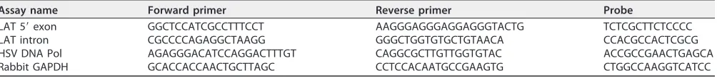
[image:8.585.43.545.83.138.2]Real-time PCR.cDNA samples from transfection experiments were analyzed by TaqMan quantitative real-time PCR on a StepOnePlus thermocycler (Applied Biosystems). Quantities were normalized to a cellular housekeeping gene, rabbit GAPDH gene. Custom TaqMan assay sequences are given in Table 2. Immunofluorescence.Slides containing sections of rabbit TG were deparaffinized, hydrated, and peroxidase blocked through a series of washes that included (i) two xylene washes for 5 min each, (ii) two 100% ethanol washes for 2 min each, (iii) one 3% hydrogen peroxide wash (30% hydrogen peroxide was diluted 1:10 in 100% methanol) for 10 min, (iv) one 95% ethanol wash for 3 min, (v) one 70% ethanol wash for 1 min, and (vi) one Tris-buffered saline with Tween 20 (TBST) wash for 1 min. Epitope retrieval was done using the IHC-Tek epitope retrieval steamer set. The retrieval jar was filled with retrieval solution, slides were placed inside, and the jar was placed in the steamer for 1 h. The lid was removed
TABLE 2Custom TaqMan assay sequences
Assay name Forward primer Reverse primer Probe
LAT 5=exon GGCTCCATCGCCTTTCCT AAGGGAGGGAGGAGGGTACTG TCTCGCTTCTCCCC
LAT intron CGCCCCAGAGGCTAAGG GGGCTGGTGTGCTGTAACA CCACGCCACTCGCG
HSV DNA Pol AGAGGGACATCCAGGACTTTGT CAGGCGCTTGTTGGTGTAC ACCGCCGAACTGAGCA
Rabbit GAPDH GCACCACCAACTGCTTAGC CCTCCACAATGCCGAAGTG CTGGCCAAGGTCATCC
on November 6, 2019 by guest
http://jvi.asm.org/
from the jar, and the solution was cooled to room temperature. Slides were washed with TBST and placed in a humidified chamber. Slides were blocked using 1.5% normal serum (goat) for 30 min and washed with TBST. Sections were then incubated with primary antibody (1:200 of mouse anti-GFP) for 24 h at 4°C. Secondary antibody incubation was then performed using 1:1,000 goat anti-mouse Alexa Fluor 548 (Invitrogen) for 2 h at room temperature, followed by a TBST wash. The slides were mounted using ProLong gold mounting medium (Invitrogen) and imaged using a Leica deconvolution microscope equipped with Slidebook 5.0.
ACKNOWLEDGMENTS
This work was supported in part by grants R01 AI48633 and R56 AI101174 from the
NIH and an unrestricted grant from Research to Prevent Blindness to A.S.L. and
S.S.T. D.M.P. was supported by a Clinical Research Training Award from the NIH (TL1
TR000066). We also thank William Hauswirth and the Center for Vision Research Vector
Core lab for providing some of the AAV vectors used in these experiments. This core
was supported by a core grant from the National Eye Institute (P30 EY02172).
REFERENCES
1. Liesegang TJ. 2001. Herpes simplex virus epidemiology and ocular im-portance. Cornea 20:1–13.https://doi.org/10.1097/00003226-200101000 -00001.
2. Stevens JG, Wagner EK, Devi RGB, Cook ML, Feldman LT. 1987. RNA complementary to a herpesvirus alpha gene mRNA is prominent in latently infected neurons. Science 235:1056 –1059. https://doi.org/10 .1126/science.2434993.
3. Deatly AM, Spivack JG, Lavi E, Fraser NW. 1987. RNA from an immediate early region of the type 1 herpes simplex virus genome is present in the trigeminal ganglia of latently infected mice. Proc Natl Acad Sci U S A 84:3204 –3208.
4. Rock DL, Nesburn AB, Ghaisi H, Ong J, Lewis TL, Lokensgard JR, Wechsler SL. 1987. Detection of latency-related viral RNAs in trigeminal ganglia of rabbits latently infected with herpes simplex virus. J Virol 61:3820 –3826. 5. Farrell MJ, Dobson AT, Feldman LT. 1991. Herpes simplex virus latency-associated transcript is a stable intron. Proc Natl Acad Sci U S A 88: 790 –794.
6. Thomas DL, Lock M, Zabolotny JM, Mohan BR, Fraser NW. 2002. The 2-kilobase intron of the herpes simplex virus type 1 latency-associated transcript has a half-life of approximately 24 hours in SY5Y and COS-1 cells. J Virol 76:532–540.https://doi.org/10.1128/JVI.76.2.532-540.2002. 7. Mehta A, Maggioncalda J, Bagasra O, Thikkavarapu S, Saikumari P,
Valyi-Nagy T, Fraser NW, Block TM. 1995. In situ DNA PCR and RNA hybridization of herpes simplex virus sequences in trigeminal ganglia of latently infected mice. Virology 206:633– 640.https://doi.org/10.1016/ S0042-6822(95)80080-8.
8. Chen XP, Mata M, Kelley M, Glorioso JC, Fink DJ. 2002. The relation-ship of herpes simplex virus latency associated transcript expression to genome copy number: a quantitative study using laser capture microdissection. J Neurovirol 8:204 –210. https://doi.org/10.1080/ 13550280290049642.
9. Perng GC, Jones C, Ciacci-Zanella J, Stone M, Henderson G, Yukht A, Slanina SM, Hofman FM, Ghiasi H, Nesburn AB, Wechsler SL. 2000. Virus-induced neuronal apoptosis blocked by the herpes simplex virus latency-associated transcript. Science 287:1500 –1503.https://doi.org/10 .1126/science.287.5457.1500.
10. Margolis TP, Imai Y, Yang L, Vallas V, Krause PR. 2007. Herpes simplex virus type 2 (HSV-2) establishes latent infection in a different population of ganglionic neurons than HSV-1: role of latency-associated transcripts. J Virol 81:1872–1878.https://doi.org/10.1128/JVI.02110-06.
11. Leib DA, Bogard CL, Kosz VM, Hicks KA, Coen DM, Knipe DM, Schaffer PA. 1989. A deletion mutant of the latency-associated transcript of herpes simplex virus type 1 reactivates from the latent state with reduced frequency. J Virol 63:2893–2900.
12. Hill JM, Sedarati F, Javier RT, Wagner EK, Stevens JG. 1990. Herpes simplex virus latent phase transcription facilitates in vivo reactivation. Virology 174:117–125.https://doi.org/10.1016/0042-6822(90)90060-5. 13. Perng G-C, Dunkel EC, Geary PA, Slanina SM, Ghiasi H, Kaiwar R, Nesburn
AB, Wechsler SL. 1994. The latency-associated transcript gene of herpes simplex virus type 1 (HSV-1) is required for efficient in vivo spontaneous reactivation of HSV-1 from latency. J Virol 68:8045– 8055.
14. Bloom DC, Devi-Rao GB, Hill JM, Stevens JG, Wagner EK. 1994. Molecular
analysis of herpes simplex virus type 1 during epinephrine induced reactivation of latently infected rabbits in vivo. J Virol 68:1283–1292. 15. Giordani NV, Neumann DM, Kwiatkowski DL, Bhattacharjee PS, McAnany
PK, Hill JM, Bloom DC. 2008. During HSV-1 infection of rabbits, the ability to express the LAT increases latent-phase transcription of lytic genes. J Virol 82:6056 – 6060.https://doi.org/10.1128/JVI.02661-07.
16. Cliffe AR, Garber DA, Knipe DM. 2009. Transcription of the herpes simplex virus latency-associated transcript promotes the formation of facultative heterochromatin on lytic promoters. J Virol 83:8182– 8190.
https://doi.org/10.1128/JVI.00712-09.
17. Kwiatkowski DL, Thompson HW, Bloom DC. 2009. The polycomb group protein Bmi1 binds to the herpes simplex virus 1 latent genome and maintains repressive histone marks during latency. J Virol 83:8173– 8181.
https://doi.org/10.1128/JVI.00686-09.
18. Phelan D, Barrozo ER, Bloom DC. 2017. HSV1 latent transcription and non-coding RNA: a critical retrospective. J Neuroimmunol 308:65–101.
https://doi.org/10.1016/j.jneuroim.2017.03.002.
19. Perng GC, Slanina SM, Yukht A, Ghiasi H, Nesburn AB, Wechsler SL. 2000. The latency-associated transcript gene enhances establishment of her-pes simplex virus type 1 latency in rabbits. J Virol 74:1885–1891.https:// doi.org/10.1128/JVI.74.4.1885-1891.2000.
20. Thompson RL, Sawtell NM. 2001. Herpes simplex virus type 1 latency-associated transcript gene promotes neuronal survival. J Virol 75: 6660 – 6675.https://doi.org/10.1128/JVI.75.14.6660-6675.2001. 21. Sawtell NM, Poon DK, Tansky CS, Thompson RL. 1998. The latent herpes
simplex virus type 1 genome copy number in individual neurons is virus strain specific and correlates with reactivation. J Virol 72:5343–5350. 22. Watson ZL, Ertel MK, Lewin AS, Tuli SS, Schultz GS, Neumann DM, Bloom
DC. 2016. Adeno-associated virus vectors efficiently transduce mouse and rabbit sensory neurons coinfected with herpes simplex virus 1 following peripheral inoculation. J Virol 90:7894 –7901.https://doi.org/ 10.1128/JVI.01028-16.
23. Washington SD, Edenfield SI, Lieux C, Watson ZL, Taasan SM, Dhummak-upt A, Bloom DC, Neumann DM. 2018. Depletion of the insulator protein CTCF results in herpes simplex virus 1 reactivation in vivo. J Virol 92:e00173-18.https://doi.org/10.1128/JVI.00173-18.
24. Citti L, Rainaldi G. 2005. Synthetic hammerhead ribozymes as therapeu-tic tools to control disease genes. Curr Gene Ther 5:11–24.https://doi .org/10.2174/1566523052997541.
25. Uhlenbeck OC. 1987. A small catalytic oligoribonucleotide. Nature 328: 596 – 600.https://doi.org/10.1038/328596a0.
26. Haseloff J, Gerlach WL. 1992. Simple RNA enzymes with new and highly specific endoribonuclease activities. 1988. Biotechnology 24:264 –269. 27. Liu J, Lewin AS, Tuli SS, Ghivizzani SC, Schultz GS, Bloom DC. 2008.
Reduction in severity of a herpes simplex virus type 1 murine infection by treatment with a ribozyme targeting the UL20 gene RNA. J Virol 82:7467–7474.https://doi.org/10.1128/JVI.02720-07.
28. Bloom DC, Hill JT, Wagner EK, Feldman LF, Stevens JG. 1996. A 348-bp region in the latency associated transcript facilitates herpes simplex virus type 1 reactivation. J Virol 70:2449 –2459.
29. Hill JM, Haruta Y, Rootman DS. 1987. Adrenergically induced recurrent
In VivoKnockdown of HSV-1 LAT Reduces Reactivation Journal of Virology
on November 6, 2019 by guest
http://jvi.asm.org/
HSV-1 corneal epithelial lessions. Curr Eye Res 6:1065–1071.https://doi .org/10.3109/02713688709034878.
30. Nesburn AB, Elliot JH, Leibowitz HM. 1967. Spontaneous reactivation of experimental herpes simplex keratitis in rabbits. Arch Ophthalmol 78: 523–529.https://doi.org/10.1001/archopht.1967.00980030525021. 31. Berman EJ, Hill JM. 1985. Spontaneous ocular shedding of herpes
sim-plex virus 1 in latently infected rabbits. Invest Ophthalmol Vis Sci 26:587–590.
32. Sawtell NM, Thompson RL. 1992. Herpes simplex virus type 1 latency associated transcription unit promotes anatomical site-dependent es-tablishment and reactivation from latency. J Virol 66:2157–2169. 33. Nicoll MP, Proenca JT, Connor V, Efstathiou S. 2012. Influence of herpes
simplex virus 1 latency-associated transcripts on the establishment and maintenance of latency in the ROSA26R reporter mouse model. J Virol 86:8848 – 8858.https://doi.org/10.1128/JVI.00652-12.
34. Nicoll MP, Hann W, Shivkumar M, Harman LE, Connor V, Coleman HM, Proenca JT, Efstathiou S. 2016. The HSV-1 latency-associated transcript functions to repress latent phase lytic gene expression and suppress virus reactivation from latently infected neurons. PLoS Pathog 12: e1005539.https://doi.org/10.1371/journal.ppat.1005539.
35. Henderson G, Peng W, Jin L, Perng GC, Nesburn AB, Wechsler SL, Jones C. 2002. Regulation of caspase 8- and caspase 9-induced apoptosis by the herpes simplex virus type 1 latency-associated transcript. J Neurovirol 8(Suppl 2):S103–S111.
36. Thompson RL, Sawtell NM. 1997. The herpes simplex virus type 1 latency-associated transcript gene regulates the establishment of la-tency. J Virol 71:5432–5440.
37. Perng G-C, Ghiasi H, Slanina SM, Nesburn AB, Wechsler SL. 1996. The spontaneous reactivation function of the herpes simplex virus type 1 LAT gene resides completely within the first 1.5 kilobases of the 8.3-kilobase primary transcript. J Virol 70:976 –984.
38. Ahmed M, Lock M, Miller CG, Fraser NW. 2002. Regions of the herpes
simplex virus type 1 latency-associated transcript that protect cells from apoptosis in vitro and protect neural cells in vivo. J Virol 76:717–729.
https://doi.org/10.1128/JVI.76.2.717-729.2002.
39. Thompson RL, Sawtell NM. 2011. The herpes simplex virus type 1 latency associated transcript locus is required for the maintenance of reactiva-tion competent latent infecreactiva-tions. J Neurovirol 17:552–558.https://doi .org/10.1007/s13365-011-0071-0.
40. Grimm D, Buning H. 2017. Small but increasingly mighty: latest advances in AAV vector research, design, and evolution. Hum Gene Ther 28: 1075–1086.https://doi.org/10.1089/hum.2017.172.
41. Sehara Y, Fujimoto KI, Ikeguchi K, Katakai Y, Ono F, Takino N, Ito M, Ozawa K, Muramatsu SI. 2017. Persistent expression of dopamine-synthesizing enzymes 15 years after gene transfer in a primate model of Parkinson’s disease. Hum Gene Ther Clin Dev 28:74 –79.https://doi.org/ 10.1089/humc.2017.010.
42. Bloom DC, Stevens JG. 1994. Neuronal specific restriction of an HSV recombinant maps to the UL5 gene. J Virol 68:3761–3772.
43. Zuker M. 2003. Mfold web server for Nucleic acid folding and hybrid-ization prediction. Nucleic Acids Res 31:3406 –3415.https://doi.org/10 .1093/nar/gkg595.
44. McCarty DM, Fu H, Monahan PE, Toulson CE, Naik P, Samulski RJ. 2003. Adeno-associated virus terminal repeat (TR) mutant generates self-complementary vectors to overcome the rate-limiting step to transduction in vivo. Gene Ther 10:2112–2118.https://doi.org/10.1038/sj.gt.3302134. 45. Gorbatyuk MS, Pang JJ, Thomas J, Jr, Hauswirth WW, Lewin AS. 2005.
Knockdown of wild-type mouse rhodopsin using an AAV vectored ri-bozyme as part of an RNA replacement approach. Mol Vis 11:648 – 656. 46. Creech CC, Neumann DM. 2010. Changes to euchromatin on LAT and ICP4 following reactivation are more prevalent in an efficiently reacti-vating strain of HSV-1. PLoS One 5:e15416. https://doi.org/10.1371/ journal.pone.0015416.
on November 6, 2019 by guest
http://jvi.asm.org/