in Solution
M. Alejandra Tortorici,a,bStéphane Duquerroy,a,b,cJane Kwok,a,dClemens Vonrhein,eJavier Perez,fBenjamin Lamp,g Gerard Bricogne,eTill Rümenapf,gPatrice Vachette,hFélix A. Reya,b
Unité de Virologie Structurale, Département de Virologie, Institut Pasteur, Paris, Francea
; CNRS, UMR 3569, Paris, Franceb
; Université Paris Sud, Gif sur Yvette, Francec ; Department of Physiology, Li Ka Shing Faculty of Medicine, University of Hong Kong, Hong Kongd
; Global Phasing Ltd., Cambridge, United Kingdome
; Synchrotron Soleil, Gif sur Yvette, Francef
; Institute of Virology, University of Veterinary Medicine, Vienna, Austriag
; Institut de Biologie Intégrative de la Cellule, UMR 9198, Gif-sur-Yvette, Franceh
ABSTRACT
Pestiviruses form a genus in the
Flaviviridae
family of small enveloped viruses with a positive-sense single-stranded RNA
ge-nome. Viral replication in this family requires the activity of a superfamily 2 RNA helicase contained in the C-terminal domain
of nonstructural protein 3 (NS3). NS3 features two conserved RecA-like domains (D1 and D2) with ATPase activity, plus a third
domain (D3) that is important for unwinding nucleic acid duplexes. We report here the X-ray structure of the pestivirus NS3
helicase domain (pNS3h) at a 2.5-Å resolution. The structure deviates significantly from that of NS3 of other genera in the
Flavi-viridae
family in D3, as it contains two important insertions that result in a narrower nucleic acid binding groove. We also show
that mutations in pNS3h that rescue viruses from which the core protein is deleted map to D3, suggesting that this domain may
be involved in interactions that facilitate particle assembly. Finally, structural comparisons of the enzyme in different crystalline
environments, together with the findings of small-angle X-ray-scattering studies in solution, show that D2 is mobile with respect
to the rest of the enzyme, oscillating between closed and open conformations. Binding of a nonhydrolyzable ATP analog locks
pNS3h in a conformation that is more compact than the closest apo-form in our crystals. Together, our results provide new
in-sight and bring up new questions about pNS3h function during pestivirus replication.
IMPORTANCE
Although pestivirus infections impose an important toll on the livestock industry worldwide, little information is available
about the nonstructural proteins essential for viral replication, such as the NS3 helicase. We provide here a comparative
struc-tural and functional analysis of pNS3h with respect to its orthologs in other viruses of the same family, the flaviviruses and
hepa-titis C virus. Our studies reveal differences in the nucleic acid binding groove that could have implications for understanding the
unwinding specificity of pNS3h, which is active only on RNA duplexes. We also show that pNS3h has a highly dynamic
behav-ior—a characteristic probably shared with NS3 helicases from all
Flaviviridae
members—that could be targeted for drug design
by using recent algorithms to specifically block molecular motion. Compounds that lock the enzyme in a single conformation or
limit its dynamic range of conformations are indeed likely to block its helicase function.
P
estiviruses infect a wide range of cloven-hoofed animals, wild
and domestic, causing serious disease. The most studied are
the classical swine fever virus (CSFV) (
1
) and the bovine viral
diarrhea virus (BVDV), which impose important economic losses
to the livestock industry worldwide (
2
). They form a genus within
the
Flaviviridae
family of single-stranded RNA viruses, which also
includes medically important pathogens in the flavi- and
hepaci-virus genera. Recent studies also revealed that hepaci- and
pegivi-ruses (the fourth genus in the family) cause infections in horses
and other domestic animals (
3
,
4
).
The pestivirus genome is a single mRNA molecule of about
12.3 kb with a single large open reading frame (ORF) coding for a
polyprotein precursor of about 3,900 residues. This long ORF is
flanked by 5
=
and 3
=
untranslated regions with
cis
-active elements
essential for virus translation and replication (
2
). The polyprotein
is co- and posttranslationally processed by cellular and virus
ge-nome-encoded proteases to yield the individual mature viral
pro-teins in the order shown in
Fig. 1A
(
2
,
5
,
6
). Structural studies are
available for nonstructural protein 5B (NS5B) (
7
), E
rns(
8
), E2 (
9
,
10
), and N
pro(
11
,
12
). N
proand E
rnsare unique to pestiviruses,
while all the others have their counterparts in the other genera.
Particle morphogenesis in the infected cell occurs when the core
protein forms a ribonucleoprotein (RNP) complex with the
genomic RNA, which then buds into the lumen of an internal
compartment, thereby acquiring a lipid envelope in which are
incorporated the three envelope proteins E
rns, E1, and E2. The
nonstructural proteins NS2 through NS5B are present only in
Received30 October 2014Accepted27 January 2015 Accepted manuscript posted online4 February 2015
CitationTortorici MA, Duquerroy S, Kwok J, Vonrhein C, Perez J, Lamp B, Bricogne G, Rümenapf T, Vachette P, Rey FA. 2015. X-ray structure of the pestivirus NS3 helicase and its conformation in solution. J Virol 89:4356 –4371.
doi:10.1128/JVI.03165-14. Editor:M. S. Diamond
Address correspondence to M. Alejandra Tortorici, tortoric@pasteur.fr, or Félix A. Rey, rey@pasteur.fr.
Supplemental material for this article may be found athttp://dx.doi.org/10.1128 /JVI.03165-14.
Copyright © 2015, American Society for Microbiology. All Rights Reserved. doi:10.1128/JVI.03165-14
on November 7, 2019 by guest
http://jvi.asm.org/
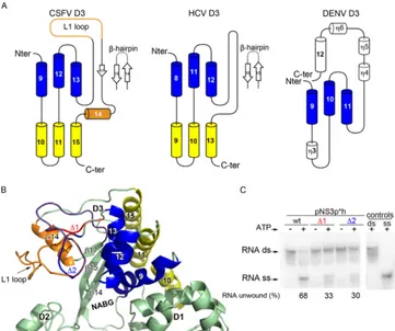
FIG 1Biochemical characterization of recombinant pNS3 and pNS3h from CSFV. (A) Schematic diagram of the pestivirus precursor polyprotein organization, indicating the individual mature proteins (boxes) and proteolytic processing sites (arrowheads or curved arrows for autoproteolytic cleavages). A diagram of the different constructs used in this study is shown below. (B) SDS-PAGE analysis of purified recombinant proteins stained with Coomassie blue. The lanes labeled
on November 7, 2019 by guest
http://jvi.asm.org/
[image:2.585.43.538.68.697.2]infected cells and are essential for virus replication. Among them,
NS2 (a cysteine protease), NS5B (the RNA-dependent RNA
poly-merase), and NS3 have enzymatic activities. NS3 has a molecular
mass of about 76 kDa and features two main functional domains,
each with different enzymatic activities. The N-terminal domain is
a chymotrypsin-like serine protease, which is responsible for most
of the maturation cleavages of the polyprotein precursor in the
cytosolic side of the endoplasmic reticulum membrane (
Fig. 1A
);
the exception is the NS2/NS3 junction (
2
). The C-terminal
do-main, about two-thirds of NS3, is a helicase belonging to
super-family 2 (SF2) that displays characteristic sequence motifs that
constitute the SF2 signature (
13
). The helicase activity is thought
to be important for unwinding highly structured regions of the
RNA genome during replication. While NS3 from hepatitis C
vi-rus (HCV) unwinds both RNA and DNA double-stranded helices,
pesti- and flavivirus helicases unwind only RNA duplexes (
14
,
15
).
The protease and helicase domains of NS3 (NS3p and NS3h,
re-spectively) are believed to function at different stages of the
repli-cation cycle, although in the case of HCV, the two domains were
shown to enhance each other’s activities (
16
,
17
). NS3 was recently
shown to be autoproteolyzed in CSFV-infected cells to release
NS3h (
6
). In addition, NS3 was shown to play other functions in
the virus cycle, such as interfering with the innate immune system
in the case of HCV (
18
) and modulating fatty acid synthesis in the
case of dengue virus (DENV) (
19
). It was also shown to participate
in genome packaging and particle assembly in HCV (
20
,
21
),
yel-low fever virus (
22
,
23
), and pestiviruses (
24
). Indeed, an apparent
interplay between genomic RNP formation and interactions with
NS3 has been reported: CSFV virions lacking the core protein
were found to be viable only when NS3 is mutated at specific
locations.
Studies on HCV and several flaviviruses have provided
impor-tant insight into the translocation mechanism of NS3 along
nu-cleic acid polymers (
25–30
), but very little is known about the
pestivirus counterpart (
15
,
31
). For clarity, here we use “pNS3” to
specify the pestivirus enzyme. No three-dimensional (3D)
struc-ture is available for pNS3, except for a homology model based on
HCV NS3h that was recently published (
32
). Given that the amino
acid sequence identity between NS3 from HCV and the
pestivi-ruses is low (less than 20%), the relevance of the proposed 3D
model remains to be tested.
We report here an enzymatic characterization and the X-ray
structure of the pNS3 helicase domain (pNS3h) from CSFV. We
found that pNS3p or portions of it modulate the unwinding
ac-tivity of pNS3, as is the case for NS3 from the other genera. The
crystal structure showed that the RecA-like pNS3h domains D1
and D2 are quite similar to those of HCV and DENV NS3h. In
contrast, there are important differences in the nucleic acid
bind-ing groove (NABG), which result from two insertions in D3.
Fur-thermore, the various reported mutations affecting pestivirus
genomic RNA encapsidation map to D3, although they do not
make a specific surface patch or affect the RNA unwinding activity
of pNS3. Our crystallographic analyses, together with small-angle
X-ray-scattering (SAXS) experiments in solution, further show
that pNS3h has a highly dynamic behavior that is in line with its
expected motor-like translocation activity along the pestivirus
RNA genome.
MATERIALS AND METHODS
Cloning, expression, and purification.The oligonucleotide sequences used in this study are displayed in Table S1 in the supplemental material. To express pNS3h we modified the construct described previously (6) by introducing the tobacco etch virus (TEV) protease cleavage site (ENLY FQG) between a histidine (His) tag and the beginning of the pNS3h se-quence to have the ability to remove the N-terminal His tag. The resulting vector allows the expression of pNS3h1, which is a 516-residue protein comprising pNS3 residues 193 to 683 (corresponding to residues 1782 to 2272 in the polyprotein) fused to a His tag plus the TEV protease cleavage site at the N terminus and the first 8 residues of NS4A at the C terminus (Fig. 1A). We also modified this construct to express pNS3h2, a 536-residue protein comprising pNS3 (536-residues 173 to 683) (33). To express full-length pNS3ph, we used the construct described elsewhere (6), which includes the NS3-activating sequence of NS4A (residues 21 to 55) fused to the N terminus of NS3 (residues 1 to 683) through a GSGS linker. As for the pNS3h constructs, the His tag plus the TEV site and the first 8 residues of NS4A were fused to the N and C termini, respectively. All the mutations that we describe in this work were introduced into the corresponding plasmids by outward PCR. Identical protocols were used to express and purify all the proteins used in this study. TransformedEscherichia coli
Rosetta(DE3)pLysS cells (Novagen) were grown at 37°C in Luria-Bertani medium (LB) containing 100 mg ml⫺1ampicillin, 35 mg ml⫺1 chloram-phenicol, and 0.3% glucose to an optical density at 600 nm of 0.6 to 0.8. Protein expression at 30°C was induced with 1 mM isopropyl--D -1-thiogalactopyranoside. Selenomethionine (SeMet)-labeled pNS3h pro-tein was expressed inE. colistrain B834(DE3) (Novagen), a methionine auxotroph, in a deep liver medium (DLM) containing 0.2 g liter⫺1SeMet. After 4 h induction, the cells were harvested by centrifugation and stored at⫺80°C. Frozen cells pellets from a 1-liter culture were resuspended in 40 ml of lysis buffer (50 mM NaHPO4, pH 7.5, 300 mM NaCl, 1% Triton X-100, 2 mM dithiothreitol [DTT], 40 mM imidazole) supplemented with a protease inhibitor cocktail (Complete, EDTA free; Roche) and lysed by high pressure using an EmulsiFlex-C5 homogenizer (Avestin). The lysate was clarified by centrifugation at 30,000⫻gfor 30 min at 4°C. The supernatant was filtered (pore size, 0.22m; Millipore), loaded onto a nickel-affinity resin (His-Trap HP; GE Healthcare), washed with 50 mM NaHPO4, pH 7.5, 300 mM NaCl, 40 mM imidazole, and eluted with a 40 to 500 mM imidazole gradient. Fractions containing His-tagged protein were pooled and loaded into a gel filtration column (Superdex 200; GE Healthcare) that had previously been equilibrated in 20 mM Tris-HCl, pH 7.5, 100 mM NaCl, 2 mM DTT. The His tag was removed by TEV cleavage in a reaction mixture containing His-tagged pNS3h and His-tagged TEV at a molar ratio of 1:3 in TEV buffer (50 mM Tris-HCl [pH 8.0], 0.5 mM EDTA, and 1 mM DTT). The reaction mix was incubated at 4°C overnight and reloaded into a His-Trap column to remove uncleaved protein and
1 through 6 correspond to the various constructs listed in panel A. The middle lane displays prestained molecular mass markers. (C) The ATP hydrolysis activity of the proteins was measured by determination of the release of inorganic phosphate (yaxis) and was plotted as a function of the ATP concentration (xaxis). (D) Summary of the ATP hydrolysis properties of the different pNS3ph and pNS3h constructs. (E) The ATPase rate was measured in the presence of 0.2 mM ATP and the indicated concentrations of nonhydrolyzable ATP analogs (ADP·BeF3, AMPPcP, and ATP␥S). Plotted is the amount of hydrolyzed ATP (yaxis) against the ATP analog concentration (xaxis). The standard deviations in panels C and E were estimated from two independent experiments. (F) TheKmfor ATP hydrolysis
was determined by fitting the data from panel C with the Michaelis-Menten equation and is presented here in comparison to the corresponding values obtained for NS3 of otherFlaviviridaemembers available in the literature. (G) The unwinding activities of pNS3ph and pNS3h were measured under multiple turnover assay conditions. The percentage of unwound RNA was calculated by dividing the amount of single-stranded RNA (RNA ss) by the total amount of RNA (single-stranded RNA plus double-stranded [RNA ds]). nd, not detected.
on November 7, 2019 by guest
http://jvi.asm.org/
the tagged TEV protease. The cleaved protein was further purified on a gel filtration column as described above for the tagged protein. The samples were kept at 4°C until concentration to about 6 to 7 mg ml⫺1on a Vivas-pin apparatus, performed immediately before the crystallization trials.
ATPase assay.The steady-state rate of ATP hydrolysis was measured by determining the release of inorganic phosphate, initially by using ra-dioactive [␥-32
P]ATP and, later, for more accurate results at high ATP concentrations (Fig. 1C), by using a colorimetric assay from Innova Bio-sciences following the manufacturer’s instructions. Briefly, 10 nM protein was incubated with different amounts of ATP in ATPase buffer [50 mM Tris-HCl, pH 7.5, 2.5 mM MgCl2, 50 mM NaCl, 1 mM tris(2-carboxy-ethyl) phosphine (TCEP)] for 15 min at room temperature. The absor-bance at 630 nm was converted to the inorganic phosphate concentration using a standard curve. TheKmand the turnover number (kcat) of the enzymes were determined from measurements of the initial rates at dif-ferent ATP concentrations with GraphPad Prism software.
Screening of ATP analogs by inhibition of ATPase activity.For screening the effect of ATP analogs (Fig. 1E), we used a radioactivity assay. Briefly, pNS3h (0.33M) was incubated for 15 min at room temperature in a 20-l reaction mixture in ATPase buffer containing 1Ci/l of [␥-32P]ATP (3,000 Ci/mmol), 200M cold ATP, and different concen-trations (0.5 to 50 mM) of ground-state ATP analogs, such as ADP·BeF3, ,␥-methyleneadenosine 5=-triphosphate (AMPPcP), and adenosine 5= -(␥-thio) triphosphate (ATP␥S). One-microliter aliquots were spotted onto polyethylenimine-cellulose membranes (Merck), and the reaction products were resolved by ascending thin-layer chromatography in 1 M formic acid– 0.5 M LiCl. Radiolabeled spots on the membranes were de-tected by autoradiography and quantified with a phosphorimager.
Unwinding assays.To generate substrates for helicase assays, two syn-thetic oligonucleotides were annealed in 50 mM Tris-HCl, pH 8.0, 55 mM KCl, and 5 mM MgCl2by heating to 95°C and allowing them to cool slowly to room temperature. Before annealing, the shorter strand of the duplex substrate was32P labeled using polynucleotide kinase. The duplex DNA substrate consisted of the shorter DNA oligonucleotide (5=-[32P] GCCTCGCTGCCGTCGCCA-3=) annealed to the longer DNA oligo-nucleotide (5=-TGGCGACGGCAGCGAGGCTTTTTTTTTTTT-3=). The duplex RNA substrate consisted of the shorter RNA oligonucleotide (5= -[32P]CGACUCUAGAGAGGUG-3=) annealed to the longer RNA oligo-nucleotide (5=-CACCUCUCUAGAGUCGACCUGCAGGCAUCG-3=). The DNA or RNA substrate (5 nM) and 400 nM protein were incubated in 20-l reaction mixtures including 25 mM sodium phosphate buffer, pH 7.5, 1 mM DTT, 3 mM MgCl2, 0.1 mg ml⫺1bovine serum albumin, and 3 U RNasin (RNase inhibitor) for 15 min at 25°C. Reactions were initiated by the addition of 5 mM ATP and terminated by the addition of 5l of loading buffer (20 mM Tris-HCl, pH 7.5, 4 mM EDTA, 0.1% SDS, 50% glycerol, 0.01% bromophenol blue, 0.01% xylene cyanol) after 30 min at 37°C. Multiple turnover unwinding reactions were performed by adding 5 M excess unlabeled short DNA or RNA oligonucleotide as an enzyme trap at the end of the reaction. Reaction products were separated by elec-trophoresis on a 10% acrylamide native gel and detected and quantified with a phosphorimager.
Crystallization and structure determination. Crystals of native pNS3h (wild type or mutant) and the SeMet-substituted protein were grown at 20°C by vapor diffusion under the conditions listed inTable 1. For data collection, the crystals were flash cooled in liquid nitrogen using 20% glycerol as a cryoprotectant. Diffraction data were collected at syn-chrotron beam line PX1 of the Swiss Light Source (SLS X06SA) using a Pilatus 6M detector (34,35). Our initial attempts to determine the struc-ture by molecular replacement using the available models of NS3h from HCV or the flaviviruses failed. We therefore grew crystals of SeMet-sub-stituted protein to obtain experimental phases. These correspond to crys-tal form 2 (Table 1). As pNS3h has 21 methionines, the 4 molecules in the asymmetric unit (AU) amounted to 84 selenium sites, the coordinates of which needed to be determined to calculate phases. This required anad hocdata collection strategy to measure anomalous and dispersive pairs of
reflections as close in time as possible to minimize errors introduced by radiation damage. We collected the required diffraction data in 5-degree slices in inverse beam mode and switched wavelengths between the peak and the inflection points of the absorption edge within each slice. The diffraction data were indexed and processed with the XDS program suite (36) (the MOSFLM program was used for visualizing the indexing) and scaled using SCALA software (37). The program SHELXC was used to estimate heavy atom structure factors from the 2-wavelength anomalous dispersion data (peak and inflection). These data were then used in the SHELXD program to a resolution of 5 Å to find the Se sites. On purpose, a much lower estimate of the number of sites was used (21 out of 84 sites in the 4 molecules of the AU), since initial trials using the full number of expected sites were unsuccessful. Those initial 21 sites were used in the SHARP program for parameter refinement and calculation of log-likeli-hood gradient (LLG) residual maps. Inspection of the resulting electron density maps allowed the identification of further sites, and this was fol-lowed by additional SHARP runs. Iterating these steps gave a final heavy atom model containing 66 sites. Density modification using the SOLO-MON program gave a partially interpretable map using a solvent content of 52%. The density-modified map was used in the Buccaneer program (38) to build an initial model of 1,536 residues corresponding to the 4 molecules of the AU. The best map from auto-SHARP was used as a starting point in 2-fold noncrystallographic symmetry (NCS) averaging with the DM program (39). The program Buccaneer was then able to build 1,841 residues (out of 2,064 residues) into the 2-fold averaged map, with most of them being assigned in sequence correctly. The model was subsequently manually modified with the Coot program (40) and refined with the BUSTER-TNT program (41). This atomic model was then used for molecular replacement with the AMoRe program (42) to determine the structures of pNS3h in crystals forms 1 and 3, as well as crystals 4 and 5 (corresponding to the N588Y and Q600K mutants, respectively).
Normal modes analysis (NMA).To analyze the transition between the various conformations of pNS3h observed in the crystals, we used the two extreme conformations observed in the AU of crystal form 2 to cal-culate normal modes with the NOMAD-Ref and elNemo (http://www .sciences.univ-nantes.fr/elnemo/) servers (43,44). The programs com-pute the low-frequency normal modes for a given protein structure, suggesting potential conformational changes.
Small-angle X-ray scattering.X-ray-scattering data were collected at the SWING and ID14-3 beamlines of the Soleil synchrotron (Saint Aubin, France) and the European Synchrotron Radiation Facility (ESRF; Grenoble, France), respectively. The SWING data were recorded with a charge-coupled-device-based detector (AVIEX) at an X-ray wavelength () of 1.033 Å at 15°C on TEV protease-treated pNS3h to remove the His tag. We did the measurements over a range of protein concentrations ranging from 0.5 to 8 mg ml⫺1in SAXS buffer (20 mM Tris-HCl, pH 7.5, 100 mM NaCl) with the addition of either 5 mM TCEP (Pierce) or 2 mM DTT. The samples were circulated through a 1.8-mm-diameter quartz capillary using an automatic sample changer (Agilent). In addition, we also made measurements using 40l of an 8-mg ml⫺1solution of pNS3h loaded onto a Superdex 200 size exclusion column mounted on a high-pressure liquid chromatography (HPLC) system (Agilent) online with an SAXS measuring cell (size exclusion chromatography [SEC]-SAXS) (45). The absorbance at 280 nm in the elution profile exhibited only a faint trace of small oligomers (representing about 2% of the total protein in solution) before the peak corresponding to the monomeric fraction (data not shown). In both cases, the flow rate was high enough to minimize radia-tion damage. The resulting data were processed with the PRIMUS package (46). The forward scattering [I(0)] and the radius of gyration (Rg) were evaluated using the Guinier approximation (47). Frames over the elution peak recorded using the size exclusion HPLC were analyzed individually before averaging the appropriate subset, yielding identicalRgandI(0)/c (wherecis the protein concentration) values. The most concentrated samples were used to measure scattering data only at higher angles be-cause the curve exhibited distortions at smallqvalues (q⫽4sin/or
on November 7, 2019 by guest
http://jvi.asm.org/
momentum transfer, where 2is the scattering angle andis the wave-length of the X rays), with a Guinier plot yielding an apparentRgvalue of 31.8 Å (which was 15% higher than the value from the SEC-SAXS data). This was due to the presence of the small amount of oligomers revealed by
gel filtration and to the existence of attractive interactions between mol-ecules at a high concentration. Therefore, the average curve from the SEC-SAXS data was spliced with that for the most concentrated sample (after scaling to the same protein concentration) to yield a complete
scat-TABLE 1Crystallographic statistics for CSFV NS3h crystal formsa
Parameter
Value for the following crystal:
Crystal form 1 Crystal form 2 Crystal form 3 N588Y crystal Q600K crystal
PDB accession no. 4CBH 4CBG 4CBI 4CBL 4CBM
Crystallization conditions 0.1 M Na cacodylate, pH 6.5, 0.2 M magnesium acetate, 12% PEG 8000
1 M Na cacodylate, pH 6.5, 0.2 M magnesium acetate, 12% PEG 8000
0.1 M MES, pH 6.5, 0.6 M NaCl, 20% PEG 4000, 1 mM AMPPcPb
0.1 M Na cacodylate, pH 6.5, 0.15 M calcium acetate, 12% PEG 3350
0.1 M NaAc, pH 4.6, 0.025 M calcium acetate, 9% PEG 4000
Data collection statistics
Space group P21 P21 P1 P21 P21
Unit cell dimensions
a,b,c(Å) 70.07, 144.4, 118.690 70.455, 168.360, 98.8
69.63, 96.303, 97.611 70.027, 156.820, 99.52 70.1, 159.585, 99.227
␣ ⫽  ⫽ ␥(°) 92.94 98.522 91.23, 110.86, 107.62 97.39 97.24
Resolution range (Å) 44.34–2.51
(2.58–2.51)
49.57–2.82 (2.89– 2.82)/49.57–2.98 (3.16–2.98)c
46.83–3.00 (3.16–3.00) 39.21–3.05 (3.21–3.05) 37.98–3.27 (3.45–3.27)
No. of reflections
Measured 277,586 402,065/192,825c 84,132 208,052 77,233
Unique 71,319 49,724/43,835c 40,172 38,441 31,533
Completeness (%) 88.63 91.5/96.4c 89.8 94.9 94.4
Multiplicity 4.0 (2.3) 8.1 (3.4)/4.4 (2.1)c 2.1 (1.9) 5.4 (4.1) 2.4 (1.8)
I/I 15.4 (2.0) 16.2 (2.0)/12.2
(1.9)c
7.2 (1.7) 10.4 (1.8) 7.0 (1.9)
Rmerge(%)d 6.9 (39) 13.3 (34.9)/11.8
(23.4)c
8.9 (40.9) 12.3 (64.5) 2.8 (55.4)
Ranom(%)e 8.2 (28.4)/7.6
(24.1)c
Anomalous completeness (%) 87.2 (58.3)/85.1
(50.3)c
Anomalous signal up to the indicated resolution (Å)
3.99/5.15c
Refinement statistics
No. of reflections in refinement (free)
71,319 (3,572) 49,698 (2,532) 40,150 (1,996) 33,223 (1,647) 31,359 (1,585)
No. of molecules per asymmetric unit
4 4 4 4 4
Rwork(%)
f 20.41 16.61 19.21 24.6 23.30
Rfree(%)
f 23.75 20.49 24.67 25.66 27.0
No. of:
Protein atoms 11,032 14,731 13,820 14,530 14,595
Water molecules 928 122 105 0.007 0.01
RMSD
Bond length (Å) 0.010 0.01 0.01 1.0 1.12
Bond angle (°) 1.28 1.19 1.21 79.0 64.57
Overall avg B factor (Å2) 60.78 66.05 65.06 95.81 96.1
Ramachandran plot (%)g 97.4 96.24 3.05 3.18
Favored regions 1.93 97.14 2.85 1.13 0.72
Allowed regions 0.67 2.26 0.9
Outlier regions 0.6
aData in parentheses are for the last shell. PEG, polyethylene glycol; MES, morpholineethanesulfonic acid; NaAc, sodium acetate.
b
No electron density corresponding to AMPPcP was found in crystal form 3. cData are those for the peak point/inflection point.
d
Rmerge⫽冱(冱|Ii⫺ 具I典|/冱|I|), where the first冱is the sum over all reflections, and the second冱is the sum over all measurements of a given reflection, withIibeing theith measurement of the intensity of the reflection and具I典the average intensity of that reflection.
e
Ranom⫽ ⌺|Mn(I⫹)⫺Mn(I⫺)|/⌺[Mn(I⫹)⫹Mn(I⫺)], whereIis intensity and the⫹and⫺correspond to the Friedel-Bijvoet pair for the sameI, summed over all reflections.
fR
work/Rfree⫽冱(|Fo|⫺ 具|Fc|典)/冱|Fo|, where具|Fc|典is the expectation of |Fc| under the probability model used to define the likelihood function. The sum is overall reflections.
g
Calculated by use of the MolProbity program.
on November 7, 2019 by guest
http://jvi.asm.org/
[image:5.585.42.540.84.612.2]on November 7, 2019 by guest
http://jvi.asm.org/
tering curve (see Fig. S1A in the supplemental material). The pair distance distribution function [p(r)] was determined using the indirect Fourier transform method implemented in the program GNOM (48). The mo-lecular mass of the protein was evaluated by comparison of the forward scattering with that of water recorded in the same capillary using a value of 0.00164 cm⫺1for the theoretical scattering intensity of water. To calculate the theoretical scattering curves using the program CRYSOL (49), we used the atomic coordinates of the conformations observed in crystal forms 2 and 3, in which dummy coordinates for the missing disordered residues (see Table S2 in the supplemental material) were added manually. The agreement between our models and the experimental data were evaluated by examining the distribution of reduced residuals as a function ofq
[R(q)], given by the following expression: [Iexp(q)⫺Icalc(q)]/exp(q), whereIexp(q) is the experimental value ofIas a function ofq,Icalc(q) is the
calculated value ofIas a function ofq, andexp(q) is the experimental standard deviation as a function ofq.
The splicing of the experimental curves did not interfere with the fitting of the models, while it allowed us to take advantage of the better statistical quality of the high concentration data at highqvalues. The SAXS data showed that pNS3h is a monomer and not the dimer present in all our crystal forms (see Results). We were not able to test the oligomeric state under the crystallization conditions listed inTable 1to see if they induce dimerization because of the poor solubility of the protein under those conditions and the high viscosity introduced by the high concentra-tion of high-molecular-weight polyethylene glycol.
To investigate the effect of nonhydrolyzable ATP analogs, we used ADP·BeF3, which showed a strong inhibitory effect, as did AMPPcP, on the ATPase activity of pNS3h (Fig. 1E). SAXS measurements of pNS3h in
FIG 2Structural comparison of NS3h orthologs. (A) Ribbon diagram of the pNS3h structure in its closed conformation from crystal form 2 (PDB accession no. 4CBG) (left), the homology model (http://www.bioacademy.gr/bioinformatics/csfv/Welcome.html) (second panel), HCV NS3h (PDB accession no.1A1V) (third panel), and DENV-4 NS3h (PDB accession no.2JLQ) (right). The proteins were superposed on D1 using the PDBefold program and are displayed in the same orientation. The three globular domains are colored green (D1), pink (D2), and blue (D3). (B) Structure-based alignment (produced using the Multalign and ESPript, version 2.2, programs) of pNS3h with its HCV and flavivirus counterparts. The CSFV (strain Alfort; GenBank accession no.J04358.2), HCV (strain H77; GenBank accession no.ACA48642), and DENV-4 (GenBank accession no.AAW30973) amino acid sequences of NS3h were retrieved from GenBank. The secondary structure elements of the CSFV, HCV, and DENV-4 NS3h proteins are displayed above the alignment. Identical or chemically similar residues are indicated at each position with a red background or red font, respectively. The residues in NS3 that allow the formation of core-less infectious pestivirus particles when substituted (described inFig. 4) are highlighted in green. The residues deleted in the⌬1,⌬2, and⌬14 mutants (described inFig. 3) are indicated by horizontal lines on top of the alignment. (C) The histograms show the percent amino acid sequence identity, the percentage of residues aligned, and the RMSD (Å) after the structure-based alignment between CSFV and HCV, CSFV and DENV, and HCV and DENV.
FIG 3Domain 3 comparison with its NS3h orthologs. (A) Topology diagram ofFlaviviridaeNS3h domain 3. In pNS3h D3, the common core of three␣helices shared with HCV and DENV NS3h is shown in blue, and the additional three␣helices shared between CSFV and HCV D3 are shown in yellow. The secondary structure elements are numbered according to the structural alignment shown inFig. 2. (B) Zoom view of CSFV NS3h D3. The␣helices shared with HCV and DENV are shown using the same color code used for panel A. The two insertions in pNS3h D3, the L1 loop and helix␣14, are colored orange. (C) Unwinding activity of pNS3, using construct pNS3p*h and the deletion mutants⌬1 and⌬2 (seeFig. 2B), measured using an RNA duplex template. The percentage of unwound RNA was calculated as described in the legend toFig. 1.
on November 7, 2019 by guest
http://jvi.asm.org/
[image:7.585.111.472.66.368.2]on November 7, 2019 by guest
http://jvi.asm.org/
the presence of ADP·BeF3were recorded at beamline ID14-3 (ESRF) using a Pilatus 1M detector and X rays with aof 0.93 Å. The protein was in SAXS buffer with 0.4 mM MnCl2and 10 mM NaF in the presence or absence of 1 mM ADP and 1.5 mM BeCl2to make ADP·BeF3in solution (28). The data were processed as described above and confirmed that addition of just MnCl2and NaF with no ADP had no effect (see Fig. S1B in the supplemental material). To compare the resulting experimental scat-tering curve with the corresponding calculated curve obtained from the coordinates of the closed conformation (from crystal form 2), coordinates for the ADP·BeF3molecule were added to the pNS3h atomic model. To this end, we superposed the available X-ray structure of HCV NS3h in complex with ADP·BeF3onto that of pNS3h (superposing only on D1). Comparison of the calculated scattering curves obtained in the absence and presence of ADP·BeF3indicated a weak nucleotide contribution to the total scattering, which did not critically depend on the exact location of the nucleotide on the protein (see Fig. S1C in the supplemental material).
Illustrations. Figures were prepared using ESPript (50), PyMOL (http://pymol.sourceforge.net), and Ribbons software. The structural alignments were done using the PDBeFold program from the EMBL-EBI website (http://www.ebi.ac.uk/msd-srv/ssm/) and the LSQMAN program (Uppsala Software Factory).
Protein structure accession numbers.The atomic coordinates and structure factor amplitudes of pNS3h have been deposited at the Protein Data Bank (PDB) under accession numbers4CBH(crystal form 1, wild type),4CBG(crystal form 2, wild type),4CBI(crystal form 3, wild type), 4CBL(N588Y mutant), and4CBM(Q600K mutant).
RESULTS
Biochemical characterization of CSFV NS3.
We made
recombi-nant constructs of CSFV full-length NS3 (residues 1 to 683
con-tinued by the 8 N-terminal residues of NS4A;
Fig. 1A
) containing
protease and helicase domains, and the N termini of the
con-structs were fused with the protease cofactor, residues 21 to 55 of
NS4A, which contribute to the folded protease domain. These
constructs correspond to the wild type (pNS3ph), catalytic
mu-tants with mutation of the protease (pNS3p*h, in which catalytic
residue S163 is changed to alanine) and of the helicase (pNS3ph*,
in which the catalytic residue K232 of H1 or the Walker A motif is
replaced by alanine), and mutants with both catalytic mutations
(pNS3h*p*). We also made two constructs spanning the helicase
domain: pNS3h1 (starting at NS3 residue 193), with its
corre-sponding catalytic mutant being pNS3h*1, and pNS3h2 (starting
at NS3 residue 173). pNS3h1 spans the complete helicase and
contains the linker connecting it to the upstream pNS3p domain.
pNS3h2 has a further N-terminal extension that includes the
C-terminal portion of pNS3p (
33
). All the constructs are listed in
Fig.
1A
and were expressed in
E. coli
(
Fig. 1B
). The ATP hydrolysis
activity of the proteins, examined under steady-state conditions,
showed that they follow standard Michaelis-Menten kinetics
(
Fig. 1C
), with similar enzymatic properties being found between
full-length pNS3 and its helicase domain (
Fig. 1C
and
D
). The
corresponding helicase catalytic mutants (h*) showed no activity,
as expected, when assayed under conditions identical to those
used for the wild-type versions (data not shown) and served as
controls. We also compared the effect of nonhydrolyzable ATP
mimics on the steady-state kinetics of ATP hydrolysis of pNS3h1,
using ADP·BeF
3, AMPPcP, and ATP
␥
S. Although the assay was
not designed to provide a quantitative description, we found that
ADP·BeF
3and AMPPcP efficiently inhibited the ATPase activity,
while ATP
␥
S was much less efficient at inhibiting the ATPase
ac-tivity (
Fig. 1E
). Comparison with the findings of similar studies
using NS3h orthologs of the
Flaviviridae
family indicated that our
results are similar to those obtained with the flaviviruses Murray
valley encephalitis virus (MVEV) and dengue virus serotype 4
(DENV-4), for which the helicase domain and the full-length
pNS3 had similar
K
mvalues for ATP (
Fig. 1F
) (
27
,
51
). However,
as shown in
Fig. 1F
, in similar studies with HCV and DENV-2, the
NS3h domain was reported to have a
K
mfor ATP 1 order of
mag-nitude (or more) lower than that of the full-length NS3 (
52
,
53
).
We cannot explain the discrepancies between the different reported
K
mvalues (especially between very closely related flaviviruses). With
pNS3, however, we have reproducibly found the same results under a
number of different conditions (data not shown). We also tested the
RNA unwinding activity of the various pNS3 constructs using a
du-plex with a 14-base overhang at the 3
=
end of the loading strand (see
Materials and Methods for details) (
Fig. 1G
). In this assay, constructs
pNS3ph and pNS3p*h were active, pNS3h2 displayed reduced
activ-ity, and pNS3h1 was inactive. These results show that, like in HCV
and flavivirus NS3 (
17
,
29
), the protease domain or portions of it
modulate the pNS3 helicase activity. Our data thus reinforce the
con-cept that in the
Flaviviridae
family the two functions—protease
and helicase—are covalently linked in the same polypeptide chain
for regulatory purposes.
Crystallization and structure determination.
All our
crystal-lization studies of pNS3h were done using the construct pNS3h1
(
Fig. 1A
). We obtained crystal forms 1 and 2 with the
unsubsti-tuted protein and the selenomethionine (SeMet)-derivatized
pro-tein, respectively. In later attempts to crystallize a complex with a
nonhydrolyzable ATP analog, we obtained yet a third crystal form
(crystal form 3) of apo-NS3h (i.e., it did not show evidence of a
bound ATP analog). In addition, we crystallized two pNS3h
mu-tants, N588Y and Q600K. The N588Y and Q600K mutations had
been individually identified as allowing replication and
propaga-tion of core-less CSFV particles in tissue culture (
24
). The
struc-tures were determined and refined to a 2.5-Å resolution for the
crystal with the best diffraction (crystal form 1), as described in
Materials and Methods.
Table 1
lists the relevant crystallographic
FIG 4Mapping of the mutations in NS3 that allow formation of core-independent infectious pestivirus particles. (A) Surface representation of wild-type pNS3h1 recapitulating the locations of all the mutations (in red) that have individually been shown to rescue the viability of core-less CSFV particles. (B) Crystal structure of the pNS3h N588Y mutant in surface representation and in the same orientation as the wild-type enzyme in panel A. (C) (Left) Structure of the Q600K mutant. Because residue Q600 is buried, the surface is shown semitransparently. (Right) Zoom view of the Q600K mutant (the backbone is a green C-␣trace; side chains are yellow sticks) superposed onto wild-type pNS3h (red, C-␣trace; white sticks, side chains) as viewed from the ATP binding site (arrows in panels A and B). In wild-type pNS3h, the Q600 side chain makes a bidentate hydrogen bond with the E651 side chain (which is protonated at neutral pH, as it displays a pKa of 7.5). Both are represented as white sticks. In the Q600K mutant, the K600 side chain interacts in the same way with E651 (both side chains are shown as yellow sticks), but in this mutant it makes a salt bridge and compensates for the E651 negative charge (in this case, the pKaof E651 is 5.5). As the distances between the C-␣atoms at positions 600 and 651 remain the same, the longer K600 side chain is in a slightly strained conformation. The side chain corresponding to other mutations with the same phenotype are also indicated in wild-type pNS3h (white sticks). (D) (Left) Unwinding activity of wild-type and mutant pNS3p*h. (Right) The bar graph represents the results of two independent RNA unwinding experiments made with two different preparations of proteins. The percentage of RNA unwound was calculated as described in the legend toFig. 1.
on November 7, 2019 by guest
http://jvi.asm.org/
statistics, and Table S2 in the supplemental material lists the
dis-ordered segments observed in the different independent
mole-cules of the asymmetric units of the crystals.
Overall structure.
The X-ray structure of apo-pNS3h is
dis-played in
Fig. 2A
, next to the reported homology model based on
the structure of the HCV NS3 helicase (
32
). For comparison, the
X-ray structures of NS3h from HCV and NS3h from DENV are
also displayed. The corresponding structural alignment is shown
in
Fig. 2B
, along with the secondary structure elements. pNS3h has
an arrowhead shape, with its three domains, D1 (193 to 356), D2
(residues 357 to 527), and D3 (residues 528 to 683), being
sepa-rated by deep clefts (
Fig. 2A
). The helicase signature amino acid
sequence motifs present in D1 and D2 are indicated with the same
color code in
Fig. 2B
and
5C
. D3, which is less conserved, is
pre-dominantly
␣
helical. The interface between the D1-D2 RecA-like
core and D3 forms a long and basic NABG running across one face
of the molecule (
Fig. 2A
). The major interdomain contacts
in-clude helix
␣
6 in D1 packing against helices
␣
9 and
␣
10 in D3 and
a long

hairpin (

14 and

15), structurally conserved across the
members of the
Flaviviridae
family, that projects from D2 into D3
(
Fig. 2A
). D3, together with the conserved

hairpin, is believed to
play a key role in nucleic acid unwinding by disrupting the
double-stranded region of a partially duplex nucleic acid (
54
,
55
).
D1 and D2 exhibit roughly 30 and 20% amino acid sequence
similarity with their counterparts from HCV and DENV NS3
he-licases, respectively (
Fig. 2C
), and can be individually superposed
to within a root mean square deviation (RMSD) of 2 Å, with about
80% of the residues being aligned (
Fig. 2C
). In contrast, D3 is
more distant and displays less than 10% sequence identity to its
counterparts from HCV and DENV NS3 helicases (
Fig. 2C
). There
is a common D3 core of three
␣
helices (encompassing about 20%
of the aligned residues) that is shared between the three genera
(shown in blue in
Fig. 3A
and
B
). This core is extended to six
helices (with the addition of the yellow helices in
Fig. 3A
and
B
,
amounting to 40% aligned residues) when only pestiviruses and
HCV are compared (
Fig. 2B
and
3A
). In spite of being closer to
HCV, however, D3 in pNS3 is substantially different, in particular
because of two important insertions (L1 and helix
␣
14;
Fig. 2B
and
3A
) which were not accounted for in the recently published
ho-mology model of CSFV NS3h (
32
) (
Fig. 2A
; compare the first and
second panels). The L1 loop projects into the NABG, taking up a
similar spatial location as the C-terminal helix of DENV D3,
which also projects within the NABG (
Fig. 2A
; compare the first
and fourth panels). In HCV NS3, the NABG is unoccluded (
Fig.
2A
, third panel). Because pestivirus and DENV NS3 unwind only
RNA duplexes, in contrast to HCV NS3, which can unwind RNA,
DNA, and DNA/RNA hybrid double-stranded molecules, it is
possible that the differences observed in NABG could account for
the differences in unwinding specificity. In an attempt to test this
hypothesis, we generated proteins carrying either a complete or a
partial deletion of the L1 loop:
⌬
1, which contained a deletion of
residues V612 to A634, and
⌬
2, which contained a deletion of
residues F614 to D627. Since pNS3ph and pNS3p*h exhibit
simi-lar RNA unwinding levels (
Fig. 1G
), we chose to introduce the
mutations in the background of the latter construct to avoid
pos-sible artifacts due to the autocatalytic activity described for
pNS3ph by Lamp et al. (
6
). Both deletion mutants were active and
able to support RNA unwinding activity (
Fig. 3C
). However, the
deletion of the L1 loop did not allow the enzyme to unwind DNA
duplexes (data not shown), suggesting that additional features and
not just a more open NABG are involved. Proteins harboring a
deletion of helix
␣
14 or a deletion of both helix
␣
14 and loop L1
were defective in 3D folding and were unstable (data not shown),
and so we were not able to test them for DNA unwinding. In
summary, our results indicate that the presence of the L1 loop is
dispensable for RNA unwinding, but its absence by itself does not
result in a broader unwinding specificity.
Putative genome-packaging function.
Protein NS3 has been
implicated in genome encapsidation as an RNP complex with the
core protein to allow efficient budding of infectious particles. Both
in HCV (
20
,
21
,
56
) and in yellow fever virus (
22
,
23
), the NS3
mutations rescuing impaired genomic RNP formation map to D1
and D2. In contrast, the mutations that rescue infectious
pestivi-rus particle formation in the absence of a functional core protein
(
24
) all map to D3 (
Fig. 4A
). However, we observed that they do
not make a clear cluster and are relatively distant from each other
at the surface of the molecule (
Fig. 4A
). Among them, one is
lo-cated by the NABG (N588Y), and another one (Q600K) is internal
in the structure, introducing an internal charge (
Fig. 4A
). Because
these mutations could affect the structure in a way that cannot be
predicted, we crystallized and determined the X-ray structures of
these two mutants (
Fig. 4B
and
C
). Q600 is buried in D3 (
Fig. 4C
,
left), with its side chain making a bidentate hydrogen bond with
the carboxylate group of E651, such that both side chains face each
other in an extended conformation (
Fig. 4C
, right). In the Q600K
mutant, the lysine side chain makes a salt bridge and hydrogen
bond with the carboxylate group of E651, maintaining the
dis-tance between the corresponding C-
␣
atoms. This involves a
rel-atively strained rotamer of the lysine side chain, which is longer
than that of glutamine (
Fig. 4C
, right). We used the PROPKA web
interface (
57
) to calculate the pK
aof E651. In the wild-type
en-zyme, the pK
ais shifted to 7.5, while in the mutant it is 5.5, a value
more in line with the pK
aof the glutamate side chain when it is
accessible to solvent. As a consequence, in spite of the
introduc-tion of a positive charge with the Q600K mutaintroduc-tion, the overall
charge does not change because E651 is not protonated like it is in
the wild-type enzyme and the salt bridge with K600 compensates
for its negative charge. Because this change is in the D3 interior, it
does not result in any obvious new surface feature, such as a
change in the surface hydrophobicity or in the surface electrostatic
potential with respect to the values for the wild-type enzyme.
Taken together, the structures show that the mutations do not
map to a specific surface site on the pNS3 helicase that could be
involved in protein/protein interactions with host partners. We
also compared the RNA unwinding activity of wild-type and
mu-tant proteins in the context of the full-length pNS3p*h construct.
Again, we found no significant differences (
Fig. 4D
), leaving open
the debate for this intriguing role of NS3 in virus encapsidation.
Conformational dynamics.
Comparison of the various
inde-pendent copies of pNS3h within and across crystal forms
(includ-ing the two mutants) indicated that all crystals have 4 molecules in
the asymmetric unit organized in two pairs. The molecules within
each pair are related by a local 2-fold axis about which D1 and D3
make a head-to-tail dimer interaction (
Fig. 5A
), burying from
solvent a surface area of about 2,400 Å
2with a surface
comple-mentarity coefficient (
58
) of 0.62. Within each pair, one protomer
adopts a closed conformation and the other adopts an open
con-formation with different orientations of D2 (
Fig. 5B
). Although
they are crystallographically independent, the two pairs found in
each crystal form are very similar to each other (see Table S3A in
on November 7, 2019 by guest
http://jvi.asm.org/
FIG 5pNS3h and its conformational dynamics. (A) The crystallographic dimer observed in the various pNS3h crystal forms. pNS3h molecules within the AU of the crystals are related by a local 2-fold axis about which they make head-to-tail dimer interactions. This axis is perpendicular to the plane of the figure, and its intersection with this plane is represented here by a central black dot. The plot shows the 10 independent copies of the dimers superposed on the local 2-fold axes, showing that D1 and D3 superpose well but D2 has different orientations in each case. (B) Distances between the centers of mass (COM) of D1 and D2 (measured in Å). The distances were calculated using the same number of atoms for each domain in all molecules. The two columns of data correspond to the results for the two molecules within the crystallographic dimer displayed in panel A. In total, there are 16 protomers in which D2 was ordered and for which the distances between centers of mass could be determined. (C) The two conformations in crystal form 2. (Top) pNS3h in surface representation highlighting its characteristic arrowhead shape. Crystal form 2 displays the most open (left, light blue) and closed (right, pale green) conformations of pNS3h, shown here with D1 and D3 in the same orientation. D2 rotates by about 40 degrees as a rigid body around a hinge axis tilted by roughly 30 degrees with respect to the plane of the paper (the projection on the plane of the paper of the hinge axis is drawn as a thin black arrow). (Bottom) The same molecules from the top panels viewed down the hinge axis, with an X denoting the intersection of the hinge axis with the plane of the surface. The conserved helicase motifs are colored as described in the legend toFig. 2B: cyan (H1 or Walker A), green (H1a), orange (H1b), red (H1c), pink (H2 or Walker B), light orange (H3), blue (H4), yellow (H4a), light purple (H5), and purple (H6). Motif Y, present only in the pesti- and hepacivirus genera, is shown in dark pink.
on November 7, 2019 by guest
[image:11.585.137.449.39.585.2]on November 7, 2019 by guest
http://jvi.asm.org/
the supplemental material). The head-to-tail dimer was present
in all crystal forms, including form 1, which displayed no electron
density for D2. In the other forms, D2 deviates from the local 2-fold
axis, with dimers of D1-D3 superposing with an RMSD of under 0.6
Å in all the structures, whereas inclusion of D2 brings the RMSD
values between dimers to about 3 Å (see Table S3A in the
supplemen-tal material). The individual D2 domains superpose separately with
an RMSD of about 0.4 Å, indicating that this domain moves as a rigid
body (see Table S4 in the supplemental material).
Each D2 conformation is stabilized by specific crystal-packing
interactions. The crystals of the N588Y and Q600Y mutants are
very similar to crystal form 2, differing essentially in the b axis by
about 10 Å (
Table 1
). Correspondingly, the individual open and
closed conformations of pNS3h observed in these crystals are very
similar to each other. The transformation from open to closed
corresponds to a roughly 40-degree rotation of D2 (
Fig. 5C
) about
an axis tilted 30 degrees with respect to the plane of the figure. In
crystal form 3, D2 adopts an intermediate orientation between the
two extreme conformations of crystal form 2 (
Fig. 5B
; see also
Table S3A in the supplemental material). We used normal modes
analysis (NMA) to study the conformational change, taking the
most open and closed conformations of crystal form 2 (
Fig. 5C
) as
references (see Materials and Methods). The results showed that
the two lowest-frequency modes account for 90% of the observed
open-to-closed transition (see Movie S1 in the supplemental
ma-terial). Applying the transformation corresponding to these two
harmonic modes to the open form yielded a closed form with an
RMSD of 1.1 Å with respect to the observed closed form
(com-pared to an RMSD of roughly 5 Å between the experimentally
observed open and closed forms; see Table S3B in the
supplemen-tal material). We also found that the two intermediate
conforma-tions (
Fig. 5B
) observed in crystal form 3 were within the path
predicted by NMA (data not shown).
Conformation in solution.
The SAXS analysis of pNS3h
(al-ways using the pNS3h1 construct;
Fig. 1A
) in solution (
Fig. 6A
to
D
) reveled a Guinier plot corresponding to a radius of gyration
(
R
g) of 27.5
⫾
0.3 Å and an estimated molecular mass of 57.5 kDa,
in agreement with the value calculated from the amino acid
se-quence, 55.9 kDa (
Fig. 6A
, inset). This indicates that in spite of the
large surface area buried in the dimer observed in the crystals (
Fig.
5A
), pNS3h dimerizes only under the particular crystallization
conditions used (listed in
Table 1
) and not under more
physiolog-ical conditions, such as those used in the SAXS experiments (20
mM Tris-HCl, pH 7.5, 100 mM NaCl, 2 mM DTT). We also
con-firmed the monomeric conformation of pNS3h in solution by
analytical ultracentrifugation (data not shown). The pair distance
distribution function [
p
(
r
)] (
Fig. 6B
) calculated from the
scatter-ing curve (
Fig. 6A
) yielded a maximum intramolecular distance
(
D
max) of 88 Å and an accompanying
R
gvalue of 27.2 Å (
Fig. 6B
,
black curve), very close to the values obtained by the Guinier
de-termination indicated above.
We compared the experimental scattering pattern with the
curve calculated from the atomic coordinates of pNS3h from the
various structures (both by visual inspection and by the
chi-square test). The conformations observed in crystal form 3, which
are intermediate between the open and closed conformations of
crystal form 2 (
Fig. 5B
), fit much better (
2⫽
2.4) than the
indi-vidual open (
2⫽
28.0) and closed (
2⫽
29.5) conformations of
crystal form 2 (see Fig. S1D in the supplemental material). We
obtained the best fit (
2⫽
2.0) by using a linear combination of
three forms, open, closed (from crystal form 2), plus either
con-formation of crystal form 3 (
Fig. 6C
). However, the increase in the
number of degrees of freedom by adding more models may
arti-ficially improve the match between calculated and experimental
curves, so these data must be interpreted cautiously.
The most conspicuous variation among the calculated curves
occurred in the region of the shoulder around a
q
value of 0.16 Å
⫺1(see Fig. S1D in the supplemental material), which corresponds to
about 40 Å. This is roughly the distance between the centers of
mass of D1 and D2 (
Fig. 5B
), suggesting that the variation is due to
the interdomain vectors. So, the shoulder in the scattering curve
appears to report about the position of D2.
The scattering from a solution containing a mixture of
confor-mations is the average of all scattering curves weighted by the
respective fractional concentration of each conformation. SAXS
cannot unambiguously discriminate between an ensemble of
con-formations in solution and a single conformation whose
scatter-ing curve is identical to the experimental average. In the case of
small amplitude differences, as observed here, such a
conforma-tion would be close to the centroid of the ensemble. This is also the
case for the conformations found in crystal form 3, in which D2 is
about midway between the two more extreme conformations (
Fig.
5B
). The better agreement with the experimental SAXS data of the
scattering curves calculated from these intermediate
conforma-tions may thus be fortuitous, in particular since D2 is maintained
in this intermediate conformation essentially through
crystal-packing contacts. Another sign of its high mobility is that in crystal
form 3 D2 displays mean B factors that are significantly higher
than those of the other domains (see Table S5 in the supplemental
material) and displays very poor electron density in the two open
conformations of this crystal form.
Nucleotide binding.
Structural studies of HCV NS3 have
shown that binding of a nonhydrolyzable ATP analog, such as
ADP·BeF
3, results in a closed conformation of the molecule that is
FIG 6SAXS analysis. (A and B) Experimental scattering curves (A) andp(r) (B) of pNS3h in the apo-form (black curves) and in complex with ADP·BeF3(red curves). (Inset in panel A) Guinier plots of the two samples. The regression lines are shown as cyan and gray, while the data used for the fit are indicated as black and red dots, respectively. Theyaxis of one data set was shifted vertically for clarity. a.u., arbitrary units. (C) The linear combination of the scattering curves derived from the conformations observed in crystal forms 2 and 3 together (magenta,2⫽2.0) fits the experimental scattering curve from apo-pNS3h (black) slightly better than the calculated scattering curve obtained with either of the intermediate forms of crystal form 3 (green,2⫽2.4), as indicated by the reduction of the corresponding residuals (bottom). (D) Experimental scattering curve of pNS3h in complex with ADP·BeF3(red) superposed on the calculated scattering curve from the most closed conformation observed in crystal form 2 (purple), showing that the complex has a more compact conformation. (E) The remaining rearrangement necessary to generate the nucleotide-binding site in pNS3h. (Left) Apo-pNS3h in its closed conformation (pale green); (right) HCV NS3h in complex with ADP·BeF3(PDB accession no.3O8D) (blue). The structures were superposed on D1 and D3 using PDBfold. The D1-D2 linker is colored red in both structures. Dashed lines indicate disordered residues in the pNS3h structure. ADP·BeF3and residues interacting with it in HCV NS3h are shown as orange and yellow sticks, respectively. The equivalent residues in pNS3h are also displayed in the same way, to show that a rotation of D2 of about 37 degrees (blue arrow) would be necessary to bring R506 and R509 into superposition with their HCV counterparts that bind the nucleotide, while displacing the D1-D2 linker out of the way.
on November 7, 2019 by guest
http://jvi.asm.org/
different from the conformation of the available apo-forms (
28
,
59
). Although our attempts to crystallize a similar complex with
pNS3h failed, we observed that its X-ray-scattering pattern in
so-lution was clearly different in the presence of ADP·BeF
3(
Fig. 6
).
The observed differences are best visualized when comparing the
p
(
r
) profiles (
Fig. 6B
). The presence of ADP·BeF
3results in an
R
greduced by 1 Å and a
D
maxreduced by about 8 Å as the
p
(
r
) profile
becomes distinctly narrower, with its right-hand side shifting
left-ward by about 3 Å. These observations suggest that pNS3h
be-comes more compact when in complex with ADP·BeF
3(
Fig. 6A
and
B
). To confirm this, we compared the experimental SAXS
curve with the one calculated with the coordinates of the closed form
(complemented by modeling an ADP·BeF
3molecule guided by the
structure of the HCV NS3h complex). The results indicate that the
nucleotide-bound conformation is different and more compact than
the most closed apo-form found in our crystals (
Fig. 6D
).
Structural studies of HCV and DENV NS3h have shown that
the incoming nucleotide binds at the interface between D1 and D2
through residues of motifs H1, H2, and H3 in D1 and H5 and H6
in D2 (
28
,
60
). The structure of the closed apo-pNS3h
conforma-tion has the D1-D2 linker intercalated between these motifs (
Fig.
6E
). Comparison with HCV NS3h in complex with ADP·BeF
3suggests that the side chains of Tyr 266 and Thr 459 in pNS3h
would sandwich the ADP adenine base from the nonhydrolyzable
ATP analog, playing the same role as Tyr 242 and Thr 419 in HCV
NS3h (
28
). However, in order to reach such a conformation, a
rotation of D2 of about 37 degrees from its location in the
apo-pNS3h conformation is necessary (indicated by the blue arrow in
Fig. 6E
, lower left). Such a rotation would simultaneously displace
the D1-D2 linker to a location similar to that of its counterpart in
HCV NS3h (
Fig. 6
, lower right), resulting in a more compact
form, in line with the findings of SAXS analyses.
DISCUSSION
In this work, we structurally and functionally characterized the
pestivirus NS3 helicase domain. In isolation, the pNS3h domain
efficiently hydrolyzes ATP but is not active in unwinding duplex
RNA, in contrast to full-length pNS3 or an N-terminally extended
version of pNS3h that includes parts of the protease domain,
which actively unwound RNA in our assays. Modulation of the
helicase activity by the protease domain or by parts of it has
al-ready been described for flaviviruses and for HCV NS3 (
17
,
29
). As
proposed for DENV-4, the presence of basic residues in the
pro-tease domain could facilitate the diffusion of ATP toward the
nu-cleotide binding site, optimizing its hydrolysis rate (
61
). Indeed,
our ATPase assay showed that despite having similar affinities for
ATP, full-length pNS3 and the pNS3h2 construct have a
k
catvalue
that is twice that of pNS3h1, indicating that they hydrolyze ATP
more efficiently and suggesting that a certain threshold in the rate
of ATP hydrolysis may be necessary for the unwinding activity.
The conserved modulation of the helicase activity by the protease
domain across the
Flaviviridae
family suggests that this domain
may play a regulatory role during virus replication. The recently
reported autocatalytic proteolysis between the protease and
heli-case domains (
6
) may represent a way to control helicase activity
during infection. Indeed, this natural cleavage results in two
forms, one beginning at M193 (corresponding to our pNS3h1
construct) and the other beginning at K160, i.e., a form longer
than the pNS3h2 construct (
Fig. 1
), which has intermediate
un-winding activity.
The nucleic acid unwinding mechanism of NS3 is not yet
un-derstood, in part because it is not clear what motifs are involved in
separating the two strands of the nucleic acid duplex. The crystal
structure of the DNA helicase Hel308 from
Archaeoglobus fulgidus
in complex with a partially unwound DNA duplex provided
im-portant structural insight about its unwinding mechanism (
54
).
Extrapolation from this work would suggest that both the

hair-pin and the D2-D3 interface are key players for nucleic acid
un-winding in pNS3h. Indeed, the

hairpin was shown to be
impor-tant for the helicase activity of HCV NS3 (
62
). Functionally, the
pestivirus helicase domain resembles the flavivirus NS3 helicase
more than it resembles the HCV counterpart, since both
pesti-and flaviviruses exclusively unwind RNA duplexes, whereas HCV
NS3h also unwinds DNA and DNA/RNA hybrid double-stranded
nucleic acids. Although pNS3 and the HCV NS3 are structurally
closer to each other than to flavivirus NS3 (
Fig. 2
and
3
), the two
insertions in D3—the L1 loop and helix
␣
14 —partially occlude
the NABG, as does the C-terminal helix
␣
12 of DENV NS3h, while
in HCV NS3h the NABG is unoccluded. Deletion of the L1 loop
showed that it is not required for RNA unwinding activity but did
not allow a gain of function, i.e., to unwind DNA duplexes. With
regard to this RNA-only specificity, the DENV NS3h residues that
make specific contact with the 2
=
OH moieties of the bound
single-stranded RNA (ssRNA) (
60
) are conserved in both pNS3h and
HCV NS3h (see Table S6 in the supplemental material), but in the
latter they do not make specific 2
=
OH interactions (
59
). Thus, the
predictions of the pNS3h residues that may specifically contact
the 2
=
OH moiety are inconclusive.
The NS3 helicase domain has also been implicated in virus
morphogenesis, although the mechanism behind it remains
un-known. In this work, we mapped the mutations conferring
viabil-ity to core-less pestivirus particles to D3, but we observed that they
do not make any obvious surface patch that could be interpreted
as the binding site for a protein or other molecular partner and do
not change the surface hydrophobicity or the electrostatic
poten-tial with respect to the values for the wild-type enzyme. Also, they
do not change the
in vitro
capacity of pNS3 to unwind RNA (
Fig.
4
), suggesting that, similar to yellow fever virus (
23
), the NS3 role
during virus assembly is independent of its enzymatic functions.
These structural and functional data, although negative, could
steer future efforts toward the elucidation of this intriguing role of
NS3 in the
Flaviviridae
family during the virus cycle.
Our data suggest that pNS3h does not adopt a unique,
well-defined apo-conformation in solution but explores a dynamic
en-semble of conformations sampled by the various crystal forms.
Because it is about midway between the open and closed forms,
the intermediate conformations observed in crystal form 3 appear
to be close to the experimental average of the ensemble of
confor-mations sampled by the molecule in solution, explaining the
bet-ter agreement between the scatbet-tering patbet-tern calculated from
these forms and the experimental SAXS curve. Binding of a
non-hydrolyzable ATP analog stabilizes the protein in a different
con-formation that is more compact than the closed apo-form
identi-fied in our crystals. Structures displaying similar closed and open
conformations of the apo-NS3 enzyme from HCV have also been
reported, albeit from NS3 belonging to different HCV genotypes
(
25
,
63–65
). Recent molecular dynamics simulations indicated
that DENV apo-NS3h also appears to oscillate between open and
closed conformations (
66
), again suggesting an intrinsically
dy-namic behavior, although the structures of the apo-form of the
on November 7, 2019 by guest
http://jvi.asm.org/
enzyme in different conformations are not available. SAXS
analy-ses of HCV and flavivirus NS3h in the presence or absence of an
ATP analog might provide useful comparative information.
In conclusion, our data fill a gap by providing the structure of
the pestivirus helicase, which could not have been accurately
pre-dicted by homology modeling on the basis of its orthologs of
known structure. Our results also support the idea that the
un-winding and translocation functions of NS3 require a highly
dy-namic interplay between alternative conformations, with limited
structural transitions being essential for function. This flexibility
could therefore constitute a valuable target to block the replication
cycle of these viruses by using antiviral compounds that stabilize
the molecule in a single conformation, thereby interfering with its
unwinding activity. Recent approaches designed to act
allosteri-cally to target motion in motor proteins (
67
) might finally result in
specific inhibitors of the NS3 helicase function, which have
re-mained elusive, in spite of intensive efforts targeting HCV NS3.
ACKNOWLEDGMENTS
J.K. was awarded an internship from the L’Oreal Scholarship for Research for performing part of this work at the Institut Pasteur, Paris, France.
We are grateful to A. Haouz (Plate-Forme de Cristallographie, Pasteur Institute) for help with crystallization trials, Bertrand Raynal (Plate-Forme de Biophysique des Macromolecules, Pasteur Institute) for exper-tise in analytical ultracentrifugation, Christine Girard-Blanc (Plate-Forme, de Protéines Recombinantes en Cellules Eucaryotes, Pasteur Institute) for assistance with protein purifications, and Marie-Christine Vaney for her expert help during data collection. We thank Pablo Guar-dado-Calvo and Alexander Rouvinski for helpful discussions and Evelyne Richet and Raphael Laurenceau for help setting up the ATPase assay. We also thank the SLS, ESRF, and Soleil synchrotron staff for providing access to synchrotron facilities and L. Zerrad and P. Pernot for help with mea-surements at ID14-3 (ESRF, Grenoble, France).
REFERENCES
1.Rossi S, Toigo C, Hars J, Pol F, Hamann JL, Depner K, Le Potier MF.
2011. New insights on the management of wildlife diseases using multi-state recapture models: the case of classical swine fever in wild boar. PLoS One6:e24257.http://dx.doi.org/10.1371/journal.pone.0024257. 2.Lindenbach BM, Murray CL,Thiel HJ, Rice CM.2013. Flaviviridae, p
712–746. Knipe DM, Howley PM, Cohen JI, Griffin DE, Lamb RA, Martin MA, Racaniello VR, Roizman B (ed), Fields virology, 6th ed. Lippincott Williams & Wilkins, Philadelphia, PA.
3.Burbelo PD, Dubovi EJ, Simmonds P, Medina JL, Henriquez JA, Mishra N, Wagner J, Tokarz R, Cullen JM, Iadarola MJ, Rice CM, Lipkin WI, Kapoor A.2012. Serology-enabled discovery of genetically diverse hepaciviruses in a new host. J Virol86:6171– 6178.http://dx.doi .org/10.1128/JVI.00250-12.
4.Kapoor A, Simmonds P, Cullen JM, Scheel TK, Medina JL, Giannitti F, Nishiuchi E, Brock KV, Burbelo PD, Rice CM, Lipkin WI. 2013. Identification of a pegivirus (GB virus-like virus) that infects horses. J Virol87:7185–7190.http://dx.doi.org/10.1128/JVI.00324-13.
5.Lamp B, Riedel C, Roman-Sosa G, Heimann M, Jacobi S, Becher P, Thiel HJ, Rumenapf T.2011. Biosynthesis of classical swine fever virus nonstru