A study of pupil response components in human vision
143
0
0
Full text
Figure


+7


Outline
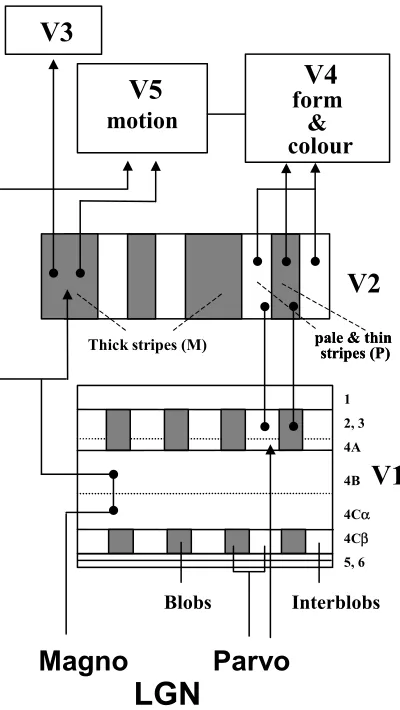
Extra-striate areas V2, V3, V4 and V5
Models for visual perception
Factors affecting pupil size
Disorders of the pupil
Basic Photometry and Colorimetry
Computer displays: luminance and colour calibration
Discussion
Experiments and results
Data analysis and results: Group 1
Data analysis and results: Group 2
Related documents