vents.
.
White Rose Research Online URL for this paper:
http://eprints.whiterose.ac.uk/83486/
Version: Published Version
Article:
Georgieva, MN, Little, CTS, Glover, AG et al. (1 more author) (2015) Mineralization of
Alvinella polychaete tubes at hydrothermal vents. Geobiology, 13 (2). 152 - 169. ISSN
1472-4677
https://doi.org/10.1111/gbi.12123
eprints@whiterose.ac.uk
https://eprints.whiterose.ac.uk/
Reuse
Unless indicated otherwise, fulltext items are protected by copyright with all rights reserved. The copyright
exception in section 29 of the Copyright, Designs and Patents Act 1988 allows the making of a single copy
solely for the purpose of non-commercial research or private study within the limits of fair dealing. The
publisher or other rights-holder may allow further reproduction and re-use of this version - refer to the White
Rose Research Online record for this item. Where records identify the publisher as the copyright holder,
users can verify any specific terms of use on the publisher’s website.
Takedown
If you consider content in White Rose Research Online to be in breach of UK law, please notify us by
Mineralization of
Alvinella
polychaete tubes at
hydrothermal vents
M . N . G E O R G I E V A ,1 , 2 C . T . S . L I T T L E ,1 A . D . B A L L3 A N D A . G . G L O V E R2
1School of Earth and Environment, University of Leeds, Leeds, UK
2Life Sciences Department, The Natural History Museum, London, UK
3Imaging and Analysis Centre, The Natural History Museum, London, UK
ABSTRACT
Alvinellid polychaete worms form multilayered organic tubes in the hottest and most rapidly growing areas of deep-sea hydrothermal vent chimneys. Over short periods of time, these tubes can become entirely min-eralized within this environment. Documenting the nature of this process in terms of the stages of minerali-zation, as well as the mineral textures and end products that result, is essential for our understanding of the fossilization of polychaetes at hydrothermal vents. Here, we report in detail the full mineralization of
Alvinellaspp. tubes collected from the East Pacific Rise, determined through the use of a wide range of imaging and analytical techniques. We propose a new model for tube mineralization, whereby mineraliza-tion begins as templating of tube layer and sublayer surfaces and results in fully mineralized tubes com-prised of multiple concentric, colloform, pyrite bands. Silica appeared to preserve organic tube layers in some samples. Fine-scale features such as protein fibres, extracellular polymeric substances and two types of filamentous microbial colonies were also found to be well preserved within a subset of the tubes. The fully mineralizedAlvinellaspp. tubes do not closely resemble known ancient hydrothermal vent tube fos-sils, corroborating molecular evidence suggesting that the alvinellids are a relatively recent polychaete line-age. We also compare pyrite and silica preservation of organic tissues within hydrothermal vents to soft tissue preservation in sediments and hot springs.
Received 10 April 2014; accepted 29 November 2014
Corresponding author: M. N. Georgieva. Tel.: +44 2079425643; fax: +44 1133432846; e-mail: eemng@leeds.ac.uk
INTRODUCTION
The annelid worms are an ancient lineage of animals dating to at least the earliest Cambrian period,~540 Ma (Conway
Morris & Peel, 2008; Vinther et al., 2011). Over evolu-tionary time, they have radiated into almost all marine hab-itats including deep-sea hydrothermal vents. Many vent sites in the Pacific are characterized by spectacular colonies of tube-dwelling polychaetes in the families Siboglinidae and Alvinellidae (Van Dover, 2000). Our understanding of the evolutionary history of these polychaetes and the vent ecosystems more generally is limited by a poor fossil record of soft-bodied organisms. Typically, preservation of soft tis-sues occurs through early authigenic mineralization (the impregnation and/or replication of an organic structure by minerals) and usually involves the minerals phosphate,
car-bonate, pyrite or silica (Briggset al., 1991; Akahaneet al., 2004). Much research has focused on organic tissue miner-alization within soft sediments and terrestrial hot springs (e.g. Raffet al., 2008; Farrellet al., 2013), but mineraliza-tion of organic animal and prokaryotic remains within hydrothermal vent environments, which also involves pyrite and silica (Cook & Stakes, 1995; Maginn et al., 2002; Boyce et al., 2003), is poorly understood. Documenting this process at modern hydrothermal vents is key to under-standing taphonomy within this chemically distinct setting and to improving the interpretation of ancient vent fossils.
Worm tube fossils with diverse morphologies are known from vent sites in the geological record back to the early Silurian period,~430 million years ago (Littleet al., 1998,
1999, 2004, 2007; Hilario et al., 2011), but little is known about the animals that formed them. Although
some have been assigned to extant vent polychaete groups, morphological identifications are not generally consistent with estimates of molecular divergence (Little & Vrijen-hoek, 2003; VrijenVrijen-hoek, 2013) and there is potential con-fusion with morphologically similar polychaete tubes (Kiel & Dando, 2009).
An endemic tube-forming polychaete genus within extant hydrothermal vents on the East Pacific Rise (EPR) is Alvi-nella(Desbruyeres & Laubier, 1986), comprising two spe-cies: Alvinella pompejana and A. caudata. Both are renowned for their occupation of high-temperature vent chimneys and role as biogeoengineers within this habitat (Desbruyereset al., 1998; Le Bris & Gaill, 2007). After col-onization,Alvinellaspp. can alter local vent fluid flow and composition, creating a range of micro-environments that allow the establishment of other hydrothermal vent biota less tolerant to high temperatures, and also promoting addi-tional mineral precipitation and thus modifying chimney morphology (Juniper & Martineu, 1995; Pradillon et al., 2005). In part, this biological habitat modification arises fromAlvinellaspp. irrigating the interior of their tubes with cool sea water from above the alvinellid colony. This results in an inner tube environment with a lower temperature and a more neutral pH (temp. of~29
–81°C, pH~7) compared
to conditions on the surface of the vent chimney substrate (temp. of~120°C, pH~4) (Di Meo-Savoieet al., 2004; Le
Bris et al., 2005) and creates buffered micro-niches which are colonized by micro-organisms (Le Briset al., 2005).
Colonization of fresh vent chimneys byAlvinellaspp. is considered to be strongly dependent on the properties of their unique tubes, which are attached directly onto vent chimney walls. These tubes possess high thermal and chemi-cal stability (Gaill & Hunt, 1986) and can be secreted incredibly quickly, at a maximum rate of 1 cm day1 in
length (Pradillonet al., 2009). The tubes ofA. pompejana andA. caudataare identical in appearance and are formed from granules primarily composed of protein (Vovelle & Gaill, 1986). The resulting tubes are fibrous and concentri-cally multilayered, with each tube layer comprised of super-imposed sublayers of parallel fibrils that vary in direction between adjacent sublayers (Gaill & Hunt, 1986; Des-bruyereset al., 1998). Both the inner and outer surfaces of Alvinella spp. tubes are covered by a patchy, but dense microbial community that includes filamentous, rod-shaped and coccoid forms (Desbruyeres et al., 1985), belonging primarily to the epsilon subdivision of the proteobacteria (Haddad et al., 1995; Campbell & Cary, 2001; Campbell et al., 2003). Micro-organisms on the insides of the tubes can become trapped between the tube layers as more organic material is deposited during tube growth, to form distinctive microbial layers within the tube wall (Gaill & Hunt, 1986; Zbindenet al., 2001; Maginnet al., 2002).
Within the extreme environment of the EPR hydrother-mal vents (see Fornariet al.(2012) and references therein
for an overview of the EPR spreading centre), minerals can precipitate onto occupied Alvinella tubes remarkably quickly, such that an 11-day-old alvinellid colony can have 88% mineral content (Pradillon et al., 2009). During the early stages of this mineralization, minerals progressively coat the inner and outer tube surfaces (Gaill & Hunt, 1991) and accumulate within the tube walls, where they occur as nanocrystalline iron or zinc sulphides that assem-ble along sublayer surfaces (Zbinden et al., 2001, 2003; Maginnet al., 2002; Le Briset al., 2008). Mineral precipi-tation has been observed particularly in tube layers contain-ing trapped micro-organisms, and pyrite may occasionally replace organic tube layers (Maginn et al., 2002). Over time, Alvinella spp. tubes can become entirely mineral in composition (fully mineralized) (Haymon et al., 1984; Haymon & Koski, 1985). Full mineralization of originally organic polychaete tubes has also been observed by Cook & Stakes (1995) for siboglinid worm tubes at vent sites on the Juan de Fuca Ridge (JdFR), but the details of how Al-vinellaspp. tubes are fully mineralized, including the gross and fine-scale mineral textures and distributions, have not been documented.
Here, we provide a detailed account of the complete mineralization process ofAlvinellaspp. tubes to show how polychaete tubes can be fossilized at vent sites such as the EPR. A large number of Alvinella spp. tube specimens exhibiting varying degrees of mineralization have been analysed to better understand (i) the identity of the main minerals replacing the tubes, (ii) the nature and distribu-tion of the mineral textures and (iii) the stages and timing of mineralization of the tubes. The identification of prob-lematic tubular fossils from ancient vent sites is discussed, and mineralization of Alvinella spp. tubes is compared to preservation of organic tissues by silica and sulphide miner-als within other environments.
METHODS
Sample collection and storage
The studied samples comprised vent chimney material con-taining Alvinella spp. tubes exhibiting varying degrees of mineralization. These were collected from the tops of nine active vent chimneys and one inactive chimney (Alvinellid Pillar) located along the EPR axial summit trough at depths of~2500 m (Fig. 1). The material was collected on
during the same RV Atlantis cruises [outlined in Little (2009); see Methods S1]. After recovery from the sea floor, theAlvinellaspp. tubes that were largely non-miner-alized were removed from the vent chimneys and preserved in 95% ethanol (hereafter referred to as partially mineral-izedAlvinella spp. tubes). Samples of vent chimney sulp-hides with fully mineralizedAlvinellaspp. tubes were dried and stored at room temperature post-collection (Fig. 2A). During post-collection storage, some of the sulphide chim-ney samples started to oxidize, forming secondary sulphate minerals; these were washed off prior to analyses. This oxi-dation may have resulted in the formation of iron oxides in addition to those formedin situ (in situ iron oxides were
evidenced by a red colour on recovery; Fig. 2A), and we hence excluded iron oxide analysis from the study.
Micro-CT analyses
Five partially mineralized Alvinella spp. tubes (Specimens 44, 46, 47, 48, 49; Table 1) were initially scanned using a Metris X-Tek HMX ST 225 micro-computed tomography (l-CT) system at the Natural History Museum, London, UK (NHM), to visualize the distribution of minerals on and within the tubes. Data volumes were constructed using CT Pro ver. 2.1 (Metris X-Tek, Tring, UK) and analysed using Drishti ver. 2.0 (Limaye, 2006). All five tube scans had a res-olution of 71lm or better. Mineral and organic tube com-ponents were separated based on grayscale values that represent X-ray attenuation, which closely corresponds to material density. To verify that the two were being accurately distinguished, one of the scanned specimens (Specimen 44) was embedded in resin, cross-sectioned and polished for reflected light microscopy to cross-reference the presence/ absence of minerals with the CT scan reconstruction.
Microscopic and chemical analyses
[image:4.595.66.291.250.446.2]The ethanol-preserved tubes were critically point dried and, along with the fully mineralized tubes, were cut, impreg-nated in resin and made into polished blocks of both trans-verse and longitudinal tube sections. Polished blocks of mineralizedAlvinellaspp. tubes contained both tubes and a section of the surrounding vent chimney matrix. The pol-ished blocks were coated with an approximately 17 nm car-bon layer and imaged using the following scanning electron microscopes (SEM) with backscattered electron detectors: a LEO 1455VP SEM, a Carl Zeiss Ultra Plus Field Emission SEM, and an FEI Quanta 650 FEG-ESEM both at the NHM and at the University of Leeds, UK (Leeds). Two fully
Table 1Information on theAlvinellaspp. tube material used for this study. Specimen numbers were assigned during this study
Vent Alvin dive
Collection date
Latitude of vent site
Longitude of vent site
Depth of
vent site (m) Temp. (°C) pH Specimens
Fossilization cage sample?
Bio90 4274 24-Nov-06 N9°50.311 W104°17.480 2509 382 4.4 45, 66, 69 No Bio9 4274 24-Nov-06 N9°50.312 W104°17.484 2509 388 3.6 55, 67, 71 No
4375 11-Dec-07 N9°50.312 W104°17.484 2509 358 3.9 47, 56, 61, 70, 72 Yes–370 days L-vent 4276 26-Nov-06 N9°46.256 W104°16.749 2519 341 4.4 46, 54 No
4377 13-Dec-07 N9°46.256 W104°16.749 2519 279 3.6 62, 65 No
4467 01-Nov-08 N9°46.256 W104°16.749 2519 – – 57, 60 Yes–319 days P-vent 4278 28-Nov-06 N9°50.280 W104°17.473 2509 392 4.5 44 No
Alvinellid Pillar 4281 01-Dec-06 N9°50.125 W104°17.456 2504 – – 68 No Biovent 4374 10-Dec-07 N9°50.963 W104°17.617 2501 349 4.1 49 No A-vent 4377 13-Dec-07 N9°46.500 W104°16.810 2541 136 5.4 74 No V-vent 4378 14-Dec-07 N9°47.231 W104°16.989 2517 363 3.6 48, 58 No S-vent 4379 15-Dec-07 N9°39.816 W104°15.714 2510 326 4.3 59 No
Vent location, depth, temperature and pH data were obtained from the Marine Geoscience Data System (Bryceet al., 2007, 2008) (http://www.marine-geo.org/).
[image:4.595.64.542.540.694.2]A
B
C
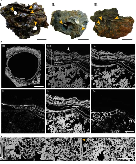
Fig. 2Fully mineralizedAlvinellaspp. tubes and associated vent chimney fragments. (A), Blocks of sulphide from vent chimneys containing fully mineralized
Alvinellaspp. tubes (orange arrows). (i, ii) Chimney blocks containing complex intertwinedAlvinellaspp. tubes, some of which have been mineralized com-pletely as cylindrical structures; (iii) sulphide block withAlvinellaspp. tubes mineralized on the surfaces that were attached to the vent chimney wall. (i) Spec-imen 74 (coated in epoxy resin); (ii) SpecSpec-imen 57; (iii) SpecSpec-imen 61. All scales in (A) are 30 mm. (B), WDS elemental mapping of a fully mineralizedAlvinella
spp. tube in transverse section (Polished Block 57.1) and associated vent chimney minerals. TS, Transverse section with the area analysed highlighted with white box; scale=5 mm. BSE, Backscatter electron image of the analysed area; scale=500lm. Fe to Zn show the distribution of four elements within the area analysed (Fe–iron, Si–silicon, S–sulphur, Zn–zinc). (C), Backscatter SEM composite of Polished Block 60.2 showing variation in mineral texture and
[image:5.595.74.521.80.618.2]mineralized Alvinella spp. tubes (Specimens 54 and 55; Table 1) were also imaged uncoated in the environmental chamber of a Philips XL 30 FEG-SEM at Leeds (UK).
The elemental composition of mineral phases, and ele-mental distribution were determined using both energy-dis-persive X-ray spectroscopy (EDX) within the SEMs above, and wavelength-dispersive spectrometry (WDS) using a Cameca SX-100 electron microprobe (EPMA) at the NHM. An accelerating voltage of 20 kV was used for EDX point-analyses and maps, whereas in the EPMA, these were carried out using an accelerating voltage of 15 kV and a probe cur-rent of 40 nA for mapping and 20 nA for point analyses. Reflected light microscopy was used to identify the mineral phases within approximately half of the specimens (Table S1). X-ray diffraction (XRD) was performed on a Bruker D8 instrument (Bruker, Karlsruhe, Germany) (Cu Karadiation source, 40 kV voltage and 40 mA of current) in Leeds on bulk material from a single vent chimney section and attached fully mineralizedAlvinellaspp. tube (Specimen 57) to identify the crystalline form of the zinc sulphide phase. In addition, confocal laser scanning microscopy (CLSM) using a Nikon A1-Si Confocal microscope at the NHM and oper-ated in spectral imaging mode, was used to visualize the structure of the organic tube layers and microbial filaments on the inner surface of anAlvinellaspp. tube (Specimen 44) by laser-induced autofluorescence.
Measurements of mineral textures
The dimensions of mineral textures preserved within Alvi-nella spp. tubes were measured from SEM images using the program IMAGEJ (version 1.46r; National Institutes of Health, USA; http://rsb.info.nih.gov/ij). Pores and fila-ments were prevalent mineral textures within the samples, which are likely to be fossilized microbial filaments (see later). When measuring the dimensions of these textures, only pores with a distinctly circular or elliptical transverse section, that is those likely to be biogenic in origin, were measured. For statistical tests, diameter measurements from pore and filament textures were grouped into two types. Shapiro–Wilk normality tests were used to determine
whether diameter measurements were normally distributed, andF-tests to compare variances between data pairs. Two-sample Kolmogorov–Smirnov tests were subsequently used
to compare the cumulative distributions between pairs of diameter measurements. All three types of statistical test were performed inR(R Core Team, 2013).
RESULTS
Vent chimney minerals aroundAlvinellaspp. tubes
The fragments of vent chimneys onto whichAlvinella spp. tubes were attached (Fig. 2A) were formed largely of iron
(pyrite, marcasite), zinc and copper (chalcopyrite, minor is-ocubanite) sulphides, silica, anhydrite and galena (Fig. 2B, C). An XRD trace for a vent chimney sample with an attached tube (Specimen 57) showed the zinc sulphide to be sphalerite, but it is likely that both sphalerite and wurtz-ite were common in the samples (these polymorphs are dif-ficult to discriminate when intergrown). The distribution of mineral phases within these vent chimney fragments was variable, but generally fine-grained marcasite occurred directly adjacent toAlvinellaspp. tube walls on the outside of vent chimneys, which was sometimes overgrown by zinc sulphides (Fig. 2B). This was succeeded by zinc sulphides and amorphous silica further into the vent chimney, which in turn was succeeded by larger-grained marcasite or zinc sulphides, then chalcopyrite or anhydrite (Fig. 2C). The vent chimney minerals exhibited crystalline morphologies and porosity associated with fine-grained marcasite growth, while colloform (finely concentric and radiating) textures were rare and did not delineate consistent shapes. An exception were continuous thin bands of colloform iron sulphide (Fig. 2C), found on the interiors of three chim-ney sections (Polished Blocks 57.3, 60.2 and 62.1). These were similar to the mineral layers comprising fully mineral-izedAlvinellaspp. tubes (see later).
Partially mineralizedAlvinellaspp. tubes
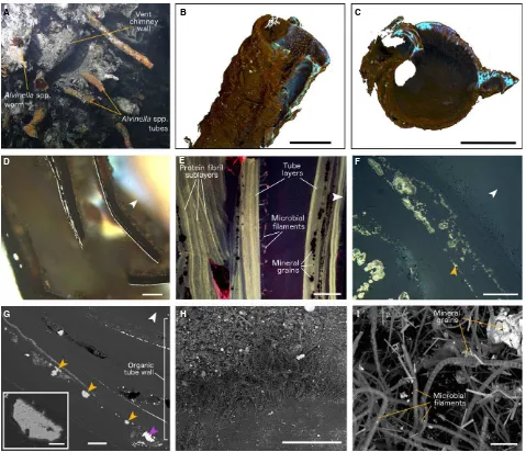
Examples of in situ partially mineralized Alvinella spp. tubes are shown in Fig. 3A. Three-dimensional l-CT reconstructions of partially mineralized Alvinella spp. tubes showed that minerals were often concentrated along a longitudinal surface of the tubes (Fig. 3B) (in Specimens 44, 46, 48, 49), which in one tube (Speci-men 44) was known to have been the side that was directly attached to the vent chimney. Minerals occurred as grains and crusts coating inner and outer tube wall surfaces and were also abundant between the concentric organic layers that comprise the Alvinella spp. tube walls (Fig. 3C). Detailed microscopy revealed that minerals were templating (here defined as the growth of minerals on a surface) certain organic tube layer and sublayer sur-faces (Fig. 3D–G), where mineral growth appears to
begin as small cores, often <1lm in diameter. These
cores appeared to fuse with adjacent cores following fur-ther mineral precipitation, to form multiple bands of mineralization parallel to the natural layering of the tubes (Fig. 3D–G). The composition of the minerals
iron sulphide growth. Large mineral grains, as well as large grains of elemental sulphur (Fig. 3G), also occurred between organic tube layers and on both inner and outer tube surfaces. The elemental sulphur grains were usually 10 s of micrometres in size, but some were up to 468lm across. They had a pitted texture (Fig. 3G, insert) and were rarely observed in the fully mineralized tubes.
Fully mineralizedAlvinellaspp. tubes
The fully mineralized Alvinellaspp. tubes occurred in two forms: as tubes fully enclosed within vent chimney sulp-hides, in which the entire circumference of the tube wall had been preserved and tube interiors were mostly hollow (Fig. 2A-i,ii), or as partial tube walls attached to the sur-faces of vent chimney fragments (Fig. 2A-iii). The fully
A B C
D E F
G H I
Fig. 3Partially mineralizedAlvinellaspp. tubes. (A)Alvinellaspp. tubes on a hydrothermal vent chimney (L-vent, AT15-27, Alvin dive 4382), with an Alvi-nellaspp. worm at its tube opening. Image credit: Woods Hole Oceanographic Institution. (B, C) Reconstructions of a singleAlvinellaspp. tube (Specimen 46) using micro-computed tomography (l-CT). Blues and purples highlight dense areas where minerals have precipitated, while browns constitute the organic tube wall; scales=10 mm. (B) tube in oblique side view; (C) tube in transverse section. (D) Bands of mineral growth within and on surfaces of organic
Alvi-nellaspp. tube layers, scale=300lm (Polished Block 46.1). (E) Confocal image of a transverse section through anAlvinellaspp. tube (Polished Block 44.1), showing organic tube layers, microbial filaments trapped between layers, and the texture of protein fibrils within the organic tube. Scale=100lm. (F) Detail of an organic tube layer where mineralization begins as small iron sulphide cores, which join up upon further mineral precipitation to form distinct colloform pyrite bands. Cores and bands often occur along distinct surfaces within the organic layers (orange arrow) (Polished Block 44.1); scale=50lm. (G) Trans-verse section of anAlvinellaspp. tube (Polished Block 44.1) with mineral grain (purple arrow) and elemental sulphur grains (orange arrows); scale=100lm.
[image:7.595.56.535.72.484.2]mineralized Alvinella spp. tubes that were obtained from the fossilization experiment lasting approximately 1 year (319 and 370 days; Table 1; Methods S1) demonstrate that full tube mineralization can occur within this time period.
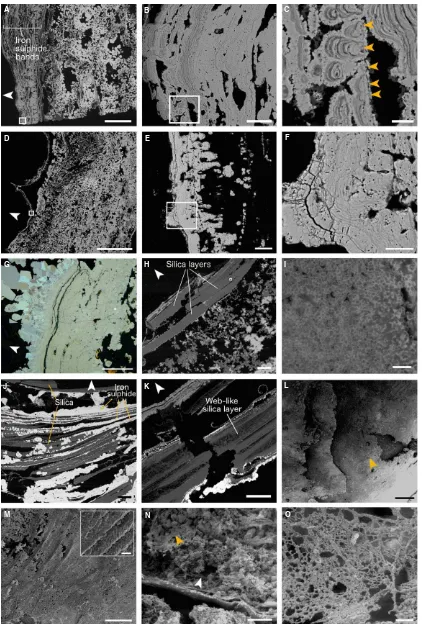
The composition of fully mineralized Alvinella spp. tubes also reflected the mineralogy of adjacent vent chim-ney fragments. Mineral Alvinella spp. tube walls were mainly iron sulphide (pyrite and marcasite) and amorphous silica (Fig. 2B; Table S1) in composition, with small quan-tities of zinc sulphides (sphalerite and/or wurtzite), and minor quantities of copper containing sulphides (chalcopy-rite, isocubanite), galena and anhydrite. The majority of fully mineralized Alvinella spp. tubes were comprised of multilayered iron sulphide (pyrite) sheets that broadly mir-rored the layering of organic tube walls, which appeared as concentric pyrite bands or horizons in transverse and lon-gitudinal section (Figs 2B,C and 4A–F). The pyrite bands
occasionally showed weak anisotropy and contained over-growths of crystalline marcasite that increased in crystal size away from the tube wall (Fig. 4G).
The number of pyrite bands comprising fully mineralized Alvinella spp. tubes varied greatly between different tube samples (Table S1; Figs 2B and 4A,D). Pyrite band num-ber, thickness and the degree to which they joined with adjacent bands also varied between different parts of the same tube. The pyrite bands were characterized by collo-form textures, the development of which could in some instances be traced to small iron sulphide cores very similar to those recorded within partially mineralized Alvinella spp. tubes (Figs 3D–F and 4C,F). Sustained mineral
pre-cipitation onto the iron sulphide cores appears to have resulted in the formation of colloform micro-stromatolitic structures, up to 218lm in length, comprised of fine-scale pyrite layers<1lm thick (Fig. 4B,C,E,F). Colloform
layer-ing tended to become increaslayer-ingly sheet-like with distance away from the cores and as adjacent micro-stromatolitic structures fused (Fig. 4B,E,G). Most micro-stromatolitic iron sulphide structures were oriented towards the inside of the tubes. Electron microprobe transects through the colloform textures (Tables S2–S5; Figs S2–S5) showed
sili-con or zinc to be present within some of the fine mineral layers; however, the small size of individual layers made it difficult to determine their elemental composition indepen-dently of the surrounding layers.
Amorphous silica was often present filling voids between pyrite bands in the fully mineralized Alvinella spp. tubes (Fig. 2B). However, in four of the polished blocks (num-bers 57.2, 57.3, 58, 62.1), the mineral tube wall was mainly comprised of layers of silica that resembled organic tube layers in thickness and shape (Fig. 4H,I). These silica layers also contained rows of iron sulphide cores, much like those observed to have grown within the organic walls of partially mineralized Alvinella spp. tubes (Fig. 4J). The
silica layers were made up of small (<1lm diameter) silica
spheres, and in some places, the thick silica layers exhibited web-like or stringy textures (Fig. 4J,K).
Additional mineral textures found within fully mineral-ized Alvinella spp. tubes comprised mainly of pyrite include a texture of cross-cutting and/or bundled ‘fibres’ (Fig. 4L,M), which occurred on the external surface of a tube. Under higher magnification, these bundled ‘fibres’ showed a surface covering of smaller cross-hatched stria-tions<1lm in width (Fig. 4M, insert). Another tube also
contained mineral infilling of pyrite crystals, and a fine mesh-like structure also formed of pyrite (Fig. 4N,O), while anhydrite was observed to have overgrown the inside of a different fully mineralized Alvinella spp. tube (Fig. 4P).
Pore and filament textures
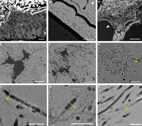
Another texture prevalent in the fully mineralized tube samples was porosity. The pyrite minerals of several tubes contained circular pores 0.1lm to several micrometres in diameter, in association with sinuous, unbranched filaments of a uniform diameter (Fig. 4F,Q,S–U) (hereafter referred
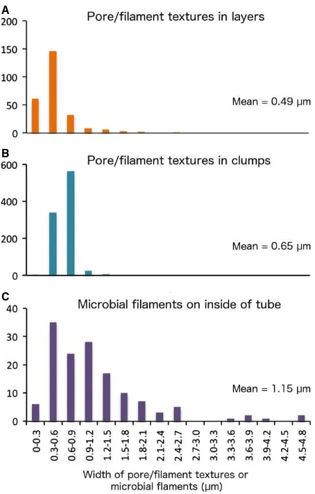
to as pore and filament associations). A few of these fila-ments contained cross-walls resembling septae (Fig. 4V, W). Pore and filament textures were found to crosscut col-loform structures, and in some samples, the filaments appeared ‘rooted’ to individual pyrite layers (Fig. 4Q,U). Two types of pore and filament associations were identi-fied, based on their mode of occurrence. The first (associa-tion 1) occurred within the pyrite layers comprising fully mineralizedAlvinellaspp. tubes (Fig. 4F,Q) and had pores and filaments ranging from 0.13 to 2.62lm in diameter (mean=0.49 lm; Fig. 5) that were in some instances very
densely packed (Table S6). Association 1 filaments were hollow. The second type of pore and filament association (association 2) occurred as clumps of pores and filaments preserved within pyrite minerals adjacent to the outer lay-ers of fully mineralized Alvinella spp. tubes (Fig. 4R–U).
Association 2 pores and filaments had diameters of a smal-ler size range (0.26–1.36lm; mean=0.65 lm) (Fig. 5)
and were often more densely packed than pores and fila-ments in association 1 (Table S6). Association 2 filafila-ments at times also exhibited changes in orientation within the clumps, appearing in transverse section towards the centre of the clumps and in longitudinal section towards the clump perimeters (Fig. 4S,U). The filaments were mostly hollow but some were infilled by pyrite (Fig. 4X).
A B C
D E F
G H I
J K L
M N O
[image:9.595.84.506.71.701.2]P Q R
S T U
V W X
Fig. 4Fully mineralizedAlvinellaspp. tubes. (A) Longitudinal section of a tube (Polished Block 60.3) with a large number of iron sulphide (pyrite) bands replacing the tube wall; scale=1 mm. White box shows location of (B). (B) Detail of boxed area in (A) showing bands of colloform pyrite; scale=50lm. White box shows location of (C). (C) Detail of boxed area in (B) showing colloform micro-stromatolitic structures with orange arrows pointing towards the cores from which they originate; scale=10lm. (D) Transverse section through two adjacent Alvinella spp. tubes; white box shows location of (E) scale=4 mm. (E) Bands of pyrite comprising the mineralized tube, white box shows location of (F); scale=50lm. (F) Pore and filament textures within col-loform pyrite (association 1); scale=20lm. (G) Bands of colloform pyrite overgrown by marcasite; scale=100lm (Polished Block 70.1). (H) Amorphous sil-ica appears to be replacing organic tube layers (Polished Block 58.1). Scale=200lm; white box shows location of (I). (I) Detail of tube in (H) showing small silica spheres that comprise some of the amorphous silica layers; scale=4lm. (J) Interlaminated silica and pyrite, where silica appears to have preserved parts of disintegrating organic tube layers and surrounds iron sulphide cores (Polished Block 57.2). Scale=200lm. (K) Silica layer within a mineralizedAlvinella spp. tube (Polished Block 62.1) exhibiting a web-like texture; scale=200lm. (L) View of the external wall of a fully mineralizedAlvinellaspp. tube (Speci-men 54) showing four mineralized layers; orange arrow points to texture in (M). Scale=500lm. (M) detail of iron sulphide ‘fibres’ that are cross-cutting and/or bundled; scale=250lm.Insert, Detail of adjacent fibres showing surface covering of small cross-hatched striations; scale=10lm. (N) Interior of a mineralizedAlvinellaspp. tube (Specimen 55) which has been partially filled by minerals. The mineralized tube wall runs horizontally along the bottom third of the image; orange arrow points towards location of EPS-like mineral texture. Scale=1 mm. (O) Detail of EPS-like mineral texture from the tube in (N); scale=50lm. (P), Anhydrite growing on the inside of anAlvinellaspp. tube (orange arrow); scale=500lm. (Q), Pore and filament texture (association 1) occurring within one of the outer pyrite bands of a fully mineralizedAlvinellaspp. tube (Polished Block 57.1); scale=20lm. (R), Transverse section of
[image:10.595.68.541.75.495.2]filaments in clumps; and microbial filaments from an inner tube surface) were not normally distributed, and F-tests revealed the variances to be significantly different between all three data types (Table 2). Subsequent two-sample Kol-mogorov–Smirnov tests between the data type pairs were
all significant (Table 2). However, theP-values are approx-imate due to the presence of ties in the data.
INTERPRETATIONS AND DISCUSSION
Alvinellaspp. tube mineralization process
Model of mineralization
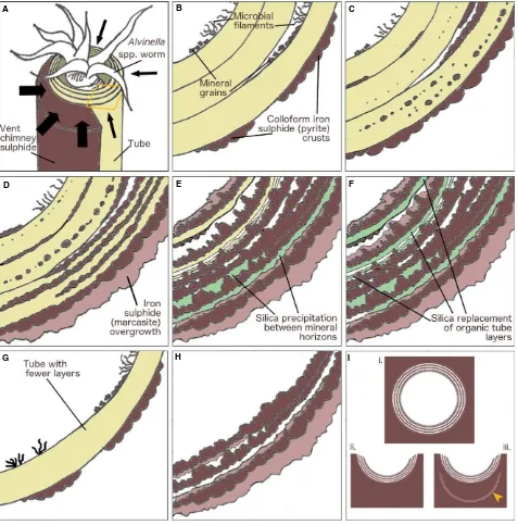
Micro-CT reconstructions (Fig. 3B,C) of the partially min-eralizedAlvinellaspp. tubes show that tube mineralization begins preferentially along the longitudinal surfaces of the tubes which are attached to, or nearest to the vent chim-ney walls (Fig. 6A). From these surfaces, mineralization likely spreads through the remainder of the tubes. This directional mineralization appears to result from a greater supply of mineral ions from the vent chimney. The greater amounts of sulphide minerals in the outer tube layers of partially mineralized tubes suggests that, at least initially, mineralization begins along the exterior surfaces of tubes. Mineralization appears to then progress within the organic walls ofAlvinellaspp. tubes, where fine iron sulphide (plus or minus a zinc and/or copper content) cores template tube layer and sublayer surfaces (Figs 3D–F and 6C,D).
Space to accommodate the growth of these cores may be provided by breaks between adjacent protein sublayers, possibly created by a poorer organization of their protein fibrils (Zbindenet al., 2001). The sulphide cores may also form within these particular layers because of an accumula-tion of metal ions (e.g. iron, Le Briset al., 2008), and/or by seeding on mineral grains (including elemental sulphur) trapped between tube layers (Maginn et al., 2002) (Figs 3G and 6B,C). In addition, sulphide mineralization within Alvinella spp. tube walls may also be aided by the creation of oxygen-poor micro-environments between tube layers. Newly secreted Alvinella spp. tubes are permeable to hydrogen sulphide (Le Bris et al., 2008). If hydrogen sulphide is trapped within such crevices as it diffuses into the tube, a greater influence of anoxic vent fluid over sea water may favour the precipitation of sulphide minerals in tube wall interspaces.
The small sulphide mineral cores formed along the Alvi-nellaspp. organic tube layer and sublayer surfaces continue to grow in a directed manner, amalgamating to produce bands of iron sulphides (pyrite). These run in parallel with the organic tube layering (Figs 3C–F, 4A–F and 6D) and
are often more numerous than the original organic tube layers. These pyrite bands show an increasingly colloform texture as they thicken, and can preserve fine-scale details of the original fibrous structure of the organic tubes
(Fig. 4L,M), possibly even individual protein fibres (Fig. 4M, insert). The growing sulphide bands may also incorporate and fossilize the microbial community present between the organic tube layers, leading to the formation of layers of pyrite with pore and filament textures (as pro-posed by Maginn et al., 2002). Delamination of adjacent protein sublayers, probably resulting from iron sulphide growth and/or decay of the organic material, may expose additional surfaces for further templating by pyrite. This process could account for the very large number of pyrite bands observed in some of the fully mineralized tubes (Fig. 4A,B). In other samples where the fully mineralized tubes comprise of only up to 3–5 mineral bands (Figs 4D–
F and 6G,H), the original organic tubes may not have been very thick, perhaps the result of a tube which was built and vacated fairly quickly by the worm [Zbinden et al.(2003) found 3–6 layers in 70-day-oldAlvinellaspp.
tubes], or a tube that was being rapidly elongated to keep pace with rapid chimney growth as suggested by previous authors (Gaill & Hunt, 1991; Chevaldonne & Jollivet, 1993).
As the organic tube walls eventually decompose, sul-phide mineral precipitation continues through the accre-tion of thin colloform pyrite layers onto existing mineral bands (Figs 4B,C,E,F and 6E). At this stage, mineral pre-cipitation appears to be greater on the inside of the tubes, which is shown by the orientation of micro-stromatolitic iron sulphide structures towards the tube interiors. The colloform pyrite bands that at this stage comprise the Alvi-nella spp. tube wall are subsequently overgrown by more crystalline marcasite (Figs 4G and 6E,F,H). The mineral precipitation orientation change, and the growth of later marcasite minerals, can be explained by the absence of the worms at this stage of tube mineralization. During the life of Alvinella spp., the fluids in the tube are both cooler than end-member vent fluids and less acidic (pH~7 inside
the tube compared to pH 4–5 outside; Le Bris et al.,
At this stage, silica may form layers between the pyrite bands (Fig. 6E,F). This likely occurs due to convective cooling, which induces silica saturation and promotes its precipitation after the tube has already been mineralized by iron sulphides (Hannington & Scott, 1988; Juniper & Fouquet, 1988; Halbachet al., 2002). However, the silica layers that resemble organic tube layers and contain bands of iron sulphide cores (Fig. 4H–K) suggest that the iron
sulphide cores initially templated organic tube layers, which were later directly preserved by silica. These observations therefore suggest that silica can be intimately involved in the tube fossilization process, rather than occurring just as a late-stage mineral phase. These silica layers are unlikely to have formed through replacement (i.e. substitution of a mineral phase by another phase, or of organic matter by a mineral) of iron sulphides, as there is no evidence in our samples of the oxidized products (i.e. no iron oxides) that one might expect from this process.
The fully mineralized Alvinella spp. tubes may be pre-served within the vent chimneys in several different forms (Fig. 6I). Rapid mineral precipitation may surround entire tubes to create the porous structures in Fig. 2A-i,ii, which are analogous to those observed by previous studies of EPR chimney sulphides (Hekinian et al., 1980; Haymon & Kastner, 1981; Haymon & Koski, 1985). Alternatively, only the sides of the tubes that are attached to the vent chimney may be mineralized, resulting in the partial tubes observed on mineral blocks used in this study (Fig. 2A-iii). The concentric continuous layers of colloform pyrite that comprise fully mineralized Alvinellaspp. tubes clearly dis-tinguish them from vent chimney minerals, and such layers inside some of the vent chimney samples likely represent the mineralization of earlier tubes, which have been subse-quently overgrown by vent chimney sulphides and other Alvinella spp. tubes (Fig. 2C). Tubes may eventually be infilled by the precipitation of later-stage sulphides, includ-ing higher grade minerals (e.g. copper-rich sulphides), as noted by Cook & Stakes (1995) for mineralized siboglinid tubes from the JdFR.
The fully mineralizedAlvinellaspp. tubes collected from the fossilization experiment provide a maximum-time esti-mate for annelid tubes to be completely mineralized at hydrothermal vents (~1 year). However, one should be
cautious about deriving an actual rate of mineralization from the above, as it is not known at what point during the deployment period Alvinella spp. colonized the fossil-ization cages. Mineralfossil-ization rates are also likely to be highly variable on small spatial scales due to the heteroge-neous nature of vent environments (Van Dover, 2000). It is therefore very likely that Alvinella spp. tubes can become fully mineralized much more rapidly than within 1 year, as suggested by the very high mineral content observed within an 11-day-old alvinellid colony (Pradillon et al., 2009).
A
B
C
Fig. 5Frequency distribution plots showing diameter measurements for (A), pores and filaments occurring within mineralized bands ofAlvinella
[image:12.595.65.292.72.427.2]spp. tubes (association 1); (B) those occurring as clumps (association 2); and (C) confirmed microbial filaments from the inside of anAlvinellaspp. tube (Specimen 44).
Table 2Results of statistical tests performed on pore and filament mineral textures preserved in mineral layers and as clumps, and microbial filaments from the inner surface of anAlvinellaspp. tube (Specimen 44)
P/F in layers P/F in clumps Microbial filaments
Shapiro–Wilk test
n 259 939 142
W 0.7337 0.9550 0.8181
P <2.2E-16 2.34E-16 5.47E-12
P/F in clumps vs. P/F in layers
P/F in layers vs. microbial filaments
P/F in clumps vs. microbial filaments
F-test
F 0.1861 0.1392 0.0259
P 0.000 0.000 0.000
Kolmogorov–Smirnov test
D 0.5678 0.5584 0.5103
P <2.2E-16* <2.2E-16* <2.2E-16*
[image:12.595.63.292.534.695.2]Microbial preservation
We interpret the pore and filament textures in pyrite found within and around the mineralizedAlvinella spp. tubes to
be the fossils of microbial filaments. This is because they demonstrate all of the suggested characteristics ofbona fide microbial fossils (Westall, 1999; Schopf et al., 2010), are
A B C
D E F
G H I
Fig. 6Stages ofAlvinellaspp. tube mineralization. (A)Alvinellaspp. worm inside multilayered organic tube attached to vent chimney sulphide. Mineraliza-tion (black arrows) progresses from the outside of the tube towards the inside and is more prevalent from the vent chimney side. Orange area shows trans-verse section in (B–F). (B) Transverse section of tube in (A). Mineralization begins as colloform iron sulphide (pyrite) crusts forming on outer layers, and
mineral grains becoming trapped in microbial filaments acting as nuclei for further mineral precipitation. (C), Minerals precipitate onto distinct surfaces within organic layers and sublayers, as small predominantly iron sulphide cores. (D), Further mineral precipitation results in the formation of concentric colloform pyr-ite bands. More crystalline iron sulphide (marcaspyr-ite) overgrows initial iron sulphides depospyr-ited on the outside of the tube. (E), Organic matter degrades and silica occasionally fills gaps where it previously occurred. The supply of dissolved minerals changes to being from inside the tube, and growing colloform iron sulphides orient towards the tube interior. (F), In some instances, silica may directly preserve the degrading organic matter. Further overgrowth of more crys-talline iron sulphide occurs. (G, H), Mineralization of a tube with fewer layers and without silica; (G) early stage; (H), late stage. (I) End products ofAlvinella
[image:13.595.58.535.74.557.2]morphologically closely analogous to the microbial com-munity associated with partially mineralizedAlvinella spp. tubes (Gaill et al., 1987; Desbruyeres et al., 1998; Zbin-den et al., 2001; Maginn et al., 2002) and occur in a range of orientations throughout the matrix of the iron sulphides in which they are present (cf. pyrite leaching/ oxidation by microbes; Veratiet al., 1999; Edwards et al., 2003).
The microbial fossils appear to be mostly external fila-ment moulds, but exhibit some evidence of replacefila-ment of septae and external sheaths by iron sulphide (Fig. 4V–X),
demonstrating that extremely fine-scale preservation can occur at hydrothermal vent sites. The difference in diame-ter distributions (Fig. 5; Table 2) between the fossil and the non-fossil microbial filaments may occur due to min-eral replacement of microbial sheaths in the former. The significantly different diameter distributions between the fossil filaments occurring in iron sulphide bands (associa-tion 1) of the Alvinella spp. mineralized tube wall and those occurring as clumps around the tube (association 2) (Fig. 5; Table 2) are likely due to the fossilization of two distinct microbial communities. Micro-organisms sampled from wholeA. pompejanatubes have been shown to differ to those sampled from tube interiors through molecular studies (Campbell et al., 2003), which likely reflects the variation in microhabitats between the tube interior and exterior. Microbial mats are commonly encountered on hydrothermal vent chimneys in areas colonized by Alvinel-la spp. (Taylor et al., 1999), and the position of the clumped filament fossils relative to theAlvinellaspp. tubes in our samples indicates that the microbial filaments inhab-ited crevices next to the tubes, which may have provided some protection from thermal and chemical extremes. The marked differences in temperature and pH along an Alvi-nellaspp. tube (temp. of~20°C, pH~8 at tube openings;
temp. of ~120°C, pH ~4 adjacent to vent chimney
sub-strate) (Le Briset al., 2005) may explain the patchy distri-bution of the clumped filaments in our samples and why they were only found within a few of the examined speci-mens. The observation that the filaments in clumps are sometimes ‘rooted’ onto specific iron sulphide layers (Fig. 4U) indicates a complex intergrowth of microbial mats and sulphide mineral precipitation.
The mesh-like pyritized structure associated with sul-phide minerals precipitated in the internal space of the fully mineralized Alvinella spp. tube shown in Fig. 4N,O likely represents mineralized EPS, due to the irregular sizes of the fibres comprising the mesh. In addition, the mesh in Fig. 4O bears a strong resemblance to mineralized microbial EPS textures observed in hot spring deposits (Handleyet al., 2008; Tobleret al., 2008; Peng & Jones, 2012). Many hydrothermal vent micro-organisms are known to secrete EPS (Raguenes et al., 1996, 1997b), including those associated with the integument of
A. pompejana (Vincent et al., 1994; Raguenes et al., 1997a; Cambon-Bonavita et al., 2002). However, as the mesh observed in this study is positioned on top of miner-als infilling anAlvinellaspp. tube, we hypothesize that the mesh was created in the absence of an Alvinella spp. polychaete.
Comparison with previous accounts of vent tube mineralization
Our observations of early-stage mineral precipitation in Al-vinella spp. tubes are similar to that reported by Maginn et al.(2002), as we also observed early-stage iron sulphide mineralization along sublayers in Alvinella spp. tubes. However, we did not find pyrite to be directly replacing the organic walls of Alvinella spp. tubes in our samples, finding instead more evidence for the growth of sulphides on the organic layer surfaces (i.e. templating). The small iron sulphide cores we observed along tube sublayer sur-faces appear analogous to the nanocrystalline zinc–iron
sulphides reported by Zbindenet al. (2001, 2003). How-ever, the general absence of zinc sulphides in our tube samples may be explained by different chemical and/or thermal characteristics of the vent fluids in the 9°N EPR
area at the time that our samples were collected, compared to when theAlvinellaspp. tubes studied by Zbindenet al. (2001, 2003) were obtained, as it is well established that EPR 9°N vent fluid temperature and chemistry vary
tem-porally (Von Damm, 2000, 2004; Von Damm & Lilley, 2004). Previous studies on early-stage mineralization have also proposed that the presence of trapped micro-organ-isms within the organic tube walls ofAlvinella spp. (Mag-inn et al., 2002) and siboglinid tubes (Peng et al., 2008, 2009) may have a direct control on their mineralization. However, mineral growth along the organic sublayers within our Alvinella spp. tubes often did not originate where trapped microbial filaments occurred, suggesting that mineralization may also take place in the absence of micro-organisms, in spaces between the protein fibril layer-ing of Alvinella spp. tubes. This may also account for the absence of fossilized microbial filaments in many of the pyrite bands.
Comparison with ancient hydrothermal vent tubeworm fossils
Our mineralized Alvinella spp. tubes differ in several respects from all fossil vent tubes described to date (Little et al., 1998). The Silurian vent tube speciesEoalvinellodes annulatus(Littleet al., 1999) occupied a similar ecological habitat asAlvinellaspp., being found on the exterior of vent chimney walls, but the mineralized tubes of E. annulatus are formed of a single layer (0.04–0.60 mm thick) of
framb-oidal and/or colloform pyrite, that is externally smooth but with internal ornament of concentric and subconcentric ann-ulations. Further, E. annulatustubes are generally smaller thanAlvinellaspp. tubes. The tubes of the Silurian vent spe-ciesYamankasia rifeia(Littleet al., 1997, 1999) are equiv-alent in size to Alvinella spp. tubes and have tube walls formed of several thin layers (0.01–0.08 mm thick) of
arse-nian pyrite; however, they show an external ornament of concentric growth lines, fine longitudinal ridges that are absent inAlvinellaspp. tubes. However, someY. rifeiatube walls have a thick coating (up to 2.7 mm) of micro-lami-nated colloform pyrite with ~1lm diameter holes (Little
et al., 1997), which are very much like the microbial fossils inAlvinellaspp. tubes.
Haymonet al.(1984) and Haymon & Koski (1985) com-pared fully mineralizedAlvinellaspp. tubes from 21°N EPR
with Upper Cretaceous (Bayda, Oman) tubular fossils formed of 2–3 concentric pyrite layers, with interlaminations
of silica or void space. While these fossil tubes are morpho-logically similar to ourAlvinellaspp. tubes in terms of size and the presence of multilayering in the tube wall, they do not show all the characteristics outlined above forAlvinella spp. tubes, such as some tube walls being comprised of many more than three pyrite layers. In addition, the Bayda tubes exhibit annulations and longitudinal ornamentation which are more closely associated with siboglinid or chaetopterid tubes (Kiel & Dando, 2009; Hilario et al., 2011) and are generally absent fromAlvinellaspp. tubes. Because of this, we suggest that no known ancient vent fossil tubes are equivalent to present dayAlvinellaspp. tubes. This interpre-tation is currently supported by molecular evidence suggest-ing that modern alvinellids diverged from 41 to 51 Ma (Eocene) (Vrijenhoek, 2013), whereas the majority of well-preserved tubular vent fossils are Mesozoic or Palaeozoic in age (Littleet al., 1997, 1998, 1999).
Soft tissue preservation by silica and pyrite
Our study shows that the proteinaceous organic tube walls ofAlvinellaspp. and associated microbial cells can be rap-idly preserved by both sulphide minerals and silica at vent sites. Soft tissue preservation by pyrite is on the whole rare and only seen at sites of exceptional preservation, for exam-ple Beecher’s Trilobite Bed, Upper Ordovician, New York
State, USA, or the Hunsruckschiefer, Devonian, western€ Germany (Briggset al., 1991; Briggs & Bartels, 2001; Rai-swellet al., 2008; Farrellet al., 2013). In these cases, soft tissues are preserved by an infilling or templating of pyrite occurring as framboids, pyritohedra and euhedral crystals generally <20lm in size (Briggs et al., 1991, 1996).
Sul-phide mineralization within this setting is biogenically mediated as it results from the decay of the soft tissues and leads to the localization of sulphide mineral precipitation on and around these tissues. Within hydrothermal vent environments, mineral precipitation generally occurs abio-genically as a result of the mixing of hot, mineral-rich vent fluid with cooler sea water. However, the role of decay products on mineralization within vents is largely unknown. Elevated dissolved sulphide, low oxygen and low pH conditions that may be especially concentrated around decomposingAlvinellaspp. tube surfaces could act to induce local supersaturation of iron and sulphide at these sites and thereby promote more intense iron sulphide precipitation. The micro-stromatolitic, fine-scale nature of pyrite that preserves Alvinella spp. tubes could be indica-tive of this process, as small pyrite grain sizes suggest abundant nucleation, which is considered a product of high degrees of supersaturation and the formation of collo-form pyrite (Barrie et al., 2009). Decay-induced local supersaturation may thereby account for the widespread association and organization of colloform iron sulphide textures with Alvinellaspp. tubes, compared to their rela-tive rarity within vent chimney minerals. The role of decay products in hydrothermal vent mineralization therefore could have similarities to (albeit different products to) pyri-tization within soft sediments, but warrants further investi-gation.
modes of organic matter preservation by silica may be occurring simultaneously within hydrothermal vents, but if silica templating is a pathway through whichAlvinellaspp. tubes are mineralizing, it seems to be occurring in an alto-gether different manner to pyrite templating.
CONCLUSIONS
Documenting in detail the mineralization of modern poly-chaete tubes is critical to ensuring the validity of fossil-modern comparisons and to advancing current understand-ing of the taphonomy and palaeontology of polychaete worms. We have shown how Alvinella tubes can be fully mineralized within a modern hydrothermal vent setting as multiple concentric pyrite bands that include fine-scale fea-tures such as protein fibres and associated micro-organ-isms. Our ability to interpret ancient fossils will always be limited by paucity of material and diagenetic alteration, and it is important to be mindful of these factors when comparing ancient to recently mineralized material. Fortu-nately, many ancient hydrothermal vent tube fossils appear well preserved, with some of the oldest specimens exhibit-ing fine colloform layerexhibit-ing which suggests that they have not been significantly altered. These tubes will make ideal targets for investigating the preservation and nature of interactions between micro-organisms and tubeworms, a largely unknown aspect of the ecology of ancient vent communities.
ACKNOWLEDGMENTS
MNG is funded by a NERC CASE PhD studentship (NE/ K500847/1). For sample collection cruises to the East Pacific Rise, the authors would like to thank Karen Von Damm (deceased), Stefan Sievert and Timothy Shank spe-cifically, and the Woods Hole Oceanographic Institution in general. Funding for these cruises was from the NSF Ridge2000 project (AT15-13 and AT15-27 –
OCE03-27126; AT15-38 – MCB07-02677). NERC Small Grant
NE/C000714/1 funded the fossilization experiments of CTSL associated with these cruises. Thank you to Chris Stanley (NHM) for help with mineralogical observations and to Claire Stevenson for observations of anhydrite and rooted filaments in the Alvinella spp. tubes. Thanks also to Harri Wyn Williams, Richard Walshaw, Lesley Neve, Geoff Lloyd, David Wallis and Liane Benning (Leeds); Tomasz Goral, Farah Ahmed, Daniel Sykes and John Spratt (NHM) for guidance and assistance with analytical techniques.
REFERENCES
Akahane H, Furuno T, Miyajima H, Yoshikawa T, Yamamoto S (2004) Rapid wood silicification in hot spring water: an
explanation of silicification of wood during the Earth’s history.
Sedimentary Geology169, 219–228.
Barrie CD, Boyce AJ, Boyle AP, Williams PJ, Blake K, Ogawara T, Akai J, Prior DJ (2009) Growth controls in colloform pyrite.
American Mineralogist94, 415–429.
Benning L, Wilkin R, Barnes H (2000) Reaction pathways in the Fe–S system below 100°C.Chemical Geology167, 25–51.
Boyce AJ, Little CTS, Russell MJ (2003) A new fossil vent biota in the Ballynoe barite deposit, Silvermines, Ireland: evidence for intracratonic sea-floor hydrothermal activity about 352 Ma.
Economic Geology98, 649–656.
Briggs DEG, Bartels C (2001) New arthropods from the Lower Devonian Hunsr€uck Slate (Lower Emsian, Rhenish Massif, Western Germany).Palaeontology44, 275–303.
Briggs DEG, Bottrell SH, Raiswell R (1991) Pyritization of soft-bodied fossils: Beecher’s Trilobite Bed, Upper Ordovician, New York State.Geology19, 1221–1224.
Briggs DEG, Raiswell R, Bottrell SH, Hatfield DT, Bartels C (1996) Controls on the pyritization of exceptionally preserved fossils; an analysis of the Lower Devonian Hunsruck Slate of€
Germany.American Journal of Science296, 633–663.
Bryce J, Prado F, Von Damm K (2007) AT15-13 Chemistry: Fluid.Marine Geoscience Data System, http://www.marine-geo.org/tools/search/Files.php?data_set_uid=17364. Bryce J, Prado F, Von Damm K (2008) AT15-27 Chemistry:
Fluid.Marine Geoscience Data System, http://www.marine-geo.org/tools/search/Files.php?data_set_uid=17603. Cambon-Bonavita M-A, Raguenes G, Jean J, Vincent P,
Guezennec J (2002) A novel polymer produced by a bacterium isolated from a deep-sea hydrothermal vent polychaete annelid.
Journal of Applied Microbiology93, 310–315.
Campbell BJ, Cary SC (2001) Characterization of a novel spirochete associated with the hydrothermal vent polychaete annelid,Alvinella pompejana.Applied and Environmental Microbiology67, 110–117.
Campbell B, Stein J, Cary S (2003) Evidence of
chemolithoautotrophy in the bacterial community associated withAlvinella pompejana, a hydrothermal vent polychaete.
Applied and Environmental Microbiology69, 5070–5078.
Chevaldonne P, Jollivet D (1993) Videoscopic study of deep-sea hydrothermal vent alvinellid polychaete populations: biomass estimation and behaviour.Marine Ecology Progress Series95,
251–262.
Conway Morris S, Peel JS (2008) The earliest annelids: lower Cambrian polychaetes from the Sirius Passet Lagerst€atte, Peary Land, North Greenland.Acta Palaeontologica Polonica53, 137–
148.
Cook T, Stakes D (1995) Biogeological mineralization in deep-sea hydrothermal deposits.Science267, 1975–1979.
Desbruyeres D, Gaill F, Laubier L, Fouquet Y (1985)
Polychaetous annelids from hydrothermal vent ecosystems: an ecological overview. In:Hydrothermal Vents of the Eastern Pacific: An Overview(ed. Jones ML). Bulletin of the Biological Society of Washington, 6, pp. 103–116.
Desbruyeres D, Chevaldonne P, Alayse A-M, Jollivet D, Lallier FH, Jouin-Toulmond C, Zal F, Sarradin P-M, Cosson R, Caprais J-C, Arndt C, O’Brien J, Guezennec J, Hourdez S, Riso R, Gaill F, Laubier L, Toulmond A (1998) Biology and ecology of the ‘Pompeii worm’ (Alvinella pompejanaDesbruyeres and Laubier), a normal dweller of an extreme deep-sea environment: a synthesis of current knowledge and recent developments.Deep Sea Research II45, 383–422.
withAlvinella pompejana, a hydrothermal vent annelid.
Geochimica et Cosmochimica Acta68, 2055–2066.
Edwards KJ, McCollom T, Konishi H, Buseck P (2003) Seafloor bioalteration of sulfide minerals: results from in situ incubation studies.Geochimica et Cosmochimica Acta67, 2843–2856.
FarrellUC, Briggs DEG, Hammarlund EU, Sperling EA, Gaines RR (2013) Paleoredox and pyritization of soft-bodied fossils in the Ordovician Frankfort Shale of New York.American Journal of Science313, 452–489.
Fornari DJ, Von Damm KL, Bryce JG, Cowen JP, Ferrini V, Fundis A, Lilley MD, Luther GW, Mullineaux LS, Perfit MR, Meana-Prado MF, Rubin KH, Seyfried Jr. WE, Shank TM, Soule SA, Tolstoy M, White SM (2012) The East Pacific Rise between 9°N and 10°N: twenty-five years of integrated,
multidisciplinary oceanic spreading center studies.Oceanography 25, 18–43.
Gaill F, Hunt S (1986) Tubes of deep sea hydrothermal vent worms
Riftia pachyptila(Vestimentifera) andAlvinella pompejana
(Annelida).Marine Ecology Progress Series3, 267–274.
Gaill F, Hunt S (1991) The biology of annelid worms from high temperature hydrothermal vent regions.Reviews in Aquatic Sciences4, 107–137.
Gaill F, Desbruyeres D, Prieur D (1987) Bacterial communities associated with “Pompei Worms” from the East Pacific Rise hydrothermal vents: SEM, TEM observations.Microbial Ecology 13, 129–139.
Haddad A, Camacho F, Durand P, Cary SC (1995) Phylogenetic characterization of the epibiotic bacteria associated with the hydrothermal vent polychaeteAlvinella pompejana.Applied and Environmental Microbiology61, 1679–1687.
Halbach M, Halbach P, Luders V (2002) Sulfide-impregnated and pure silica precipitates of hydrothermal origin from the Central Indian Ocean.Chemical Geology182, 357–375.
Handley KM, Turner SJ, Campbell KA, Mountain BW (2008) Silicifying biofilm exopolymers on a hot-spring
microstromatolite: templating nanometer-thick laminae.
Astrobiology8, 747–770.
Hannington MD, Scott SD (1988) Mineralogy and geochemistry of a hydrothermal silica-sulfide-sulfate in the caldera of Axial Seamount, Juan de Fuca ridge.Canadian Mineralogist26,
603–625.
Haymon RM, Kastner M (1981) Hot spring deposits on the East Pacific Rise at 21°N: preliminary description of mineralogy and
genesis.Earth and Planetary Science Letters53, 363–381.
Haymon R, Koski R (1985) Evidence of an ancient hydrothermal vent community: fossil worm tubes in Cretaceous sulfide deposits of the Samail Ophiolite, Oman.Bulletin of the Biological Society of Washington6, 57–65.
Haymon RM, Koski RA, Sinclair C (1984) Fossils of
hydrothermal vent worms from Cretaceous sulfide ores of the Samail Ophiolite, Oman.Science223, 1407–1409.
Hekinian R, Fevrier M, Bischoff JL, Picot P, Shanks W (1980) Sulfide deposits from the East Pacific Rise near 21°N.Science 207, 1433–1444.
Hilario A, Capa M, Dahlgren TG, Halanych KM, Little CTS, Thornhill DJ, Verna C, Glover AG (2011) New perspectives on the ecology and evolution of siboglinid tubeworms.PLoS ONE 6, e16309.
Jones B, Renaut RW (2003) Hot spring and geyser sinters: the integrated product of precipitation, replacement, and
deposition.Canadian Journal of Earth Sciences40, 1549–1569.
Jones B, Renaut RW, Rosen MR (1997) Biogenicity of silica precipitation around geysers and hot-spring vents, North Island, New Zealand.Journal of Sedimentary Research67, 88–104.
Jones B, Renaut RW, Rosen MR (1998) Microbial biofacies in hot-spring sinters: a model based on Ohaaki Pool, North Island, New Zealand.Journal of Sedimentary Research68, 413–434.
Juniper SK, Fouquet Y (1988) Filamentous iron-silica deposits from modern and ancient hydrothermal sites.Canadian Mineralogist26, 859–869.
Juniper S, Martineu P (1995) Alvinellids and sulfides at hydrothermal vents of the eastern Pacific: a review.American Zoologist35, 174–185.
Juniper SK, Sarrazin J (1995) Interaction of vent biota and hydrothermal deposits: present evidence and future experimentation. InSeafloor Hydrothermal Systems: Physical, Chemical, Biological, and Geological Interactions(eds Humphris SE, Zierenberg RA, Mullineaux LS, Thomson RE). American Geophysical Union, Washington, D.C., pp. 178–193.
Kiel S, Dando PR (2009) Chaetopterid tubes from vent and seep sites: implications for fossil record and evolutionary history of vent and seep annelids.Acta Palaeontologica Polonica54, 443–
448.
Konhauser KO, Phoenix RR, Bottrell SH, Adams DG, Head IM (2001) Microbial–silica interactions in Icelandic hot spring
sinter: possible analogues for some Precambrian siliceous stromatolites.Sedimentology48, 415–433.
Le Bris N, Gaill F (2007) How does the annelidAlvinella pompejanadeal with an extreme hydrothermal environment?
Reviews in Environmental Science and Biotechnology6, 197–
221.
Le Bris N, Zbinden M, Gaill F (2005) Processes controlling the physico-chemical micro-environments associated with Pompeii worms.Deep Sea Research I52, 1071–1083.
Le Bris N, Anderson L, Chever F, Gaill F (2008) Sulfide diffusion and chemoautotrophy requirements in an
extremophilic worm tube. InBiological Oceanography Research Trends(ed. Mertens LP). Nova Science Publishers Inc, New York, pp. 157–175.
Limaye A (2006)Drishti: Volume Exploration and Presentation Tool. Australian National University, Canberra, ACT.
Little CTS (2009) Hot stuff in the deep sea.Planet Earth Online. NERC, http://planetearth.nerc.ac.uk/features/story.aspx? id=576.
Little CTS, Vrijenhoek RC (2003) Are hydrothermal vent animals living fossils?Trends in Ecology & Evolution18, 582–588.
Little CTS, Herrington RJ, Maslennikov V, Morris NJ, Zaykov V (1997) Silurian hydrothermal-vent community from the southern Urals, Russia.Nature385, 146–148.
Little CTS, Herrington RJ, Maslennikov V, Zaykov V (1998) The fossil record of hydrothermal vent communities.Geological Society, London, Special Publications148, 259–270.
Little CTS, Maslennikov V, Morris NJ, Gubanov AP (1999) Two Palaeozoic hydrothermal vent communities from the southern Ural mountains, Russia.Palaeontology42, 1043–1078.
Little CTS, Danelian T, Herrington RJ, Haymon R (2004) Early Jurassic hydrothermal vent community from the Franciscan Complex, California.Journal of Paleontology78, 542–559.
Little CTS, Magalashvili A, Banks D (2007) Neotethyan Late Cretaceous volcanic arc hydrothermal vent fauna.Geology35,
835–838.
Maginn E, Little CTS, Herrington R, Mills R (2002) Sulphide mineralisation in the deep sea hydrothermal vent polychaete,
Alvinella pompejana: implications for fossil preservation.Marine Geology181, 337–356.
Murowchick J, Barnes H (1986) Marcasite precipitation from hydrothermal solutions.Geochimica et Cosmochimica Acta50,

