Available online: http://edupediapublications.org/journals/index.php/IJR/ P a g e | 1101
Dentin Hypersensitivity: A Review
Dr. Mehak Sharma; Dr. Arundeep Singh; Dr. Dax Abraham; Dr. Ravjot Ahuja & Dr.Abhinav Kumar
Dr. Mehak Sharma, Post Graduate, Department of Conservative dentistry and Endodontics, Manav Rachna Dental College, Faridabad.
Dr. Arundeep Singh, Principal, Professor and Head of the Department, Department of Conservative dentistry and Endodontics, Manav Rachna Dental College, Faridabad.
Dr. Dax Abraham, Professor, Department of Conservative dentistry and Endodontics, Manav Rachna Dental College, Faridabad.
Dr. Ravjot Ahuja, Reader, Department of Conservative dentistry and Endodontics, Manav Rachna Dental College, Faridabad.
Dr. Abhinav Kumar, Reader, Department of Conservative dentistry and Endodontics, Manav Rachna Dental College, Faridabad.
ABSTRACT
Dentin hypersensitivity is a widespread
painful dental problem that is
characterized by short sharp pain. The management of this clinical condition requires a good understanding of the complexity of the problem as well as the variety of treatments available. The objective of this review article is to provide a brief overview of the diagnosis, etiology and clinical management of dentin hypersensitivity and to discuss technical approaches to relieve sensitivity. We have used PubMed to find relevant literature published up to date.
Keywords: dentin hypesensitivity; dentin;
sensitivity; exposed tubules; diagnosis; iontophoresis.
INTRODUCTION
Dentin hypersensitivity (DH) is a significant global clinical oral health problem in the adult population (Bekes 2014). It is clinically described as an exaggerated response to application of a stimulus to exposed dentine, regardless of its location (Miglani, Aggarwal, and Ahuja 2010). The terms DS or DH have been used interchangeably to describe the same
clinical condition. True hypersensitivity can develop due to pulpal inflammation and can present the clinical features of irreversible pulpitis, i.e., severe and persistent pain, as compared with typical short sharp pain of DH (Miglani, Aggarwal, and Ahuja 2010). All these terms convey the same meaning and can
be used interchangeably (Table 1) (Davari,
Ataei, and Assarzadeh 2013).
1- DEFINITION
According to the international workshop on DH, it is defined as a “short, sharp pain arising from exposed dentine in response to stimuli, typically thermal, evaporative, tactile, osmotic, or chemical, which cannot be ascribed to any other form of dental defect or pathology.” A modification to this definition was suggested by the Canadian Advisory Board on Dentine Hypersensitivity in 2003, who proposed that “disease” should be substituted for “pathology.”
Available online: http://edupediapublications.org/journals/index.php/IJR/ P a g e | 1102 diagnosis, given that other conditions may
have identical symptoms but require different management strategies (Bekes 2014).
2- PREVALENCE AND
EPIDEMIOLOGY
Tooth sensitivity is often reported in
clinical dental practice. The
epidemiological studies of the prevalence of tooth sensitivity have produced conflicting data with figures ranging from 1.34% to 98% (Bamise CT, Olusile AO, Oginni AO 2007) (Chabanski MB, Gillam DG, Bulman JS 1997)(Chabanski MB, Gillam DG, Bulman JS 1997).The heterogeneity of prevalence data may be related to assessment methods, ranging from questionnaires to clinical detection, and could possibly be related to study location as well. It appears that the
incidence of true DH in most general populations ranges from 10% to 30% (Bekes 2014).
3- DISTRIBUTION
The higher incidence of DHS is reported in females than in males which may reflect hormonal influence and dietary practices (Sufyan Garoushi 2015). The reasons for any differences are not yet clear, but they have been presumed to be possibly related to womens’ better overall health care and oral hygiene awareness, which would make them more aware of DH. Furthermore, women have been found to be more sensitive to pain, and this physiological phenomenon may be another possible reason for any gender difference in DH prevalence (Bekes 2014). The disease is prevalent in the patient with the age range of 20-50 years. However, it is more prevalent in the patient with the age range of 30-40 and more prevalent in female individuals that would probably be related to their dental hygiene and dietary (Davari, Ataei, and Assarzadeh 2013). This observation may be attributable to the
natural processes of aging. After the age of approximately 40 years, secondary or
reparative dentine develops and
subsequent sclerosis of the dentinal tubules is accompanied by reduced sensitivity (Bekes 2014). Concerning the type of teeth involved, canines and premolars of both the arches are the most affected teeth. Also it was reported that buccal aspect of cervical area is the commonly affected site (Sufyan Garoushi 2015). Teeth with lower plaque scores are associated with DH, suggesting a connection between regular (possibly overzealous) toothbrushing and the onset of sensitivity. The most common stimuli that cause DH are, in order, cold drinks, hot drinks, toothbrushing, and sour substances (Bekes 2014).
4- ETIOLOGY AND RISK FACTORS
DH can manifest when dentin is exposed by enamel loss (lesions of abrasion, erosion or corrosion) followed by the constant action of acids, which keep the tubules open on the dentin surface, or because the root surface has been denuded due to loss of structures such as cementum, which is easily removed by brushing or periodontal treatment ,or more commonly, by the association of two or more of these factors. It may also be caused by gingival recession which occurs with aging, chronic periodontal disease and patient’s deleterious habits (Porto, Andrade, and Montes 2009).
Available online: http://edupediapublications.org/journals/index.php/IJR/ P a g e | 1103 2. Low level of oral hygiene.
Patients with a low level of oral hygiene have a high degree of periodontal tissue destruction, loss of supporting bone tissue and root exposure
3. Periodontal therapy has been associated with DH due to the exposure of dentinal tubules after the removal of supra and/or subgingival calculi.
4. Exposure to non bacterial acids in the diet, chemical products, medication, drugs or endogenous acids from reflux or regurgitation of stomach acid; that is, substances with low pH lead to the loss of dental structure by chemical dissolution without bacterial involvement. This process, called erosion, produces a more softened enamel zone. In the cervical area, the thinner enamel can be gradually dissolved and dentin becomes exposed to the oral environment.
5. Occlusal contact with excessive force and premature occlusal contact.
Excessive occlusal forces have been related to tooth deformation and flexion, resulting in fracture of the enamel crystals in the cervical region, contributing to the exposure of coronal dentin, and in more severe cases, of coronal and root dentin.
6. Physiological causes.
The increase in the number of teeth with root exposure is evident, as age advances. Dental extrusion, in the absence of an antagonist tooth, results in root exposure, which may lead to DH.
5- ETIOPATHOGENESIS
5a- Anatomy of the tooth and dentin-pulp complex
Dentin is considered as a vital tissue and has the capacity to respond to physiologic and pathologic stimuli. Dentin is sensitive to stimuli due to the lesion extension of
odontoblastic process and formation of dentin-pulp complex. Dentin and pulp are histologically different. However, they have the same embryonic origin; ectomesenchymal origin. The formation of dentin-pulp causes dentin to be affected by pulp and vice versa. Dentin has very minute tubules which are filled with odontoblastic process. The processes are also surrounded by dentinal fluid which forms about 22% of the total volume of dentin. The fluid is completely filtrated and originates from the blood vessels of the pulp (Davari, Ataei, and Assarzadeh 2013).
5b- Pathogenesis
Based on the studies, DH is developed in two phases (Dababneh RH, Khouri AT 1999)(Borges A, Barcellos D 2012): 1. Lesion localization
2. Lesion initiation
In the first phase, dentinal tubules, due to loss of enamels, are exposed by attrition,
abrasion, erosion, and abfraction.
However, dentinal exposure mostly occurs due to gingival recession along with the loss of cementum on the root surface of canines and premolars in the buccal surface. It is worth noticing that not all the exposed dentins are sensitive. However, their calcified smear layer, as compared to non sensitive dentin, is thin and this leads to an increase in the fluid movement and consequently the pain response.
In the second phase, for the exposed dentin to be sensitized, the tubular plugs and the smear layer are removed and consequently, dentinal tubular and pulp are exposed to the external environment.
Available online: http://edupediapublications.org/journals/index.php/IJR/ P a g e | 1104
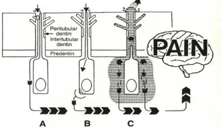
6- MECHANISM (Sufyan Garoushi 2015)
Three main mechanisms of dentin sensitivity are proposed: (Figure 1).
Figure 1: The schematic picture of the propped theories on DH
Odontoblastic transduction theory
The odontoblast transducer theory
proposed by Rapp et al. postulated that odontoblasts act as receptor cells, and transmit impulses via synaptic junctions to the nerve terminals causing the sensation of pain from the nerve endings located in
the pulpodentine border. However,
evidence for the odontoblast transducer mechanism theory is deficient and unconvincing. This is because the majority of studies have shown that odontoblasts are matrix forming cells and they are not considered to be excitable cells, and no synapses have been revealed between odontoblasts and nerve terminals.
Neural theory
Available online: http://edupediapublications.org/journals/index.php/IJR/ P a g e | 1105 Hydrodynamic theory
The currently accepted mechanism of
dentine hypersensitivity is the
hydrodynamic theory which has been proposed by Brännström in 1964. According to this theory, when the exposed dentin surface is subjected to thermal, chemical, tactile or evaporative stimuli, the fluid flow within the dentine tubules there will be increased. This fluid movement within the dentine tubules causes an alteration in pressure and excites pressure-sensitive nerve receptors across the dentine. So the response of the excited pulpal nerves, mainly in intradentine fibers, will be depended upon the intensity of stimuli in pain production. Scanning electron microscopic (SEM) examination of hypersensitive dentin surface reviles the presence of widely open dentine tubules which is considered consistent with the hydrodynamic theory. Accordingly, the number and the diameter of the dentine tubules are considered important factors in initiating pain from DHS.
Hence, the higher the number and greater the diameter of the open dentine tubules the more intense will be the pain from DHS. It has been noted that triggers such as cold stimuli stimulate fluid to flow away from the pulp creating more rapid and rigorous neural responses than heat stimuli, which cause somewhat sluggish fluid flow towards the pulp. This is aligned with the observation that dentine
hypersensitivity patients are more
frequently complain of pain in response to cold stimuli than to heat.
7- CLINICAL FEATURES
Classically, the pain experienced with DH is of rapid onset, short and sharp in character, and of duration equal to that of the applied stimuli, although it can persist as a dull throbbing ache for variable periods. It may be localized or generalized, affecting one or more tooth surfaces simultaneously (Bekes 2014).
8- DIFERENTIAL DIAGNOSIS
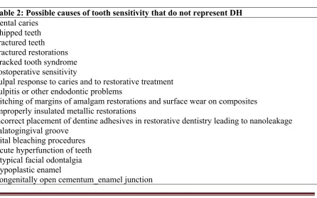
A number of clinical conditions may elicit the short, sharp tooth sensitivity that is
characteristic of DH (Table 2) (Bekes 2014)
Table 2: Possible causes of tooth sensitivity that do not represent DH
Dental caries Chipped teeth Fractured teeth Fractured restorations Cracked tooth syndrome Postoperative sensitivity
Pulpal response to caries and to restorative treatment Pulpitis or other endodontic problems
Ditching of margins of amalgam restorations and surface wear on composites Improperly insulated metallic restorations
Incorrect placement of dentine adhesives in restorative dentistry leading to nanoleakage Palatogingival groove
Vital bleaching procedures Acute hyperfunction of teeth Atypical facial odontalgia Hypoplastic enamel
Available online: http://edupediapublications.org/journals/index.php/IJR/ P a g e | 1106
9- ASSESSMENT OF DHS (Sufyan
Garoushi 2015)
Commonly, DHS might either be assessed in terms of a stimulus intensity required to
elicit pain called stimulus-based
assessment or as a subjective evaluation of the pain caused by a distinct stimulus named response-based assessment.
Commonly, DHS might either be assessed in terms of a stimulus intensity required to
elicit pain called stimulus-based
assessment or as a subjective evaluation of the pain caused by a distinct stimulus named response-based assessment
Alternative thermal or electrical devices such as electrical pulp testers, dental pulp stethoscope and others have been used for applying graded thermal or electrical stimuli
It was realized that, these stimulus-based methods have certain drawbacks such as repeated painful stimulation may cause a change in sensitivity and influence the outcome.
On the other hand, the response-based methods assess pain severity after application of a constant, standardized, consistent, and reproducible stimulus like a timed airblast. The air will be directed for 1 s from a distance of approximately 1 cm at the exposed buccal surface of the hypersensitive tooth after its isolation from
the adjacent teeth Directly after
stimulation, the subject response can be quantified by using a verbal rating, or visual analog scales or a validated graphic pain scale, such as the Faces Pain Scale Several studies recommended the use of the Schiff cold air sensitivity scale to assess subject response to a stimulus like air or evaporative. This scale is composed of several distinct scores which are:
i. Subject does not respond to air stimulus.
ii. Subject responds to air stimulus but does not request discontinuation of stimulus.
iii. Subject responds to air stimulus and requests discontinuation or moves from stimulus.
iv. Subject responds to air stimulus, considers stimulus to be painful, and requests discontinuation of the stimulus.
Verbal rating scales
For measuring of DHS, verbal rating
scales (VRS) are used to grade the level of
pain experience. Most pain scales utilize
several pain descriptors, including ‘no
pain’, ‘weak’, ‘mild’, ‘moderate, ‘strong’, ‘intense’, and ‘agonizing’. Numerical
scores (0, 1, 2, 3, etc.) have been attached
to these descriptors, and mean values are calculated
Visual analogue scale
The visual analogue scale (VAS) utilizes a
line of 10 cm length anchored at the 2
extremes with descriptors representing the
absolute minimum and the absolute
maximum of pain a patient can experience from an external stimulus
Objective evaluation
This evaluation may include application of thermal and evaporative stimuli such as a short blast of cold air from the 3-in-1 syringe, mechanical or tactile stimuli such as running a sharp explorer over the area of exposed dentine or chemical stimuli such as using hypertonic
Available online: http://edupediapublications.org/journals/index.php/IJR/ P a g e | 1107 sever dentine hypersensitivity On other
hands, if several stimuli are applied, the least sever stimulus should be always performed first to avoid a negative impact on the results of the stimulation. Furthermore, the interval between stimulus applications should be sufficient to prevent interactions between both stimuli.
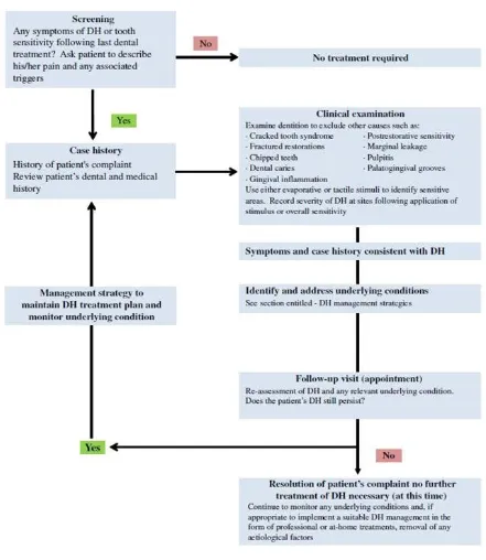
10- MANAGEMENT OF DH
A recent UK Expert Forum on DH produced simple guidelines that can be readily applied in general practice. The Forum acknowledged that a single approach to DH may not satisfy everyone (Figure 2) (Gillam and Talioti 2014).
Available online: http://edupediapublications.org/journals/index.php/IJR/ P a g e | 1108 The proposed scheme links the diagnosis,
prevention, and treatment strategies to three specific groups of people rather than recommending a blanket management for all patients with DH.
These groups include:
(1) people with gingival recession caused by mechanical trauma,
(2) those with tooth wear lesions,
(3) those with periodontal disease and receiving periodontal treatment
An essential step in the initial management of DH is to correctly diagnose the
underlying condition by excluding
conditions with a similar clinical
presentation (Table 3).
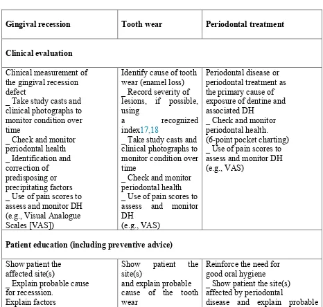
Table 3. Overall management strategy options for treating DH
Gingival recession
Tooth wear Periodontal treatment
Clinical evaluation
Clinical measurement of the gingival recession defect
_ Take study casts and clinical photographs to monitor condition over time
_ Check and monitor periodontal health _ Identification and correction of predisposing or precipitating factors _ Use of pain scores to assess and monitor DH (e.g., Visual Analogue Scales [VAS])
Identify cause of tooth wear (enamel loss) _ Record severity of lesions, if possible, using
a recognized
index17,18
_ Take study casts and clinical photographs to monitor condition over time
_ Check and monitor periodontal health _ Use of pain scores to assess and monitor DH
(e.g., VAS)
Periodontal disease or periodontal treatment as the primary cause of exposure of dentine and associated DH
_ Check and monitor periodontal health. (6-point pocket charting) _ Use of pain scores to assess and monitor DH (e.g., VAS)
Patient education (including preventive advice)
Show patient the affected site(s)
_ Explain probable cause for recesssion.
Explain factors
triggering sensitive teeth episodes
_ Encourage patients to
Show patient the
site(s)
and explain probable cause of the tooth wear
lesion(s).
Recommend an oral hygiene regimen to
Reinforce the need for good oral hygiene
_ Show patient the site(s) affected by periodontal
Available online: http://edupediapublications.org/journals/index.php/IJR/ P a g e | 1109 modify their oral
hygiene regimen in order to reduce damage to gingivae (e.g.,
reducing brushing force, correction of toothbrush technique)
_ Reduce excessive consumption of acid foods and drinks
minimize risk of
further tooth wear.
_ Where appropriate recommend reducing frequency of
consumption of acidic food and drink.
hygiene regimen.
_ Instruction on measures of reducing periodontal risk factors, for example, diabetes, smoking, and obesity.
Corrective clinical outcomes
Reduce excessive consumption of acid foods and drinks
_ Manufacture of silicone gingival veneers
_ Orthodontic treatment _ Restorative correction of recession defect and subgingival margins of fillings and crowns _ Polymers: sealants/ varnishes/resins/dentine bonding agents
_ Laser obturation of dentinal tubules _ Use of desensitizing polishing pastes
_ Pulpal extirpation (root canal treatment)
Provide high fluoride remineralizing treatment
(pre-emptive phase) _ Provide professional desensitizing
treatment to relieve DH
_ Encourage patient to seek advice from medical practitioner, if tooth wear caused by working environment or
reflux/excesssive vomiting (psychiatric evaluation may also be appropriate)
_ Restorative
correction in
the form of composite build up, crowns may also be appropiate
Initial phase
Nonsurgical periodontal procedure(s).
DH treatment (including desensitizing polishing pastes/fluoride
varnishes) Reevaluation
Follow-up assessment on periodontal status and DH
Corrective phase _ Surgical periodontal procedure(s)
_ DH treatment (including desensitizing polishing pastes/fluoride
varnishes)
Follow-up management maintenance phase _ Supportive periodontal therapy
_ Ongoing monitoring of periodontal health DH treatment (including desensitizing polishing pastes/fluoride
varnishes)
_ Oral hygiene advice
Recommendations for home use (including toothpaste/mouth rinses)
Oral hygiene
Available online: http://edupediapublications.org/journals/index.php/IJR/ P a g e | 1110 recommendation
_ Strontium chloride/ strontium acetate _ Potassium nitrate/ chloride/citrate/oxalate _ Calcium compounds: _ Calcium carbonate and arginine and caesin phosphopeptide1 amorphous calcium phosphate
_ Bioactive glass _ Nano/hydroxyapatite _ Fluoride in higher concentration (2800/ 5000 ppm F
[prescription])
_ Amine/stannous fluoride
recommendation _ Toothpastes and mouth
rinses (see
recommendations for gingival recession)
recommendation
_ Regular brushing with an antibacterial
toothpaste to aid plaque control
_ Short period, the use of a 0.2% chlorhexidine solution for plaque control
_ Use of a desensitizing mouthrinse twice daily for DH control (when appropriate)
CLASSIFICATION OF
DESENSITIZING AGENTS (Miglani,
Aggarwal, and Ahuja 2010)
I. Mode of administration
At home desensitizing agents In-office treatment
II. On the basis of mechanism of action
Nerve desensitization
Potassium nitrate
Protein precipitation
Gluteraldehyde
Silver nitrate
Zinc chloride
Strontium chloride hexahydrate
Plugging dentinal tubules
Sodium fluoride
Stannous fluoride
Strontium chloride
Potassium oxalate
Calcium phosphate
Calcium carbonate
Bio active glasses (SiO2–P2O5–CaO–
Na2O)
Dentine adhesive sealers
Fluoride varnishes
Oxalic acid and resin
Glass ionomer cements
Composites
Dentin bonding agents
Lasers
Neodymium:yttrium aluminum garnet
(Nd–YAG) laser
GaAlAs (galium–aluminium–arsenide
laser)
Erbium–YAG laser
Homeopathic medication
Propolis
Ideal requirements of a desensitizing agent:
Grossman listed the requirements for an ideal dentine desensitizing agent as:
rapidly acting with long-term effects,
non-irritant to pulp,
painless
easy to apply,
Available online: http://edupediapublications.org/journals/index.php/IJR/ P a g e | 1111 At home desensitizing therapy
Traditionally, the therapy for management of DH is primarily aimed at occluding the dentinal tubules or making coagulates inside the tubules. Patients are often prescribed over-the-counter desensitizing agents. These “at home” desensitizing agents include
toothpastes, mouthwashes and chewing gums.
Majority of the toothpastes contain
potassium salts (potassium nitrate,
potassium chloride or potassium citrate), sodium fluoride, strontium chloride, dibasic sodium citrate, formaldehyde, sodium monofluorphosphate and stannous fluoride. Potassium salts act by diffusion along the dentinal tubules and decreasing the excitability of the intradental nerve fibers by blocking the axonic action. Various clinical studies have shown the efficacy of potassium salts in controlling the DH. It has been shown that toothpastes containing 5% potassium nitrate and 0.454% stannous significantly reduced the
DH. Also, toothpastes containing
potassium nitrate and fluorides have been shown to reduce post-bleaching sensitivity. The desensitizing toothpastes should be used with the help of a toothbrush with soft bristles. Patients should be advised to use minimal amount of water to prevent the dilution of the active agent. Along with
the desensitizing toothpastes,
mouthwashes and chewing gums
containing potassium nitrate, sodium fluoride or potassium citrate are also recommended. The results of “at-home” desensitizing therapy should be reviewed after every 3–4 weeks. If there is no relief in DH, “in-office” therapy should be initiated.
In-office desensitizing agents
Theoretically, the in-office desensitizing therapy should provide an immediate relief from the symptoms of DH. The in-office
desensitizing agents can be classified as the materials which undergo a setting
reaction (glass ionomer cement,
composites) and which do not undergo a setting reaction (varnishes, oxalates).
Fluorides
Traditionally, fluorides have been used as a caries preventive material which can help in remineralization of enamel/dentin. Also, various clinical trials have shown that application of fluoride solution can decrease the DH. Fluorides decrease the dentinal permeability by precipitation of calcium fluoride crystals inside the dentinal tubules. These crystals are partially insoluble in saliva. SEM revealed granular precipitates in the peritubular dentin after application of fluorides. Various fluoride formulations are used to treat DH. These include sodium fluoride,
stannous fluoride, sodium
monofluorophosphate, fluorosilicates and fluoride combined with iontophoresis. Sodium fluoride has been used in dentifrices or may be professional applied in a concentration of 2%. The precipitates formed by sodium fluoride can be mechanically removed by the action of saliva or mechanical action. Therefore, an
addition of acid formulation is
Available online: http://edupediapublications.org/journals/index.php/IJR/ P a g e | 1112 the exposed dentine. Fluorosilicates act by
formation of precipitates of calcium phosphates from saliva. Ammonium hexafluorosilicate has been used as a desensitizing agent. It can present a continuous effect of dentinal tubule occlusion via precipitation of a mixture of calcium fluoride and fluoridated apatite. If the precipitate is predominantly composed of fluoridated apatite, it can form stable crystals deposited deep inside the dentinal tubules. These crystals are resistant to removal from the action of saliva, brushing or action of dietary substances.
Oxalates
Oxalates can reduce dentinal permeability and occlude dentinal tubules. Thirty percent potassium oxalate had shown a 98% reduction in dentinal permeability. Also, topical application of 3% potassium oxalate reduced DH after periodontal therapy. The oxalate reacts with the calcium ions of dentine and forms calcium oxalate crystals inside the dentinal tubules as well as on the dentinal surface. This results in a better sealing as compared with an intact smear layer. It has been shown that the effect of oxalates on DH diminishes over a period of time. This can be attributed to the removal of the calcium oxalate crystals by brushing or dietary acids. The condition can be improved by acid etching of the dentinal surface, thus increasing the penetration of calcium oxalate crystals deep into the dentinal tubules. Many vegetables like rhubarb, spinach and mint contain oxalates. It has been shown that phytocomplexes obtained from these natural products can reduce the dentinal permeability. This can also be followed by covering the exposed surface with a dental adhesive. Potassium oxalate can lead to gastric irritation. Therefore it should not be used with a tray with prolonged placement. Varnishes are commonly used useful in-office measures
to treat DH. Copal varnish can be applied to cover the exposed dentinal surface. But its effect is for short term and is not recommended for long term management of DH. To improve its efficacy, removal of smear layer is advocated. Also, the varnishes can act as a vehicle for fluoride. The fluoride varnishes can be acidulated to increase the penetration of ions.
Adhesive materials
Resin-based dental adhesive systems can provide a more durable and long lasting dentine desensitizing effect. The adhesive resins can seal the dentinal tubules effectively by forming a hybrid layer. Various clinical studies have demonstrated the effectiveness of adhesives in management of DH. Traditionally, resin composites or dentin bonding agents are used as desensitizing agents. The conventional dentin bonding agents (DBA) removes the smear layer, etches the dentinal surface and forms deep dentinal resin tags inside the dentinal tubules. The combined dentin–resin layer (consisting of penetrating resinous tags) has been termed as hybrid layer. It effectively seals the dentinal tubules and prevents DH. Newer bonding agents modify the smear layer and incorporate it in into the hybrid layer. Recently, some dentin bonding agents have been introduced in the market with the sole purpose of treating DH. Gluma Desensitizer (Heraeus Kulzer, Hanau,
Germany) contains hydroxyethyl
Available online: http://edupediapublications.org/journals/index.php/IJR/ P a g e | 1113 Bioglass
Bioglass was developed to stimulate the formation of new bone. It is used in orthopedics to cover the implants to promote union between implant and bone. It has been used in dentistry to fill up the
osseous defects during periodontal
surgery. It has been reported that a formulation of bioglass can promote infiltration and remineralization of dentinal tubules. The basic component is silica, which acts as a nucleation site for precipitation of calcium and phosphate. SEM analysis has shown that bioglass application forms an apatite layer, which occludes the dentinal tubules. The use of bioglass in management of DH has been shown by some products such as NovaMin (NovaMin Technology Inc., FL, USA).
Portland cement
Some authors have shown that calcium silicate cement derived from Portland cement can help in the management of DH. It helps to occlude the dentinal tubules by remineralization.
Laser
Laser is an acronym for light amplification by stimulated emission of radiations. It has been shown in various studies that lasers can be used in the effective management of DH. The mechanism of action of lasers in treating DH is not very clear. Some authors have shown that Nd–YAG laser application occluded the dentinal tubules. GaAlA laser is thought to act by affecting the neural transmission in the dentinal tubules. It has also been proposed that lasers coagulate the proteins inside the dentinal tubules and block the movement of fluid.
Casein phosphopeptide–amorphous
calcium phosphate
Recently, milk protein casein has been used to develop a remineralizing agent
(GC Tooth Mousse). The casein
phosphopeptide (CPP) contains
phosphoseryl sequences which get
attached and stabilized with amorphous calcium phosphate (ACP). The stabilized CPP–ACP prevents the dissolution of calcium and phosphate ions and maintains a supersaturated solution of bioavailable calcium and phosphates. Various studies have shown that CPP– ACP can effectively remineralize the enamel subsurface lesions. By virtue of its remineralizing capacity, it has also been proposed by the manufacturers that it can also help in prevention and treatment of DH.
MANAGEMENT STRATEGY
Take a detailed clinical and dietary
history.
Differentially diagnose the condition
from other dental pain conditions.
Identify and manage etiological and
predisposing factors.
In case of mild-to-moderate sensitivity,
advice at-home desensitizing therapy.
If there is no relief or in case of severe
sensitivity, initiate in-office treatment.
In extreme cases, if patient does not
respond to the therapy and there are
individual teeth exhibiting the
symptoms, then endodontic therapy can be initiated.
A regular review should be made with
an emphasis on prevention of the condition.
REFERENCES
Available online: http://edupediapublications.org/journals/index.php/IJR/ P a g e | 1114 Bekes, Katrin. 2014. Clinical Presentation
and Physiological Mechanisms of Dentine Hypersensitivity. Dentine Hypersensitivity: Developing a Person-Centred Approach to Oral Health. Elsevier Inc.
Borges A, Barcellos D, Gomes C. 2012. “Dentin Hypersensitivity- Etiology, Treatment Possibilities and Other Related Factors: A Literature
Review.” World Journal of Dentistry 3: 60–67.
Chabanski MB, Gillam DG, Bulman JS, Newman HN. 1997. “Clinical Evaluation of Cervical Dentine Sensitivity in a Population of Patients Referred to a Specialist
Periodontology Department: A Pilot Study.” J Oral Rehabil 24: 666–72.
Dababneh RH, Khouri AT, Addy M. 1999. “Dentine Hypersensitivity - an
Enigma? A Review of Terminology, Mechanisms, Aetiology and
Management.” Br Dent J 187: 606– 11.
Davari, Ar, E Ataei, and H Assarzadeh. 2013. “Dentin Hypersensitivity: Etiology, Diagnosis and Treatment; A Literature Review.” Journal of
Dentistry (Shiraz, Iran) 14 (3): 136– 45.
Gillam, David, and Elena Talioti. 2014. The Management of Dentine Hypersensitivity. Dentine Hypersensitivity: Developing a Person-Centred Approach to Oral Health. Elsevier Inc.
Miglani, Sanjay, Vivek Aggarwal, and Bhoomika Ahuja. 2010. “Dentin Hypersensitivity: Recent Trends in
Management.” Journal of
Conservative Dentistry 13 (4): 218.
Porto, Isabel C C M, Ana K M Andrade, and Marcos a J R Montes. 2009. “Diagnosis and Treatment of Dentinal Hypersensitivity.” Journal of Oral Science 51 (3): 323–32.