1 | P a g e Title: Hydrocortisone, Ascorbic acid and Thiamine (HAT therapy) for the
treatment of sepsis. Focus on Ascorbic Acid
Author: Paul E. Marik, MD, FCCM, FCCP
Affiliation: Division of Pulmonary and Critical Care Medicine
Eastern Virginia Medical School, Norfolk, VA
Date: October 11, 2018
COI: The author has no real or potential conflicts of interest concerning this manuscript.
Key words: sepsis; septic shock; hydrocortisone; vitamin C; ascorbic acid; thiamine
Word count: 4084
Version: HAT-2018g
Running title: HAT therapy and sepsis
Funding: None
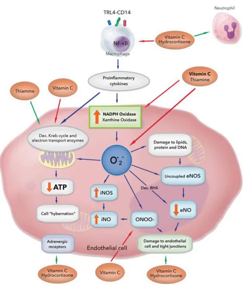
Acknowledgements: The author acknowledges the exceptional work of Heather Cousineau (cmedillustration) for creating Figure 1.
Address for Correspondence: Paul E Marik, MD
Chief, Division of Pulmonary and Critical Care Medicine Eastern Virginia Medical School
825 Fairfax Ave, Suite 410 Norfolk VA,23517, USA Email: [email protected]
2 | P a g e Abstract:
Sepsis is a devastating disease that carries an enormous toll in terms of human suffering and lives
lost. Over 100 novel pharmacologic agents which targeted specific molecules or pathways have
failed to improve the outcome of sepsis. Preliminary data suggests that the combination of
Hydrocortisone, Ascorbic Acid and Thiamine (HAT therapy) may reduce organ failure and
mortality in patients with sepsis and septic shock. HAT therapy is based on the concept that a
combination of readily available, safe and cheap agents which target multiple components of the
host’s response to an infectious agent will synergistically restore the dysregulated immune
response and thereby prevent organ failure and death. This paper reviews the rationale for HAT
3 | P a g e The global burden of sepsis is substantial with an estimated 32 million cases and 5.3
million deaths per year per year with most of these cases occurring in low income countries. [1]
Data from the US and Australia demonstrates that over the last 2 decades the annual incidence
of sepsis has increased by approximately 13% with a decrease in in-hospital mortality from about
35 to 20%.[2-4] In 2013 over 1.3 million patients were hospitalized in the USA with a diagnosis
of sepsis of whom over 300 000 died.[5] In addition to short-term mortality, septic patients
suffer from numerous long-term complications with a reduced quality of life and an increased
risk of death up to five years following the acute event.[6-10] Approximately 50% of sepsis
survivors develop the post-sepsis syndrome characterized by the development of new psychiatric
and cognitive deficits. [11] The post sepsis-syndrome is similar in many respects to the
post-traumatic stress disorder (PTSD); patients suffer memory impairment, abnormalities of higher
executive function, nightmares, anxiety disorders and depression.[12] Apart from the enormous
financial costs of sepsis, the human toll of this disease is staggering and new interventions that
limit the ravages of this disease are urgently required.
In patients with sepsis organ failure and death usually result from the host’s response to
the infecting pathogen rather than from the infecting pathogen itself. This was first recognized by
Sir William Osler who commented that “except on few occasions, the patient appears to die
from the body’s response to infection rather than from the infection.” [13] Sepsis is
fundamentally an inflammatory disease mediated by the activation of the innate immune system
by both pathogen-associated molecular patterns (PAMPs) and damage-associated molecular
patterns (DAMPs). Calvano et al demonstrated that exposure of blood leukocytes to bacterial
4 | P a g e anti-inflammatory cytokines, chemokines, adhesion molecules, transcription factors, enzymes,
clotting factors, stress proteins and anti-apoptotic molecules.[15] These inflammatory mediators
have widespread pathophysiologic consequences, including vasoplegic shock, myocardial
dysfunction, altered microvascular flow and a diffuse endothelial injury.[16,17] However,
fundamentally sepsis is characterized by the excessive production of reactive oxygen species
(ROS) by the induction of enzymes such as nicotinamide adenine dinucleotide
phosphate-oxidase (NOX) and the uncoupling of mitochondrial oxidative phosphorylation (see figure
1).[18] In addition, ROS are produced by xanthine oxidase, lipoxygenase and cyclooxygenase.
Important ROS in sepsis pathogenesis includes hydrogen peroxide (H2O2), superoxide (O.2-),
hydroxyl radicals (HO.).peroxynitrite (ONOO-) and hypochlorous acid (HOCl).
The excess production of ROS underlie many of the pathological processes characteristic
of sepsis.[19] ROS has been shown to modulate the lipopolysaccharide-Toll like receptor 4
(LPS-TLR4) signaling pathway. ROS activate nuclear factor kappa-B (NF-κB) by activation of
inhibitory kappa-B kinase (IκB kinase). NF-κB increase the transcription of multiple
pro-inflammatory mediators (see Figure1). When the hosts antioxidant defenses are overwhelmed,
ROS can induce injury to lipids, proteins, nucleic acids thereby resulting in widespread
endothelial dysfunction, mitochondrial dysfunction, cellular injury and multiple organ
dysfunction.[19] Isoprostanes are products of lipid peroxidation that are formed
non-enzymatically and can be measured in blood, urine or tissues. Because of their stability and high
specificity, the F2-isoprostanes are currently considered to be the most reliable biomarkers of in
vivo oxidative stress and lipid peroxidation. In a cohort of patients with sepsis Ware et al
5 | P a g e in those who died. [20] Similarly, Kumar et al reported augmented levels of oxidants in patients
with sepsis as demonstrated by DMPO nitrone adduct formation and plasma myeloperoxidase
(MPO) level activity.[21] In this study the SOFA and APACHE II scores correlated linearly with
the MPO levels and inversely with the levels of antioxidants. This study supports the concept
that the imbalance between oxidant and antioxidants plays key role in the pathophysiology of
organ failure in sepsis.
The unbalanced production of mitochondrial ROS impairs mitochondrial structure,
enzymatic function and biogenesis. In an in vitro model of sepsis-induced kidney injury, Quoilin
et al demonstrated that LPS induces NADPH oxidase and inducible nitric oxide synthetase
(iNOS) expression. Following this, uncoupling of the mitochondrial respiratory chain due to the
inhibition of complex IV and reduction in ATP levels were observed.[22] Mitochondrial
dysfunction was associated with cytochrome c release and the loss of mitochondrial membrane
potential. Alterations in fatty acid metabolism with decreased beta-oxidation and abnormalities
of the citric acid cycle appear to be a characteristic feature of the mitochondrial dysfunction in
sepsis. [23,24] These changes lead to decreased ATP production. In the heart, myocyte oxidative
injury is accompanied by increased proteolysis, mitochondrial damage, dysregulated nitric oxide
metabolism, β-adrenoceptor down-regulation and calcium mishandling. [25-27] The unbalanced
production of mitochondrial ROS impairs mitochondrial structure, enzymatic function and
biogenesis and play a role in the metabolic failure of sepsis. These data suggest that excessive
production of ROS plays a pivotal role in the pathophysiology of sepsis and that interventions
that neutralize oxidants would have a protective role in sepsis. Furthermore, proinflammatory
6 | P a g e inactivate the pyruvate dehydrogenase complex (PDC).[28,29] Inactivation of the PDC prevents
pyruvate entering the Krebs cycle potentiating the metabolic failure caused by mitochondrial
dysfunction. This phenomenon is compounded by thiamine deficiency and cytokine mediated
down regulation of expression of the PDC.[28]
Over the last 3 decades, over 100 phase II and phase III clinical trials have been
performed testing various novel pharmacologic agents and therapeutic interventions in an
attempt to improve the outcome of patients with severe sepsis and septic shock; all of these
efforts ultimately failed to produce a novel pharmacologic agent that reduced organ failure and
improved the survival of patients with sepsis.[30] All these studies used a single agent that
targeted a specific molecule or pathway; due to the thousands of mediators involved and the
redundancy of the multiple pathways such an approach was doomed to fail. Furthermore, the
central role of ROS was ignored. Dr Aird has commented that “the best hope for therapeutic
advances [in sepsis] will depend on broad-base targeting, in which multiple components are
targeted at the same time”. [31] We believe that the combination of Hydrocortisone, Ascorbic
Acid and Thiamine (HAT therapy) achieves these goals. The premise behind HAT therapy is the
use of a combination of readily available, safe and cheap agents that target multiple components
of the host’s response to an infectious agent such that they synergistically restore the
dysregulated host immune response, neutralize damaging oxidants and restore mitochondrial
function. We believe this unique and novel approach has the potential to reduce the global
burden of sepsis, reduce the post-sepsis syndrome without side-effects and be highly cost
7 | P a g e and millions of life-years in the United States.[32] This paper reviews the rationale for HAT
therapy with a focus on vitamin C.
Vitamin C
It has been known for over 20 years that acute illness, endotoxemia and sepsis result in an
acute deficiency of vitamin C, characterized by low serum and intracellular levels of the
vitamin.[33-40] Critically ill septic patients typically have very low or undetectable serum levels
of vitamin C resulting in an acute scorbutic condition.[35,41-43] Recently Carr and coauthors
demonstrated that 100% of septic patients had low vitamin C levels, 88% had hypovitaminosis C
(< 23 μmol/L), while 38% had serum levels compatible with acute scurvy (< 11 μmol/L). [44]
Low vitamin C levels in critically ill patients are associated with increased vasopressor
requirements, kidney injury, multiple organ dysfunction (higher SOFA scores) and increased
mortality. [35,43] The underlying cause of the vitamin C deficiency is likely due to increased
oxidation (metabolic consumption), decreased absorption and increased urinary losses of the
vitamin. In a murine caecal-ligation and perforation model (CLP), Armour et al reported that the
plasma ascorbate level fell rapidly by 50% and this was associated with a 1 000% increase in the
urine ascorbate concentration.[36] Sepsis induced glomerular hyperfiltration and/or tubular
dysfunction results in decreased tubular reabsorption of filtered vitamin C with increased urinary
losses.[45]
Vitamin C is a potent antioxidant which directly scavenges oxygen free radicals, restores
co-8 | P a g e factor for iron and copper containing enzymes. [18,46] Vitamin C is a key cellular antioxidant,
detoxifying exogenous oxidants radical species that have entered cells or which have arisen
within cells due to excess superoxide generation by mitochondrial metabolism, by NADPH
oxidase, xanthine oxidase or by uncoupled nitric oxide synthase (NOS).[18,47] The low electron
reduction potential of both vitamin C and its one-electron oxidation product, the ascorbyl
radical, enable them to reduce virtually all clinically important radicals and oxidants.
Dehydroascorbic acid is transported via the GLUT1 transporter into mitochondria, where
it converted to ascorbic acid and acts as a potent antioxidant limiting mitochondrial oxidant
injury.[48,49] Considering that the mitochondrial respiratory chain is a main source of ROS in
live cells and mitochondrial dysfunction plays a prominent role in sepsis pathogenesis,
antioxidants targeting the intra-mitochondrial environment could be pivotal role in the treatment
of sepsis. Furthermore, ascorbic acid is required for the synthesis of carnitine, which is required
for the transport of fatty acids into the mitochondrial matrix and for beta-oxidation.[23,48] In an
experimental model, Dhar-Mascareno and colleagues demonstrated that oxidant induced
mitochondrial damage and apoptosis in human endothelial cells were inhibited by vitamin C.[50]
In a CLP sepsis model, Kim et al administered 100mg/kg ascorbic acid immediately after sepsis
induction.[51] In this study, vitamin C attenuated the elevation in serum aminotransferase and
hepatic lipid peroxide levels. Studies using N-acetylcysteine (a synthetic anti-oxidant) have
proven to be ineffective and potentially harmful in patients with sepsis possibly due to the
limited ability of this drug to enter into the mitochondria and its inability to regenerate
9 | P a g e Vitamin C suppresses activation of NF-κB by inhibiting tumor necrosis factor-α (TNFα)
induced phosphorylation of inhibitory kappa-B kinase (IκB kinase).[54] Ascorbic acid decreases
high mobility group box 1(HMGB1) secretion;[55] HMGB1 is an important late
pro-inflammatory cytokine. Vitamin C has been demonstrated to decrease the synthesis and to
inactivate histamine; [56] histamine has been shown to play an important role in sepsis.[57]
Vitamin C is an essential co-factor for the synthesis of norepinephrine, epinephrine and
vasopressin; in addition vitamin C increases adrenergic transmission.[58] Vitamin C may
decrease the immunosuppression associated with sepsis. It has been known for over 60 years that
vitamin C has immune enhancing properties. In 1949 Dr Fred Klenner, from Reidsville, North
Carolina, reported on the use of intravenous vitamin C in the treatment of polio and other viral
illnesses.[59] It was initially assumed that vitamin C was directly viricidal (in vivo) and this
mistaken belief underlies the recommendations of Linus Pauling who promoted the use of large
doses of oral vitamin C (up to 18g/day) for the prevention and treatment of the common
cold.[60] However, a number of RCTs have reported that vitamin C supplementation had no
effect on the incidence of the common cold. [61] While high dose vitamin C has in-vitro viricidal
properties,[62,63] there is no data or physiologic rationale to suggest that this occurs in vivo.
Rather, the “anti-viral” effect of vitamin C are likely due to that fact that vitamin C has specific
immune enhancing effects. Vitamin C is concentrated in leucocytes, lymphocytes and
macrophages reaching high concentrations in these cells.[64] Vitamin C improves
chemotaxis, enhances neutrophil phagocytic capacity and oxidative killing, stimulates
interferon production and supports lymphocyte proliferation.[65-67] The major presumed
10 | P a g e Vitamin C: Dose response and pro-oxidant effect.
While vitamin C is the most potent and important anti-oxidant in mammals, in the
presence of transition metals vitamin C may paradoxically be associated with a pro-oxidant
effect.[68] In the presence of free iron vitamin C may reduce free ferric iron to the ferrous
form. The ferrous form then undergoes a Fenton-type reaction with hydrogen peroxide
yielding hydroxyl or hydroxyl-like reactions. Vitamin C may generate ROS in in-vitro, cell
culture or tissue incubation experiments, where free metal ions might exist. [69] Normally, iron
is tightly bound to protein and does not exist in the free form. However, in conditions such as
hypoxia (ischemia/reperfusion) and sepsis free iron may be released from ferritin.
Furthermore, sepsis is associated with hemolysis and the release of free heme. Free heme can
be highly cytotoxic in the presence of proinflammatory mediators.[70,71] The divalent iron
atom contained within its protoporphyrin IX ring can promote the production of free radicals.
In the presence of free iron or free heme and depending on the dose of vitamin C and the
timing of the administration of the dose in relationship to inciting event, vitamin C may
either act as an anti-oxidant or pro-oxidant. This concept was elegantly demonstrated in a
hepatic ischemia/reperfusion model where Seo and Lee demonstrate that an infusion of 30
mg/kg and 100 mg/kg of vitamin C decreased markers of oxidative injury whereas these
markers were increased with a dose of 300 mg/kg and 1000 mg/kg.[72] Similarly, in a
hepatic ischemia/reperfusion model Park and colleagues demonstrated that the oxidative
injury was attenuated at ascorbic acid concentrations of 0.25 and 0.5 mM, however they were
augmented at a concentration of 2 mM.[73] This concept is further supported by a number of
11 | P a g e in a dose of 50 and 100 mg/kg IV decreased myocardial damage, improved neurological
outcome and the survival rate.[68,69] However, using a similar model, Motl and colleagues
reported that a 250 mg/kg dose was harmful.[74] In a murine CLP model, Tyml et al
demonstrated that the delayed (24 hours) administration of vitamin C (in a dose of 76 mg/kg)
restored blood pressure and microvascular perfusion.[39] Using human umbilical vein
endothelial cells Kuck and colleagues demonstrated that cell free hemoglobin increased
endothelial permeability in part through depletion of intracellular ascorbate.[75] In this study
the addition of ascorbate up to a concentration of 60 umol/l (physiologic concentrations)
attenuated the increase in permeability mediated by cell free hemoglobin.[75] This study
reaffirms that low dose vitamin C may reduce the toxicity of cell free hemoglobin.
In the absence of free iron or heme, vitamin C acts as an oxidant only in extremely
high pharmacologic doses (> 100g) when used as adjunctive treatment in patients with
cancer.[76-79] Repeated intravenous injection of 750–7500 mg/day of vitamin C for 6 days in
healthy volunteers did not induce a pro-oxidant change in plasma markers.[80] Furthermore,
intravenous infusion of very high-dose ascorbate (1584 mg/kg over 24 hours) lowered serum
malondialdehyde concentration (a marker of oxidative stress) in severely burned
patients.[69] When used prophylactically (before the inciting event) prior to the oxidant
injury with release of free iron, it is likely that a lower dose of vitamin C may act as a
powerful antioxidant. Basili et al demonstrated that a single 1 g infusion of vitamin C prior
to the performance of percutaneous coronory reperfusion significantly improved
12 | P a g e Similarly, Wang and colleagues demonstrated that 3g vitamin C IV prior to elective
percutaneous coronory reperfusion was associated with less myocardial injury.[82] A
metaanalysis, which included 8 randomized controlled trials, demonstrated that vitamin C up
to a dose of 2g IV given pre-operatively significantly reduced the risk of atrial fibrillation in
patients undergoing cardiac surgery.[83] Oxidant injury is postulated to play a role in
contrast-induced renal dysfunction. A systematic review by Xu and colleagues demonstrated
that prophylactic vitamin C (oral and IV up to a total dose of 7g) significantly reduced the
risk of renal impairment. [84]
These data suggest that in the setting of sepsis and ischemia/reperfusion a narrow
dose response curve exists. In preliminary observational data in patients with severe sepsis
and septic shock and using procalcitonin as a biomarker we have similarly observed a narrow
dose response curve; the optimal daily dose appears to be approximately 6g/day with an
attenuated effect at a dose of 2g and 10g respectively (personal observations). In treating
over 800 patients with a 6g/day dose we are unaware of any patient who had a pro-oxidant
effect (as reflected by the procalcitonin trajectory) even when the treatment was delayed. It is
possible that delayed treatment with a higher dose may have a pro-oxidant deleterious effect.
It should be recognized that there is a delicate balance between protective oxidant signaling
and the detrimental effects of ROS. Additional studies are required to further elucidate the
dose response of vitamin C in various clinical situations. Furthermore, we endorse the
recommendation of Spoelstra-de Man and colleagues who suggest administering “high-dose
vitamin C for a short course of four days only, e.g., during the overwhelming oxidative stress
13 | P a g e vitamin C can be continued in a low (nutritional) dose to allow generation of low
concentrations of ROS, which are essential for physiological signaling and repair.” [68]
Hydrocortisone
Glucocorticoids have diverse anti-inflammatory properties. These are briefly reviewed
here; the reader is referred to recent publications for a more comprehensive review of this
topic.[85-88] Classically, glucocorticoid binding to the glucocorticoid receptor (GR) activates or
represses gene transcription, with glucocorticoids regulating up to 20% of the genome.[89]
Glucocorticoids affect nearly every cell of the immune system. Glucocorticoids suppress
inflammation by multiple mechanisms that impact both the innate and adaptive immune
responses. The primary anti-inflammatory action of glucocorticoids is to repress a large number
of pro-inflammatory genes which encode cytokines, chemokines, inflammatory enzymes, cell
adhesion molecules coagulation factors and receptors. GR mediated repression of the
transcriptional activity of NF-κB and AP-1 play a major role in mediating the anti-inflammatory
actions of glucocorticoids. In addition to attenuating the pro-inflammatory response, low-dose
glucocorticoids have immune-stimulating effects which may limit the anti-inflammatory
immunosuppressive state.[90] The immune enhancing effects of glucocorticoids and the balance
between the immune suppressing and enhancing effects of the drug are critically dependent on
the dose and duration of treatment as well as the state of immune activation of the host.
Over the last 40 years, 22 randomized controlled trail have been conducted investigating
the benefits of glucocorticoids in patients with septic shock.[91] Many of these studies are
14 | P a g e a 40-year period during which the treatment for sepsis has improved and the mortality from
sepsis and septic shock has decreased significantly.[2,4] The earlier studies used a short course
of high-dose corticosteroid (30mg/kg methylprednisone for up to 4 doses); this approach
increased mortality and complications and was abandoned. [92] This was followed by numerous
studies using a prolonged course (5-7 days) of physiologic “stress-doses” of glucocorticoids
(typically 200-300 mg hydrocortisone/day). The results of these studies were mixed with some
demonstrating a survival benefit while other did not. [93,94] In 2018, two large randomized
controlled trials (RCTs) were published evaluating the role of hydrocortisone in patients with
septic shock. [95,96] The Activated Protein C and Corticosteroids for Human Septic Shock
(APROCCHSS) study demonstrated a reduction in 90-day mortality whereas the Adjunctive
Corticosteroid Treatment in Critically Ill Patients with Septic Shock (ADRENAL) study
demonstrated no mortality benefit. Both studies however demonstrated a reduction in
vasopressor dependency, duration of mechanical ventilation and ICU stay with no increased risk
of complications. These studies indicate that while glucocorticoids (alone) have a biological
effect in patients with septic shock their effect on patient centered outcomes is limited. However
as indicated below we believe that glucocorticoids act synergistically with both vitamin C and
thiamine to reduce the complications and mortality and associated with sepsis.
Thiamine
Thiamine is the precursor of thiamine pyrophosphate (TPP), the essential coenzyme of
several decarboxylases required for glucose metabolism, the Krebs cycle, the generation of ATP,
15 | P a g e the pyruvate dehydrogenase complex, the rate-limiting step in the citric acid cycle.[97]
Thiamine play an important role in many enzymatic processes involved in brain function,
maintenance, and interneuronal communication.[98] Thiamine is involved in nerve tissue repair,
myelin synthesis and nerve signal modulation. Thiamine play a role in the uptake of serotonin,
which in turns affects the activity of the cerebellum, the hypothalamus, and hippocampus.[98] In
addition, thiamine has anti-inflammatory effects suppressing the oxidative stress-induced
activation of NF-κB.[98]
Thiamine deficiency is common among septic patients, with a range in prevalence
between 20 and 70%, depending on measurement techniques and inclusion criteria.[99-101] A
deficiency in thiamine leads to decreased activity of thiamine dependent enzymes that triggers a
sequence of metabolic events leading to energy compromise and decreased ATP production.
Thiamine deficiency is associated with excitotoxic-mediated neuronal cell death. [102]
Furthermore, thiamine deficiency is associated with increased production of ROS as well as
increased expression of heme oxygenase (HO-1) and eNOS.[102-104] Thiamine can reverse
oxidative stress that is not related to thiamine deficiency, suggesting that thiamine may act as a
site-directed antioxidant. [103] It is therefore likely that thiamine deficiency compounds the
oxidative mitochondrial injury and bioenergetic failure caused by vitamin C depletion.
In a pilot randomized controlled trial, Donnino et al randomized 88 patients with septic
shock to receive 200 mg thiamine twice daily for 7 days. [101] In the predefined subgroup of
patients with thiamine deficiency, those in the thiamine treatment group had statistically
16 | P a g e secondary analysis of this study the need for renal replacement therapy and the serum creatinine
were greater in the placebo group.[105] Similarly, in a propensity matched observational study in
patients with septic shock Woolum et al demonstrated that thiamine supplementation increased
lactate clearance and decreased 28-day mortality.[106]
Hydrocortisone, Ascorbic Acid and Thiamine (HAT) in Combination
The overlapping anti-inflammatory properties of glucocorticoids and vitamin C reduce
the production of pro-inflammatory mediators and ROS which are associated with endothelial
injury, mitochondrial damage and organ failure characteristic of sepsis (see Figure 1).
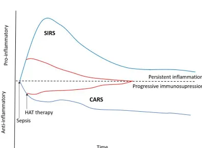
Furthermore, both agents have immuno-enhancing effects which limit the immunosuppression
that occurs in patients with prolonged sepsis. These agents may synergistically to restore the
dysregulated immune system which characterizes sepsis (see Figure 2).[107] Thiamine may act
synergistically with glucocorticoids and vitamin C to limit mitochondrial oxidative injury and
restore mitochondrial function and energy production. The anti-inflammatory properties of these
agents likely restores the activity of the PDC thereby improving ATP production. Thiamine may
reduce the production of oxalate thereby reducing the risk of hyper-oxalosis associated with the
administration of vitamin C. [108,109] However, the interaction between thiamine and ascorbic
acid is complex, and likely dependent on the clinical context and ascorbic acid dosing.
17 | P a g e The synergy between glucocorticoids and vitamin C has been established in
experimental studies. Barabutis et al have demonstrated that hydrocortisone together with
vitamin C protects the vascular endothelium from damage by endotoxin while neither agent
alone had this effect.[110] Azari et al compared the protective effects of Vitamin C (in a dose of
50 mg/kg), vitamin E and hydrocortisone alone and in combination, in a murine renal and
intestinal ischemia-reperfusion model.[111,112] In these studies both vitamin C and
hydrocortisone reduced the ischemia-reperfusion injury with the combination having synergistic
protective effects. In a small, retrospective, before-after-study we demonstrated that the
combination of hydrocortisone, ascorbic acid (6 g/day) and thiamine (HAT Rx) improved organ
function (as reflected by the SOFA score) with a significant reduction in mortality.[42] In a
similar before-after, propensity adjusted observational study Kim et al demonstrated a significant
reduction of mortality in patients with severe pneumonia using the same treatment strategy (HAT
Rx).[113] According to the U.S. National Library of Medicine’s ClinicalTrials.gov website
(https://clinicaltrials.gov/) in excess of 12 randomized controlled trials are currently underway
testing vitamin C alone and in combination with hydrocortisone and thiamine in patients with
severe sepsis and or septic shock. The results of these studies should provide definite information
on the role of this treatment strategy in the management of patients with severe sepsis and septic
18 | P a g e Conclusions:
Glucocorticoids, vitamin C and thiamine have important biological effects in patients
with sepsis and septic shock. Due to the overlapping and synergistic effects of these
remarkably safe and inexpensive drugs, the combination of these agents (HAT therapy)
likely restores the dysregulated immune system and bioenergetic failure that characterizes
sepsis. We therefore propose that HAT therapy will improve both the short term (mortality)
and long term (post-sepsis syndrome) outcome of patients with sepsis and septic shock.
19 | P a g e
Key Role Mechanism
Antioxidant Scavenges extracellular, intracellular and mitochondrial ROS; limits oxidation of
mitochondrial proteins, enzymes, lipoproteins, cell membrane, etc.
Anti-inflammatory Inhibits activation of NFκB, decreases HMGB1, inhibits histamine, prevents NETosis,
inactivates HIF-1α
Microcirculation increases eNOS, decreases iNOS, preserves tight junctions
Immune function Supports lymphocyte proliferation, increases neutrophil bacteriocidal action, improves
chemotaxis, stimulates interferon production, decreases T regulatory cells (Tregs)
Anti-thrombotic Decreases platelet activation and tissue factor expression, increases thrombomodulin
Synthesis of catecholamines Acts cofactor in synthesis of epinephrine, dopamine and vasopressin.
Increases adrenergic sensitivity
Wound Healing Hydroxylation of procollagen, increased expression of collagen mRNA
20 | P a g e Legends for Figures.
Figure 1. Multiple and overlapping effects of hydrocortisone, vitamin C and thiamine in the setting of bacterial sepsis.
23 | P a g e References
1. Fleischmann C, Scherag A, Adhikari NK et al. Assessment of global incidence and mortality of hospital-treated sepsis. Current estimates and limitations. Am J Respir Crit Care Med 2016; 193:259-72.
2. Gaieski DF, Edwards JM, Kallan MJ et al. Benchmarking the incidence and mortality of severe sepsis in the United States. Crit Care Med 2013; 41:1167-74.
3. Stevenson EK, Rubenstein AR, Radin GT et al. Two decadesof mortality trends among patients with severe sepsis: A comparative meta-analysis. Crit Care Med 2014; 42:625-31.
4. Kaukonen KM, Bailey M, Suzuki S et al. Mortality related to severe sepsis and septic shock among critically ill patients in Australia and New Zealand, 2000-2012. JAMA 2014; 311:1308-16.
5. Torio, C. M. and Moore, B. J. National Inpatient Hospital Costs: The Most expensive Conditions by Payer, 2013:Statistical Brief #240. Healthcare Costs and Utilization Project (HCUP) Statistical Briefs. 2016. Rockville, MD, Agency for Healthcare Research and Quality. 2016.
6. Karlsson S, Ruokonen E, Varpula T et al. Long-term outcome and quality-adjusted life years after severe sepsis. Crit Care Med 2009; 37:1268-74.
7. Yende S, Austin S, Rhodes A et al. Long-term qualityof life among survivors of severe sepsis: Analyses of two international trials. Crit Care Med 2016; 44:1461-67.
8. Ou SM, Chu H, Chao PW et al. Long-term mortality and major adverse cardiovascular events in sepsis survivors. A Nationwide population-based study. Am J Respir Crit Care Med 2016; 194:209-17.
9. Prescott HC, Langa KM, Liu V et al. Increased 1-year healthcare use in survivors of severe sepsis. Am J Respir Crit Care Med 2014; 190:62-69.
10. Liu V, Lei X, Prescott HC et al. Hospital readmission and healthcare utilization following sepsis in community settings. Journal of Hospital Medicine (Online) 2014; 9:502-7.
11. Shah FA, Pike F, Alvarez K et al. Bidirectional relationship between cognitive function and pneumonia. Am J Respir Crit Care Med 2013; 188:586-92.
12. Prescott JC, Angus DC. Enhancing recovery from sepsis. JAMA 2018; 319:62-75.
24 | P a g e 14. Calvano SE, Xiao W, Richards DR et al. A network-based analysis of systemic
inflammation in humans. Nature 2005; 437:1032-37.
15. Tang BM, Huang SJ, McLean AS. Genome-wide transcription profiling of human sepsis: a systematic review. Crit Care 2010; 14:R237.
16. Landry DW, Oliver JA. Pathogenesis of vasodilatory shock. N Engl J Med 2001; 345:588-95.
17. Lee WL, Slutsky AS. Sepsis and endothelial permeability. N Engl J Med 2010; 363:689-91.
18. May JM, Harrison FE. Role of vitamin C in the function of the vascular endothelium. Antioxid Redox Signal 2013; 19:2068-83.
19. Prauchner CA. Oxidative stress in sepsis: Pathophysiological implications justifying antioxidant co-therapy. Burns 2017; 43:471-85.
20. Ware LB, Fessel JP, May AK et al. Plasma biomarkers of oxidant stress and development of organ failure in severe sepsis. Shock 2011; 36:12-17.
21. Kumar S, Gupta E, Kaushik S et al. Evaluation of oxidative stress and antioxidant status: Correlation with the severity of sepsis. Scand J Immunol 2018; 87:e12653.
22. Quoilin C, Mouithys-Mickalad A, Lecart S et al. Evidence of oxidative stress and
mitochondrial respiratory chain dysfunction in an in vitro model of sepsis-induced kidney injury. Biochimica et Biophysica Acta 2014; 1837:1790-1800.
23. Langley RJ, Tsalik EL, van Velkinburgh JC et al. An integrated clinico-metabolomic model improves prediction of death in sepsis. Science Translational Medicine 2013; 5:195ra195.
24. Langley RJ, Tipper JL, Bruse S et al. Integrative "omic" analysis of experimental
bacteremia identifies a metabolic signature that distinguishes human sepsis from systemic inflammatory response syndromes. Am J Respir Crit Care Med 2014; 190:445-55.
25. Beesley SJ, Weber G, Sarge T et al. Septic cardiomyopathy. Crit Care Med 2018; 46:625-34.
26. Haileselassie B, Su E, Pozios I et al. Myocardial oxidative stress correlates with left ventricular dysfunction on strain echocardiography in a rodent model of sepsis. Intensive Care Med Exp 2017; 5:21.
25 | P a g e 28. Alamdari N, Constantin-Teodosiu D, Murton AJ et al. Temporal changes in the
involvement of pyruvate dehydrogenase complex in muscle lactate accumulation during lipopolysaccharide infusion in rats. J Physiol 2008; 586:1767-75.
29. Thomas GW. Potential dysregulation of the pyruvate dehydrogenase complex by bacterial toxins and insulin. J Trauma 2009; 67:628-33.
30. Artenstein AW, Higgins TL, Opal SM. Sepsis and scientific revolutions. Crit Care Med 2013; 41:2770-2772.
31. Aird WC. The role of the endothelium in severe sepsis and multiple organ dysfunction syndrome. Blood 2003; 101:3765-77.
32. Blythe R, Cook D, Graves N. Septicemia: The impact on the health system and patients of dealying new treatments with uncertain evidence; a case study of the sepsis bundle. F1000Research 2018; 7.
33. Rojas C, Cadenas S, Herrero A et al. Endotoxin depletes ascorbate in the guinea pig heart. Protective effects of vitamins C and E against oxidative stress. Life Sci 1996; 59:649-57.
34. Galley HF, Davies MJ, Webster NR. Ascorbyl radical formation in patients with sepsis: effect of ascorbate loading. Free Radic Biol Med 1996; 20:139-43.
35. Borrelli E, Roux-Lombard P, Grau GE et al. Plasma concentrations of cytokines, their soluble receptors, and antioxidant vitamins can predict the development of multiple organ failure in patients at risk. Crit Care Med 1996; 24:392-97.
36. Armour J, Tyml K, Lidington D et al. Ascorbate prevents microvascular dysfunction in the skeletal muscle of the septic rat. J Appl Physiol 2001; 90:795-803.
37. Victor VM, Guayerbas N, Puerto M et al. Changes in the ascorbic acid levels of peritoneal lymphocytes and macrophages of mice with endotoxin-induced oxidative stress. Free Radic Res 2001; 35:907-16.
38. Wu F, Wilson JX, Tyml K. Ascorbate inhibits iNOS expression and preserves
vasoconstrictor responsiveness in skeletal muscle of septic mice. Am J Physiol Regul Integr Comp Physiol 2003; 285:R50-R56.
39. Tyml K, Li F, Wilson JX. Delayed ascorbate bolus protects against maldistribution of microvascular blood flow in septic rat skeletal muscle. Crit Care Med 2005; 33:1823-28.
40. Evans-Olders R, Eintracht S, Hoffer LJ. Metabolic origin of hypovitaminosis C in acutely hospitalized patients. Nutrition 2010; 26:1070-1074.
26 | P a g e 42. Marik PE, Khangoora V, Rivera R et al. Hydrocortisone, Vitamin C and Thiamine for the
treatment of severe sepsis and septic shock: A retrospective before-after study. Chest 2017; 151:1229-38.
43. De Grooth HM, Spoeistra-de Man AM, Oudermans-van Straaten HM. Early plasma vitamin C concentration, organ dysfunction and ICU mortality [Abstract]. Intensive Care Med 2014; 40:S199.
44. Carr AC, Rosengrave PC, Bayer S et al. Hypovitaminosis C and vitamin C deficiency in critically ill patients despite recommended enteral and parenteral intakes. Crit Care 2017; 21:300.
45. de Grooth HJ, Manubulu-Choo WP, Zandvliet A et al. Vitamin C pharmacokinetics in critically ill patients: a randomized trial of four intravenous regimens. Chest 2018; 153:1368-77.
46. Wilson JX. Evaluation of vitamin C for adjuvant sepsis therapy. Antioxid Redox Signal 2013; 19:2129-40.
47. Oudemans-van Straaten HM, Spoelstra-de Man AM, de Waard MC. Vitamin C revisited. Crit Care 2014; 18:460.
48. Sagun KC, Carcamo JM, Golde DN. Vitamin C enters mitochondria via facilitative glucose transporter 1 (Glut1) and confers mitochondrial protection against oxidative injury. FASEB J 2005; 19:1657-67.
49. Lowes DA, Webster NR, Galley HF. Dehydroascorbic acid as pre-conditioner: protection from lipopolysaccharide induced mitochondrial damage. Free Radical Research 2010; 44:283-92.
50. Dhar-Mascareno M, Carcamo JM, Golde DW. Hypoxia-reoxygenation-induced mitochondrial damage and apotosis in human endothelial cells inhibited by vitamin C. Free Radic Biol Med 2005; 38:1311-22.
51. Kim JY, Lee SM. Effect of ascorbic acid on hepatic vasoregulatory gene expression during polymicrobial sepsis. Life Sci 2004; 75:2015-26.
52. Szakmany T. N-acetylcysteine for sepsis and systemic inflammatory response in adults. Cochrane Database of Systematic Reviews 2012; 9:CD006616.
53. Molnar Z. N-acetylcysteine as the magic bullet: too good to be true. Crit Care Med 2008; 36:645-46.
27 | P a g e 55. Kim SR, Kim YM, Park EJ et al. Ascorbic acid reduces HMGB1 secretion in
lipopolysaccharide-activated RAW 264.7 cells and improves survival rate in septic mice by activation of Nrf2/HO-1 signals. Biochem Pharmacol 2015; 95:279-89.
56. Hagel AF, Layritz CM, Hagel WH et al. Intravenous infusion of ascorbic acid decreases serum histamine concentrations in patients with allergic and non-allergic diseases. Naunyn-Schmiedebergs Arch Pharmacol 2013; 386:789-93.
57. Hattori M, Yamazaki M, Ohashi W et al. Critical role of endogenous histamine in promoting end-organ tissue injury in sepsis. Intensive Care Med Exp 2016; 4:36.
58. Carr AC, Shaw G, Fowler AA et al. Ascorbate-dependent vasopressor synthesis- a rationale for vitamin C administration in severe sepsis and septic shock? Crit Care 2015; 19:418.
59. Klenner FR. The treatment of poliomyelitis and other virus diseases with vitamin C. South Med Surg 1949; 111:209-14.
60. Pauling L. Ascorbic acid and the common cold. Am J Clin Nutr 1971; 24:1294.
61. Hemila H, Chalker E. Vitamin C for preventing and treating the common cold. Cochrane Database of Syst Rev 2013; 1:CD000980.
62. Furuya A, Uozaki M, Yamasaki H et al. Antiviral effects of ascorbic and dehydroascorbic acids in vitro. Int J Mol Med 2008; 22:541-45.
63. Madhusudana SN. In vitro inactivation of the rabies virus by ascorbic acid. International Journal of Infectious Diseases 2004; 8:21-25.
64. Anderson R. Vitamin C and cellular immune functions. Protection against hypochlorous acid-mediated inactivation of glyceraldehyde-3-phosphate dehydrogenase and ATP generation in human leukocytes as a possible mechanism of ascorbate-mediated immunostimulation. Ann N Y Acad Sci 1990; 587:34-48.
65. Manzella JP, Roberts NJ. Human macrophage and lymphocyte responses to mitogen stimulation after exposure to influenza virus, ascorbic acid, and hyperthermia. J Immunol 1979; 123:1940-1944.
66. Siegel BV. Enhancement of interferon production by poly(rI)-poly(rC) in mouse cell cultures by ascorbic acid. Nature 1975; 254:531-32.
67. Jeong YJ, Kim JH, Hong JM et al. Vitamin C treatment of mouse bone marrow-derived dendritic cells enhanced CD8(+) memory T cell production capacity of these cells in vivo. Immunobiology 2014; 219:554-64.
28 | P a g e 69. Tanaka H, Matsuda T, Miyagantani Y et al. Reduction of resuscitation fluid volumes in
severely burned patients using ascorbic acid administration: a randomized, prospective study. Arch Surg 2000; 135:326-31.
70. Larsen R, Gozzelino R, Jeney V et al. A central role for free heme in the pathogenesis of severe sepsis. Science Translational Medicine 2010; 2:51ra71.
71. Jeney V, Balla J, Yachie A et al. Pro-oxidant and cytotoxic effects of circulating heme. Blood 2002; 100:879-87.
72. Seo MY, Lee SM. Protective effect of low dose of ascorbic acid on hepatobiliary function in hepatic ischemia/reperfusion in rats. J Hepatol 2002; 36:72-77.
73. Park SW, Lee SM. Antioxidant and prooxidant properties of ascorbic acid on hepatic dysfunction induced by cold ischemia/reperfusion. Eur J Pharmacol 2008; 580:401-6.
74. Motl J, Radhakrishnan J, Ayoub IM et al. Vitamin C compromises cardiac resuscitability in a rat model of ventricular fibrillation. American Journal of Therapeutics 2014; 21:352-57.
75. Kuck JL, Bastarache JA, Shaver CM et al. Ascorbic acid attenuates endothelial
permeability triggered by cell-free hemoglobin. Biochemical and Biophysical Research Communications 2018; 495:433-37.
76. Chen Q, Espey MG, Sun Ay et al. Ascorbate in pharmacologic concentrations selectively generates ascorbate radical and hydrogen peroxide in extracellular fluid in vivo. Proc Natl Acad Sci U S A 2007; 104:8749-54.
77. Chen Q, Espey MG, Krishna MC et al. Pharmacologic ascorbic acid concentrations selectively kill cancer cells: action as a pro-drug to deliver hydrogen peroxide to tissues. Proc Natl Acad Sci U S A 2005; 102:13604-9.
78. Chen Q, Espey MG, Sun Ay et al. Pharmacologic doses of ascorbate act as a prooxidant and decrease growth of aggressive tumor xenografts in mice. Proc Natl Acad Sci U S A 2008; 105:11105-9.
79. Levine M, Padayatty SJ, Espey MG. Vitamin C: a concentration-function approach yields pharmacology and therapeutic discoveries. Advances in Nutrition 2011; 2:78-88.
80. Muhlhofer A, Mrosek S, Schlegel B et al. High-dose intravenous vitamin C is not associated with an increase of pro-oxidative biomarkers. Eur J Clin Nutr 2004; 58:1151-58.
81. Basili S, Tanzilli G, Mangieri E et al. Intravenous ascorbic acid infusion improves myocardial perfusion grade during elective percutaneous coronary intervention:
29 | P a g e 82. Wang ZJ. The effect of intravenous vitamin C infusion on periprocedural myocardial
injury for patients undergoing elective percutaneous coronary intervention. Canadian Journal of Cardiology 2014; 30:96-101.
83. Hu X. Efficacy and safety of vitamin C for atrial fibrillation after cardiac surgery: A meta-analysis with trial sequential analysis of randomized controlled trials. International Journal Of Surgery 2017; 37:58-64.
84. Xu Y, Zheng X, Liang B et al. Vitamins for prevention of contrast-induced acute kidney injury: A systematic review and trial sequential analysis. Am J Cardiovasc Drugs 2018; ePub: https://doi.org/10.1007/s40256-018-0274-3.
85. Cain DW, Cidlowski JA. Immune regulation by glucocorticoids. Nature Reviews 2017; Immunology. 17:233-47.
86. Cruz-Topete D, Cidlowski JA. One hormone, two actions: Anti-and pro-inflammatory effects of glucocorticoids. Neuroimmunomodulation 2015; 22:20-32.
87. Busillo JM, Cidlowski JA. The five Rs of glucocorticoid action during inflammation: ready, reinforce, repress, resolve and restore. Trends Endocrinol Metab 2013; 24:109-19.
88. Barnes PJ. Glucocorticosteroids: current and future directions. Br J Pharmacol 2011; 163:29-43.
89. Galon J, Franchimont D, Hiroi N et al. Gene profiling reveals unknown enhancing and suppressive actions of glucocorticoids on immune cells. FASEB Journal 2002; 16:61-71.
90. Keh D, Boehnke T, Weber-Cartens S et al. Immunologic and hemodynamic effects of "low-dose" hydrocortisone in septic shock: a double-blind, randomized, placebo-controlled, crossover study. Am J Respir Crit Care Med 2003; 167:512-20.
91. Rygard SL, Butler E, Granholm A et al. Low-dose corticosteroids for adults with septic shock: a systematic review with meta-analysis and trial sequential analysis. Intensive Care Med 2018; 44:1003-16.
92. Minneci PC, Deans KJ, Eichacker PQ et al. The effects of steroids during sepsis depend on dose and severity of illness: an updated meta-analysis. Clin Microbiol Infect 2009; 15:308-18.
93. Volbeda M, Wetterslev J, Gluud C et al. Glucocorticosteroids for sepsis: systematic review with meta-analysis and trial sequential analysis. Intensive Care Med 2015; 41:1220-1234.
30 | P a g e 95. Venkatesh B, Finfer S, Cohen J et al. Adjunctive glucocorticoid therapy in patients with
septic shock. N Engl J Med 2018; 378:797-808.
96. Annane D, Renault A, Brub-Buisson C et al. Hydrocortisone plus fludrocortisone for adults with septic shock. N Engl J Med 2018; 378:809-18.
97. Collie JT, Greaves RF, Jones OA et al. Vitamin B1 in critically ill patents: needs and challenges. Clin Chem Lab Med 2017; 55:1652-68.
98. Manzetti S, Zhang J, van der Spoel D. Thiamin function, metabolism, uptake, and transport. Biochem 2014; 53:821-35.
99. Cruickshank AM, Telfer AB, Shenkin A. Thiamine deficiency in the critically ill. Intensive Care Med 1988; 14:384-87.
100. Donnino MW, Carney E, Cocchi MN et al. Thiamine deficiency in critically ill patients with sepsis. J Crit Care 2010; 25:567-81.
101. Donnino MW, Andersen LW, Chase M et al. Randomized, double-blind,
placebo-controlled trial of thiamine as a metabolic resuscitator in septic shock: A pilot study. Crit Care Med 2016; 44:360-367.
102. Hazell AS, Faim S, Wertheimer G et al. The impact of oxidative stress in thiamine deficiency: A multifactorial targeting issue. Neurochem Int 2013; 62:796-802.
103. Gibson GE, Zhang H. Interactions of oxidative stress with thiamine homeostasis promote neurodegeneration. Neurochemistry International 2002; 40:493-504.
104. Gioda CR, de Oliveira Barreto T, Primola-Gomes TN et al. Cardiac oxidative stress is involved in hart fialure induced by thiamine deprivation in rats. Am J Physiol Heart Circ Physiol 2010; 298:H2039-H2045.
105. Moskowitz A, Anderson LW, Cocchi MN et al. Thiamine as a renal protective agent in septic shock: A secondary analysis of a randomized, double-blind, placebo-controlled trial. Ann Am Thorac Soc 2017; 14:737-41.
106. Woolum JA, Abner EL, Kelly A et al. Effect of thiamine administration on lactate clearance and mortality in patients with septic shock. Crit Care Med 2017.
107. Singer M, Deutschman CS, Seymour CW et al. The third international consensus definitions for sepsis and septic shock (Sepsis-3). JAMA 2016; 315:801-10.
108. Sidhu H, Gupta R, Thind SK et al. Oxalate metabolism in thiamine-deficient rats. Ann Nutr Metab 1987; 31:354-61.
31 | P a g e 110. Barabutis N, Khangoora V, Marik PE et al. Hydrocortisone and Ascorbic Acid
synergistically protect and repair lipopolysaccharide-induced pulmonary endothelial barrier dysfunction. Chest 2017; 152:954-62.
111. Azari O, Kheirandish R, Azizi S et al. Protective effects of hydrocortisone, Vitamin C and E alone or in combination against renal-ischemia-reperfusion injury rat. Iran J Pathol 2015; 10:272-80.
112. Tavasoli M, Azari O, Kheirandish R. Evaluation of combination therapy with
hydrocortisone, vitamin C and vitamin E in a rat model of intestine ischemia-reperfusion injury. Comp Clin Pathol 2018; 27:433-439..
113. Kim WY, Jo EJ, Eom JS et al. Combined vitamin C, hydrocortisone, and thiamine therapy for patients with severe pneumonia who were admitted to the intensive care unit: propensity score-based analysis of a before-after cohort study. J Crit Care 2018;