_____________________________________________________________________________________________________ *Corresponding author: E-mail: [email protected];
19(6): 1-10, 2017; Article no.BJMMR.30193
ISSN: 2231-0614, NLM ID: 101570965
SCIENCEDOMAINinternational www.sciencedomain.org
Correction of Skeletal Openbite Using Zygomatic
Miniplates
Sepideh Dadgar
1, Farhad Sobouti
1*, Mehran Armin
1and Nasim Esnaashari
2 1Department of Orthodontics, Dental Faculty, Mazandaran University of Medical Sciences, Sari, Iran. 2Department of Orthodontics, Dental Faculty, Islamic Azad University of Isfahan, Isfahan, Iran.Authors’ contributions
This work was carried out in collaboration between all authors. Author SD designed the study and managed the whole treatment. Author FS wrote the protocol and managed the literature searches. Authors MA and NE wrote the first draft of the manuscript. All authors read and approved the final manuscript.
Article Information
DOI: 10.9734/BJMMR/2017/30193 Editor(s): (1) Karl Kingsley, Biomedical Sciences and Director of Student Research University of Nevada, Las Vegas - School of Dental Medicine, USA. Reviewers: (1) Himawan Halim, Trisakti University, Jakarta, Indonesia. (2) Karpal Singh Sohal, Muhimbili University of Health and Allied Sciences, Tanzania. (3) Marcel Marchiori Farret, Centro de Estudos Odontológicos Meridional (CEOM), Brazil. Complete Peer review History: http://www.sciencedomain.org/review-history/17577
Received 23rdOctober 2016 Accepted 28thDecember 2016 Published 21stJanuary 2017
ABSTRACT
Aims: Anterior open bite is often caused by excessive vertical development of the posterior maxilla. In such cases, it is hardly possible to establish absolute anchorage for molar intrusion by traditional orthodontic mechanics. The use of skeletal anchorage for orthodontic tooth movement is offering a minimally invasive treatment option for correction of skeletal anterior open bite and enhancement of facial esthetics as an alternative to major surgery.
Presentation of Case: This article reports a case of 23 year old female patient, who had a moderately severe skeletal anterior open bite, that was successfully corrected by using titanium miniplates and miniscrews. The miniplate were inserted in zygomaticomaxillary buttress area and fixed with two miniscrews on each side. Titanium miniscrews were inserted bilaterally in palatal region to preserve molar axial inclination during intrusion. An intrusion force was provided with niti coilsprings for 9 months.
Discussion: After active treatment of 24 months, The mean amount of accomplished molar intrusion was 2.8 mm ± 0.64 mm, with a rate of 0.311 mm ± 0.071 mm per month and a bite closure of 5.61 mm ± 1.23 mm. No significant buccal tip was observed in the right and left molars
upon intrusion. Her retrognathic chin and convex profiles were improved by counterclockwise rotation of the mandible.
Conclusion:Our results suggest that titanium miniplates are useful for intrusion of posterior teeth. Intrusion of the posterior teeth induced counterclockwise rotation of the mandible and, as a consequence, corrected the anteroposterior intermaxillary relationship with a significant improvement in the facial soft tissue convexity.
Keywords: Anterior open bite; molar intrusion; skeletal anchorage; titanium miniplates; molar protraction.
1. INTRODUCTION
Skeletal anterior open bite is among the most difficult cases to treat in orthodontics [1,2]. any orthodontic treatment approaches have been used to treat anterior open bites, such as extraction therapy, multiloop edgewise arch-wires (MEAW), high-pull headgear, chincups, bite-blocks, and functional appliances, but relapse is common and skeletal improvements are often poor [1–6].
Usually, treatment of severe skeletal anterior open bite, especially in adults, needs surgically repositioning of both the maxilla and the mandible [3]. However, there are some patients who strongly prefer nonsurgical approaches because they find surgery more risky [1,2].
As Anterior open bite is often caused by excessive vertical development of the posterior maxilla, intrusion of maxillary posterior teeth would allow autorotation of the mandible which helps to close anterior open bite and improves facial esthetics [4,5]. It is hardly possible to establish absolute anchorage for molar intrusion by traditional orthodontic mechanics [2,6,7].
To obtain an absolute anchorage, temporary anchorage devices (TADs), including miniplates and miniscrews, have been used recently as orthodontic anchorage to intrude the maxillary posterior teeth to allow autorotation of the mandible and bite closure [7-12]. Miniscrews are frequently used for orthodontic anchorage However, their disadvantage is an approximately 15% failure rate, which is primarily attributed to the low mechanical stability between the miniscrew and cortical bone and to the miniscrew's close proximity to the dental root [13]. Recent studies show that, The failure rate for miniplates was significantly lower than for miniscrews [14].
However, there have been few cases reported of titanium miniplates being used as orthodontic
anchorage in the treatment of severe skeletal anterior open bites [10].
The present case report demonstrates the usefulness of titanium miniplates for orthodontic anchorage to intrude the upper molars and concurrent use of titanium miniscrews as palatal anchorage to maintain molar axial inclination during intrusion in an adult patient with severe skeletal anterior open bite. Concomitantly lower molar protraction was done with the aid of minscrew to close mandibular edentulous space.
2. PRESENTATION OF CASE 2.1 Case Summery
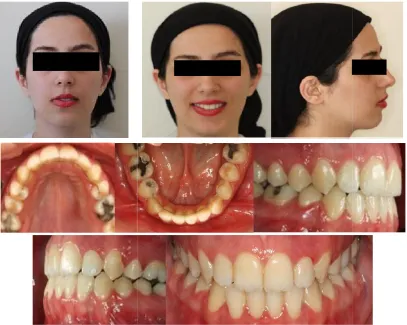
A 23 year old female presented with anterior open bite of 3 mm, increased facial height and convex profile (Fig. 1). Her chief complaints were anterior open bite and a chewing problem. Furthermore, she feels uncomfortable to smile. She had no medical complication and orthodontic treatment before attending the orthodontic department. Her facial profile was convex due to a retrognathic mandible. An increased lower facial height, and circumoral musculature strain on lip closure were noted.
An intraoral examination showed that the patient had a moderately severe anterior open bite extending from the left maxillary canine to the right first premolar with overbite of -3 mm and overjet of +3 mm.
Right maxillary first molar and left mandibular first molar both had poor prognosis. The second molar relationship was Class II on right and class I on left side, and there was a small space between the maxillary right lateral incisor and the canine. Moderate crowding was present in both upper and lower arches.
Fig. 1. Pretreatment photographs
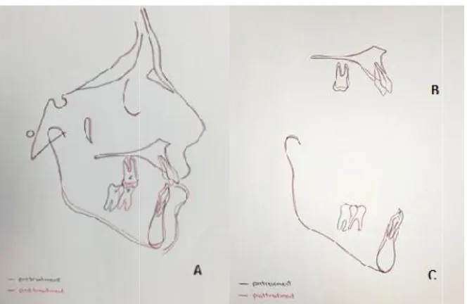
Fig. 2. (A) Pretreatment cephalograph. (B) Tracing. (C) Panoramic radiograph
Cephalometric analysis showed a Class II skeletal relationship with an ANB angle of 5.3° and a high mandibular plane angle of 40° (Fig. 2, above). The maxillary and mandibular incisors inclination were within the normal range:
U1 to FH, 115°; and IMPA, 90.1°. upper molars were significantly extruded (U6/NF 30.7). No symptoms of temporomandibular disorder were observed.
Fig. 1. Pretreatment photographs
Fig. 2. (A) Pretreatment cephalograph. (B) Tracing. (C) Panoramic radiograph
Cephalometric analysis showed a Class II skeletal relationship with an ANB angle of 5.3° and a high mandibular plane angle of 40° (Fig. 2, above). The maxillary and mandibular incisors inclination were within the normal range:
Fig. 3. (A) Panoramic radiograph (B) Intraoral photograph during treatment. Miniplates and one of the palatal screws are in place
2.2 Diagnosis, Treatment Objective and Treatment Alternatives
This case was diagnosed as having an Angle Class II Subdivision on left malocclusion, with a skeletal Class II jaw base relationship, a skeletal anterior open bite, high mandibular plane angle and missing of maxillary right and mandibular left first molar. The treatment objectives were [1] to close the anterior open bite and establish ideal overjet and overbite, [2] to obtain a functional Class I occlusion, [3] to correct the convex facial profile, [4] to close the edentulous spaces in both arches.
A surgical (maxillary posterior impaction) and nonsurgical treatment option (molar intrusion with TAD) were presented to the patient to address her skeletal anterior open bite and facial convexity.
2 options were presented to the patient for closing the mandibular edentulous space: 1. Implant insertion and prosthetic buildup followed by orthodontic treatment 2.molar protraction with the aid of TAD.
Risks and benefits of each treatment plan were explained in detail to the patient. The patient chose nonsurgical treatment combined with molar protraction because she did not want to undergo orthognathic surgery and prosthetic procedures.
2.3 Treatment Progress
Before the start of orthodontic treatment, all carious lesions were restored- all third molars, upper right and lower second molars and lower right second premolar were restored with amalgam material. Lower left and upper right first molars were extracted because they both had poor prognosis.
Titanium miniplates (L type, 0.8-mm diameter, 13.5-mm length; Dentsply-Sankin, Tokyo, Japan) were inserted bilaterally in the zygomatic process of the maxilla and were fixed with two titanium miniscrews (2-mm diameter, 5-mm length) (Fig. 3 above).
The operation was carried out under local anesthesia administered with intravenous sedation. First, a mucoperiosteal vertical incision was made at the buccal vestibul of the implantation site. After exposing the cortical bone, the appropriate size of anchor plate which was medium length for this case was contoured to fit the bone surface.
A pilot hole was drilled and one self-tapping and monocortical screw was inserted. With the insertion of the second screw, the anchor plate was firmly placed on the bone surface. Finally, the wound was closed and sutured with absorbable thread.
At the palatal side, The interradicular space between maxillary first molar and second molar was selected for miniscrew implant insertion. After administration of local anesthesia, Titanium screws (2-mm diameter, 10-mm length; Keisei Medical Industrial Co Ltd, Tokyo, Japan) were threaded 5 mm from the alveolar crest apically at an angle of about 40°-50° to the dental axis using self-drilling mechanism. In the lower arch, The interradicular space between first and second premolar was selected for miniscrew insertion. After administration of local anesthesia, Titanium miniscrews (1.6-mm diameter, 8-mm length; Keisei Medical Industrial Co Ltd., Tokyo, Japan) were threaded 5 mm from the alveolar crest apically at an angle of about 50°-60° to the dental axis using self-drilling mechanism.
Then, 0.022-inch slot, preadjusted edgewise appliances were placed in both arches. Mild exaggerated nickel-titanium archwires were placed in the upper arch during leveling and
alignment in order to prevent upper incisores extrusion.
4 weeks after miniplate insertion, loading of the intrusion force was started with elastic chains both buccally and palataly. Space closure was done on 0.019* 0.025-inch stainless steel archwires in both arches. In the lower left segment molar protraction was done using elastomeric chain directly administered from miniscrew to the molar band. In other segments, moderate anchorage was used for space closure. Eight months after the start of loading, overbite had increased to 3 mm. The total active treatment period was 24 months.
After the removal of the edgewise appliances, fixed spiral wire in addition to essix retainer –with increased thickness in the upper arch-was placed to retain both arches. The miniplate and miniscrew anchorage was stable for the entire duration of the treatment. The screws were easily removed with a screwdriver during the retention phase.
To remove miniplates a mucoperiosteal incision and subperiosteal ablation was performed at the implantation site. Then, the implanted anchor plate was exposed and taken off.
3. RESULTS
The posttreatment photographs showed a significant improvement in the facial appearance and smile when compared with the pretreatment photographs (Fig. 4). The retrognathic chin and convex profile were dramatically improved. The mandibular autorotation decreased the lower facial height, as a result facial proportions were improved. Lip strain was disappeared.
The anterior open bite had been corrected. overbite and overjet became 3 and 2 mm respectively. Upper molars were intruded 3.0 mm toward the palatal plane and lower molar protraction was done without any extrusion. The upper and lower incisors were minimally extruded.
Fig. 4. Postactive treatment photographs alignment in order to prevent upper incisores
extrusion.
4 weeks after miniplate insertion, loading of the intrusion force was started with elastic chains both buccally and palataly. Space closure was done on 0.019* 0.025-inch stainless steel archwires in both arches. In the lower left segment molar protraction was done using elastomeric chain directly administered from miniscrew to the molar band. In other segments, moderate anchorage was used for space closure. Eight months after the start of loading, overbite had increased to 3 mm. The total active treatment period was 24 months.
After the removal of the edgewise appliances, fixed spiral wire in addition to essix retainer –with increased thickness in the upper arch-was placed to retain both arches. The miniplate and miniscrew anchorage was stable for the entire duration of the treatment. The screws were easily removed with a screwdriver during the retention phase.
To remove miniplates a mucoperiosteal incision and subperiosteal ablation was performed at the implantation site. Then, the implanted anchor plate was exposed and taken off.
3. RESULTS
The posttreatment photographs showed a significant improvement in the facial appearance and smile when compared with the pretreatment photographs (Fig. 4). The retrognathic chin and convex profile were dramatically improved. The mandibular autorotation decreased the lower facial height, as a result facial proportions were improved. Lip strain was disappeared.
The anterior open bite had been corrected. overbite and overjet became 3 and 2 mm respectively. Upper molars were intruded 3.0 mm toward the palatal plane and lower molar protraction was done without any extrusion. The upper and lower incisors were minimally extruded.
The occlusion was much more stable, and ideal intercuspation of the teeth was achieved (Fig. 4). An Angle Class I canine relationship was achieved on both sides. Molar relationship was class III on right and class II on left side witch was ineluctable due to the imperfect dentition of the patient at the start of the treatment. However, the intercuspation of the teeth was still good and the occlusion was much more stable. The lower midline was shifted to the right side by 2 mm, upper midline stayed the same.
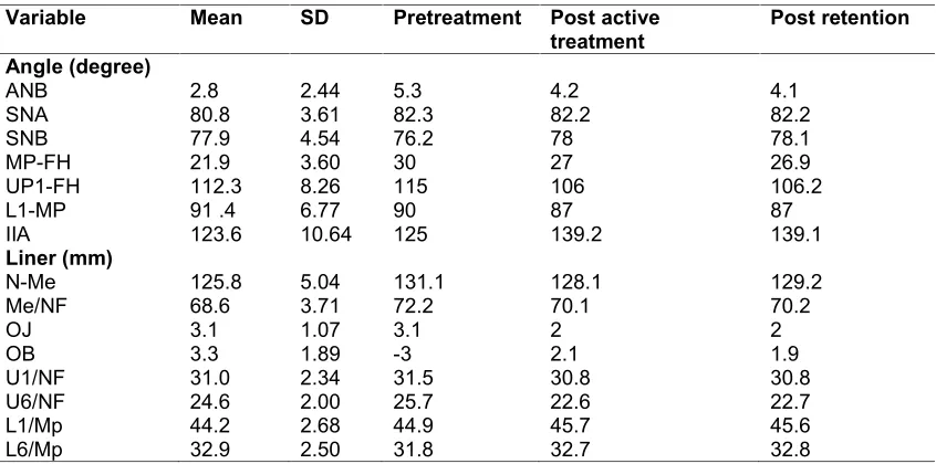
A summary of cephalometric variables of the patient is shown in Table 1. The table shows improvement in many of the variables. Post-treatment cephalometric evaluation showed a significant improvement in jaw base relationship (Figs. 5 and 6).
After active orthodontic treatment, no functional problems were observed in the examination for jaw movement.
After one year of retention, a good facial profile was retained and the occlusion was also stable (Fig. 7). No significant change in mandibular position was observed in cephalometric analysis (Table 1, Fig. 8).
4. DISCUSSION
Skeletal open bites are often related to excessive vertical growth of the dentoalveolar complex, especially in the posterior molar region [5]. The
treatment of skeletal open bites varies in growing versus adult patients [1,2]. Treatment strategies in growing patients with skeletal open bites involves vertical growth modification [1]. In adult patients, the options are more limited, and a correction of the skeletal dysplasia has been addressed primarily through orthognathic surgery [1]. Molar intrusion in nongrowing patients has always been a topic of interest [15]. The rationale for this treatment method is based on the expectation of mandibular autorotation as the molars intrude, resulting in anterior open bite closure [14,15]. For every millimeter of molar intrusion, approximately 3 mm of open bite reduction is seen in the anterior region [15].
With conventional orthodontics in patients with anterior open bite, closure of the openbite is achieved mostly through dentoalveolar changes, however, the skeletofacial complex may worsen because of extrusive mechanics [16,17]. The molars can be intruded directly from TADs to correct the vertical dimension without the need for compliance by the patient [16-18]. Molar intrusion can be accomplished with different types of TADs [13]. recent studies have shown that, The failure rate for miniplates is significantly lower than for miniscrews [13]. So in this case we used miniplates to have a more stable anchorage and No movement of miniplates took place at any time during their use or before intentional clinical removal.
Table 1. Cephalometric summary
Variable Mean SD Pretreatment Post active
treatment Post retention
Angle (degree)
ANB 2.8 2.44 5.3 4.2 4.1
SNA 80.8 3.61 82.3 82.2 82.2
SNB 77.9 4.54 76.2 78 78.1
MP-FH 21.9 3.60 30 27 26.9
UP1-FH 112.3 8.26 115 106 106.2
L1-MP 91 .4 6.77 90 87 87
IIA 123.6 10.64 125 139.2 139.1
Liner (mm)
N-Me 125.8 5.04 131.1 128.1 129.2
Me/NF 68.6 3.71 72.2 70.1 70.2
OJ 3.1 1.07 3.1 2 2
OB 3.3 1.89 -3 2.1 1.9
U1/NF 31.0 2.34 31.5 30.8 30.8
U6/NF 24.6 2.00 25.7 22.6 22.7
L1/Mp 44.2 2.68 44.9 45.7 45.6
Fig. 5. (A) Postactive treatment cephalograph. (B) Tracing. (C) Panoramic radiograph
Fig. 6. Superimposition of cephalometric tracings made before (black line) and after (red line) treatment. (A) Superimposit ion on the Sella-Nasion plane at Sella. (B) Superimposition on the
palatal plane at ANS. (C) Superimposition on the mandibular plan e at Menton
According to cifter and Sarac, balanced intrusion with minimal tipping occurred when buccal and palatal forces were applied simultaneously via TADs [19]. In this case we applied intrusive force both buccaly and palatally through miniplates and miniscrews recpectively. Our outcome shows this is a great method to maintain molar inclination during intrusion and to prevent palatal cusps extrusion.
As a result of 2.8 mm intrusion of the upper molars, 1.1 mm incisors extrusion, 1.1 degree clockwise rotation of the maxillary occlusal plane, and 6 degree counterclockwise rotation of the mandible, 3 mm of anterior openbite was corrected.
Rotation of the mandible caused advancement of the chin at pogonion by 5 mm and improved the retrognathic profile.
The anterior facial height was significantly reduced, and Lip strain during closure, disappeared. By preventing the upper anterior extrusion, an esthetic smile was achieved. We believe that the functional adaptation in circumoral musculature is an important factor in the retention of the correction of anterior open bites.
Reports of one year of retention for anterior open-bite cases treated with skeletal anchorage, shows various relapse tendencies [20].
Fig. 5. (A) Postactive treatment cephalograph. (B) Tracing. (C) Panoramic radiograph
Fig. 6. Superimposition of cephalometric tracings made before (black line) and after (red line) treatment. (A) Superimposit ion on the Sella-Nasion plane at Sella. (B) Superimposition on the
palatal plane at ANS. (C) Superimposition on the mandibular plan e at Menton
According to cifter and Sarac, balanced intrusion with minimal tipping occurred when buccal and palatal forces were applied simultaneously via TADs [19]. In this case we applied intrusive force both buccaly and palatally through miniplates and miniscrews recpectively. Our outcome shows this is a great method to maintain molar inclination during intrusion and to prevent palatal cusps extrusion.
As a result of 2.8 mm intrusion of the upper molars, 1.1 mm incisors extrusion, 1.1 degree clockwise rotation of the maxillary occlusal plane, and 6 degree counterclockwise rotation of the mandible, 3 mm of anterior openbite was corrected.
Rotation of the mandible caused advancement of the chin at pogonion by 5 mm and improved the retrognathic profile.
The anterior facial height was significantly reduced, and Lip strain during closure, disappeared. By preventing the upper anterior extrusion, an esthetic smile was achieved. We believe that the functional adaptation in circumoral musculature is an important factor in the retention of the correction of anterior open bites.
Fig. 7. One-year postretention photograph
Fig. 8. (A) One-year postretention cephalograph. (B) Panoramic radiograph
In our case, little relapse was observed after a 1-year retention period, which can be due to the functional adaptation in circumoral musculature following counterclockwise rotation of the mandible.
In this case Lower left and upper right first molars were extracted because they had both poor prognosis. To close mandibular edentulous space lower molar protraction was done with the aid of minscrew which is more cost benefit for the Fig. 7. One-year postretention photograph
Fig. 8. (A) One-year postretention cephalograph. (B) Panoramic radiograph
In our case, little relapse was observed after a 1-year retention period, which can be due to the functional adaptation in circumoral musculature following counterclockwise rotation of the mandible.
patient than the other alternative that is implant insertion. Upper right first molar space was closed using moderate anchorage. To treat Class II Subdivision on left malocclusion, correct lower midline and reveal crowding in both arches, we planed extraction of upper left and lower right first premolar.
5. CONCLUSION
Our results suggest that titanium minplates are useful for posterior dento-alveolar intrusion. Intrusion of the posterior teeth induced upward rotation of the mandible and, as a consequence, corrected the anteroposterior intermaxillary relationship with a significant improvement in the facial soft tissue convexity. This method is a safe, quick, and less expensive alternative to orthognathic surgery.
CONSENT
As per international standard or university standard, patient’s written consent has been collected and preserved by the author(s).
ETHICAL APPROVAL
All authors hereby declare that all experiments have been examined and approved by the appropriate ethics committee and have therefore been performed in accordance with the ethical standards laid down in the 1964 Declaration of Helsinki.
COMPETING INTERESTS
Authors have declared that no competing interests exist.
REFERENCES
1. Epker BN, Fish LC. Surgical-orthodontic collection of openbite deformity. Angle Orthod. 1977;71:278–299.
2. Proffit WR, Phillips C, Dann CIV. Who seeks surgical-orthodontic treatment? Int J Adult Orthod Orthognath Surg. 1990; 5:153–160.
3. Alexander CD. Open bite, dental alveolar protrusion, class I malocclusion: A successful treatment result. Am J Orthod Dentofacial Orthop. 1999;116:494– 500.
4. Smith GA. Treatment of an adult with a severe anterior open bite and mutilated
malocclusion without orthognathic surgery. Am J Orthod Dentofacial Orthop. 1996; 110:682–687.
5. Kim YH. Anterior open bite and its treatment with multiloop edgewise archwire. Angle Orthod. 1987;4:290–321. 6. Enacar A, Ugur T, Toroglu S. A method for
correction of open bite. J Clin Orthod. 1996;30:43–48.
7. Freeman CS, McNamara JA Jr, Baccetti T, Franchi L, Graff TW. Treatment effects of the bionator and high-pull facebow combination followed by fixed appliances in patients with increased vertical dimensions. Am J Orthod Dentofacial Orthop. 2007;131:184–95.
[PubMed: 17276859]
8. Sherwood KH, Burch JG, Thompson WJ. Closing anterior open bites by intruding molars with titanium miniplate anchorage. Am J Orthod Dentofacial Orthop. 2002; 122:593–600.
[PubMed: 12490869]
9. Erverdi N, Keles A, Nanda R. The use of skeletal anchorage in open bite treatment: A cephalometric evaluation. Angle Orthod. 2004;74:381–90.
[PubMed: 15264651]
10. Erverdi N, Usumez S, Solak A. New generation open-bite treatment with zygomatic anchorage. Angle Orthod. 2006;76:519–26.
[PubMed: 16637736]
11. Park H, Kwon O, Sung J. Nonextraction treatment of an open bite with microscrew implant anchorage. Am J Orthod Dentofacial Orthop. 2006;130:391–402. [PubMed: 16979500]
12. Xun C, Zeng X, Wang X. Microscrew anchorage in skeletal anterior open bite treatment. Angle Orthod. 2007;77:47–56. [PubMed: 17029531]
13. Miyawaki S, Tomonari H, Yagi T, Kuninori T, Oga Y, Kikuchi M. Development of a novel spike-like auxiliary skeletal anchorage device to enhance miniscrew stability. Am J Orthod Dentofacial Orthop. 2015;148(2):338-44.
DOI: 10.1016/j.ajodo.2015.02.030
14. Yao CC, Chang HH, Chang JZ, Lai HH, Lu SC, Chen YJ. Revisiting the stability of mini-implants used for orthodontic anchorage. J Formos Med Assoc. 2014;pii:S0929-6646(14)00232-0.
DOI: 10.1016/j.jfma.2014.08.001
anchorage. Anglc Orthod. 1968;38:340-349.
16. Buschang PH, Carrillo R, Rossouw PE. Orthopedic correction of growing hyperdivergent, retrognathic patients with miniscrew implants. J Oral Maxillofac Surg. 2011;69(3)754-762.
17. Sherwood KH, Burch JG, Thompon WJ. Closing anterior open bites by intruding molars with titanium miniplate anchorage. Am J Orthod Dentofacial Orthop. 2002; 122(6):593-600.
18. Umemori M, Sugawara J, Mitani H, Ngasaka H, Kawamura H. Skeletal anchorage system for open-bite correction.
Am J Orthod Dentofacial Orthop. 1999; 115(2):166-174.
19. Cifter M, Sarac M. Maxillary posterior intrusion mechanics with miniimplant anchorage evaluated with the finite element method. Am J Orthod Dentofacial Orthop. 2011;140(5):e233-e241.
20. Sugawara J, Baik UB, Umemori M, Takahashi I, Nagasaka H, Kawamura H, Mitani H. Treatment and post-treatment dentoalveolar changes following intrusion of mandibular molars with application of a skeletal anchorage system (SAS) for open bite correction. Int J Adult Orthod Orthognath Surg. 2002;17:243–253. _________________________________________________________________________________
© 2017 Dadgar et al.; This is an Open Access article distributed under the terms of the Creative Commons Attribution License (http://creativecommons.org/licenses/by/4.0), which permits unrestricted use, distribution, and reproduction in any medium, provided the original work is properly cited.
Peer-review history: