_____________________________________________________________________________________________________
ISSN: 2231-0614
SCIENCEDOMAIN international www.sciencedomain.org
Neuropsychiatric Complications of Patients with
Obstructive Sleep Apnea Syndrome
Ghaydaa A. Shehata
1*, Lamia Shaaban
2, Esam El-Deen M. Abdalla
3and Hossam El-Deen K. Ahmed
41Department of Neurology, Assiut University, Egypt.
2
Department of Chest Medicine, Assiut University, Egypt. 3
Department of Anesthesia, Assiut University, Egypt. 4
Department of Psychiatry, Assiut University, Egypt.
Authors’ contributions
This work was carried out in collaboration between all authors. Author GAS designed the study, assessed patients, performed the statistical analysis, wrote the protocol, wrote the final draft of the manuscript and submit it. Authors LS and HEDKA examined cases, managed the analyses of the study and wrote the first draft. Author EEDMA managed the literature searches and revised the manuscript. All authors read and approved the final manuscript.
Article Information
DOI: 10.9734/BJMMR/2015/9867 Editor(s): (1) Mohamed Essa, Department of Food Science and Nutrition, Sultan Qaboos University, Oman. Reviewers: (1) Pietro Scicchitano, Cardiology Department, University of Bari, Bari, Italy. (2) Anonymous,China. Complete Peer review History:http://www.sciencedomain.org/review-history.php?iid=949&id=12&aid=8554
Received 4th March 2014 Accepted 21st April 2014 Published 23rd March 2015
ABSTRACT
Objective: Patients with obstructive sleep apnea (OSA) associated with neuropsychological deficits and altered quality of life. This study aimed to assess cognitive functions, quality of life (QOL), and mood changes in patients with obstructive sleep apnea (OSA).
Methods: This study included 37 patients who diagnosed as obstructive sleep apnea compared with 34 subjects with age and sex matched healthy persons as control group. They were assessed by using Short-Form 36 Health Survey questionnaire (SF-36), the Epworth sleepiness scale (ESS) and sleep disorders questionnaire, Hamilton depression and anxiety scales (HADS), Cognitive Abilities Screening Instruments (CASI) and mini mental state examination (MMSE).
Results: Patients with OSA had significant lower scores in all domains of SF-36 (5.4±1.2; 10.7±2.2; 7.2±1.5; 4.5±0.8; 31.6±2.9; 3.8±0.8; 10.2±2.2 and 3.5±0.8), compared to control group. Mean scores of the Hamilton questionnaires (depression 7.7±8.3; anxiety 6.4±1.3) were
significantly higher in patients than control group. Most domains of SF-36 were also significantly correlated with AHI, slowest heart rate, sleep disorders questionnaires, and different variables of studied cognitive function.
Conclusion: Cognitive functions, mood and QOL of patients with OSA were markedly affected compared with normal control subjects. Early recognition and treatment of OSA could reduce disability and improve the quality of life of patients with OSA.
Keywords: Obstructive sleep apnea; quality of life; cognitive function; depression; anxiety; excessive daytime sleepiness; SF-36.
1. INTRODUCTION
Obstructive Sleep Apnea Syndrome (OSA) is a potentially dangerous and progressive disease of recurrent impaired ventilation episodes impacting millions of Americans [1]. It is characterized by repetitive episodes of airflow reduction due to pharyngeal narrowing which lead to acute gas exchange abnormalities, sleep fragmentation and then it resule in neurobehavioral consequences [2]. Excessive daytime sleepiness and tiredness, lack of concentration, memory impairment, and impaired vigilance are features of OSA. Accordingly, persons with OSA has higher risk for motor vehicle crashes, occupational injuries, and decreased quality of life in social, emotional, and physical domains [3].
Performance deficits during neuropsychological tests can be documented with even mild OSA. Health - related quality of life (HRQL) is an important domain for measuring the impact of chronic disease [4]. In patients with OSA excessive daytime sleepiness (EDS), may contribute to the impaired quality of life (QOL) [5]. Apart from serious and life threatening disorders such as hypertension, acute myocardial infarction, cerebrovascular accident and heart failure, OSA causes defects in cognitive functions and mood. Neuropsychological impairment, resulting from OSA, affect daily life activities and the ability to set up regular social life. Although some studies reported that anxiety and depression are more common among OSA patients, and impairing the quality of life is much severe compared to normal population, other studies report no relationship between OSA and QOL, anxiety and depression [6].
The aim of the present study was to assess the quality of life (QOL), cognitive functions and mood changes in patients with obstructive sleep apnea (OSA). Moreover, the relationship between the QOL on one hand and severity of
OSA, excessive daytime sleepiness (EDS), cognitive functions and mood on the other hand. 2. PATIENTS AND METHODS
2.1 Patients
During a 24-months period, thirty seven patients with OSA and 34 control healthy subjects were recruited from intensive care units of anesthesia and chest departments, Assiut University Hospital, Assiut, Egypt. The control group was age, sex, educational level, and socioeconomic status matched to patient group. Written consents were obtained from patients and controls or their relatives after having received oral or written information about the study. The local ethical committee of Assiut University Hospital approved the study. This study was in agreement with Helsinki declaration research ethics.
Inclusion criteria for the study were: 1) Egyptian ethnicity; 2) patients who had been referred for snoring, fatigue, and/or daytime sleepiness and diagnosed as OSA with AHI > 5 and admitted to intensive care units. 3) Ability to give written consent or the availability of patients' guardians to give consent to participate in this study; and 4) ability to have informed knowledge of the patients’ earlier and current cognitive functioning. Exclusion criteria included: 1) Previous treatment for sleep apnea with CPAP, corrective airway surgery or a mandibular advancement device; 2) refusal to perform maintenance wakefulness test; 3) the presence of cardiovascular risk factors (hypertension, diabetes, dyslipidaemia ---etc ,and 4) presence of a mental, neurological or physical impairment severe enough to participate and complete the questionnaires.
thoroughly examined and demographic data, including age, sex, educational level, occupation, and socioeconomic status [7] were collected. 2.2 Polysomnography
Patients underwent a nocturnal polysomnogram with a minimum of 4 hours in bed. The following variables were systematically monitored using a computerized sleep system (Somnostar; Sensormedics; Yorba Linda, CA): EEG (leads C3/A2, C4/A1, O1/A2, and O2/A1); electrooculogram; chin and leg electromyogram; ECG (modified V2 lead); and body position. Respiration was monitored using nasal thermistor, uncalibrated thoracic and abdominal inductive respiratory plethysmography, pulseoximetry, and neck microphone.
Sleep staging was performed according to the modified criteria of [8], that were done by American Academy of Sleep medicine, 2007 [9], as the following: Stage W (wakefulness), Stage N1 (NREM1), Stage N2 (NREM2), Stage N3 (NREM3) that represents slow wave sleep and replaces the R&K nomenclature of stage 3, 4 sleep, and stage R (REM). Respiratory events (Hypopneas, Apneas and The apnea / hypopnea index (AHI) were scored using standard criteria. As indices of sleep fragmentation, thenumber of arousals and awakenings were defined, as well as the number of sleep state transitions. As indices of nocturnal hypoxemia,the mean SaO2
and the minimal value recorded during sleep (Sa,O2min) were considered [6]. The analysis and
interpretation of the polysomnography data were performed by standard techniques. Apnea was defined as a cessation of airflow for at least 10 seconds, and hypopnea was defined as a decrease forat least 10 seconds in the amplitude of airflow to < 50%of the level before the event, with a fallin oxyhemoglobin saturation of at least 4%. The apnea hypopnea index (AHI) was calculated, as the sum of all apneas and hypopneas, divided by the TST, and were expressed as per hour. Arousals was defined as abrupt change of the EEG frequency to alpha or frequencies >16 Hz that last at least 3 seconds plus an increase in the EMG tonus (especially in REM). Two arousals have to be at least 10 seconds apart, otherwise both events will be counted as one. Time in bed (TIB)was defined as the time from the onset of sleep to the last awakening in the morning and total sleep time (TST) was definedas sleep period time minus any time the subject was awake after falling asleep Sleep efficiency is equal TST / TIB x 100.
The desaturation index and minimum Sao2 also were calculated from the polysomnography data [9]. The OSA patients were classified according to AHI to three subgroups mild (11patient with AHI 5 - <15), moderate (12 patient with AHI 15 - < 30) and severe (14 patient with AHI ≥ 30). 2.3 Daytime Sleepiness Evaluation
To evaluate the level of sleepiness, patients completed two questionnaires, First, Epworth Sleepiness Scale (ESS) which is validated questionnaire containing eight items that ask for self-reported probability of dozing in a variety of situations. The scores ranged from 0 (never) to 3 (high probability). Normal values ranged between 2 and 24, with scores >10 indicating daytime sleepiness [7]. Second, Sleep Disorders Questionnaire is valid and standardized Arabic version of sleep disorders questionnaire [10], that used to identify patients with high-risk sleep disorders (SDS), and it is formed of 175 questions.
2.4 Neuropsychiatric Assessment
Mini-Mental State Examination (MMSE) [11], a widely used scale for the screening test for dementia. It consists of a variety of questions grouped into seven categories, each representing a different cognitive domain or function (orientation to time, orientation to place, repetition of words, attention, calculation recall of words, language, and visual construction). It has a maximum score of 30 points. As most of the subjects of the this study were illiterate or with low education levels, the two testing points of reading and writing were excluded, and the full score was calculated as 28 instead of 30 points [12].
Cognitive Abilities Screening Instruments (CASI) It consists of 25 items and provides quantitative assessment on attention, concentration, orientation, memories for post knowledge and present input, language abilities, drawing and writing abilities, list generating ability, abstract thinking and judgment [13]. It took 45–60 minutes to administer. The CASI is more comprehensive than most screening tests of cognitive abilities. The cutoff of dementia was 67 [12].
more according to Michele and Bolino [15] since then, numerous authors investigated the dimensionality of the scale and demonstrated that it is multi-dimensional [16]. Hamilton anxiety scale (HAS) consists of 14 questions. The total score range from 0 to 56. A total score of 18 or more means anxiety [17].
Health Related Quality of Life was assessed with the Medical Outcomes Study short form survey that consists of 36 questions (SF-36) that covers eight health concepts: functioning, physical and social; role, physical and emotional; mental health; bodily pain; vitality; and general health perceptions [18]. Each subject red over the 36 questions or with the help of authors then marked an answer on the sheet. This process took less than 10 min for each person. For all measures of the eight health components, scores were transformed linearly to scales of 0 to 100, with 0 and 100 assigned to the lowest and highest possible scores, respectively.
2.5 Statistical Analysis
Descriptive statistics (mean, SD, and percentages) were calculated using the computer software package SPSS for Windows, Version 16. Results were analyzed using independent-sample T test that did not assume equal variances. As values for SF-36, HAS, and HDS scores were non-normally distributed so that Spearman's correlation coefficients (r) were determined to examine the degree of correlation among components of the SF-36, and different variables. ANOVA test used to detect significance between mild, moderate and severe OSA and different variables. P value of less than 0.05 was considered significant.
3. RESULTS
The study included 37 patients with OSA and 34 healthy control subjects; their demographic data were shown in Table 1 where, there were no significant differences between both groups as regard their age or sex. Meanwhile the patient group had significant higher body mass index (p< 0.002). Table 2 showed the Polysomnographic parameters, of patients with OSA.
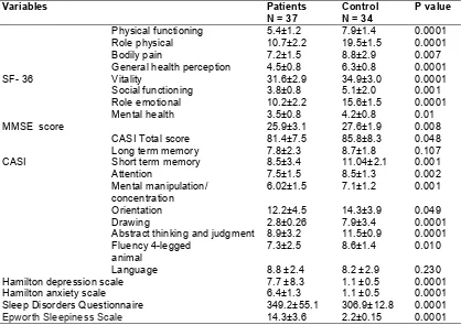
Medical Outcome Survey Short Form-36 domains scores, cognitive function, mood Scale scores, Sleep Disorders Questionnaire and Epworth Sleepiness Scale were presented in
Table 3 in which, Statistical analysis revealed that patients with OSA had significant lower scores in all domains of SF-36, when compared to normative data of control group. The greatest differences were seen for physical function, "role physical", general health perception, vitality, and "role emotional" (P value < 0.001) domains. That was followed by significant differences in the "social functioning", "bodily pain" and "mental health". Mean scores of the Hamilton questionnaires were within normal range both for the anxiety subscale (HAS) and for the depression scale (HDS), but they were significant higher than control group. Measures of sleep disorders questionnaire and Epworth sleepiness scale were significant higher among patients than control.
Comparisons between the three subgroups (mild, moderate and severe) regarding the different scales were shown in Table 4. Severe OSA patients had lower scores in all domains of SF-36 without significant difference except in role physical and bodily pain domains. However they had significant (p <0.05) higher score of Hamilton depression scale, Hamilton anxiety scale, and lower CASI score than mild and moderate OSA patients. BMI and ESS indexes were significant (p <0.05) higher in patients with severe OSA than mild and moderate OSA patients.
Individual analysis revealed that 6 cases of OSA patients suffered from depression as their Hamilton depression score was >17. All those patients were from severe subgroup and had high BMI ranging from 65-70.
significant positive correlations were noted between some domains of SF-36 and different variables of studied cognitive function.
4. DISCUSSION
Obstructive Sleep Apnea (OSA) is a common sleep-related breathing disorder affecting at least 5% of the general population [3]. Due to the high prevalence of this disease and its tremendous negative consequences, such as excessive daytime sleepiness [19], deficits in neurobehavioral performance [19], increased automobile accidents [20], and deterioration in functional status and quality of life [21]. The literature findings for OSA interventions are contained within two general themes: the immediate postoperative phase and sleep phase. The immediate postoperative phase includes the initial postoperative nursing assessment, guidelines for continuous care and monitoring, evaluation of extubation readiness, and effective pain management [1]. The sleep phase includes interventions related to sleep positioning [1]. In the present study, there were highly significant
impairment of studied cognitive function, quality of life, mood disturbance as depressed or anxious mood, and more affection of SDQ and ESS in patients with OSA than control group. These results could be attributed to multiple mechanisms, thought to contribute to sleepiness in patients with OSA. These include sleep fragmentation [22], hypoxia [23], partial chronic sleep deprivation from sleep time lost due to arousals [24], cytokine dysregulation [25], and interactions with individual adaptations [26]. Other studies had proposed the hypothesis that both daytime somnolence and hypoxemia may contribute to cognitive dysfunction in OSA patients. In particular, the impairment of executive functions, motor and visuo-constructive abilities (such as language, fluency, drawing) may be related to severity of hypoxemia. Also, attention and memory deficits may be due to excessive daytime somnolence associated with sleep fragmentation [27-29]. Other studies suggested the importance of REM sleep for memory consolidation [30,31], particularly for those individuals with insomnia [32].
Table 1. Demographic data of studied group
Patients N= 37 Control N=34 P value
Age (mean±SD) years 48.2± 1.2 45.6±7.2 0.228
BMI(mean±SD) Kg/m2 53.8±1.2 38.2±1.6 0.002
Sex
Male Female
26(70.3%) 11(29.7%)
17(50%) 17(50%)
0.066
History of smoking 22 (71.0%) 23(67.6%) 0.795
Socioeconomic scale 11.95±1.63 12.82±2.138 0.072
Data was expressed as mean ± S.D or number (percentage)
Table 2. Polysomnographic data of obstructive sleep apnea (OSA) patients
Polysomnographic data Means Standard deviation(SD)
TIB(hours) 5.2 1.6
TST(hours) 4.4 1.5
Sleep efficiency (%) 84.6 6.5
Stage REM % 23.6 16.4
Stage 1(N1) % 24.5 9.2
Stage 2(N2) % 30.1 6.8
SWS (N3) % 21.8 11.7
Basal heart rate 85.5 10.6
Slowest heart rate 55.5 14.9
Desaturation Index/h 27.04 2.19
Basal O2 saturation 89.48 6.4
Minimum O2 saturation 67.67 15.6
AHI /h 30.4 1.9
arousals index/h 9.3 6.7
Table 3. Comparison between the patient and control groups in short form-36 dimension cores, cognitive function, mood scales scores, sleep disorders questionnaire and Epworth
sleepiness scale
P value Control
N = 34 Patients
N = 37 Variables 0.0001 7.9±1.4 5.4±1.2 Physical functioning SF- 36 0.0001 19.5±1.5 10.7±2.2 Role physical 0.007 8.8±2.9 7.2±1.5 Bodily pain 0.0001 6.3±0.8 4.5±0.8 General health perception
0.0001 34.9±3.0 31.6±2.9 Vitality 0.001 5.1±2.0 3.8±0.8 Social functioning 0.0001 15.6±1.5 10.2±2.2 Role emotional 0.01 4.2±0.8 3.5±0.8 Mental health 0.008 27.6±1.9 25.9±3.1 MMSE score
0.048 85.8±8.3
81.4±7.5 CASI Total score
CASI
0.107 8.7±1.8
7.8±2.3 Long term memory
0.001 11.04±2.1
8.5±3.4
Short term memory
0.002 8.5±1.3 7.5±1.5 Attention 0.001 7.1±1.2 6.02±1.5 Mental manipulation/ concentration 0.049 14.3±3.9 12.2±4.5 Orientation 0.0001 7.9±3.4 2.8±0.26 Drawing 0.0001 11.5±0.9 8.9±3.2 Abstract thinking and judgment
0.010 8.6±1.4 7.3±2.5 Fluency 4-legged animal 0.230 8.2 ±2.9 8.8 ±2.4 Language 0.0001 1.1 ±0.5 7.7 ±8.3 Hamilton depression scale
0.0001 1.1 ±0.5 6.4±1.3
Hamilton anxiety scale
0.0001 306.9±12.8
349.2±55.1 Sleep Disorders Questionnaire
0.0001 2.2±0.15
14.3±3.6
Epworth Sleepiness Scale
Data were expressed as mean ± standard deviation, SF-36: Short Form-36, CASI: Cognitive Abilities Screening
Instruments, MMSE: Mini-Mental State Examination, Significant difference between patients and control group
considered when P < 0.05
Table 4. Comparison between mild, moderate and severe OSA in short form-36 scores, cognitive function, mood scales scores, sleep disorders questionnaire, Epworth sleepiness
scale and BMI
P value Severe
N = 14
Moderate N = 12 Mild
N = 11 Variables NS 4.9.9±1.4 5.8±1.2 5.9±1.2 Physical functioning SF- 36 0.02 10.7±2.2 10.8±2.03 19.5±1.5 Role physical 0.04 6.8±1.5 6.9±1.4 8.1±2.9 Bodily pain NS 4.3.3±0.8 4.6±0.6 4.8±0.8 General health perception
NS 31.3±3.0 31.8±2.2 32.0±2.9 Vitality NS 3.6±2.0 3.8±0.8 4.1±0.8 Social functioning NS 9.7±1.5 10.2±2.2 10.5±2.2 Role emotional NS 3.5±0.8 3.5±0.9 4.2±0.8 Mental health NS 25.7±1.9 26.03 26.1±3.1
MMSE score
0.04 77.6±1.2
84.1±1.1 84.5±1.5
CASI total score
0.02 11.8±0.5
7.6±2.5 3.6±2.3
Hamilton depression scale
0.04 9.7±0.5 6.2±2.3 3.5±2.3
Hamilton anxiety scale
NS 356.9±12.8 354±7.3
336.2±4.8 Sleep disorders questionnaire
0.01 16.5±0.15 14.05±4.2
12.5±3.6
Epworth sleepiness scale
0.01 56.06±13.5 53.02±8.3
52.2±12.9
Body mass index(BMI)
738
Table 5. Correlation Coefficient between the domains on the SF-36, and the Polysomnographic parameters in patient group
Medical outcome survey short form (SF)-36
PF RP BP GHP V SF RE MH
AHI -.253*
0.01
-.046 -.363* 0.02
-.208 -034 -.343*
0.02
-.069 -.085
Sleep efficiency % .184*
0.02
.080 .079 .066 .066 .375*
0.02
.133 .002
Stage 1% .084 -.149 -.026 -.148 .129 -.172 -.052 -.066
Stage 2% -.134 .042 -.101 -.184 -.005) .228 -.031 .062
SWS % .058 .295 .105 .093 .392*
.016
.236 .04
.170 .034
REM % -.075 -.320 -.181 -.383*
0.01
..359* 0.02
-.076 -.354* 0.01
-.140
Desaturation index -.051 -.057 -.170*
0.02
-.064 -.081 -.173*
0.02
-.302 -.032
Minimum SaO2 .263 .139 .159*
0.04
.012 .075 .146 .360*
0.02
.015
Slowest heart rat .432*
0.001
.159 .329*
0.04
.210 .147 .166*
0.02
.176 .113
Arousal index -.086 -. 037 -.022 -.037 -.205*
0.02
-.166* 0.02
-.081 -.060
ESS score -.230*
0.01
-.419* 0.01
-.0479* 0.00
-.157 -.368* 0.02
-.213* 0.00
.-.070 .-.255* 0.02 SDQ
-.231 -.348*
0.005
-.212 -.349* 0.004
-.567* 0.001
-.300* 0.005
-.029 -.033 Significant data are expressed as r (P value), PF: physical functioning; RP: role physical; GH: general health; V: vitality; SF: social functioning; RE: role emotional; BP: bodily
739
Table 6. Correlation coefficient between the domains on the SF-36, cognitive function and mood scales scores
Variables Medical outcome survey short form (SF)-36
PF RP BP GHP V SF RE MH
Age(years) NS NS NS NS NS NS NS NS
BMI NS NS NS NS NS NS NS NS
MMSE
.265*
0.032
NS NS NS .244*
0.050
NS NS NS
CASI total score NS NS NS NS NS NS NS NS
CASI-long term memory NS NS NS NS NS NS NS NS
CASI-short term memory .279* 0.025
.494** 0.000
.367** 0.003
.284* 0.012
.273* 0.028
NS .301*
0.015
NS
CASI-attention .297*
0.016
.523** 0.000
.370** 0.002
.260* 0.036
.278* 0.025
NS NS NS
CASI-mental manipulation/ concentration
NS .418
0.001
NS .450**
0.000
NS NS .405**
0.001
.273** 0.002
CASI-orientation NS NS NS NS NS NS NS NS
CASI-drawing .305*
0.014
NS .328**
0.005
.386** 0.002
.274* 0.027
NS .296*
0.017
.304* 0.014 CASI-abstract thinking and
judgment
NS NS NS .413**
0.001
.397** 0.001
NS .381**
0.002
.282* 0.023 CASI-fluency
4-legged animals
NS NS NS NS NS NS NS NS
CASI-language .257*
0.043
NS NS NS NS NS NS NS
HDS
NS -.435**
0.000
.317* 0.010
-.413** 0.000
-.276*
0.026
-.245* 0.049
-.390** 0.001
-.321** 0.005
HAS NS .435**
0.000
-.317* 0.01
-.418** 0.000
-.276* 0.02
-.245 0.04
-.390** 0.001
-.321** 0.005 Significant data are expressed as r (P value), NS: Non Significant, PF: physical functioning; RP: role physical; GH: general health; V: vitality; SF: social functioning; RE: role emotional; BP: bodily pain; MH: mental health; BMI: body mass index; CASI: Cognitive Abilities Screening Instruments, and MMSE: Mini-Mental State; HAS: Hamilton anxiety
In addition, untreated OSA, the episodic fluctuations of hypoxemia and hypercarbia produce a physiologic stress response in the individual, which ultimately cascades into systemic derangements [33]. Vascular inflammation and atherosclerosis have been linked to the stresses induced by the hypoxia-reoxygenation episodes of OSA [34,35]. Several studies showed that OSA is responsible for increased carotid intermediate thinking [36-38]. The two primary mechanisms underlying endothelial abnormalities and atherosclerosis changes in blood vessels among OSA are represented by repetitive episodes of hypoxia / reoxygenation associated with transient cessation of breath during sleep, and sleep fragmentation / deprivation [38,39].
The resulting increase in catecholamines activates the reninangiotensin- aldosterone axis, leading to sodium retention and further increases in vasoconstriction with the possibility of renal failure [33].
Since sleep disturbance and depression were associated with cognitive impairment [40,41] further delineation of the inter-relationships between these potential mediators of performance is warranted. Other studies have also shown that slow wave sleep modulates hypothalamic–pituitary-adrenal axis function which, in turn, plays a vital role in cortisol secretion, the hippocampus network and memory [42]. Thus, it is possible that there are complex inter-relationships between sleep, depression, and cognition—particularly memory. Importantly, sleep disturbance may be a prodromal feature, a risk factor for recurrence [43] and a potential mediator of mood and neuropsychological functioning. That matched with previous study by Millman et al. [44], demonstrated that patients with OSA have depression and anxiety.
As excessive day sleepiness (EDS) is a primary symptom in patients with OSA and an important symptom for activities of daily living, it appears to be related to the decrease of the QOL. Therefore, a number of studies [5,21,45], have examined the relationship between the EDS and QOL and found that the EDS, whether evaluated subjectively or objectively, were correlated with the decrease in the QOL. These agree with the results of this study as most domains of SF-36 were negative significantly correlated with Epworth Sleepiness Scale and sleep disorders questionnaires.
This may suggest that some of the psycho-physiological consequences of OSA do not reflect a general psychological and mood effect but rather the specific consequences of sleepiness and impaired alertness. This could suggest that the sleepiness reported by OSA patients translates both the inability to stay awake, as measured by sleepiness questionnaires, as well as a subjective feeling of "loss of energy" [46]. In addition, the impairment of quality of life among OSA is correlated with cognitive impairments and increased risk for depression and anxiety, it also correlated with AHI, minimum Sao2% and disturbed sleep architecture. However, the pathways producing alterations in behavioral outcomes are complex and may involve both chronic and acute insults acting on cerebral structure or function [47]. Experimental evidence suggests that mild to moderate hypoxia can reduce turnover of acetylcholine [48], potentially producing global and diffuse cortical slowing. In more severe hypoxemia, loci of cerebral anoxic damage could be produced. One speculative mechanism involves both sleep fragmentation and hypoxemia acting synergistically to produce irreversible neuronal damage. Such cerebral damage in OSA might be mediated by abnormalities in ventilatory drive, potentiated by sleep disruption, leading to alterations to intracranial haemodynamic. These path physiological changes might potentially act in a positive feedback loop to produce cerebral ischemia, an agent of irreversible damage to neuronal structure and function.
Since the severity of OSA was not always associated with reductions in the QOL, as shown in present study, and associated more with mood and cognitive disability, a careful evaluation of the mood or depressive state of a patient will be needed to improve the QOL of patients with severe OSA.
5. CONCLUSION AND RECOMMENDATIONS
6. LIMITATION OF STUDY
Small number of patients was included in this study as the cost of polysomnography and there were no fund support this work.
CONSENT
All authors declare that ‘written informed consent was obtained from the patient.
ETHICAL APPROVAL
All authors declare that all patients and control groups have been examined and approved by the local ethical committee of Assiut University Hospital approved the study. This study was in agreement with Helsinki declaration research ethics.
COMPETING INTERESTS
Authors have declared that no competing interests exist.
REFERENCES
1. Gammon BT, Ricker KF. An evidence-based checklist for the postoperative management of obstructive sleep apnea. J Perianesth Nurs. 2012;27(5):316-22. 2. Schwengel DA, Sterni LM, Tunkel DE,
Heitmiller ES. Perioperative management of children with obstructive sleep apnea. Anesthesia and analgesia. 2009;109(1):60-75.
3. Young T, Peppard PE, Gottlieb DJ. Epidemiology of obstructive sleep apnea: A population health perspective. American journal of respiratory and critical care medicine. 2002;165(9):1217-39.
4. Saunamäki T, Himanen S, Polo O, MJ. Executive Dysfunction in Patients with Obstructive Sleep Apnea Syndrome. Eur Neurol. 2009;62:237-42.
5. Briones B, Adams N, Strauss M, Rosenberg C, Whalen C, Carskadon M, et al. Relationship between sleepiness and general health status. Sleep. 1996;19(7):583-8.
6. Fiahbaz S, Til O, Nönü H, BÖ. Quality of life, frequency of anxiety and depression in obstructive sleep apnea syndrome. Tur Toraks Der. 2008;9:141-5.
7. Fahmy SI, El-Sherbini AF. Determining simple parameters for social classification
for health research. Bull High Inst Public Health. 1983;8:98-108.
8. Rechtschaffen Kales. eds. Scoring system for sleep stages of human subjects. US Government Printing Office; 1968.
9. American, Academy, Sleep o, medicine, eds. A Manual for Scoring Sleep; 2007. 10. Douglass AB, Bornstein R, Nino-Murcia G,
Keenan S, Miles L, Zarcone VP Jr., et al. The sleep disorders questionnaire. I: Creation and multivariate structure of SDQ. Sleep. 1994;17(2):160-7.
11. Folstein MF, Folstein SE, McHugh PR. "Mini-mental state". A practical method for grading the cognitive state of patients for the clinician. Journal of Psychiatric Research. 1975;12(3):189-98.
12. Khedr EM, Hamed SA, El-Shereef HK, Shawky OA, Mohamed KA, Awad EM, et al. Cognitive impairment after cerebrovascular stroke: Relationship to vascular risk factors. Neuropsychiatric Disease and Treatment. 2009;5:103-16. 13. Evans DA, Beckett LA, Albert MS, Hebert
LE, Scherr PA, Funkenstein HH, et al. Level of education and change in cognitive function in a community population of older persons. Annals of Epidemiology. 1993;3(1):71-7.
14. Hamilton M. A rating scale for depression. Journal of neurology, neurosurgery, and psychiatry. 1960;23:56-62.
15. Michele VD, Bolino F. Post stroke depression. The British Journal of Psychiatrists. 2000;176:94-5.
16. Moller HJ. Methodological aspects in the assessment of severity of depression by the Hamilton Depression Scale. European Archives of Psychiatry and Clinical Neuroscience. 2001;251(Suppl 2):13-20. 17. Hamilton A. Diagnosis and rating of
anxiety. British Journal of Psychiatry; Special Publication. 1969;3:76-9.
18. Ware JE Jr., Sherbourne CD. The MOS 36-item short-form health survey (SF-36). I. Conceptual framework and item selection. Medical Care. 1992;30(6):473-83.
19. Black J. Sleepiness and residual sleepiness in adults with obstructive sleep apnea. Respiratory Physiology & Neurobiology. 2003;136(2-3):211-20. 20. Sassani A, Findley LJ, Kryger M, Goldlust
21. Baldwin CM, Griffith KA, Nieto FJ, O’Connor GT, Walsleben JA, Redline S. The association of sleep-disordered breathing and sleep symptoms with quality of life in the Sleep Heart Health Study. Sleep. 2001;24(1):96-105.
22. Bonnet MH, Doghramji K, Roehrs T, Stepanski EJ, Sheldon SH, Walters AS, et al. The scoring of arousal in sleep: reliability, validity, and alternatives. J Clin Sleep Med. 2007;3(2):133-45.
23. Veasey SC, Zhan G, Fenik P, Pratico D. Long-term intermittent hypoxia: reduced excitatory hypoglossal nerve output. American Journal of Respiratory and Critical Care Medicine. 2004;170(6):665-72.
24. Schwartz DJ, Moxley P. On the potential clinical relevance of the length of arousals from sleep in patients with obstructive sleep apnea. J Clin Sleep Med. 2006;2(2):175-80.
25. Vgontzas AN, Zoumakis E, Lin HM, Bixler EO, Trakada G, Chrousos GP. Marked decrease in sleepiness in patients with sleep apnea by etanercept, a tumor necrosis factor-alpha antagonist. The Journal of Clinical Endocrinology and Metabolism. 2004;89(9):4409-13.
26. Kapur VK, Baldwin CM, Resnick HE, Gottlieb DJ, Nieto FJ. Sleepiness in patients with moderate to severe sleep-disordered breathing. Sleep. 2005;28(4): 472-7.
27. Roehrs T, Merrion M, Pedrosi B, Stepanski E, Zorick F, Roth T. Neuropsychological function in obstructive sleep apnea syndrome (OSAS) compared to chronic obstructive pulmonary disease (COPD). Sleep. 1995;18(5):382-8.
28. Naegele B, Thouvard V, Pepin JL, Levy P, Bonnet C, Perret JE, et al. Deficits of cognitive executive functions in patients with sleep apnea syndrome. Sleep. 1995;18(1):43-52.
29. Doran SM, Van Dongen HP, Dinges DF. Sustained attention performance during sleep deprivation: evidence of state instability. Archives Italiennes de Biologie. 2001;139(3):253-67.
30. Fogel SM, Smith CT, Cote KA. Dissociable learning-dependent changes in REM and non-REM sleep in declarative and procedural memory systems. Behavioural Brain Research. 2007;180(1):48-61. 31. Rauchs G, Bertran F, Guillery-Girard B,
Desgranges B, Kerrouche N, Denise P, et
al. Consolidation of strictly episodic memories mainly requires rapid eye movement sleep. Sleep. 2004;27(3):395-401.
32. Backhaus J, Junghanns K, Born J, Hohaus K, Faasch F, Hohagen F. Impaired declarative memory consolidation during sleep in patients with primary insomnia: Influence of sleep architecture and nocturnal cortisol release. Biological Psychiatry. 2006;60(12):1324-30.
33. Diffee PD, Beach MM, Cuellar NG. Caring for the patient with obstructive sleep apnea: implications for health care providers in postanesthesia care. J Perianesth Nurs. 2012;27(5):329-40. 34. Rudra A, Chatterjee S, Das T, Sengupta S,
Maitra G, Kumar P. Obstructive sleep apnoea and anaesthesia. Indian J Crit Care Med. 2008;12(3):116-23.
35. Park JG, Ramar K, Olson EJ. Updates on definition, consequences, and management of obstructive sleep apnea. Mayo Clinic Proceedings. 2011;86(6):549-54; quiz 54-5.
36. Schulz R, Seeger W, Fegbeutel C, Husken H, Bodeker RH, Tillmanns H, et al. Changes in extracranial arteries in obstructive sleep apnoea. The European Respiratory Journal. 2005;25(1):69-74. 37. Suzuki T, Nakano H, Maekawa J, Okamoto
Y, Ohnishi Y, Yamauchi M, et al. Obstructive sleep apnea and carotid-artery intima-media thickness. Sleep. 2004;27(1):129-33.
38. Ciccone MM, Scicchitano P, Zito A, Cortese F, Boninfante B, Falcone VA, et al. Correlation between inflammatory markers of atherosclerosis and carotid intima-media thickness in Obstructive Sleep Apnea. Molecules (Basel, Switzerland). 2014; 19(2):1651-62.
39. Ciccone MM, Scicchitano P, Mitacchione G, Zito A, Gesualdo M, Caputo P, et al. Is there a correlation between OSAS duration/severity and carotid intima-media thickness? Respiratory Medicine. 2012; 106(5):740-6.
40. Naismith SL, Hickie IB, Turner K, Little CL, Winter V, Ward PB, et al. Neuropsychological performance in patients with depression is associated with clinical, etiological and genetic risk factors. Journal of Clinical and Experimental Neuropsychology. 2003;25(6):866-77. 41. Naismith S, Winter V, Gotsopoulos H,
functioning in obstructive sleep apnea: Differential effects of sleep quality, hypoxemia and subjective sleepiness. Journal of Clinical and Experimental Neuropsychology. 2004;26(1):43-54. 42. Buckley TM, Schatzberg AF. On the
interactions of the hypothalamic-pituitary-adrenal (HPA) axis and sleep: normal HPA axis activity and circadian rhythm, exemplary sleep disorders. The Journal of Clinical Endocrinology and Metabolism. 2005;90(5):3106-14.
43. Cho HJ, Lavretsky H, Olmstead R, Levin MJ, Oxman MN, Irwin MR. Sleep disturbance and depression recurrence in community-dwelling older adults: a prospective study. The American Journal of Psychiatry. 2008;165(12):1543-50. 44. Millman RP, Fogel BS, McNamara ME,
Carlisle CC. Depression as a manifestation of obstructive sleep apnea: reversal with nasal continuous positive airway pressure.
The Journal of Clinical Psychiatry. 1989;50(9):348-51.
45. Jenkinson C, Stradling J, Petersen S. Comparison of three measures of quality of life outcome in the evaluation of continuous positive airways pressure therapy for sleep apnoea. Journal of Sleep Research. 1997;6(3):199-204.
46. Sforza E, de Saint Hilaire Z, Pelissolo A, Rochat T, Ibanez V. Personality, anxiety and mood traits in patients with sleep-related breathing disorders: Effect of reduced daytime alertness. Sleep medicine. 2002;3(2):139-45.
47. Engleman H, Joffe D. Neuropsychological function in obstructive sleep apnoea. Sleep Medicine Reviews. 1999;3(1):59-78. 48. Gibson GE, Pulsinelli W, Blass JP, Duffy
TE. Brain dysfunction in mild to moderate hypoxia. The American Journal of Medicine. 1981;70(6):1247-54.
© 2015 Shehata et al.; This is an Open Access article distributed under the terms of the Creative Commons Attribution License (http://creativecommons.org/licenses/by/4.0), which permits unrestricted use, distribution, and reproduction in any medium, provided the original work is properly cited.
Peer-review history: