_____________________________________________________________________________________________________
*Corresponding author: E-mail: solnce-sun@ukr.net;
(Past name: British Journal of Medicine and Medical Research, Past ISSN: 2231-0614, NLM ID: 101570965)
Common Bile Duct Stone Exploration: T-Tube or
Biliary
V. Grubnik
1, V. Ilyashenko
1, A. Tkachenko
1, A. Kovalchuk
2, K. Vorotyntseva
1*and Grubnik Victor
11Odessa National Medical University, Ukraine.
2I.Horbachevsky Ternopil State Medical University, Ukraine.
Authors’ contributions This work was carried out in collaboration between all authors. Authors GV, KA and IV designed the study,
approved the protocol, managed the literature searches and wrote the manuscript. Authors TA and VK executed the surgery and managed the analyses of the study. All authors read, corrected and approved the final manuscript.
Article Information
DOI: 10.9734/JAMMR/2018/36378
Editor(s):
(1)Aniket Kishor Sakharpe, Easton Hospital & Drexel University, Easton, USA.
Reviewers:
(1)Jurij Janež, University Medical Centre Ljubljana, Slovenia. (2)Huseyin Eken, Erzincan University, Turkey. Complete Peer review History:http://www.sciencedomain.org/review-history/23617
Received 26th August 2017 Accepted 24th February 2018 Published 14th March 2018
ABSTRACT
Introduction: Laparoscopic common bile duct exploration (LCBDE) for choledocholithiasis is a popular option in many surgical institutes. Decompression of biliary system via T-tube post supra-duodenal choledochotomy has been the traditional surgical practice. Primary closure of common bile duct (CBD) has been shown to reduce hospital stay but bears a risk of bile leak. We conducted a prospective randomized trial to compare complications and length of stay in patients undergoing biliary stent insertion versus T-tube drainage following LCBDE via choledochotomy.
Methods and Procedures: The study involves 52 patients with choledocholithiasis who underwent LCBDE and decompression of the biliary system by either antegrade biliary stent or T-tube insertion. A 7 French biliary stent (9 “10 cm long) have been placed in 27 patients (group I), T-tube insertion have been used for 25 patients (group II). The length of hospital stay and complications were recorded. All transcystic explorations were excluded.
Results: There were no significant differences between groups with respect to age, sex, comorbidities, number and size of CBD stones. Postoperative complications have been observed in 4 patients (16%) in the T-tube group (one patient needed reoperation for dislocation of T-tube),
Grubnik et al.; JAMMR, 25(8): 1-9, 2018; Article no.JAMMR.36378
and in 1 patient (3.7%) in the biliary stent group (p < 0.05). The mean postoperative hospital stay was 3.2 ± 1.2 days for group I, and 6.2 ± 1.7 days for group II (p < 0.05).
Conclusions: Our results showed a reduction of length of hospital stay and morbidity following stent insertion compared to T-tube drainage. Also, the use of biliary stent after LCBDE can reduce costs and increase patient satisfaction.
Keywords: Choledocholithiasis; laparoscopic common bile duct exploration; T-tube drainage; biliary stenting.
1. INTRODUCTION
Laparoscopic common bile duct exploration (LCBDE) for choledocholithiasis is feasible and has become increasingly popular [1,2,3,4,5,6,7]. The LCBDE procedure can be performed transcystically or by choledochotomy.
Transcystic approach is preferred whenever but may be limited by either number and size of gallstones, or small diameter of the cystic duct, or anatomical variation of bile ducts.
In such cases laparoscopic choledochotomy is an alternative solution [8,3,4,7] but it may carry higher morbidity rates, prolong recovery and increase hospital stay [9,10,8,1,11,2,12].
Disadvantages associated with the use of a T-tube led some authors to attempt laparoscopic primary duct closure, which was demonstrated to be safe [13,12,4]. However, following primary closure bile leaks may be observed due to retained stones, stenosis of ampulla of Vater, oedema secondary to surgical manipulation [14,15,4]. To avoid such complications, some authors proposed ante-grade biliary stent insertion with laparoscopic primary closure of choledoch [16,17,18]. The advantages of biliary stent placement were recently demonstrated by Lyon et al. [19]. However, no prospective randomised cohort studies are comparing primary closure with ante-grade biliary stent insertion versus T-tube drainage of CBD following laparoscopic choledochotomy to date.
We conducted a randomised study to compare the postoperative course and outcome of primary closure with ante-grade biliary stent insertion and T-tube drainage of the CBD after laparoscopic choledochotomy.
2. MATERIALS AND METHODS
Between January 2009 and January 2014, a total of 125 patients with CBD stones underwent elective and emergency laparoscopic common
bile duct exploration (LCBDE). 122 cases were successful, remaining three cases required conversion to open surgery due to LCBDE failure. Of the 122 successfully treated patients, 70 underwent laparoscopic transcystic stone extraction and 52 required laparoscopic choledochotomy. CBD stones were diagnosed on history, physical examination, biochemical tests and transabdominal ultrasound followed by MRCP/CT cholangiography. Intraoperative cholangiography (IOC) was performed in all patients.
On preoperative assessment the patients were classified according to the American society of Anesthesiologist (ASA) classification.
Patients 18 years or older who had undergone a laparoscopic choledochotomy were included in the study. Exclusion criteria were acute suppurative cholangitis, severe acute biliary pancreatitis, ampullary stenosis, previous gastrectomy, gastric bypass or failure of endoscopic retrograde cholangiopancreato-graphy (ERCP). Eight patients who had undergone laparoscopic choledochotomy were excluded due to ineligibility (one patient younger than 18 years, three with acute suppurative cholangitis, two with acute biliary pancreatitis, one with ampullary stenosis and one with ERCP failure).
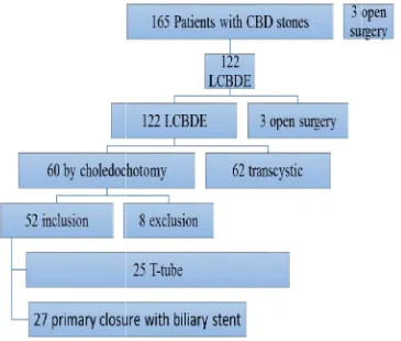
The 52 eligible patients were randomly assigned to two groups: the first group (27 patients) underwent antegrade biliary stent insertion for biliary tree drainage with primary closure of choledochus; the second group (25 patients) underwent LCBDE with T-tube insertion (Fig. 1).
Informed consent for randomisation to primary closure with a biliary stent or T-tube drainage was obtained. Randomization was performed with the use of a computer-generated randomisation schedule.
complications were recorded on an excel spread sheet. Data on late complication was recorded at outpatient clinic visits (removal of stent), and unplanned hospital re-admissions. Patients who underwent trans-cystic CBD exploration and open CBD exploration were excluded
study.
2.1 Operative Techniques
All operations were performed by the same experienced laparoscopic surgeon under general anaesthesia. Patients were positioned supine. All patients received prophylactic intravenous antibiotic (cephalosporins, 2nd generation). The standard four port cholecystectomy technique was used in all operations. Transcystic IOC was performed by introducing a special instrument with cholangiocatheter through 5 mm port in the right upper quadrant. The catheter was then inserted into a small incision in the cystic duct and secured in place with a clamp. Contrast solution was injected under fluoroscopy for visualization of the biliary ducts. Biliary anatomy as well as the number, size and location of bile
Fig. 1. Study design
were recorded on an excel spread sheet. Data on late complication was recorded at outpatient clinic visits (removal of stent), and admissions. Patients who cystic CBD exploration and open CBD exploration were excluded from this
All operations were performed by the same experienced laparoscopic surgeon under general anaesthesia. Patients were positioned supine. All patients received prophylactic intravenous 2nd generation). The standard four port cholecystectomy technique was used in all operations. Transcystic IOC was performed by introducing a special instrument with cholangiocatheter through 5 mm port in the right upper quadrant. The catheter was then rted into a small incision in the cystic duct and secured in place with a clamp. Contrast solution was injected under fluoroscopy for visualization of the biliary ducts. Biliary anatomy as well as the number, size and location of bile
duct stones was considered in choosing transcystic approach or choledochotomy.
After imaging of CBD stones a vertical supraduodenal choledochotomy was performed with laparoscopic scissors to allow for choledochoscopy. CBD exploration and visualization was performed with a 5 m choledochoscope (Olympus, Tokyo, Japan) with normal saline irrigation. Stones were extracted under vision using Dormia or Natanson baskets, or irrigating balloon catheter. Biliary
was used if necessary to fragment large stones or stones impacted at the ampulla. After removal of stones choledochoscope was used to visualize the CBD from ampulla of vater to the hepatic ducts to confirm clearance. A
T-stent was inserted then prior to closure. For the biliary stent group a 7 Fr straight (9
duodenal curve biliary stent (Balton, Poland) was placed through the choledochotomy into CBD and blindly directed across the ampulla of Vater. Choledochoscopy or fluoroscopy was performed to confirm position. The longitudinal choledocho tomy was then closed with 4-0 vicryl. All patients
dered in choosing transcystic approach or choledochotomy.
After imaging of CBD stones a vertical supraduodenal choledochotomy was performed with laparoscopic scissors to allow for choledochoscopy. CBD exploration and visualization was performed with a 5 mm (Olympus, Tokyo, Japan) with normal saline irrigation. Stones were extracted under vision using Dormia or Natanson baskets, or irrigating balloon catheter. Biliary lithotripsy was used if necessary to fragment large stones or stones impacted at the ampulla. After removal of stones choledochoscope was used to visualize the CBD from ampulla of vater to the hepatic -tube or biliary was inserted then prior to closure. For the biliary stent group a 7 Fr straight (9-11 cm) or duodenal curve biliary stent (Balton, Poland) was placed through the choledochotomy into CBD and blindly directed across the ampulla of Vater. luoroscopy was performed to confirm position. The longitudinal
Grubnik et al.; JAMMR, 25(8): 1-9, 2018; Article no.JAMMR.36378
post LBCDE had a drainage placed in the subhepatic space. Biliary stents were removed endoscopically in 4-6 weeks after operation. T-tubes were removed in clinic 5-8 weeks postoperatively after T-tube cholangiogram confirmed duct clearance.
2.2 Statistical Analysis
Student’s paired t-test was used to compare means between two groups. Nonparametric, Fisher’s and χ² tests for independent data were also used to analyze the clinical outcomes the two groups. The null hypothesis was declined for p>0.05. Beta-error was calculated to minimize false negative results. For all statistical procedures there were used the standard options of MS Excel tables.
3. RESULTS
The study included 52 patients with choledocho-lithiasis who underwent LCBDE: 25 patients in the T-tube group and 27 patients in the biliary stent group.
There were no significant differences in the demographic data (Table 1) and clinical presentation of CBD stones in the two groups.
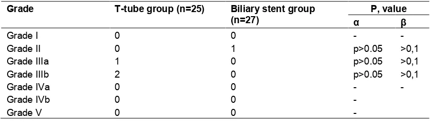
There was no postoperative mortality in either group. Postoperative complications (Table 2)
were observed in 3 (11,4 %) patients in the T-tube group, and only in 1 (3,7 %) patient in the biliary stent group (Grade II by Clavien-Dindo classification) (2=0,36 p>0,05).
Two patients were re-operated for biliary peritonitis: one was due to accidental T-tube dislocation on the fifth postoperative day (Grade IIIa by Clavien-Dindo classification) and another was re-operated after the planned removal of T-tube drain five weeks after initial procedure (Grade IIIb by Clavien-Dindo classification), see Table 3.
Bile leak around the T-tube drain was found in one patient, but it stopped spontaneously. This patient required percutaneous drain insertion for subhepatic bile collection.
Transient acute pancreatitis developed in one patient in biliary stent group, and responded to conservative treatment. No bile leaks were detected in the biliary stent group.
The mean postoperative hospital stay was 6,2 ± 1,7 days in the T-tube group, and 3,2 ± 1,2 days in the biliary stent group (p<0,05).
The total follow-up rate was 96,2% and the follow-up period was 6 to 50 months (average 24 months). There were no bile duct stones or strictures in either group.
Table 1. Characteristic of patients
Patients characteristics T-tube group (n=25)
Biliary stent group (n=27)
p
α β
Age, years Mean ± SD 50,6 ±11,5 48,9±10 p>0,1 >0,1
Range (29-72) (27-69)
Sex, (n %) Male 7 (28,0%) 8 (29,6%) p>0,1 >0,1
female 18 (72,0%) 19 (70,4%)
Jaundice, (n %) 10 (40,0%) 9 (33,3%) p>0,1 >0,1
CBD diameters (cm) Mean ± SD 2,1 ± 0,4 1,9 ± 0,5 p>0,1 >0,1
Range 1,3-2,9 1,0-3,0
No of CBD stones Mean ± SD 2,5 ± 1,3 2 ± 1,0 p>0,05 >0,1
Range 0-5 0-4
Table 2. Patients outcomes
Patients outcomes T-tube group (n=25)
Biliary stent group (n=27)
p
α β
Operative time (minutes) 102 ± 18 114 ± 21 p>0,1 <0,0001
Time to removal of drain (days) 4,0 ± 0,6 2,8 ± 0,8 p>0,05 >0,1 Postoperative hospital stay (days) 6,2 ± 1,7 3,2 ± 1,2 p<0,05 >0,1 Complications by Clavidien-Dindo
classification (n, %)
Table 3. Complications by Clavien-Dindo classification.
Grade T-tube group (n=25) Biliary stent group (n=27)
P, value
α β
Grade I 0 0 - -
Grade II 0 1 p>0.05 >0,1
Grade IIIa 1 0 p>0.05 >0,1
Grade IIIb 2 0 p>0.05 >0,1
Grade IVa 0 0 - -
Grade IVb 0 0 -
Grade V 0 0 -
4. DISCUSSION
T-tube drain has been used routinely for biliary drainage after open or laparoscopic choledochotomy. T-tube placement helps decompress the biliary system, minimize the risk of bile leaks and provide access for follow-up imaging of biliary tree and extraction of retained stones [20,21]. Despite these advantages, specific morbidity related to T-tube usage is reported to occur in up to 6,3% in series of open choledochotomy [22,23,24]. Accidental T-tube displacement leading to CBD obstruction [9,25], bile leakage around T-tube [21], duodenal erosion [26], persistent biliary fistula [25,5], wound cellulitis around T-tube [5], excoriation of the skin, and cholangitis caused by bacteria entering through the T-tube [24] may retard recovery and prolong hospital stay. Indwelling T-tubes are uncomfortable, require continuous management and restrict patient’s activity because of the risk of dislodgement [27]. Patients with an open T-tube are at risk of dehydration and saline depletion [28]. CBD stenosis has been reported as a long-term postoperative complication following T-tube removal [29,25].
LCBDE and cholecystectomy as a single-stage treatment of choledocholithiasis has been shown to be superior when compared to two-stage management [1,30]. The best result are achieved with trans-cystic clearance, however in many cases CBD exploration via choledochotomy is indicated [16,31,17,32]. Drainage of the biliary tree post CBD exploration is common in the laparoscopic era.
Multiple articles report morbidity rates of between 10 and 15 % when LCBDE is combined with T-tube drainage [8,33,34,17]. A recent Cochrane review has discouraged the use of T-tube drains due to significantly longer operative times, prolonged hospital stays and increased
complication rates when compared with primary closure for laparoscopic choledochotomy [18]. Due to this, some experts try to avoid T-tube use for decompression of the CBD after laparoscopic surgery [20].
Primary closure of choledochotomy after CBD exploration decreases operative time, significantly reduces hospital stay, postoperative complications and expenses when compared to T-tube decompression [35,34,18]. Decreased morbidity rates are believed to be due to avoiding complications directly related to the presence and removal of T-tubes [34,17,36]. Unfortunately, primary closure of choledochotomy does not provide biliary decompression which may be critical in patients with retained stones. Recent large series suggest that retained stone rates for single-stage surgical management of choledocholithiasis are between 3,3 and 11 % [33,37,4,18]. Associated morbidity has been documented in 6,1 %, with bile leaks occurring in 5 % of patients post primary closure [33,38, 17,18].
Ante-grade biliary stent insertion prior to choledochotomy closure combines the benefits of T-tube decompression with the reduced morbidity of primary CBD closure. Biliary stent placement is a relatively simple technique that helps decompress the biliary tree [39]. Published results demonstrate that this technique decreases surgical time, morbidity, hospital stay and increases patient comfort [11,16,40, 41,3,42,32,36]. In patients with retained stones biliary stents prevent biliary leakage and biliary peritonitis. Stents facilitate CBD cannulation via ERCP improving the success rate of postoperative ERCP stone extraction from 82 % to almost 100 % [10,27,31].
Stent related complications documented in the
Grubnik et al.; JAMMR, 25(8): 1-9, 2018; Article no.JAMMR.36378
migration and duodenal erosion. Stents in situ for longer than 30 days have been associated with ampullary stenosis and stent
migration leading to intestinal perforation [43,16,44,45,32].
The present study is one of the first randomized cohort studies which compares outcomes and length of stay in patients undergoing ante-grade biliary stenting versus T-tube drainage after LCBDE via choledochotomy. The study was performed in a single centre by a group of surgeons experienced in laparoscopic biliary surgery.
During the study period 125 patients with CBD stones underwent LCBDE. Of these patients only 56 required laparoscopic choledochotomy. The 52 eligible patients were randomly assigned to two groups: 27 patients underwent antegrade biliary stent insertion with primary closure of choledochus; 25 patients underwent LCBDE with T-tube insertion.
Both groups were comparable with respect to age, sex, comorbidities, number and size of CBD stones. There was no significant differences in operative time in the two groups. Postoperative complications were observed in 11,4% of patients in the T-tube group, and only in 3,7% of patients in the biliary stent group (p<0,05).
Complication rate in the T-tube group (11,4%) was in keeping with the current literature on T-tube associated morbidity (10-15%). Complica-tions encountered in this study were consistent with known complications associated with T-tube decompression. It is remarkable that at 5 weeks post LCBDE planned removal of T-tube caused biliary peritonitis. This may be due to the reduced number of adhesions after laparoscopic operations.
There was only one complication in biliary stent group. One patient developed transient acute pancreatitis, which responded to conservative treatment. Importantly, there were no bile leaks in the biliary stent group. This is consist with Lyon et al. [19] who reported no complications in the ante-grade biliary stent drainage group. Potential complications described in the literature, such as erosion of adjacent organs, ampullary stenosis, intestinal perforation were not observed in our study. No complications occurred during endoscopic stent removal. In 9 (33,3 %) patients stents spontaneously
migrated to the duodenum at 2-3 weeks postoperatively. There was statistically significant difference in complication rates by Clavien-Dindo classifica-tion in the two groups, supporting ante-grade biliary stent insertion as the preferred method of biliary tree decompression, however we consider that this trend could be important and better expressed in bigger clinical groups.
Primary closure of the CBD with acute-grade biliary stent insertion decreases hospital stay when compared to T-tube decompression [31,37].
The present study identified a statistically significant difference in the length of hospital stay between the two groups: mean hospital stay in the biliary stent group was 3,2 ± 1,2 days compared to the T-tube group of 6,2 ± 1,7 days (p<0,05). Shorter hospital stay decreases costs and improves patient satisfaction.
5. CONCLUSION
This randomized study demonstrates that there is a statistically significant reduction of hospital stay and post-surgery complications in patients treated with antegrade biliary stent decompression of CBD post LCBDE via choledochotomy compared to patients treated with T-tube drainage.
This study shows that ante-grade biliary stent insertion during LCBDE is one of the options for primary CBD closure, however, this problem requires more studies
CONSENT
As per international standard or university standard, patient’s written consent has been collected and preserved by the authors.
COMPETING INTERESTS
Authors have declared that no competing interests exist.
REFERENCES
randomized trial comparing two-stage vs single-stage management. Surg Endosc. 1996;10:1130–1135.
2. Franklin ME Jr, Pharand D, Rosenthal D. Laparoscopic common bile duct explora-tion. Surg Laparosc Endosc. 1994;4:119– 124.
3. Mangla V, Chander J, Vindal A, Lal P, Ramteke VK. A randomized trial comparing the use of endobiliary stent and T-tube for biliary decompression after laparoscopic common bile duct explora-tion. Surg Laparosc. 2012;22:345–348. 4. Martin IJ, Bailey IS, Rhodes M, O’Rourke
N, Nathanson L, Fielding G. Towards T-tube free laparoscopic bile duct explora-tion: A methodologic evolution during 300 consecutive procedures. Ann Surg. 1998; 228:29–34.
5. Rhodes M, Nathanson L, O’Rourke N, Fielding G. Laparoscopic exploration of the common bile duct: lessons learned from 129 consecutive cases. Br J Surg. 1995; 82:666–668.
6. Rhodes M, Sussman L, Cohen L, Lewis MP. Randomised trial of laparoscopic exploration of common bile duct versus postoperative endoscopic retrograde cholangiography for common bile duct stones. Lancet. 1998;351:159–161. 7. Tokumura H, Umezawa A, Cao H,
Sakamoto N, Imaoka Y, Ouchi A, Yamamoto K. Laparoscopic management of common bile duct stones: transcystic approach and choledochotomy. J Hepa-tobiliary Pancreat Surg. 2002;9:206–212. 8. Cuschieri A, Lezoche E, Morino M, Croce
E, Lacy A, Toouli J, Faggioni A, Ribeiro VM, Jakimowicz J, Visa J, Hanna GB. EAES multicenter prospective randomized trial comparing two-stage vs single-stage management of patients with gallstone disease and ductal calculi. Surg Endosc. 1999;13:952–957.
9. Bernstein DE, Goldberg RI, Unger SW. Common bile duct obstruction following T-tube placement at laparoscopic cholecystectomy. Gastrointest Endosc. 1994;40:362–365.
10. Chen CC, Wu SD, Tian Y, Zeng XT, Siwo EA, Xian GZ. The fading role of T-tube in laparoscopic choledochotomy: primary choledochorrhaphy and over pigtail j and endonasobiliary drainage tubes. J
Laparoendosc Adv Surg Tech. 2010;20: 807–811.
11. DePaula AL, Hashiba K, Bafutto M, Machado C, Ferrari A, Machado MM. Results of the routine use of a modified endoprosthesis to drain the common bile duct after laparoscopic choledochotomy. Surg Endosc. 1998;12:933–935.
12. Ha JP, Tang CN, Siu WT, Chau CH, Li MK. Primary closure versus T-tube drainage after laparoscopic choledocho-tomy for common bile duct stones. Hepa-togastroenterology. 2004;51:1605–1608. 13. Decker G, Borie F, Millat B, Berthou JC,
Deleuze A, Drouard F, Guillon F, Rodier JG, Fingerhut A. One hundred laparoscopic choledochotomies with primary closure of the common bile duct. Surg Endosc. 2003;17:12–18.
14. Holdsworth RJ, Sadek SA, Ambikar S, Cuschieri A, Becker JM. Dynamics of bile flow through the human choledochal sphincter following exploration of the common bile duct. World J Surg. 1989; 13:300–306.
15. Huang J, Zhu J. Spontaneously removed endobiliary J stent drainage after laparoscopic common bile duct explora-tion. Surg Endosc. 2009;23:1398–1402. 16. Gurusamy KS, Koti R, Davidson BR.
T-tube drainage versus primary closure after open common bile duct exploration. The Cochrane Database of Systematic Reviews. 2013;6:CD005640.
17. Tranter SE, Thompson MH. Comparison of endoscopic sphincterotomy and laparos-copic exploration of the common bile duct. Br J Surg. 2002;89:1495–1504.
18. Wu JS, Soper NJ. Comparison of laparoscopic choledochotomy closure techniques. Surg Endosc. 2002;16:1309– 1313.
19. Lyon M, Menon S, Lain A, Kumar H. Use of biliary stent in laparoscopic common bile exploration. Surg Endosc. 2015;29:1094-1098.
20. DeRoover D, Vanderveken M, Gerard Y. Choledochotomy: Primary closure vs T-tube. A prospective trial. Acta Chir Belg 1989;89:320–324.
Grubnik et al.; JAMMR, 25(8): 1-9, 2018; Article no.JAMMR.36378
duct exploration. J Laparoendosc Adv Surg Tech A. 2001;11:391–400.
22. Moreaux J. Traditional surgical management of common bile duct stones: A prospective study during a 20-year experience. Am J Surg. 1995;169:220– 226.
23. Pappas TN, Slimane Brooks DC. Brooks DC. 100 consecutive common duct explorations without mortality. Ann Surg. 1990;211: 260–262.
24. Sheridan WG, Williams HO, Lewis MH. Morbidity and mortality of common bile duct exploration. Br J Surg. 1987;74:1095– 1099.
25. Qi Wei, Hong-Jie Hhu, Xiao-Yan Cai, Wei Q, Hu HJ, Cai XY, Li LB, Wang GY. Biliary drainage after laparoscopic choledocho-tomy. World J Gastroenterol. 2004;10: 3175–3178.
26. Mosimann F, Schneider R, Mir A, Gillet M. Erosion of the duodenum by a biliary T-tube: an unusual complication of liver transplantation. Transplant Proc 1994;26: 3550–3551.
27. Gersin KS, Fanelli RD. Laparoscopic endobiliary stenting as an adjunct to common bile duct exploration. Surg Endosc. 1998;12:301–304.
28. Wills VL, Gibson K, Karihaloot C, Jorgensen JO. Complications of biliary T-tubes after choledochotomy. ANZ J Surg. 2002;72:177–180.
29. Perez G, Escalona A, Jarufe N, Ibanez L, Viviani P, Garcia C, Salvado J. Prospective randomized study of T-tube versus biliary stent for common bile duct decompression after open choledocotomy. World J Surg. 2005;29:869–872.
30. Millat B, Atger J, Deleuze A, Briandet H, Fingerhut A, Guillon F, Marrel E, De Seguin C, Soulier P. Laparoscopic treatment for choledocholithiasis: A prospective evaluation in 247 consecutive unselected patients. Hepatogastroentero-logy. 1997;44:28–34.
31. Isla AM, Griniatsos J, Karvounis E, Arbuckle JD. Advantages of laparoscopic stented choledochorrhaphy over T-tube placement. Br J Surg. 2004;91:862–866. 32. Yin Z, Xu K, Sun J, Zhang J, Xiao Z, Wang
J, Li Y. Is the end of the T-tube drainage era in laparoscopic choledochotomy for common bile duct stones is coming? A
systematic review and meta-analysis. Ann Surg. 2013;257:54–66.
33. Griniatsos J, Karvounis E, Arbuckle J, Isla AM. Cost-effective method for laparos-copic choledochotomy. Aust N Z J Surg. 2005;75:35–38.
34. Kelly MD. Results of laparoscopic bile duct exploration via choledochotomy. Aust N Z J Surg. 2010;80:694–698.
35. Jameel M, Darmas B, Baker AL. Trend towards primary closure following laparos-copic exploration of the common bile duct. Ann R Coll Surg Engl. 2008;90:29–35. 36. Zhang WJ, Xu GF, WuG Z, Li JM, Dong
ZT, Mo XD. Laparoscopic exploration of common bile duct with primary closure versus T-tube drainage: a randomized clinical trial. J Surg Res. 2009;157:1–5. 37. Khaled YS, Malde DJ, De Souza C, Kalia
A, Ammori BJ. Laparoscopic bile duct exploration via choledochotomy followed by primary duct closure is feasible and safe for the treatment of choledo-cholithiasis. Surg Endosc. 2013;27:4164– 4170.
38. Kacker LK, Mittal BR, Sikora SS, Ali W, Kapoor VK, Saxena R, Das BK, Kaushik SP. Bile leak after T-tube removal: A scintigraphic study. Hepatogastroentero-logy. 1995;42:975–978.
39. Croce E, Golia M, Azzola M, Russo R, Crozzoli L, Olm S. Laparoscopic choledochotomy with primary closure. Follow-up (5–44 months) of 31 patients. Surg Endosc. 1996;10:1064–1068.
40. Kuroki T, Tajima Y, Tsuneoka N, Kitasato A, Adachi T, Kosaka T, Kanematsu T. Placement of a plastic biliary stent tube with primary closure of the common bile duct after laparoscopic common bile duct exploration. Hepatogastroenterology 2010; 57:1034–1036.
41. Lee LS, Loon LC. Laparoscopic common bile exploration using V-loc suture with insertion of endobiliary stent. Surg Endosc. 2016;30:2530-2534.
42. Tang CN, Tai CK, Ha JPY, Tsui KK, Wong DCT, Li MKW. Antegrade biliary stenting versus T-tube drainage after laparoscopic choledochotomy—A comparative cohort study. Hepatogastroenterology. 2006;53: 330–334.
significantly improves success of post operatve endoscopic retrograde choladn-giopancreatography in low volume centres. Surg Endosc. 2002;16:487–491.
44. Johanson JF, Schmalz MJ, Greenen JE. Incidence and risk factors for biliary and
pancreatic stent migration. Gastrointest Endosc. 1992;38:341–346.
45. Lenzo NP, Garas G. Biliary stent migration with colonic diverticular perforation. Gastrointest Endosc. 1998;47:543–544.
_________________________________________________________________________________ © 2018 Grubnik et al.; This is an Open Access article distributed under the terms of the Creative Commons Attribution License
(http://creativecommons.org/licenses/by/4.0), which permits unrestricted use, distribution, and reproduction in any medium,
provided the original work is properly cited.
Peer-review history: