Copyright 0 1991 by the Genetics Society of America
Mapping Point Mutations in the Drosophila
rosy
Locus Using Denaturing
Gradient Gel Blots
Mark Gray,*.+ April Charpentier: Kathleen Walsh,' Pearl Wut and Welcome Bender*
*Department of Biological Chemistry and Molecular Pharmacology, Harvard Medical School, Boston, Massachusetts 021 15, and ?Division of Reproductive Endocrinology, Department of Obstetrics and Gynecology, Tufts University School of Medicine,
Boston, Massachusetts 021 11
Manuscript received September 20, 1990 Accepted for publication September 24, 1990
ABSTRACT
Mutations within the rosy locus of Drosophila were mapped using blots of genomic DNA fragments separated on denaturing gradient gels. DNA sequence differences between otherwise identical small
rosy D N A fragments were detected among the mutants as mobility shifts on the blots. Mutations were
mapped to within a few hundred base pairs of rosy sequence in 100 of 130 mutants tested-a 77%
detection rate. The sequence changes in 43 rosy mutations are presented; all but six of these were
single base changes. Thirty-four of 36 sequenced mutations induced by the alkylating agents N-ethyl-
N-nitrosourea and ethyl methanesulfonate were transitions. All of the mutations mapped in the rosy transcription unit. Twenty-three of the 43 sequenced mutations change the predicted rosy gene polypeptide sequence; the remainder would interrupt protein translation (17), or disrupt mRNA processing (3).
M
ANY of the induced mutations used in experi- mental genetics are point mutations. For the purposes of this study, we define a point mutation as any DNA lesion not detectable by conventional blot- ting of DNA fragments electrophoresed in agarose gels. Typically, deletions or insertions of less than 50 bp are not detected by this method. Ethyl methane- sulfonate (EMS) and N-ethyl-N-nitrosourea (ENU), the most commonly used chemical mutagens for Dro- sophila, appear to generate such point mutations (COTE et al. 1986). Also, point mutations are especially useful for studying mechanisms of gene expression, because they d o not produce complex phenotypes. In contrast, chromosomal rearrangement mutations in- terrupt genes or move them to locations that change gene expression by position effect. Transposon inser- tion mutations are complicated even further by tran- scription originating within the transposon. In spiteof their usefulness, only a small number of point mutations have been sequenced, because they are difficult to locate on a molecular map. Most of the molecular descriptions of point mutations have re- quired the sequencing of cloned DNA from mutant and wild-type organisms. This approach can be im- practical for characterizing many mutations in large genes, because of the effort required.
We have been studying mutations in the rosy locus of the fruit fly Drosophila rnelanogaster (CHOVNICK, GELBART and MCCARRON 1977). rosy mutant flies are deficient in the activity of xanthine dehydrogenase (XDH), an enzyme of the purine degradation path- way. XDH also affects, in an unknown way, the syn-
<;cnetics 127: 139-149 (January, 1991)
thesis of the eye pigments (REAUME, CLARK and CHOV- NICK 1988). Adult rosy flies have a brown-red eye color instead of the wild-type bright red. A genetic fine structure map of the rosy locus has been con- structed using a variety of mutant alleles and poly- morphic markers (CLARK, HILLIKER and CHOVNICK
1986).
Most of the rosy mutations that have been studied were induced by chemical mutagens. In our earlier study of 83 rosy mutants using Southern blots, 70 were indistinguishable from wild type in having no changes in the lengths of the expected rosy restriction fragments. These 70 mutations were probably point mutations (COTE et al. 1986). Only one of the 36 rosy mutations induced by the alkylating agent EMS had any mutant DNA lesion detectable on Southern blots. Initially, we were interested in finding mutations that affect the regulation of rosy locus transcription. In our previous experiments, 61 ENU-induced rosy mutants that fit several criteria for having regulatory mutations were examined (LEE et al. 1987). T h e mu- tations were genetically mapped against markers within the rosy locus; DNA from six strains with mu- tations mapping near the 5' end of the XDH gene was cloned and sequenced. All six mutations were found within the transcribed region of the gene, and either altered XDH polypeptide synthesis or mRNA processing.
I40 M. Gray et al.
than that used in our earlier studies. Denaturing gra- dient gel electrophoresis can be used to find mutations by detecting small differences in the melting behavior of short (200-700 bp) DNA fragments (FISCHER and LERMAN 1983). DNA fragments subjected to an in- creasingly denaturing physical environment partially melt. Rather than melting in a continuous zipper-like manner, most fragments melt step-wise, with discrete domains of the fragment becoming single-stranded in a very narrow range of denaturing conditions. When
DNA fragments are electrophoresed in an acrylamide gel that contains an increasing gradient of denaturing solvents, their mobilities sharply decrease when they partially melt, because their shapes become more com- plex. The denaturants used are heat (a constant tem- perature of 60") and a linear gradient of formamide (ranging from 0 to 40%) and urea (ranging from 0 to
7
M). The denaturant concentration, and position in the gel, at which a fragment partially melts (and thus nearly stops migrating) is dependent on the DNA sequence of the melted domain. This characteristic gradient concentration, and gel position, is often dif- ferent even when only a single base in the sequence of the melted region is changed (FISCHER and LERMAN1983). When a mixture of many different fragments is electrophoresed, each will stop at a sequence-de- pendent position in the denaturing gradient. Frag- ments containing an A
+
T-rich domain typically stop at relatively low denaturant concentrations (high on the gel); conversely, the more stable uniformly G+
C-rich fragments usually run low in the gel.
In the experiments described below, rosy mutations were physically mapped to within several hundred bp of rosy sequence, by analyzing genomic DNA blots prepared from denaturing gradient gels. For some alleles, we cloned and sequenced only the small mu- tant portions of the gene, rather than the entire -7.0- kb gene. The mutations sequenced include many left unresolved in our previous study, as well as mutations used in early genetic mapping experiments.
MATERIALS AND METHODS
Genetic strains: Virtually all rosy mutant strains tested in these experiments were isolated in mutagenesis experiments in the laboratory of ARTHUR CHOVNICK, with one exception, rf ", a gift of E. B. LEWIS. T h e mutant alleles are numbered
so that the first digit indicates the wild-type rosy locus background used to induce the mutation. For example, ry""
was induced on the ry+2 background, and rytio6, on the ry+"
background (CHOVNICK, GELBART and MCCARRON 1977). In all of these experiments, mutants were compared to their parental wild-type counterparts. Most of the mutations (1 02
o f 135) were induced by ENU mutagenesis of the ry+" wild- type strain; the remainder were induced by other mutagens, such as EMS, nitrogen mustard (HN2), triethylene mela- mine (TEM), diepoxybutane (DEB), and X-irradiation, as indicated below (MCCARRON and CHOVNICK 1981; LEE et al. 1987; LINDSLEY and ZIMM 1990).
Preparation of genomic DNA samples: Drosophila ge-
nomic DNA was prepared from adult flies using a rapid procedure (BENDER, SPIERER and HOGNESS 1983) and ex- tracted once with phenol/chloroform (1 : 1) before precipi- tation in ethanol. Each electrophoresis sample contained 3
/.tg of genomic DNA digested in a total volume of 10 PI. Denaturing gradient gel electrophoresis: Denaturing gradient gels were prepared as previously described, with only minor modifications (FISCHER and LERMAN 1983). The apparatus used for the preparation and electrophoresis of
the denaturing gradient gels was designed by LEONARD LERMAN, and built from plans supplied by RICHARD MYERS
(FISCHER and LERMAN 1979). All of the denaturing gradient gels were 6.5% acrylamide (37.5: 1 acrylamide: bisacrylam- ide) in TAE buffer (40 mM tris-acetate, 20 mM sodium acetate, 1 mM EDTA, pH 7.4). All gels were 0.6 mm thick and 13 cm long, with 20 wells, each 4 mm wide and 15 mm deep. Most of the gels had a linear gradient of denaturant
of at least 50% (where 100% denaturant concentration is 7
M urea
+
40% formamide; see FISCHER and LERMAN 1983). DNA samples were electrophoresed at 65-85 V, for 16-1 8 hr, at 60". Afterward, the gels were stained in ethidium bromide ( 1 Fg/ml) for 5 min and examined over a long- wave ultraviolet light source.Preparation of DNA blots: T h e destained denaturing gradient gels were soaked in 0.5 M NaOH for 5 min, 0.5 M Tris, pH 8.0 for 5 min, and then in transfer buffer (20 mM Tris, 1 mM EDTA, pH 8.0) for at least 5 min. Next, each gel was placed in a stack with a nylon hybridization mem- brane (Nytran; Schleicher and Schuell) and the stack in- serted into an electrophoretic transfer apparatus. DNA frag- ments were transferred by electrophoresis at 600 mA for 2 h. After electrotransfer, the DNA blots were rinsed briefly in 6 X SSPE (0.15 M NaCI, 10 mM NaHrPO.,. H20, 1 mM EDTA, pH 7.4), and baked under vacuum at 80" for 1-2 h.
Preparation of probes: Most of the blots were hybridized with single-stranded DNA probes. Single-strand recombi- nant pEMBL plasmids containing inserts up to 8.2 kb were prepared by standard methods (DENTE, CESARENI and CORTESE 1983). These DNAs were labeled by primer exten- sion so that new strand synthesis occurs only in the vector portion of the molecule, leaving the insert region single- stranded (Hu and MESSING 1982). Some DNA probes were labeled with "P by nick-translation of purified DNA frag- ments and recombinant plasmids (RIGBY et al. 1977). Other probes were "'P-labeled DNA fragments prepared by primer extension after hybridization with random primers (FEIN-
BERG and VOGELSTEIN 1983).
Hybridization of blots: T h e blots were soaked in 6 X
SSPE for 5 min, and then prehybridized for at least 1 hr at 42" in a mixture of 50% formamide, 5 X SSPE, 1% SDS, 5 x Denhardt's solution (MANIATIS, FRITSCH and SAMBROOK 1982) and 100 pg/ml denatured sonicated salmon sperm DNA. Blots were hybridized at 42" for 16-20 hr in a mixture containing 10' cpm/ml S'P-labeled DNA probe, 50% formamide, 5 X SSPE, 10% dextran sulfate (500,000
Mr), 1 % SDS, 1 X Denhardt's solution, and 100 gg/ml
denatured sonicated salmon sperm DNA. Blots were washed as follows: (a) 2 X SSPE
+
1 % SDS at 22" for 30 min, (b) 1X SSPE
+
1 % SDS at 68" for 30 min, and (c) 0.1 X SSPE+
1% SDS at 68" for 30 min. Blots were dried briefly and exposed to X-ray film at -70" in the presence of an inten- sifying screen, for 2-20 hr.Mapping rosy Mutations 141
library was reduced approximately 10-fold. For rosy genes with mutations left of the PstI site at +l.I-kb (see Figure
2), the 4.0-kb PstI fragment (at -2.9 to +1.1) was cloned from genomic DNA. DNA was first digested with SalI,
KpnI, ApaI, and HindIII, before digestion with PstI. For
genes with mutations to the right of the +1.1 PstI site, the 3.0-kb PstIIHindIII fragment (at +1.1 to +4.1) was cloned (Figure 2). Genomic DNA (20 Pg) was first digested with XbaI, X h o I , SalI, KpnI, and ApaI, and then Hind111 and PstI, before fractionation by size. Usually, only -2000 in- sert-containing colonies were screened to identify at least one with the desired rosy genomic fragment. The fragments were ligated with pEMBL8+ plasmid DNA (DENTE, CES-
ARENI and CORTFSE 1983), and the mixture used to trans-
form J M l O l or K802 host bacteria (HANAHAN 1983). Trans- formant colonies were screened for rosy DNA inserts by colony hybridization (GRUNSTEIN and HOGNESS 1975). Plas- mid DNAs from positive colonies were prepared from l-ml cultures (ISH-HOROWICZ and BURKE 1981). The clone's identity was rechecked by electrophoresis of the appropriate restriction fragments on a denaturing gradient gel.
Some of the rosy mutant DNAs were isolated in recom- binant plasmid clones after DNA amplification by the polymerase chain reaction (PCR) strategy (SAIKI et al. 1988). A top strand oligonucleotide primer (5'-GA- ATTCCAGCCCTTGGATCC-3'; starting at nucleotide -1) was used together with a bottom strand primer (5'-
CATTCCGTTCAGATCGGATCC-3'; at +2969), to am- plify the intervening 2.9 kb of rosy DNA from ry' genomic DNA. A different top strand oligonucleotide primer (5'-
CGAGCTCAAGTCCTATTTCC-3'; at +1529) and a 3' end bottom strand primer (5"CTTCGAAACATACCTT- GAGT-3'; at +4178) were used to amplify the downstream 2.6 kb portion of the rosy gene from the mutant strains ry'~"', ry", and ry4'. PCR amplifications were done in a volume of 25 rl with 100 ng genomic DNA, 0.1 PM each primer, 200 PM deoxynucleotides, and 1 unit of Taq DNA polymerase. Denaturation for each of 30 cycles was at 94" for 1 min, annealing at 54" (62" for ry') for 1 min, and then synthesis at 72" for 1.5-2 min, using a Tempcycler model 50 (Coy Corp., Ann Arbor, Michigan). Amplified fragments were then cloned in the appropriate pEMBL plasmid vectors.
DNA sequencing For most of the cloned mutant genes, small pEMBL subclones that placed the mutant DNA within 300 bp of the sequencing primer site were made. Single- strand DNA templates were prepared from the appropriate subclones and used in dideoxynucleotide chain termination sequencing reactions (SANGER, NICKLEN and COULSON
1977). For other cloned mutant genes, templates made from the entire pEMBL genomic clone were hybridized with complementary rosy oligonucleotide primers before the se- quencing reactions.
Nondenaturing acryiamide gel electrophoresis: Ge- nomic D N A samples prepared as described above were electrophoresed for 4-5 hr at 100 V in 4.5% acrylamide/
Blots were prepared and hybridized as described above.
T B E gels (MANIATIS, JEFFREY and V A N DE SANDE 1975).
RESULTS
Mapping rosy mutants using denaturing gradient gel blots
Selection of rosy mutants for study: Most of t h e
135 mutants selected were expected to be point mu- tants, because most were induced by mutagenic agents
thought to give predominantly single base change substitutions, such as ENU a n d EMS. We were partic- ularly interested in identifying mutations at sites in the gene that are signals for normal regulation of
mRNA transcription and processing. Some mutants fit several biochemical and genetic criteria that sug-
gested their mutations might affect transcription
(MCCARRON a n d CHOVNICK 1981; LEE et al. 1987). Other mutants, such as those from earlier X-irradia- tion and EMS mutagenesis experiments, were in- cluded because of their frequent use as markers in recombination experiments. Because of o u r selection bias, the mutants included in this study may not rep- resent a typical g r o u p (see DISCUSSION). Most (1 16/
135) of the mutations were induced on the ry+5 wild- type background (CHOVNICK, GELBART a n d MC- CARRON 1977). Others were induced on the ry+', ry+',
ry+', and other well-characterized rosy wild-type strains (CHOVNICK, GELBART a n d MCCARRON 1977). I n all cases, DNA from the mutant was compared to
DNA from the appropriate parent strain.
Denaturing gradient gel blot strategy: Genomic
DNA was prepared from rosy mutant adult flies, and analyzed by denaturing gradient gel electrophoresis, as follows. Genomic DNA samples (3 P g ) were di- gested with a t least three of t h e five restriction en- zymes with 4 bp recognition sequences: AluI, HaeIII, H h a I , MspI, or RsaI. These enzymes cut Drosophila DNA frequently, producing many 200-700 b p DNA
fragments. The digested DNA samples were electro- phoresed in denaturing gradient gels with a wide range of denaturant concentration, such as 20-90%
or 2 5 4 5 % (see MATERIALS AND METHODS). After
electrophoresis, most rosy DNA fragments reached a position in the gradient where their first melting domains denatured. The electrophoresed fragments were then transferred from the gel t o nylon hybridi- zation membranes, by electroblotting. The DNA blots were hybridized with radioactive rosy DNA probes and the rosy fragments visualized by autoradiography (Figure 1).
The order, from top to bottom, of t h e DNA frag- ments in denaturing gradient gels is not determined by molecular weight. Instead, the relative stabilities
of the first melting domains in each fragment establish the fragment order in the gels. The molecular map positions of the rosy fragments detected on the dena- turing gradient gel blots were identified by two meth- ods. In the first, the blots were hybridized with probes prepared from short, cloned rosy fragments. In the second approach, cloned rosy fragments were electro- phoresed in perpendicular denaturing gradient gels
142 M . Gray et al.
"
" o n n - r
n n n n n n n
(r
c
1 1 6
1 4 1
2 8 1
Mapping of mutant fragments: Among the 135 mutant DNAs tested, 100 had at least one rosy restric- tion fragment with mutant (i.e., different from its wild-type parent) melting behavior; their map posi- tions are shown in Figure 2. In many of the mutant strains, a nlutant band was found in more than one of the four or five different restriction digests tested. In these mutants, all of the altered restriction fragments overlapped each other, suggesting that all of the melt- ing behavior alterations found in one mutant gene were caused by the same base change. Five mutants had mobility shifts i n fragments from all regions within and outside of the rosy gene, suggesting that they were induced on an unidentified chromosomal background; these mutants were not investigated fur- ther. For 30 of the mutants, fragment mobility shifts were not found among t h e 4-5 restriction enzymes tested. I n total, 602 digests of genomic D N A from the 135 mutants were analyzed in these denaturing gradient gel blot experiments.
In order to assess the molecular weights of rosy fragments, all mutant genomic DNAs with at least one
shifted fragment were analyzed using conventional blots prepared from nondenaturing acrylamide/TBE gels. Nylon blots were made from these gels and hybridized with radioactive rosy probes. Only four mutants had changes in the sizes of rosy fragments. Three of these, ry."'", ry5."!', and ry"?', had small dele- tions. T h e fourth, ryJ"'7, had a chromosomal re- arrangement with a breakpoint in the first intron of the XDH transcription unit.; this mutant was not in- vestigated further. No alterations in the molecular weight of any rosy restriction fragments were found in the remaining mutants tested; these were expected to have point mutations. Excluding the five mutants on an unknown background, and the four re-
arrangement/deletion mutants, t h e rate of detection of point mutations, using denaturing gradient gel blots, was 76% (96/126).
Melting behavior changes in rosy mutants: Dena- turing gradient gels w i t h broad ranges of denaturant concentration were chosen i n order to simultaneously compare fragments with very different stabilities. AS
a result, the mobility shifts of mutant fragments were often small-only 1 m m or less (see Table 1). T h e average magnitude of all of the mobility shifts was 2 mm. T h e shifts were always much greater (up to 10 m m ) when mutant D N A fragments were electropho- resed in gels with the appropriate narrow range (30%
or less) of denaturant concentration. Most of the DNA fragment shifts were changes in the position of a sharp
these, about one-third of the melting differences were changes in the position or the shape of diffuse, smeared bands (ry"", ry51i4, ry,','.'; Figure 1). Fragments with a high G+C content often produced diffuse bands, usually in the most denaturing region of the gradient. Most of the GC-rich rosy fragments never completely stopped during the course of electropho- resis. We suspect that the diffuse band phenotype was caused by the simultaneous melting of two or more melting domains in one DNA fragment. Each of the five restriction digests tested revealed mutant melting differences; mutant shifts were found in 26-36% of the D N A samples in each set of digests. Mutant frag- ments were found in two or more different restriction digests in 53 of the 100 mutant DNAs with an altered fragment.
Mutant fragments found by denaturing gradient gel blots were mapped throughout the rosy transcrip- tion unit; most were entirely within the large protein- coding second and third exons (Figure 2). In ry"'"",
part of the small first exon. In r~?"", all of the over- lapping mutant D N A fragments map downstream from the XDH open reading frame. No mutant frag- ments mapped completely outside of transcribed DNA, or entirely within introns. However, examples band ( e . g . , ry5.i.'3, r y 5 Y / 4 , r y 5 / / i ; Figure 1). In contrast to
,-
. .
Mapping rosy Mutations 143
-3.0 -2.0 -1.0 O L b 1 .o 2.0 3 .O 4.0
I I I I I I I 1
PSI1 Ezl I1 E c o K E g t U W I
I I I I I I 1 I I I
HI Sac1
Sal1 HiniIII EgIII EgJU SacIPnl Sac1 E m H I B m H l HindIII EmRI
5331 SI45
-
5101
-+
531
-
5163 "c5177
-
Sf17 5318
-
S135 5161
+
2902
-
5107
+
1314
-
sli"
534
-
546
-
5135 t-
5141
-+-
5141 -+"5 9 s
-
5114
5131 +
5151 +. 606 +
; z
5147 5164 "--c-
574550
-
-575591
-
5116
-
-
s6a-
5 1 s )S l l l
-
-
SI44-
51315 3 9
+*
554 4 564 _t511s __3.
511s _t_ 551
-
41 I 605
-
-
5154s 9 1
-
5114
-
-
S I 0 8-
5 1 0 9-
5158547
-
-
5315556
-
517s
-
S311
5191
+
J ? l-
.
416 0 1
-
qy
513s
-
.
1 1 35 1 9
-
-
5315 1 1
-
+
573S I 8
-
-
110a5111 d
53s
-
S I I J
+
5311-
561 "-+ 611
-
7
E
:
'
5187
5144 -+ 51.5 + A 6 0 9
SI60
-
5143- -
6115119
-
104 b
5 0 9 (>- 533
-
553 "c
1 0 1
+
544 + __c 5105
I 6 "-t
-
5111"t L.19
S l I t
--
1 0 35314 -t 5256
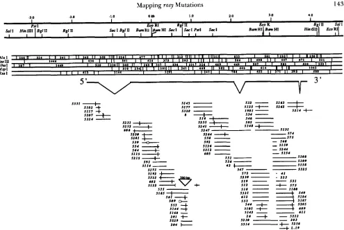
FIGURE 2.-Molecular map positions of rosy mutations found by denaturing gradient gel electrophoresis. At the top is a restriction map of the rosy locus. T h e bases are numbered as in previous studies ( C u R r r s et al. 1987; KEITH et al. 1987). T h e structure of the X D H mRNA is diagrammed below the restriction maps. T h e molecular positions of restriction fragments with altered melting behavior in rosy mutants is aligned with the restriction map, at the bottom. When a melting behavior shift was found in two or more restriction digests of the same mutant D N A , the position of the mutation was narrowed down to the region of overlap of the mutant fragments. The exact positions of sequenced mutations are indicated by the vertical slash marks. Four mutants had small deletions; these are indicated on the diagram at their respective map positions by lines with gaps indicating the lengths of the deletions (estimated by nondenaturing gel electrophoresis for $"!').
'l'he position of the rearrangement mutation ry""" is marked by the croaked line.
of mutations in these regions have been found and are described elsewhere (DUTTON and CHOVNICK
1988; CURTIS et al. 1989).
Some of the mutants analyzed with denaturing gra- dient gel blots were among the earliest mutations mapped genetically within the rosy gene (CHOVNICK
1966; CHOVNICK, GELBART and MCCARRON 1977). T h e genetic order of these mutations (ry606, ry602,
rym?04, T y m l , ry', ry4', ryZ6, ry4' and $ I 9 ) is consistent
with the physical map order of the mutant fragments detected in genomic DNA.
DNA sequence changes in selected rosy mutations Isolation of rosy mutant DNA: Genomic DNA from about one-half of the mapped rosy mutant genes was cloned and sequenced in the region of the restric- tion fragments with altered melting behavior (Figure 2). Since the molecular map positions of the mutations were localized to a few hundred base pairs, only the mutant portions of the genes were sequenced. In most of the experiments, rosy mutant DNA fragments were isolated in plasmid clones using the strategy of frag-
ment enrichment (NICHOLLS et al. 1985). Some mu- tant DNA fragments were cloned in plasmids after PCR DNA amplification from genomic DNA (SAIKI
et al. 1988).
T o verify that each plasmid clone contained the correct mutant DNA, the clones were digested with the appropriate restriction enzyme(s), and electropho- resed in denaturing gradient gels similar to those used to detect the mutant fragment in genomic DNA. T h e melting behavior shifts of cloned fragments were iden- tical to their genomic counterparts, with only one group of exceptions: all of the restriction fragments overlapping the +2.8-kb map position (Figure 2). Each of these fragments, when cloned in bacteria, stopped considerably higher (1 -3 cm) in the denaturing gra- dient than the equivalent Drosophila genomic frag-
ment. An EcoK recognition site (GCACGGAG-
144 M. Gray et al.
Eco K endonuclease and methylase activity ( KESSLER,
NEUMAIER and WOLF 1985). Genomic cloning in the
host strain K802 was successful; this strain lacks the EcoK endonuclease, but retains methylase activity. Methylation (by host bacteria) most probably caused the large differences in the melting behavior of cloned fragments that include the EcoK site. This is consistent with reports of DNA fragment destabilization associ- ated with adenine methylation (COLLINS and MYERS
1987).
Types of base changes and effect of mutagens:
Forty-three different rosy mutations were sequenced (Table 1). All were within small restriction fragments with mutant melting behavior (Table 1 ; Figure 2). Most of the sequenced mutations ( 3 7 of 43) were single base changes; the remaining six changed more than one base pair. Five of these six, as well as a rearrangement and another small deletion (not se- quenced), were induced by X-irradiation, HN2, and
DEB. The ENU-induced mutant gene in ry'"' was missing -700 bp of rosy sequence and had -300 bp of D N A sequence from an unknown genomic source at the site of the deletion.
Most of the sequenced mutations were induced by ENU. All of the ENU-induced mutations, except ry5"', were single base substitutions (Table 2). All four of the EMS-induced mutations were single base substitutions (Table 1). Twenty-seven had G to A , or C to T transitions; in seven, T was changed to C. In
ry".')', an A was replaced by a G. The only transversion mutation found was the C to A change in ry'21'.
Effect of mutations on rosy locus expression: All of the sequenced rosy mutations were in the XDH transcription unit. Approximately one-half (22 of 43) change the predicted amino acid sequence of the XDH polypeptide. Over half (1 2 of 22) of these mis- sense mutations cause either the removal or insertion of a charged amino acid (Table 1). A serine residue would be removed in four mutants; 5 of the 22 missense mutants had substitutions involving a proline residue. Half of the missense mutants have a rosy null phenotype, and half have a partial-activity phenotype (Table 1). T h e remaining mutations apparently inter- rupt normal translation of the XDH mRNA, either by introducing in-frame nonsense codons or frame- shifts, or interfering with post-transcriptional process- ing of the XDH mRNA (Table I ) .
Ten mutants had single base changes resulting in a new translation termination codon. Seven nonsense mutations were amber codons (TAG), and three were opal codons (TGA). No ochre (TAA) nonsense mu- tations were found, probably because of the Drosoph- ila codon bias (O'CONNELL and ROSBASH 1984) and the limited spectrum of ENU-induced base changes (Table 2). All but one of the nonsense mutants had a rosy null phenotype. The exception, ry"'"', has less
than 5% of wild-type XDH activity (MARGARET
MCCARRON, personal communication). This mutation
creates an amber codon 23 codons upstream from the 3' end of the XDH open reading frame, leaving intact most (1 3 12 codons) of the coding sequence.
All but one of the seven sequenced deletion/inser- tion mutations placed frameshifts in the XDH open reading frame (Table 1). Frameshifts cause premature translation termination because of nonsense codons in the other two reading frames. All seven deletion/ insertion mutants have a rosy null (eye color) pheno- type. T h e ry42 mutation was exceptional because it complements with certain other y null alleles, such as ry"' (GELBART, MCCARRON and CHOVNICK, 1976). This mutation removed seven amino acid residues, including a negatively-charged aspartic acid residue, and put in three new residues, including a negatively- charged glutamic acid residue, at the site of the dele- tion. The net loss of 9 bp keeps the rosy reading frame intact, without creating any in-frame termination co- don.
Two single base change mutations, ry.51"2 and ryS1", were substitutions of the first two bases in the first intron (Table 1). These mutations change the two most conserved bases in the 5' splice donor consensus
sequence G
I
GT
- AGT (MOUNT 1982). Normalsplicing of the first and second exons of the XDH mRNA probably does not occur in these two mutants; both have a rosy null phenotype.
T h e mutation ry"'.'' places a new ATG methionine codon upstream from the normal XDH start codon; the entire XDH open reading frame is intact. The change found in the r~"~.'' mutation is identical to that described in our previous study (LEE et al. 1987).
T h e mutation ry5214 is a T to C change 88 bp downstream from the wild-type XDH TAA stop CO-
don, and 12 bp upstream from the mRNA polyade- nylation site (KEITH et al. 1987). T h e mutation is in a sequence that could form a small hairpin structure at the 3' end of the mRNA. A similar hairpin structure near the 3' end of the mouse histone H 3 pre-mRNA is bound in vitro by a non-snRNP factor when the 3' end of the molecule is cut and polyadenylated (BIRN- STIEL, BUSSLINGER and STRUB 1985; MOWRY and STEITZ 1987). T h e ry5214 mutant has a partial activity phenotype; the efficiency of 3' end XDH mRNA processing might be reduced in this mutant. No other rosy fragment melting behavior changes were found in $'14 genomic DNA, and no additional mutant base differences were found in the DNA sequence of the 3.0-kb PstI/HindIII fragment (Figure 2). We found no mutants with more than a single altered site, al- though such have been found and reported in our earlier study (LEE et al. 1987).
(:
I
Mapping rosy Mutations 145
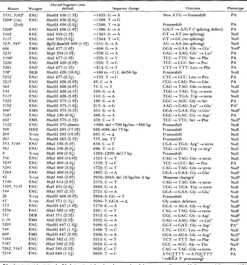
TABLE 1
Summary of DNA sequence changes in rosy mutations detected on denaturing gradient gel blots
Mutant Mutagen
Altered fragment (mm
shifted) Sequence change Outcome Phenotype
5 3 3 1 , 5 1 8 2 5204" (1 st)
(2nd) 5208" 5102 5117 527,545" 606 523 1 5198" 5220 5281 5 38" 5252 5215 564 554 51 15 5322 5192 406" 5185 602 5122 509 204 20 1 553, 5144 56 I
8 516 5235 5241 5264 42 5148 5105, 5135 544 26 41' 573 5256 531
L. I9
5205 549 609
5 184
5187 5262, 5163 5214
E N U E N U
E N U E N U E N U E N U EMS E N U E N U E N U E N U
DEB
E N U EN U E N U E N U ENU E N U E N U EMS E N U
EMS
EN U
H N 2
X-ray
E N U X-ray
E N U X-ray E N U E N U
ENU
E N U
E N U E N U EN U X-ray
X-ray X-ray E N U EN U DEB EMS E N U E N U
EMS
E N U E N U E N U EN U
Hue111 436 (1.5f) -1435: G + A Hue111 436 (2.01) -1388:T+G H a e l l l 436 (2.01) - 1 3 8 6 : T - A Hue111 436 ( 1 .Of) -1366: G + A Alul 359 (1.5f) - 1 3 6 5 : G - A AluI 359 (3.01) -1364: T + C
BgllI-BamHI 409 (1.0T) -551: G + A Aiul 477 (1 .Of)
MspI 234 (1 .Of)
Alul 477 (].Of)
HaeIII 426 (2.07) Alul 477 (1.5f) Hue111 426 (10.01) Alul 477 (2.51) Hue111 426 (6.Of) Hue111 426 (4.0f) HaeIII 426 (4.5f) Hue111 426 (3.07) Hue111 375 (l.0f) Hue111 375 (1.01) Hue111 375 (0.51) H h a I 536 (6.01) Hue111 375 (1.57) HaeIII 375 absent Hue111 285 (17.07) Hue111 285 (16.0f) Hue111 285 (2.57) H h a I 536 (5.07) H h a I 536 (6.01) MspI 468 (2.01) H h a I 469 (14.0f) H h a I 469 (4.01) H h a I 469 (3.03) H h a I 469 (4.05) MspI 446 (2.0f)
MspI 614 (2.07) Rsal 451 (2.01) HhaI 507 (2.57) Hue111 408 (9.Of) Rsal 371 (1.51) Hue111 447 (1.07) H h a I 582 ( 1 .Of) RsaI 371 (2.5f) AluI 232 (2.5f) Hue111 447 (1.03) Hue111 447 (1.53) Hue111 447 (2.07) H h a I 582 (].Sf) H h a I 582 (2.5f) Rsal 395 (2.5f)
Rsal 688 (1.51)
-468: G + A -328: G + A -225: C + T -226: T + C -214: C + T
-166 to - 1 11: del56 bp -123: T + C
43: C + A 75: C + T 109: G + A 1 l O : G - A 1 8 0 : G - A 213: A - + G 4 5 1 : G + A 466: G + A 478: C + T
-500: del -700 bp/ins -300 bp 626-698: del 73 bp
685: C + A
737: ins. TT 8 1 6 : C + T 846: T + C
1283-1 299: dell 7 bp 1 5 2 1 : C - T
1 5 3 9 : T - C 1722: C
-
T 1807: G -+ A2030-2045: del 16 bp/ins. 6 bp 2573: C + T
2683: G -+ A
2 7 2 1 : G - A 2804-5: GG + T
3095-7: GGA + A
3179: G + A 3 2 2 1 : C + T 3312: G + A 3332: G + A 3486: G + A 3498: T + C 3506: G + A 3 5 1 3 : C - T 3524: G + A 3626: C + T 3850: T + C
New A T G + frameshift
Frameshift
G/GT + A/GT (? splicing defect) G T + A T ( n o splicing)
G T + GC (no splicing) AG + AA (no splicing) GGA + GAA: Gly + Glu- GAG + AAG: Glu- + Lys+ T C C + T T C : S e r -P Phe
T C C -+ CCC: Ser + Pro
C T T + T T T : Leu + Phe Frameshift
C T C + CCC: Leu * Pro CCG + CAG: Pro * Gln CAG + TAG: Gln + term T G G + T A G : T r p + term T G G + T G A : T r p + term GGC + AGC: Gly + Ser AAG + GAG: Lys+ + Glu- GGA + GAA: Gly + Glu- GGC + GAC: Gly -P Asp-
T C C + T T C : S e r -+ Phe Frameshift
Frameshift Frameshift Frameshift
CGA + TGA: Arg+ + term T G G + CGG: T r p + Arg+ Frameshift
CAG + TAG: Gln + term T C C + CCC: Ser + Pro CAG + TAG: Gln 4 term GGA + GAA: Gly + Glu-
Missense changed CAG + TAG: Gln + term TGG + T G A : T r p + tern1 GGA + GAA: Gly -P Glu-
Frameshift Gly codon deletion GGA + AGA: Gly + Arg+ CAG + TAG: Gln + term GGC + GAC: Gly + Asp- GAG + AAG: Glu- + Lys+ G G T + GAT: Gly 4 Asp-
C T C + CCC: Leu * Pro GGA + AGA: Gly * Argf T C T + T T T : Ser + Phe GCC + ACC: Ala * T h r CAG + TAG: Gln + term A T G T T T T - + A T G C T T T
? nlRNA 3' Drocessine?
PA PA PA Null Null Null Nulld PA PA PA PA Null PA PA Null Null Null PA PAd Nulld PA Nulld Null Null Null Null Null Null Null Null PA Null' Null' Nulld Null Null Null Null Null Nulld Null' Nulld Nulld PAd Null Nulld Null' Null' PA PA 0
PA refers to partial activity; the PA designation is based on the eye color of these mutants (GELBART, MCCARRON and CHOVNICK 1976; MARGARET MCCARRON, personal communication). T h e mutagens are: N-ethyl-N-nitrosourea (ENU), ethyl methanesulfonate (EMS), nitrogen mustard (HN2), diepoxybutane (DEB) and X-irradiation. The fragment with altered melting behavior listed is that with the largest shift found for the mutant; the magnitude (in mm) and the direction of the shift
(7
for upward and1
for downward) is indicated in parentheses. (As discussed in the text, other overlapping restriction fragments that include the mutation were often shifted as well). T h e positions in the gene are numbered as in Figure 2 (CURTIS et al. 1987; KEITH et al. 1987)." Described in LEE et al. (1 987). Described in CURTIS et al. (1989). Described in CURTIS and BENDER (1 990).
Complements with other rosy null alleles (GELBART, MCCARRON and CHOVNICK 1976; MARGARET MCCARRON, personal communication).
' Adult eye color is not the same as in most null mutants; some residual XDH activity might be present.
146 M. Gray et al.
TABLE 2
Types of ENU-induced single base change rosy mutations
Base change Type Examples" total" (%)
G + A Transition 17 44.7
c
-+ -1- Transition 10 26.37 ° C Transition 7 18.4
A - + G Transition 1 2.6 C + A Transversion 1 2.6
I- -+ G lransversion 1 2.6 T - + A Deletion 1 2.6 All otller transversions 0 0
Fraction O f
Includes the 34 ENU-induced mutations, and the seven ENU- induced mutations described in LEE et al. (1987).
DISCUSSION
Denaturing gradient blots for mapping mutations:
Physical mapping of the rosy mutations with denatur- ing gradient gel blots is less laborious than traditional genetic mapping and genomic cloning. Since base differences must be in the first melting domain of a DNA fragment to be detectable in denaturing gra- dient gels (FISCHER and LERMAN 1983), we expected that mutations in G
+
C-rich sequence would be missed; G + C-rich sequence is usually found in the most stable melting domains. However, to our sur- prise, many of the mutations were mapped in the rosy DNA fragments with the highest G+
C content. By using the different restriction enzymes, the melting domains of the fragments tested at any region of the gene were often rearranged, or "moved." In this way, mutations in higher melting domains of one fragment were detected by analyzing partially-overlapping low- est melting domains.In general, G to A and C to T base changes caused fragments to shift upward in the gradient, and, con- versely, A to G and T to C changes caused downward shifts. This is consistent with predictions of melting behavior changes made for all possible base changes
in a mouse @-globin promoter DNA fragment (MYERS
et al. 1985). In our experiments, exceptions to this rule were found only in fragments that produced smeared, diffuse bands in denaturing gradient gels (discussed above).
Success rate for detection of mutations: We have found
77%
of the mutations in 130 rosy mutants tested (not including the five on an undetermined back- ground). It cannot be determined from our results if any types of base changes were undetectable with denaturing gradient gel blots. In a computer simula- tion, 95% of all possible base substitutions in a 135-bp DNA fragment containing the mouse @-globin
promoter would result in detection of a melting dif- ference from the wild-type fragment, under ideal conditions of denaturing gradient gel electrophoresis (MYERS et al. 1985). The only substitutions that were predicted to be undetectable were some conservative
transversions, although most (105/135) conservative transversions would be detectable. T h e magnitude of the fragment mobility shifts caused by conservative transversions was predicted to be much less than that of transitions and nonconservative transversions (MYERS et al. 1985). Nearly all types of base changes changed the melting behavior of mutant fragments made in vitro using chemical mutagens (MYERS, LER- MAN and MANIATIS 1985). A portion of the 23% of rosy mutations not detected in our experiments could have been base changes, such as conservative trans- versions, that cause little or no melting behavior changes, as in the &globin simulation. Almost all (95%) of the ENU-induced rosy mutations sequenced after detection of denaturing gradient blots were tran- sitions. In a study of ENU-induced mutations at the
vermilion locus, 79% were shown by DNA sequencing to be transitions, and 21% transversions (PASTINK et
al. 1989). T h e vermilion mutant genes were sequenced directly, and may be a more representative sampling of the distribution of base changes caused by ENU. Thus, it seems likely that at least some of the 30 mutations not detected on denaturing gradient gel blots might have had transversions.
Some of the 30 mutations not detected were prob- ably in the higher melting domains of the fragments tested, and the best restriction digest for analyzing these regions of rosy sequence have not yet been tested. For example, the splice acceptor site mutation ry"49 (LEE et al. 1987) was not detected using the five restriction enzymes for the 135 new mutants. Most restriction fragments that include this site contain much of the A
+
T-rich first intron sequence; the mutation is at the edge of a very G+
C-rich protein- coding sequence. We deliberately tested an alternative fragment (the BglII-BamHI 409-bp fragment) that included a large portion of the G+
C-rich sequence. On denaturing gradient gels, this fragment had dis- tinctive melting behavior in genomic DNA of both ry"' and another mutant (ry".') with the same base change (Table 1). Some of the other mutations might be detected in a similar manner, by analyzing addi- tional genomic DNA digests, using different enzymes.Mapping rosy Mutations 147
ing gradient gel electrophoresis) DNA in the mutant clones was used as wild-type control templates for sequencing new cloned mutant DNAs; no additional base changes were found. Almost all of our mutant base changes create in-frame nonsense codons, or nonconservative amino acid substitutions (Table 1). Double mutations separated by more than a few hundred base pairs have never been described in any ENU-induced mutant gene.
Regulatory mutations: All of the rosy mutations sequenced in this study either change the structure of the XDH polypeptide, cause premature termination of translation, or interfere with normal mRNA proc- essing. No mutations were found in DNA upstream from the XDH gene. There are probably few point mutations in rosy regulatory sequences that would significantly alter the rate of XDH transcription by more than 50%. T h e sequences of promoters and enhancers identified in other genes are extremely variable and often repeated (SERFLING, JASIN and SCHAFFNER 1985). Most single base substitutions made
in vitro in DNA upstream from the human @-globin gene have only subtle effects on the rate of transcrip- tion in a HeLa cell transient expression assay (MYERS, TILLY and MANIATIS 1986).
Effect of mutagen: T h e ENU and EMS results reported here are similar to what others have found for Drosophila. As discussed above, our data should not be regarded as fully representative of the spec- trum of DNA lesions to be expected whenever ENU or EMS is used as the mutagen, because of the possible insensitivity of our detection scheme to conservative transversions and base changes in some regions of the gene. Also, the rosy mutants that were expected to have deletions and rearrangements were excluded from the study. ENU can certainly induce deletions and rearrangements, as in the case of ry5I2' (described above), and the five ENU-induced large deletions extending beyond rosy into adjacent complementation groups (LEE et al. 1987). We have also found an ENU- induced 1.5-kb deletion in the Ubx gene (M. GRAY and W. BENDER, unpublished experiments). As dis- cussed above, our ENU-induced rosy single base change mutations are almost exclusively (95%) tran- sitions (Table 2), in contrast to the vermilion muta- tions, where only 79% were transitions (PASTINK et al.
1989). Twenty-seven of 39 previously reported ENU- and EMS-induced mutations in the rosy, Adh, per, Ubx, Notch and vermilion genes of Drosophila are G to A and C to T transitions (LEE et al. 1987; MARTIN et al.
1985; YU et al. 1987; WEINZIERL et al. 1987; KELLEY
et al. 1987; PASTINK et al. 1989).
All five of our X-ray-induced rosy mutations change the total number of base pairs, either as small dele- tions, an insertion, or both. Similarly, five X-ray- induced white mutations had small (6-29 bp) deletions,
with one having an insertion of 10 bp of new
sequence at the site of an 18-bp deletion (PASTINK et
al. 1988). In contrast to the white mutations, our rosy
deletions do not occur between 2- and 3-bp repeats (Table 1; KEITH et al. 198'7). T h e inserted
7
bp of new DNA sequence at the site of the 16-bp deletion in ry4? is not repeated on either side of the deletion site, as in the X-ray-induced deletion/insertion w17D2(PASTINK et al. 1988).
We have analyzed two mutations induced by die- poxybutane (DEB). One of these ( r y 5 j 9 ) and one from o u r previous study (ry5") were small deletions of less than 100-bp. The DEB-induced mutation ry531 is a single base substitution. Our DEB results are consist- ent with those of a previous study of 2 1 DEB-induced rosy mutations (REARDON et al. 1987), where 43% were deletions, ranging in size from 50 bp to 8 kb.
T h e 73-bp deletion mutation ry509 and the ry""' rearrangement were induced by HN2. This mutagen, also known as mechlorethamine, is an alkylating agent that can also cross-link DNA (HOLLAENDER 1971). Our results suggest that the mutations may have arisen by DNA repair after a crosslinking event.
Point mutations and sequence polymorphisms:
Point mutations can be used for a variety of biological investigations. They are helpful for studies of the biochemistry of genetic recombination and DNA re- pair. T h e physical mapping of point mutations causing a new and well-defined mutant phenotype can be used in identifying the relevant gene in chromosomal re-
gions with many closely spaced genes. Missense mu- tations can help identify functional domains of pro- teins, by observing the biological consequences of amino acid substitutions; studies of this type are un- derway for Drosophila xanthine dehydrogenase, using rosy mutations (BRAY et al. 1990). Nonsense mutations can be used as substrates for the isolation of suppressor tRNA genes. Indeed, the rosy mutant eye color phe- notype in the ry5I6 strain has been reverted to wild type when an active suppressor tRNA gene was intro- duced into the germline (DOERIG et al. 1988).
We have also used denaturing gradient gel blots to find DNA sequence polymorphisms, as well as mutant base changes. Laboratory and wild stocks of Drosoph- ila have a high frequency of sequence polymorphisms. Between some pairs of strains, sequence differences can be found about every 100 bp, in the rosy and
Ultrabithorax genes (LEE et al. 1987; CURTIS et al.
148 M. Gray et al.
used to map the lengths of gene conversion intervals, as well as mapping sites of crossovers (CURTIS et al.
1989; CURTIS and BENDER 1990).
We thank MARGARET MCCARRON, CAROL LOVE and ARTHUR CHOVNICK for allowing us to work with their rosy mutants. We
thank RICHARD MYERS, LEONARD LERMAN and EZRA ABRAMS, and our laboratory colleagues, for their advice and comments. Sup-
ported by National Institutes of Health grants to M.G. and W.B., and a Datnon Runyon-Walter Winchell Cancer Fund Fellowship to M.G.
L I T E R A T U R E C I T E D
BENDER, W., P. SPIERER and D. HOGNESS, 1983 Chromosomal walking and jumping to isolate DNA from the Ace and rosy loci and the Bithorax Complex in Drosophila melanogaster. J. Mol. Biol. 168: 17-33.
BIRNSTIEL, M. L., M. BUSSLINGER and K. STRUB, 1985 Transcription termination and 3’ processing: The end is in site! Cell 41: 349-359.
URAY, R. C., R. K. HUGHES, W. A. DOYLE, J. R. S. WHITTLE, J. F. BURKE and A. CHOVNICK, 1990 Towards an expression sys- ten1 for site-directed mutagenesis studies of xanthine dehydro- genase (Drosophila melanogaster rosy) gene, in Flavins and Fla- voproteins, edited by G. ZANETTI, S. PONCHI and B. CURTI. W. de Gruyter, Berlin.
CHOVNICK, A,, 1966 Genetic organization in higher organisms. Proc. R. Soc. Lond. B 164: 198-208.
CHOVNICK, A , , W. GELBART and M. MCCARRON, 1977 Organization of the rosy locus in Drosophila melanogaster. Cell 11: 1-10.
CLARK, S. H., A. J. HILLIKER and A. CHOVNICK, 1986 Genetic analysis of the right (3’) end of the rosy locus in Drosophila melanogaster. Genet. Res. 47: 109- 1 16.
COLLINS, M., and R. M. MYERS, 1987 Alterations in DNA helix stability due to base modifications can be evaluated using denaturinggradient gel electrophoresis. J. Mol. Biol. 198: 737- 744.
COTE, B., W. BENDER, D. CURTIS and A. CHOVNICK, 1986 Molecular mapping of the rosy locus in Drosophila mel- anogaster. Genetics 112: 769-783.
CuRrrs, D., S. H. CLARK, A. CHOVNICK and W. BENDER, 1989 Molecular analysis of recombination events in Drosoph- i h . Genetics 122: 653-661.
CURTIS, D., and W. BENDER, 1990 Gene conversion in Drosophila and the effects of the meiotic mutations mea-9 and mei-218. Genetics (in press).
D K N I X , l.., G. CFSARENI and R. CORTESE, 1983 pEMBL: a new Fanlily of single stranded plasmids. Nucleic Acids Res. 11:
1645-1655.
DOERIG, R., B. SUTER, M. GRAY and E. KURLI, 1988 ldentification of an amber nonsense mutation in the rosy”” gene by germline transformation of an amber suppressor tRNA gene. EMBO J.
I ~ U T T O N , F. L., J R . , and A. CHOVNICK, 1988 Developmental reg- ulation of the rosy locus in Drosophila melanogaster, pp. 267- 3 16 i n Developmental Biology, Vol. 5, edited by L. W. BROWDER. Plenum, New York.
IEINBERG, A. P., and B. VOGELSTEIN, 1983 A technique for radiolabeling DNA restriction endonuclease fragments to high specific activity. Anal. Biochem. 1 3 2 6-13.
IISCHER, S. G . , and L. S. LERMAN, 1979 Two-dimensional elec- trophoretic separation of restriction enzyme fragments of
DNA. Methods Enzymol. 68: 183-191.
I I S C H K R , S. G., and L. S. LERMAN, 1983 DNA fragments differing by single base-pair substitutions are separated in denaturing 7: 2579-2584.
gradient gels: correspondence with melting theory. Proc. Natl. Acad. Sci. USA 80: 1579-1583.
GELBART, W., M. MCCARRON and A. CHOVNICK, 1976 Extension of the limits of the XDH structural element in Drosophila melanogaster. Genetics 84: 21 1-232.
GRUNSTEIN, M . , and D. HOGNESS, 1975 Colony hybridization: a method for the isolation of cloned DNAs that contain a specific gene. Proc. Natl. Acad. Sci. USA 72: 3961-3965.
HANAHAN, D., 1983 Studies on transformation of Escherichia coli with plasmids. J. Mol. Biol. 1 6 6 557-580.
HOLLAENDER, A., 1971 Chemical mutagenesis, Plenum, New York. H U , N., and J. MESSING, 1982 T h e making of strand-specific M I 3
probes. Gene 17: 271-277.
hH-HOROWICZ, D., and J. F. BURKE, 1981 Rapid and efficient cosnlid cloning. Nucleic Acids Res. 9 2989-2998.
KEITH, T. P., M . A. RILEY, M. KREITMAN, R. C. LEWONTIN, D. CURTIS and G. CHAMBERS, 1987 Sequence of the structural gene for xanthine dehydrogenase (rosy locus) in Drosophila melanogaster. Genetics 116: 67-73.
KELLEY, M. R., S. KIDD, W. A. DEUTSCH and M . W. YOUNG, 1987 Mutations altering the structure of epidermal growth factor-like coding sequences at the Drosophila Notch locus. Cell 51: 539-548.
KESSLER, C., P. N E U M A I E R and w . WOLF, 1985 Recognition se- quences of restriction endonucleases and methylases-a re- view. Gene 33: 1 - 102.
LEE, C. S., D. CURTIS, M. MCCARRON, C. LOVE, M . GRAY, W. BENDER and A. CHOVNICK, 1987 Mutations affecting expres- sion of the rosy locus in Drosophila melanogaster. Genetics 116: 55-66.
LINDSLEY, D., and G. ZIMM, 1990 l h e genome of Drosophila melanogaster. Part 4, Genes I,-Z. Drosophila Inform. Serv. 68: 176- 184.
MANIATIS, T . , E. FRITSCH and J. SAMBROOK, 1982 Molecular Cloning; A Laboratory Manual. Cold Spring Harbor Press, Cold Spring Harbor, N.Y.
MANIATIS, I-., A. JEFFREY and H. VAN DE SANDE, 1975 Chain length determination of small double- and single-stranded DNA molecules by polyacrylamide gel electrophoresis. Bio- chemistry 14: 3787-3794.
MARTIN, P. F., A. R. PLACE, E. PENTZ and W. SOFER, 1985 UGA nonsense mutation in the alcohol dehydrogenase gene of Dro- sophila melanogaster. J. Mol. Biol. 184: 221-229.
MCCARRON, M., and A. CHOVNICK, 1981 Induced control muta- tions at the rosy locus in Drosophila melanogaster. Genetics 97: s70-7 I .
MOUNT, S. M., 1982 A catalog of splice junction sequences. Nu-
MOWRY, K. L., and J. A. STEITZ, 1987 Identification of the human U7 snRNP as one of several factors involved in the 3’ end maturation of histone premessenger RNA’s. Science 238:
MYERS, R. M . , I,. S. LERMAN and T. MANIATIS, 1985 A general method for saturation mutagenesis of cloned DNA fragments. Science 229: 242-247.
MYERS, R. M., K. TILLY and T. MANIATIS, 1986 Fine structure genetic analysis of a @ globin promoter. Science 232: 61 3-61 8. MYERS, R. M . , S . G. FISCHER, L. S. LERMAN and T. MANIATIS,
1985 Nearly all single base substitutions in DNA fragments joined to a GC-clamp can be detected by denaturing gradient gel electrophoresis. Nucleic Acids Res. 13: 3 1 1 1-3 130. NICHOLLS, R. D., A. V. S. HILL, J. B. CLEGG and 1). R. HIGGS,
1985 Direct cloning of specific genomic DNA sequences in plasmid libraries following fragment enrichment. Nucleic Acids Res. 13: 7569-7578.
~ ’ C O N N E L L , P., and M. ROSBASH, 1984 Sequence, structure, and codon preference of the Drosophila ribosomal protein 49 gene. Nucleic Acids Res. 12: 5495-5513.
cleic Acids Res. 10: 459-472.
Mapping rosy Mutations 149
PASTINK, A,, C. VREEKEN, A. P. SCHALET and J. C. J. EEKEN, 1988 DNA sequence analysis of X-ray-induced mutations at the white locus of Drosophila melanogaster. Mutat. Res. 207: 23- 28.
PASTINK, A,, C. VREEKEN, M. J. M. NIVARD, L. L. SEARLES and E.
W. VOGEL, 1989 Sequence analysis of N-ethyl-N-nitrosourea- induced vermilion mutations in Drosophila melanogaster. Ge- netics 123: 123-129.
KEARDON, J. T . , C. A. LILJESTRAND-GOLDEN, R. L. DUSENBERY and P. D. SMITH, 1987 Molecular analysis of diepoxybutane-in- duced mutations at the rosy locus of Drosophila melanogaster.
Genetics 115: 323-33 1.
KEAUME, A. G . , S. H. CLARK and A. CHOVNICK, 1989 Xanthine dehydrogenase is transported to the Drosophila eye. Genetics
KIGRY, P. W. J., M. DIECKMANN, C. RHODES and P. BERG, 1977 Labeling deoxyribonucleic acid to high specific activity
in vitro by nick translation with DNA polymerase I . J. Mol. 123: 503-509.
Biol. 113: 237-251.
SAIKI, R. K., D. H. GELFAND, S. STOFFEL, S . J. SCHARF, R. HIGUCHI,
G . T. HORN, K. B. MULLIS and H. A. ERLICH, 1988 Primer directed enzymatic amplification of DNA with a thermostable DNA polymerase. Science 2 3 9 487-491.
SANGER, F., S. NICKLEN and A. R. COULSON, 1977 DNA sequenc- ing with chain-terminating inhibitors. Proc. Natl. Acad. Sci. USA 74: 5463-5467.
SERFLING, E., M. JASIN and W. SCHAFFNER, 1985 Enhancers and eukaryotic gene transcription. Trends Genet. 1: 224-230. WEINZIERL, R., J. M. AXTON, A. GHYSEN and M. AKAM,
1987 Ultrabithorax mutations in constant and variable regions
of the protein coding sequence. Genes Dev. 1: 386-397. Yu, Q., A. C. JACQUIER, Y. CITRI, M. HAMBLEN, J. C. HALLand M.
ROSBASH, 1987 Molecular mapping of point mutations in the period gene that stop or speed up biological clocks in Drosophila melanoguster. Proc. Natl. Acad. Sci. USA 84: 784-788.