OPTIMIZATION, ISOLATION AND CHARACTERIZATION OF BIOACTIVE COMPOUNDS FROM
STREPTOMYCES LAVENDULOCOLOR VHB-9
HIMA BINDU BSSN, RAJESH KUMAR MUNAGANTI, VIJAYALAKSHMI MUVVA*, KRISHNA NARAGANI,
MANI DEEPA INDUPALLI
Department of Botany and Microbiology, Acharya Nagarjuna University, Guntur - 522 510, Andhra Pradesh, India. Email: [email protected]
Received: 07 May 2018, Revised and Accepted: 07 May 2018 ABSTRACT
Objectives: Optimization, isolation, and characterization of bioactive compounds from Streptomyces lavendulocolor VHB-9isolated from granite mines of Mudigonda village of Khammam district of Telangana state.
Methods: The potent strain was identified as S. lavendulocolor VHB-9 by polyphasic taxonomy. The influence of culture conditions on growth and bioactive compounds production was investigated. Purification of bioactive compounds was done using column chromatography. The structures of the compounds were elucidated on the basis of spectroscopic analysis including Fourier transform infrared, electron spray ionization mass spectrophotometry,1H nuclear magnetic resonance (NMR), and13C NMR. The antimicrobial activity of the compounds produced by the strain was tested against both Gram-positive and Gram-negative bacteria and fungi in terms of minimum inhibitory concentration.
Results: Isolation and identification of two compounds, namely (2R, 3R)-2, 3-Butanediol (B1A), and nonadecanoic acid (B1B). Fraction B4 was isolated partially purified fraction and identified by the gas chromatography-mass spectrometry analysis. B1B compound exhibited the highest activity against Bacillus megaterium,Staphylococcus aureus, Bacillus subtilis, Pseudomonas aeruginosa, and Candida albicans when compared toB1A and B4 compounds.
Keywords: Granite mine, Streptomyces lavendulocolor, Optimization,Spectroscopy,Gas chromatography-mass spectrometry analysis, Biological assay.
INTRODUCTION
Natural products have been the largest contributors to drugs in the history of medicine. Microorganisms are attractive resources to synthesize structurally-diverse substances with various bioactivities that may be used as effective drugs or act as drug lead compounds that could be further modified and developed for higher efficacy [1]. Within the domain bacteria, actinomycetes showed unprecedented ability to produce potentially novel, clinically useful, secondary metabolites with anticancer, antioxidant, antiviral, and antibacterial compounds, the majority of these being derived from the members of the Streptomyces genus that include different classes of antibiotics including aminoglycosides, macrolides, and Beta-lactams [2].
Newer therapeutic agents such as daptomycin, linezolid, and streptogramin combination (quinupristin/dalfopristin) have entered the clinical area in the past few years to combat the multidrug-resistant bacteria [3]. However, certain undesirable side effects and the spread of pathogens with this new antimicrobial drug resistance emphasize the need for the development of other newer antimicrobial agents with activity against Gram-positive bacteria, Gram-negative environmental, and enteric organisms currently threaten patients in hospitals and communities with multidrug resistance [4]. The end result of this phenomenon is that many strains of bacteria have become resistant, and in many cases multi-resistant to these therapeutic agents, thus rendering these drugs ineffective as treatments of choice for severe infections caused by these pathogens [5]. Rising numbers of antibiotic unresponsive infectious disease agents confront patients worldwide [6], and consensus has emerged that it is essential that novel antibiotic classes be developed as part of the strategy to control the emerging drug-resistant pathogens.
Filamentous soil bacteria belonging to the genus Streptomyces are widely recognized as industrially important microorganisms and versatile producers of new secondary metabolites from different biosynthetic pathways, originate from different ecological niches that could be used to hunt for novel bioactive compounds. The great importance given to Streptomyces is partly because these are among the most numerous and most versatile soil microorganisms, given their large metabolite production rate and their biotransformation processes, their capability of degrading lignocellulose and chitin, and their fundamental role in biological cycles of organic matter. Indeed, different Streptomyces species produce about 75% of commercially and medically useful antibiotics. They have provided more than half of the naturally occurring antibiotics discovered to date and continue to be screened for useful compounds [7]. In the course of screening for new antibiotics, several studies are oriented toward isolation of Streptomycetes from different habitats.
In the view of that the strain S. lavendulocolor VHB-9 was isolated from a granite mine of Mudigonda village of Khammam district of Telangana state, India. An attempt was made in the present study to optimize the cultural parameters required for best yields of bioactive metabolites, and chemical characterization of the compounds was also investigated.
METHODS Chemicals
All solvents, reagents, and media supplements used in this study were of extra pure grade and procured from Merck (Mumbai, India).
Strain isolation
The strain, S. lavendulocolor VHB-9, was isolated on yeast extract-malt extract-dextrose (YMD) agar medium by soil dilution technique from © 2018 The Authors. Published by Innovare Academic Sciences Pvt Ltd. This is an open access article under the CC BY license (http://creativecommons. org/licenses/by/4. 0/) DOI: http://dx.doi.org/10.22159/ajpcr.2018.v11i8.25345
a soil sample collected from granite mines of Mudigonda, Khammam District, Telangana state, India. The medium was composed of malt extract (1%), yeast extract (0.4%), dextrose (0.4%), CaCO3 (0.2%), and agar (2.0%), pH 7.0±0.2. The strain was stored on YMD agar slants at 4°C.
Antimicrobial profile of bioactive metabolites produced by the strain
The antimicrobial profile of the strain S. lavendulocolor VHB-9 was studied by cultivating the strain in YMD broth at 30°C for 8 days. The antimicrobial activity of bioactive metabolites against Staphylococcus aureus (MTCC 3160), Lactobacillus casei, Bacillus megaterium (NCIM 2187), Proteus vulgaris (ATCC 6380), Pseudomonas aeruginosa (ATCC 9027), Escherichia coli (ATCC 9027), Aspergillus niger, Fusarium solani, Fusarium oxysporum, and Candida albicans (MTCC 183) was determined by agar well diffusion assay, and inhibition zones against test microbes were determined [8].
Media optimization
Attempts were made to enhance the antimicrobial activity of
S. lavendulocolor VHB-9 by optimizing the culture conditions such as pH, temperature, carbon sources, nitrogen sources, and minerals. The bioactive metabolite production of the strain was determined after 4 days of incubation. Fermentation was carried out in 250-mL Erlenmeyer flasks with constant shaking at 180 rpm. The effect of initial pH on the bioactive metabolite production was determined by adjusting pH of the production medium from 4 to 10. The optimal pH achieved at this step was fixed for further study [9]. Similarly, the optimum temperature for antimicrobial metabolite production was determined by incubating the strain at temperatures ranging from 20 to 40°C, while maintaining all other conditions at optimum levels [10]. The effect of carbon sources on bioactive metabolite production was determined by supplementing the production medium (YMD) with different carbon sources such as maltose, sucrose, mannitol, lactose, starch, cellulose, galactose, sorbitol, and fructose each at a concentration of 0.4% (w/v) replacing dextrose by keeping the other ingredients constant [11]. Influence of varying concentrations of the best carbon source (0.5–4% w/v) on bioactive metabolite production was also investigated.
Similarly, the influence of various nitrogen sources such as sodium nitrate, ammonium oxalate, ammonium sulfate, peptone, tryptophan, L-proline, tyrosine, urea, and yeast extract was studied by adding nitrogen source (0.4%) to the medium with an optimized carbon source. Further, the optimal levels of the suitable nitrogen source (0.1–1.5% w/v) for good yields of bioactive metabolites were also recorded [12]. To evaluate the effect of mineral salts, the optimized medium containing the superior carbon and nitrogen source was supplemented separately with mineral supplements such as KH2PO4, K2HPO4, NaCl, KCl, MgSO4.7H2O, FeSO4.7H2O, and MnCl2 at a concentration of 0.05% (w/v) [13].
Extraction of the metabolite and antimicrobial activity assay The strain S. lavendulocolor VHB-9 grown under optimized cultural conditions for 4 days was extracted with ethyl acetate, and concentrated in a rotary evaporator to obtain a crude extract. The antimicrobial metabolites produced were tested by agar well diffusion assay against the following test microorganisms:
Bacteria
Overnight grown cultures of S. aureus (MTCC 3160), Bacillus subtilis
(ATCC 6633), B. megaterium (NCIM 2187), Shigella flexneri (MTCC 1457),
L. casei (MTCC 1423), Lactobacillus acidophilus (MTCC 495), Proteus vulgaris (MTCC 7299), P. aeruginosa (ATCC 9027), E. coli (ATCC 35218),
Vibrio cholerae, and Streptococcus mutans (MTCC 497) were used.
Fungi
C. albicans (ATCC 10231), A. niger, Aspergillus flavus, F. solani, F. oxysporum (MTCC 3075), Penicillium citrinum, and Alternaria sp. were used as test fungi for testing antifungal activity.
Extraction, purification, and characterization of antimicrobial compounds
Fermentation
A seed culture was prepared by culturing S. lavendulocolor VHB-9 in YMD broth and incubated on a rotary shaker (180 rpm) at 30°C for 48 h. The seed culture was then transferred to fermentation broth containing malt extract - 1%, lactose - 0.5%, peptone - 0.5%, and K2HPO4 - 0.05% with pH adjusted to 7 and incubated on rotary shaker (180 rpm) at 30°C for 120 h. The bioactive compounds from the fermented broth were harvested by filtration of biomass through Whatman Filter Paper No. 42 (Merck, Mumbai, India). The culture filtrate (30 L) was extracted twice with an equal volume of ethyl acetate, pooled and the organic layer was concentrated in a Rotavac. The deep brown semi-solid compound (3.0 g) obtained was applied to a silica gel G column (25 cm×5 cm, Silica gel, Merck, Mumbai, India).
The separation of the crude extract was carried out through gradient elution system of hexane: ethyl acetate. The eluent was run over the column, and small volumes of eluent collected in test tubes were analyzed through thin-layer chromatography (TLC) using silica gel plates (Silica gel, Merck, Mumbai, India) with hexane: ethyl acetate solvent system [14]. Compounds with identical retention factors (Rf) were combined and assayed for antimicrobial activity against Gram-positive (B. megaterium), Gram-negative (E. coli) bacteria, and yeast (C. albicans) by using agar well diffusion assay [15].
Among the different fractions, two fractions B1 (polar) and B4 (nonpolar) were collected at gradient solvent system of Hexane: ethyl acetate (70-30v/v and 90-10v/v). The B1 fraction was rechromatographed (22 X 2.5 cm, Silica gel 100; Merck) to get two pure compounds ((2R, 3R)-2, 3-Butanediol (B1A), and nonadecanoic acid [B1B]). The structures of these active fractions were analyzed on the basis of Fourier transform infrared (FTIR); model: Thermo Nicolet Nexus 670 spectrophotometer with NaCl optics and electron ionization mass/electron spray ionization mass spectrophotometry (EIMS/ ESIMS); model: Micromass VG - 7070H, 70eV spectrophotometer and nuclear magnetic resonance (NMR) (1H NMR and13C NMR) model: Varian Gemini 200 and samples were made in CDCl3 with trimethyl saline as standard.
Fractions B4 obtained as a mixture of compounds analyzed on Agilent gas chromatography-mass spectroscopy (GC-MS) system. The fused silica HP-5 capillary column (30 m×0.25 mm, ID, film thickness of 0.25 μm) was directly coupled to the MS. The carrier gas was helium with a flow rate of 1.2 ml/min. Oven temperature was programmed (50°C/min), then 50–280°C (at rate of 5°C/min) and subsequently held isothermally for 20 min. The temperature of the injector port was maintained at 250°C and that of the detector at 280°C [16]. The peaks of components in gas chromatography were subjected to mass spectral analysis. The spectra were analyzed from the available library data NIST MS search (ver. 2.0) (Included with NIST’02 mass spectral library, Agilent p/n G 1033 A).
Biological assays
Minimum inhibitory concentration (MIC)
After inoculation the plates were incubated at 30°C and examined after 24–48 h of incubation for bacteria and 48–72 h for yeast and filamentous fungi. The experiment was carried out in triplicates, and the solvent (DMSO) alone was kept as a negative control. Tetracycline and Carbendazim were employed as positive controls for bacteria and fungi, respectively. The lowest concentration of the bioactive compound exhibiting antimicrobial activity against the test microbes was taken as MIC of the compound.
The MICs of the bioactive compounds (B1A, B1B, and B4) produced by the strain were determined against several opportunistic pathogenic bacteria and fungi.
Test organisms employed
The cultures of S. aureus (MTCC 3160), B. megaterium (NCIM 2187), B. subtilis (ATCC 6633), Serratia marcesens (MTCC 1457), Xanthomonas campestris (MTCC 2286), P. vulgaris (MTCC 7299), P. aeruginosa
(ATCC 9027), E. coli (ATCC 35218), Enterococcus faecalis (MTCC 439),
S. mutans (MTCC 497), L. casei (MTCC 1423), and L. acidophilus (MTCC 495) were employed for antibacterial assay. C. albicans (ATCC 10231),
A. niger (ATCC 1015), A. flavus (ATCC 9643), F. solani (MTCC 4634), F. oxysporum (MTCC 3075), and Penicillium citrinum (MTCC 6489) were used for testing antifungal activity.
Statistical analysis
The results of bioactive metabolite production by S. lavendulocolor
VHB-9 under different cultural conditions were statistically analyzed with one-way analysis of variance (ANOVA).
RESULTS AND DISCUSSION
Antimicrobial profile of bioactive metabolites produced by the strain
The growth pattern of S. lavendulocolor VHB-9 cultured in YMD broth was reported on our earlier publication [17]. The strain entered log phase after 24 h of incubation and exhibited exponential growth up to 96 h followed by stationary phase extended up to 120 h. The crude extract obtained from a 4-day-old culture exhibited high antimicrobial activity against the test microorganisms (Fig. 1). A previous report demonstrated high antimicrobial activity of crude extracts of a 4-day-old culture of Arthrobacter kerguelensis VL-RK_09 [18]. The metabolites collected from 4-day-old culture of Streptomyces griseus exhibited good antifungal activity [19]. Similarly, the extracts of 4-day-old cultures of
Streptomyces psammoticus [20], Streptomyces tendae TKVL_ 333 [21], and Nocardia levis MK-VL_113 [22] were active against test bacteria and fungi.
Crude extracts of 5-day-old cultures of Streptomyces purpeofuscus [23] and Streptomyces albidoflavus [24] were active against Gram-positive as well as Gram-negative bacteria and fungi. Secondary metabolites
extracted from 5-day-old cultures of Streptomyces sp. CDRIL-312 [25] and Streptomyces spp. [12] exhibited good antifungal activity. 5-day-old culture of Streptomyces clavuligerus was reported to produce a good yield of clavulanic acid [26] whereas 6-day-old culture of Streptomyces
sp. 201 exhibited good antimicrobial activity [27].
Impact of pH and temperature on antimicrobial activity
The influence of initial pH on growth and bioactive metabolite production of the strain was determined by adjusting the pH of YMD broth from 4 to10. Maximum growth and antimicrobial metabolite production by the strain were found at pH 7 through the strain was able to grow over a wide range of pH (Fig. 2). The optimum pH for antibiotic production by several actinomycetes was reported to be 7 for Streptomyces hygroscopicus D1.5 [28], Streptomyces torulosus KH- 4 [29], Streptomyces
sp. VITSVK 9[30], Streptomyces cellulosae VJDS-1 [31], Rhodococcus erythropolis VLK-12 [32], and A. kerguelensis VL-RK_09 [18].
The yield of bioactive metabolites of the strain was also recorded when grown at temperatures of 20-40°C, and the optimum was recorded at 30°C (Fig. 3). With the rise of incubation temperature from 20 to 30°C, there was an increase in bioactive metabolite production. However, further, increase in temperature (above 30°C) resulted in the declined production of bioactive metabolites. These results are in agreement with the earlier reports for Streptomyces [29].
Effect of carbon and nitrogen sources on antimicrobial activity The effect of various carbon sources on antimicrobial metabolite production was tested by supplementing the YMD broth with several carbon sources at a concentration of 0.4% (replacing the dextrose) while making all other ingredients of the media same and incubated for 96 h at 30°C. The effects of carbon sources on the production of bioactive metabolites by S. lavendulocolor VHB-9 are presented in Fig. 4. Among the carbon sources tested, significant production of bioactive metabolites was obtained with lactose followed by sucrose. These results are supported by the reports of S. hygroscopicus strains AK-111-81 and CH-7, which utilized lactose as a carbon source for high antimicrobial metabolite production [33,34]. Since lactose supported a high yield of bioactive metabolites, different concentrations of lactose (0.5–4%) were tested to determine the optimal concentration. Lactose at a concentration of 0.5 % supported the highest yield of bioactive metabolites (Fig. 5).
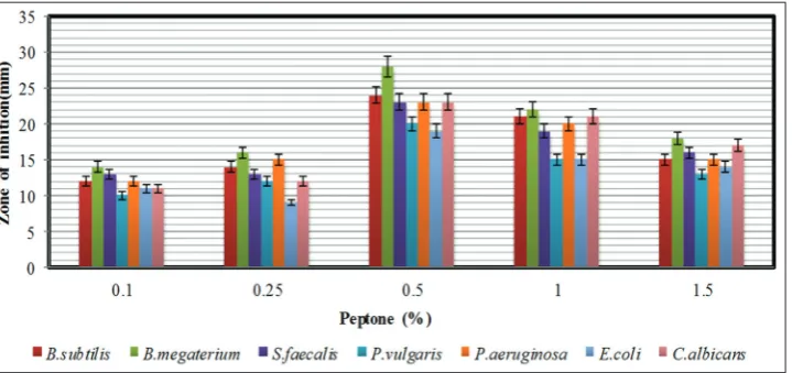
The effect of various nitrogen sources on antimicrobial metabolite production was tested by supplementing the YMD broth with several nitrogen sources at a concentration of 0.4% to the YMD broth (replacing yeast extract). Peptone was found to be good as compared to other organic and inorganic nitrogen sources tested (Fig. 6). These results are comparable with S. rochei G164 [35] and S. scabies PK-A41 [36]. Since peptone enhanced the antimicrobial metabolite production by the strain, the effects of different concentrations of peptone were tested
Fig. 1: Time course of bioactive metabolite production by Streptomyces lavendulocolor VHB-9. The data were statistically analyzed and
(Fig. 7). An enhanced level of bioactive metabolite production was found with peptone at a concentration of 0.5%. L-asparagine (0.09%) was reported as the suitable nitrogen source for optimum production of bioactive metabolites by Streptomyces sp. [27] S. rubrolavendulae ICN3 was reported to exhibit best anti-MRSA and cytotoxic activity when glucose and sodium nitrate were amended to the medium as carbon and nitrogen sources, respectively [37].
The effect of mineral salts on antimicrobial activity
The effect of mineral salts on secondary metabolite production by the strain VHB-9 is shown in Fig. 8. Among the mineral salts tested, K2HPO4 supported the highest antimicrobial activity. Similar results were reported for S. albidoflavus [24] Ripa et al. (2009) and Usha et al. (2011) reported that K2HPO4 supported antibiotic production by Streptomyces sp. RUPA-08PR and Pseudonocardia spp. [38,39].
Fig. 2: The effect of pH on the bioactive metabolite production by Streptomyces lavendulocolor VHB-9. The data were statistically analyzed
and found to be significant at a 5% level
Fig. 3: The effect of temperature on the bioactive metabolite production by Streptomyces lavendulocolor VHB-9. The data were statistically
analyzed and found to be significant at a 5% level
The strain VHB-9 is grown in the optimized fermentation medium containing lactose (0.5%), peptone (0.5%), K2HPO4 (0.05%), and CaCO3 (0.2%) with pH adjusted to 7.0 and incubated at 30°C. After 96 h of incubation, the fermentation broth extracted with ethyl acetate
exhibited good antimicrobial activity against Gram-positive as well as Gram-negative bacteria and fungi (Table 1). Among the bacteria tested,
B. megaterium was highly sensitive to the metabolites followed by
B. subtilis, S. aureus, P. aeruginosa, S. flexneri, L. Casei, and L. acidophilus.
Fig. 5: The effect of different concentrations of lactose as carbon source on antimicrobial activity of Streptomyces lavendulocolor VHB-9.
The data were statistically analyzed and found to besignificant at a 5% level
Fig. 6: The effect of various nitrogen sources supplemented in the modified yeast extract-malt extract-dextrose broth on the antimicrobial activity of Streptomyces lavendulocolor VHB-9. The data were statistically analyzed and found to be significant at a 5% level
Fig. 7: The effect of different concentrations of peptone as nitrogen source on the antimicrobial activity of Streptomyces lavendulocolor
Among the fungi tested, C. albicans exhibited high sensitivity followed by A. niger, A. flavus, F. solani, Penicillium citrinum, F. oxysporum, and
Alternaria sp.
Isolation, purification, and structural elucidation of active metabolites
The culture filtrates (30 L) collected after 96 h of incubation were extracted twice with ethyl acetate and concentrated to dryness in a Rotavac. The crude dark brown residue (3.0g) thus obtained was subjected to silica gel column chromatography. The crude extract was applied to a silica gel G column (25 cm ×5 cm, Silica gel, Merck, Mumbai, India) for the isolation and purification of bioactive compounds.
Among the fractions collected, two fractions (B1 and B4) collected at gradient solvent system of Hexane: ethyl acetate (70–30 v/v and 90–10 v/v) were analyzed. The B1 fraction was re-chromatographed (22 cm × 2.5 cm, Silica gel 100; Merck) to get two pure compounds, B1A (25 mg) and B1B (20 mg). The fraction B4, obtained as a mixture was analyzed by GC-MS system.
B1A eluted with 30% ethyl acetate appeared as light brown liquid soluble in CHCl3, MeOH, DCM, and DMSO. The IR absorption maxima Vmax at 3437/cm suggested the presence of functional OH group. In ESIMS, the compound showed molecular ions at m/z = 108 inferring
the molecular weight of C4H28O2 [M+NH3]+. The proton NMR of the compound displayed proton signals at δ 3.81 (2H, Qd, J=6.04Hz) due to methylene protons bearing hydroxyl group, two exchangeable protons at δ 1.93 (br s, OH), at δ 1.67 (br s, OH) and two methyl groups at δ 1.15 (6H, d, J=6.04 Hz).13C NMR depicted peaks at δ 70.81(2C) and δ 16.90 (2C). (α) D25 = −12.5 (c=1, CHCl3). Based on the spectral data and optical rotation, B1A was identified as B1A Fig. 9). This is the first report of this compound from S. lavendulocolor VHB-9. The second fraction B1B in pure form appeared as brown liquid soluble in CHCl3, MeOH, DCM, and DMSO. The IR absorption maxima Vmax at 1708/cm suggested the presence of the carboxylic group. In ESIMS, the compound showed molecular ions at m/z=298 inferring the molecular weight of C19H38O2 [M+1]+. The proton NMR of the compound displayed signals at δ 1.65–1.55 (30H, m), 1.25–1.99 (m, 2H) for aliphatic methylene protons, at δ 2.35 (t, 2H, J=7.2 Hz) for alpha methylene protons, at δ 1.25–1.99 (m, 2H) for methylene protons, and at δ 0.82 (t, 3H, J=6.1 Hz) for methyl protons. 13C NMR depicted peak at δ 180.8 for the carboxylic group. Based on spectral data, the B1B was identified as nonadecanoic acid (B1B) (Fig. 10). This is the first report of this compound from the strain VHB-9.
The active nonpolar fraction B4 appeared as light brown was soluble in CHCl3, MeOH, DCM, and DMSO. The proton NMR of the compound revealed the presence of a mixture of compounds. The components of partially purified fourth fraction (B4) were analyzed on Agilent GC-MS system. The peaks of components in gas chromatography were subjected to mass spectral analysis. The spectra were analyzed from the Fig. 8: The effect of mineral salts on the antimicrobial activity of Streptomyces lavendulocolor
Fig. 9: Molecular structure of (2R, 3R)-2, 3-butanediol
Fig. 10: Molecular structure of nonadecanoic acid Table: 1 Antimicrobial activity of S. lavendulocolor VHB-9 under
optimized culturing conditions
S. No. Test organisms Zone of inhibition (mm)
1 Staphylococcus aureus 27 2 Bacillus megaterium 31 3 Shigella flexneri 25 4 Bacillus subtilis 28 5 Proteus vulgaris 21 6 Pseudomonas aeruginosa 26 7 Escherichia coli 21 8 Streptococcus mutans 22 9 Vibrio cholera 21 10 Lactobacillus casei 25 11 Lactobacillus acidophilus 23
Fungi
available library data NIST MS search (ver. 2.0) (Included with NIST’02 mass spectral library, Agilent p/n G 1033 A).
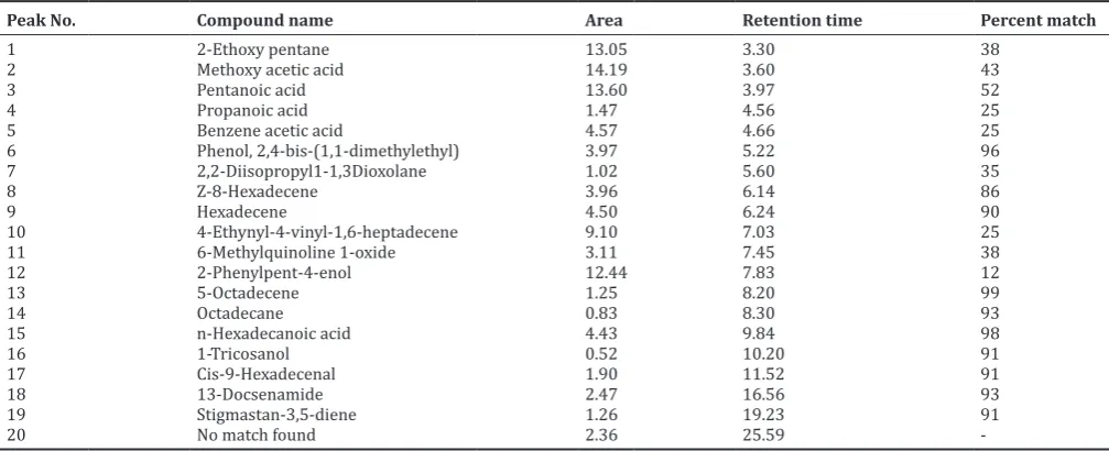
Compounds present in partially purified fraction were tentatively identified. The GC analysis revealed the presence of 20 compounds (Fig. 11). The list of compounds with their retention times is listed in Table 2.
Biological assay
MICs of compounds B1A, B1B, and B4 obtained from the strain against different microorganisms including bacteria and fungi in terms of Fig. 11: Gas chromatographic spectrum of fraction B4 produced by Streptomyces lavendulocolor VHB-9
Table 2. List of compounds obtained from GC-MS analysis of fraction B4 produced by S. lavendulocolor VHB-9
Peak No. Compound name Area Retention time Percent match
1 2-Ethoxy pentane 13.05 3.30 38
2 Methoxy acetic acid 14.19 3.60 43
3 Pentanoic acid 13.60 3.97 52
4 Propanoic acid 1.47 4.56 25
5 Benzene acetic acid 4.57 4.66 25
6 Phenol, 2,4-bis-(1,1-dimethylethyl) 3.97 5.22 96
7 2,2-Diisopropyl1-1,3Dioxolane 1.02 5.60 35
8 Z-8-Hexadecene 3.96 6.14 86
9 Hexadecene 4.50 6.24 90
10 4-Ethynyl-4-vinyl-1,6-heptadecene 9.10 7.03 25
11 6-Methylquinoline 1-oxide 3.11 7.45 38
12 2-Phenylpent-4-enol 12.44 7.83 12
13 5-Octadecene 1.25 8.20 99
14 Octadecane 0.83 8.30 93
15 n-Hexadecanoic acid 4.43 9.84 98
16 1-Tricosanol 0.52 10.20 91
17 Cis-9-Hexadecenal 1.90 11.52 91
18 13-Docsenamide 2.47 16.56 93
19 Stigmastan-3,5-diene 1.26 19.23 91
20 No match found 2.36 25.59
-GC-MS: Gas chromatography-mass spectroscopy, S. lavendulocolor: Streptomyces lavendulocolor
Test organism B1A MIC (µg/ml)
B1B B4 (PPF†) Positive
control#
Fusarium oxysporum 90 75 100 10
Fusarium solani 90 65 125 10
Penicillium citrinum 100 75 125 10 *MIC: Minimum inhibitory concentration, #Positive control: Tetracycline against bacteria, Griseofulvin against yeast and Carbendazim against fungi. B1A: (2R, 3R)-2, 3-Butanediol, B1B: Nonadecanoic acid, B4: PPF, PPF: Public provident fund, S. lavendulocolor: Streptomyces lavendulocolor
Table 3: (Continued)
Test organism B1A MIC (µg/ml)
B1B B4 (PPF†) Positive
control#
Bacteria
Bacillus megaterium 65 40 75 30
Bacillus subtilis 75 55 80 40
Serratia marcescens 90 60 100 25
Xanthomonas campestris 75 65 85 40
Proteus vulgaris 100 80 100 50
Pseudomonas aeruginosa 70 55 75 40
Escherichia coli 90 60 95 25
Enterococcus faecalis 95 75 100 25
Streptococcus mutans 75 55 90 30
Lactobacillus casei 85 55 100 25
Lactobacillus acidophilus 80 55 95 25
Staphylococcus aureus 65 50 80 25 Yeast
Candida albicans 85 55 100 50 Fungi
Aspergillus niger 90 55 125 5
Aspergillus flavus 80 70 100 10 Table 3: MIC values of the bioactive compounds produced by
S. lavendulocolor VHB-9
MIC are shown in Table 3. B1B is more effective than B1A and B4.
B. megaterium is highly sensitive to the compounds followed by S. aureus, B. subtilis, L. acidophilus, and S. mutans among the Gram-positive bacteria. P. aeruginosa is highly sensitive to the compounds followed by S. marcescens, E. faecalis, X. campestris, E. coli, and P. vulgaris among the Gram-negative bacteria. MIC values of B1A B1B, public provident fund and tetracycline against the test bacteria varied from 65 to 100 μg/ml, 40–80 μg/ml, 75–100 μg/ml, and 25–50 μg/ml, respectively. For fungi, these values ranged from 90 to 100 μg/ml for B1A, 55–75 μg/ml for B1B, 100–125 μg/ml for partially purified fraction, 5–10 μg/ml for carbendazim, and 50 μg/ml for griseofulvin. All the compounds showed good activity against Candida.
REFERENCES
1. Demain AL, Sanchez S. Microbial drug discovery: 80 years of progress. J Antibiot 2009;62:5-16.
2. de Lima Procópio RE, Da Silva IR, Martins MK, De Azevedo JL, De Araújo JM. Antibiotics produced by Streptomyces. Braz J Infect Dis 2012;16:466-71.
3. Levy SB, Marshall B. Antibacterial resistance worldwide: Causes, challenges andresponses. Nat Med 2004;10:122-9.
4. Nathwani D. Tigecycline: Clinical evidence and formulary positioning. Int J Antimicrob Agents 2005;25:185-92.
5. Alanis AJ. Resistance to antibiotics: Are we in the post-antibiotic era? Arch Med Res 2005;36:697-705.
6. Livermore DM. Bacterial resistance: origins, epidemiology and impact. Clin Infect Dis 2003;36:11-23.
7. Miyadoh S. Research on antibiotic screening in Japan over the last decade: A producing microorganisms approach. Actinomycetologica 1993;9:100-6.
8. Munaganti RK, Naragani K, Muvva V. Antimicrobial profile of
Rhodococcus erythropolis VL-RK_05 isolated from Mango Orchards. Int J Pharm Sci Res 2015;6:1805-12.
9. Srinivasan MC, Laxman RS, Deshpande MV. Physiology and nutritional aspects of actinomycetes: An overview. World J Microbiol Biotechnol 1991;7:171-84.
10. Saurav K, Kannabiran K. Diversity and optimization of process parameters for the growth of Streptomyces VITSVK 9 sp. Isolation from Bay of Bengal. India J Nat Environ Sci 2010;1:56-65.
11. Elliah P, Srinivasulu B, Adinarayana K. Optimization studies on neomycin production by a mutant strain of Streptomyces marinensis in solid state fermentation process. Biochemistry 2000;39:529-34. 12. Kathiresan K, Balagurunathan R, Selvam MM. Fungicidal activity of
marine actinomycetes against phytopathogenic fungi. Ind J Biotechnol 2005;4:271-6.
13. Farid MA, El-Enshasy HE, Ei-Diwany AI, El-sayed EA. Optimization of the cultivation medium for Natamycin production by Streptomyces netalensis. JBasic Microbiol 2000;40:157-66.
14. Konda S, Raparthi S, Bhaskar K, Munaganti RK, Guguloth V, Nagarapu L,
et al. Synthesis and antimicrobial activity of novel benzoxazine sulfonamide derivatives. Bioorg Med Chem Lett 2015;25:1643-6. 15. Naragani K, Mangamuri U, Muvva V, Poda S, Munaganti RK.
Antimicrobial potential of Streptomyces cheonanensis VUK-A from mangrove origin. Int J Pharm Pharm Sci 2016;8:53-7.
16. Boussada O, Ammar A, Saidana D, Chriaa J, Chraif I, Dami M,
et al. Chemical composition and antimicrobial activity of volatile components from capituila and aerial parts of Rhaponticum acaule DC growing wildin Tunisia. Microbial Res 2008;163:87-95.
17. Bindhu BS, Muvva VL, Munaganti RK, Naragani K, Konda S, Dorigondla KR. A study on production of antimicrobial metabolites by
Streptomyces lavendulocolor VHB-9 isolated from Granite soils. Braz Arch Biol Technol 2016;60:1-13.
18. Munaganti RK, Muvva VL, Konda S, Naragani K, Mangamuri UK, Dorigandla KR, et al. Antimicrobial profile of Arthrobacter kerguelensis
VL-RK_09. Braz J Microbiol 2016;47:1030-8.
19. Otani T, Yamawaki Y, Matsumoto H, Minami Y, Yamada Y, Marunaka T,
et al. New antibiotics. 4181-A and B from Streptomyces griseus: Taxonomy, fermentation, isolation and characterization. J Antibiot
1988;3:275-81.
20. Sujatha P, Bapiraju KV, Ramana T. Studies on a new marine streptomycete BT-408 producing polyketide antibiotic SBR-22 effective against methicillin resistant Staphylococcus aureus. Microbiol Res 2005;160:119-26.
21. Kavitha A, Vijayalakshmi M. Production of amylases by Streptomyces tendae TK-VL_333. Int J Cur Res 2010;10:110-4.
22. Kavitha A, Vijayalakshmi M. Cultural parameters affecting the production of bioactive metabolites by Nocardia levis MK-VL-113. J Appl Sci Res 2009;5:2138-47.
23. Anupama M, Narayana KJ, Vijayalakshmi M. Screening of
Streptomyces purpeofuscus for antimicrobial metabolites. Res J Microbiol 2007;4:1-3.
24. Narayana KJP, Vijayalakshmi M. Production of extracelluar α-amylase
by Streptomyces albidoflavus. Asian J Biochem 2008;3:194-7. 25. Harindran J, Gupte TE, Naik SR. HA-1-92, a new antifungal antibiotic
produced by Streptomyces CDRIL-312: Fermentation, isolation, purification and biological activity. World J Microbiol Biotechnol 1999;15:425-30.
26. Parag SS, Rekha SS. Optimization of nutrional requirements and feeding strategies for Clavulanic acid production by Streptomyces clavuligerus. Biores Technol 2007;98:2010-7.
27. Thakur D, Bora TC, Bordoloi GN, Mazumdar S. Influence of nutrition andculturing conditions for optimum growth and antimicrobial metabolite production by Streptomyces sp. 201. J Med Mycol 2009;19:161-7.
28. Battacharyya BK, Pal SC, Sen SK. Antibiotic production by
Streptomyces hygroscopicus. D1.5: Cultural effect. Rev Microbiol 1998;29:49-52.
29. Atta HM, Bayoumi R, El-Sehrawi M, Aboshady A, Al-Huminay A. Biotechnological application for producing some antimicrobial agents byactinomycetes isolates from Al-Khurmah governorate. Eur J Appl Sci 2010;2:98-107.
30. Kumar S, Krishnan K. Bioactivity guided extraction of 5-(2,4-dimethylbenzyl)pyrrolidin-2-one from marine Streptomyces
VITSVK5 spp. and its anti- Aspergillus activity against drug resistant clinical isolates. Pharm Lett 2013;5:178-84.
31. Indupalli MD, Vijayalakshmi M, Kumar MR. Streptomyces cellulosae
VJDS-1, a promising source for potential bioactive compounds. Int J Pharm Pharm Sci 2015;7:57-61.
32. Naragani K, Kumar MR, Kiranmayi MU, Vijayalakshmi M. Optimization of culture conditions for enhanced antimicrobial activity of Rhodococcus erythropolisVLK-12 isolated from South Coast of Andhra Pradesh, India. Brit Microbiol Res J 2014;4:63-79.
33. Gesheva V, Ivanova V, Gesheva R. Effect of nutrients on the production of AK-111-81 macrolide antibiotic by Streptomyces hygroscopicus. Microbiol Res 2005;160:243-8.
34. Konstantinovic SS, Veljkovic VB, Savic DS, et al. The impact of different carbon and nitrogen sources on antibiotic production by
Streptomyces hygroscopicus CH-7. Cur Res Tech Edu Top Appl Microbiol Microb Biotechn 2010;2:1337-42.
35. Chattopadhyay D, Sen SK. Optimization of cultural conditions for antifungal antibiotic accumulation by Streptomyces rochei G164. Hindustan. Antibiot Bull 1997;39:64-71.
36. Han WC, Lee JY, Park DH, Lim CK, Hwang BK. Isolation and antifungal and antioomycete activity of Streptomyces scabie strain PK-a41, the causal agent of common scab disease. Plant Pathol J 2004;20:115-26.
37. Kannan RR, Iniyan AM, Vincent SG. Production of a compound against methicillin resistant Staphylococcus aureus (MRSA) from
Streptomyces rubrolavendulae ICN3 and its evaluation in zebrafish embryos. Indian J Med Res 2014;139:913-20.
38. Ripa FA, Nikkon F, Zaman S, Khondkar P. Optimal conditions for antimicrobial metabolites production from a new Streptomyces
sp. RUPA-08PR isolated from Bangladeshi soil. Microbiology 2009;37:211-4.