Calcium signals and oocyte maturation
in marine invertebrates
RYUSAKU DEGUCHI
1, NORIYO TAKEDA
2and STEPHEN A. STRICKER*
,31Department of Biology, Miyagi University of Education, Sendai, Miyagi, Japan,
2Research Center for Marine Biology, Graduate School of Life Sciences, Tohoku University, Asamushi, Aomori, Japan
and 3Department of Biology, University of New Mexico, Albuquerque, NM, USA
ABSTRACT In various oocytes and eggs of animals, transient elevations in cytoplasmic calcium ion concentrations are known to regulate key processes during fertilization and the completion of meiosis. However, whether or not calcium transients also help to reinitiate meiotic progression at the onset of oocyte maturation remains controversial. This article summarizes reports of calcium signals playing essential roles during maturation onset (=germinal vesicle breakdown, GVBD) in several kinds of marine invertebrate oocytes. Conversely, other data from the literature, as well as previously unpublished findings for jellyfish oocytes, fail to support the view that calcium sig-nals are required for GVBD. In addition to assessing the effects of calcium transients on GVBD in marine invertebrate oocytes, the ability of maturing oocytes to enhance their calcium-releasing capabilities after GVBD is also reviewed. Furthermore, possible explanations are proposed for the contradictory results that have been obtained regarding calcium signals during oocyte maturation in marine invertebrates.
KEY WORDS:
GVBD, IP
3, meiotic reinitiation, metaphase arrest
Introduction
During oogenesis in animals, immature oocytes initially stop cell cycle progression at prophase of the first meiotic division. Prophase-arrested oocytes are characterized by a large nucleus, called the germinal vesicle (GV), and in most animal groups, GV-containing oocytes cannot undergo normal fertilization without first complet-ing complex nuclear and cytoplasmic reorganizations, collectively referred to as oocyte maturation (Voronina and Wessel, 2003).
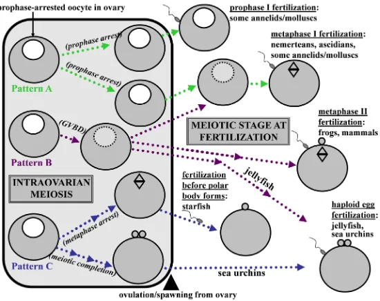
The first readily recognizable indicator of maturation onset is a process called germinal vesicle breakdown (GVBD). During GVBD, prophase arrest is overridden, and the oocyte disassembles its GV in preparation for two unequal meiotic divisions that will ultimately yield a large haploid egg with small polar bodies located at the egg’s animal pole. In many species, maturing oocytes complete their pre-fertilization phase of meiosis by entering into a secondary cell cycle arrest at metaphase I or metaphase II (Nishiyama et al., 2010; Chiba, 2011), although various exceptions to this pattern occur throughout the animal kingdom (Fig. 1).
The complex signaling pathways regulating oocyte maturation have yet to be fully elucidated, but a well documented downstream effector of both meiotic reinitiation from prophase arrest and the
www.intjdevbiol.com
*Address correspondence to: Stephen A. Stricker. Department of Biology, University of New Mexico, Albuquerque, NM 87131 USA. E-mail: sstr@unm.edu
Accepted: 20 July 2015.
ISSN: Online 1696-3547, Print 0214-6282
© 2015 UBC Press Printed in Spain
Abbreviations used in this paper: 5HT, serotonin (5-hydroxytryptamine); ASW,artificial seawater; cADPR, cyclic ADP ribose; CaFSW, calcium-free seawater; CaM, calmodulin; CaMKII,calcium/calmodulin dependent kinase II; Epac, exchan-ge protein directly activated by cAMP; DAG, diacylglycerol; ER, endoplasmic reticulum; GVBD, germinal vesicle breakdown; IP3, inositol 1,4,5 trisphosphate; MAPK, mitogen-activated protein kinase; MPF, maturation-promoting factor; NSW, natural seawater; PKC, protein kinase C; RyR, ryanodine receptor; SW, seawater; TPA, tetradecanoylphorbol-13-acetate.
establishment of a secondary metaphase arrest is the protein complex called maturation-promoting factor (=M-phase-promoting factor, MPF) (Kishimoto, 2003; Hara et al., 2012). As the names suggest, MPF induces maturation in oocytes and drives interphase to M-phase transitions in general. Accordingly, in immature oocytes, MPF activity is relatively low, whereas in response to maturation-inducing stimuli, MPF is activated to trigger GVBD and meiotic progression. Subsequently, metaphase-arrested oocytes maintain high levels of MPF activity in association with a cytostatic arrest factor (CSF) that generally depends on mitogen-activated protein kinase (MAPK) signaling (Nishiyama et al., 2010).
the important roles played by such signaling molecules as cyclic nucleotides and divalent cations (e.g. Jaffe and Norris, 2010; Wakai and Fissore, 2013). In particular, oocytes or eggs are known to elevate their concentrations of intracellular calcium ions (Ca2+)
during fertilization, typically by releasing Ca2+ from stores within
the endoplasmic reticulum (ER) via inositol-1,4,5 trisphosphate (IP3)-dependent- or IP3-independent pathways (Miyazaki, 2006).
Accordingly, in order to generate effective Ca2+ signals during
fertilization, oocytes of most animals must first enhance their calcium release capabilities during meiotic maturation (Stricker, 1999; Nader et al., 2013), and in mature, metaphase-arrested oocytes undergoing fertilization, such enhanced capabilities en-able the production of robust calcium responses that ultimately downregulate MPF and CSF activities to allow the completion of meiosis (Nishiyama et al., 2010).
However, unlike the well established functions of intraoocytic calcium transients in triggering meiotic progression after fertilization, there is little consensus as to whether or not calcium signaling also mediates the pre-fertilization reinitiation of meiosis in prophase-arrested oocytes. For example, in two intensively studied groups of vertebrates, frogs and mammals, early studies suggest either a requirement (e.g. Moreau et al., 1980; Goren et al., 1990), or a non-essential role (e.g. Cork et al., 1987; Tombes et al., 1992), for calcium signals during GVBD. Based on more recent analyses showing intraoocytic Ca2+ elevations downregulate, rather than
by intraoocytic cAMP elevations (Deguchi et al., 2011), and follow-ing spawnfollow-ing, mature haploid eggs undergo fertilization (Fig. 1). Similarly, in isolated oocytes of various hydrozoan jellyfish includ-ing Cytaeis, reinitiation from prophase I arrest is triggered by in
vitro treatments that raise intraoocytic cAMP levels (Takeda et al.,
2006; Deguchi et al., 2011), and in maturing Cytaeis oocytes that are stimulated by cAMP, a spike-like Ca2+ transient is generated
after, rather than before, GVBD (Fig. 2A). Accordingly, preload-ing Cytaeis oocytes with the Ca2+ chelator BAPTA abolishes the
cAMP-induced Ca2+ transient but does not prevent GVBD (Fig.
2B). In addition, artificial induction of a Ca2+ rise by the calcium
ionophores A23187 and ionomycin fails to trigger GVBD in Cytaeis (Fig. 2 C,D) and other hydrozoan species (Freeman and Ridgway, 1993). Thus, based on the current evidence that has been gathered for jellyfish oocytes, Ca2+ does not appear to play an essential role
in stimulating GVBD.
Nemerteans (ribbon worms)
Nemerteans constitute a phylum of about 1,300 marine spe-cies (Appeltans et al., 2012) within a larger protostome clade that includes molluscs and annelids. Nemerteans are usually dioe-cious, and gravid females typically possess numerous ovaries with relatively small (~70-200 mm) and translucent oocytes that lack follicle cells (Stricker et al., 2001). Whether nemertean oocytes in the field begin GVBD before or after being spawned has yet
Fig. 1. Patterns of meiotic progression relative to fertilization for animal groups mentioned in this article.(Pattern A) Prophase-arrested oocytes are released from the ovary. (Pattern B) Germinal vesicle breakdown (GVBD) is triggered within the ovary, and maturing oocytes are released before a secondary cell cycle arrest is attained either prior to or after meiotic divisions are completed. (Pattern C)
Meiosis is reinitiated in the ovary, and maturing oocytes reach a secondary arrest before release from the ovary. Note: optimal rates of normal fertilization in starfish occur if post-GVBD oocytes are fertilized before polar body formation, and unlike intraovarian oocytes that arrest at metaphase I before spawning, prophase-arrested starfish oocytes that are removed from ovaries and treated with the maturation inducer 1-MA in the absence of sperm can complete meiotic divisions without establishing a metaphase arrest (not shown here). This figure is based mainly on data collated by Nishiyama et al. (2010) and Chiba (2011), which contain further details regarding various subtypes within these patterns.
promote, progesterone-induced maturation in Xenopus (Sun and Machaca, 2004), it has been concluded that calcium tran-sients are not required for GVBD in frog oocytes (Machaca, 2011). Alternatively, in spite of findings that follicle-enclosed mouse oocytes can undergo GVBD independently of calcium signals (Mehlmann et al., 2006), calcium is still viewed as a key stimulator of GVBD in mammals (e.g. Silvestre et al., 2011; Wang and Machaty, 2013), although whether or not mammalian GVBD routinely requires a Ca2+ elevation
con-tinues to be debated.
Given that data for and against calcium signals controlling GVBD in vertebrate oocytes have been thoroughly summa-rized, this article attempts to expand the scope of coverage by considering oocytes produced by marine invertebrates. Thus, studies related to calcium signals during the onset of oocyte maturation in several groups of marine invertebrates are reviewed, and some new data regarding calcium signaling in jellyfish oocytes are presented. In addition, since a robust calcium response during fertilization of post-GVBD oocytes is a well documented driver of meiotic completion, evidence showing an enhanced ability to generate calcium signals in mature vs. immature oocytes is also reviewed for marine invertebrates. Finally, potential explanations for apparent discrepancies in the literature are presented.
Results and Discussion
Cnidarians (jellyfish)
to be conclusively determined. However, fully-grown nemertean oocytes isolated from the ovary in the laboratory rapidly undergo GVBD in response to natural seawater (NSW) or cAMP-elevating stimuli before eventually arresting at metaphase I (Stricker et al., 2013; Stricker, 2014). Such beneficial properties have facilitated analyses of intraoocytic calcium dynamics, and this section begins by summarizing findings related to calcium signals during meiotic reinitiation in the nemerteans Cerebratulus lacteus and Micrura
alaskensis (Stricker and Smythe, 2000).
Oocytes of C. lacteus and M. alaskensis typically remain arrested at prophase I after being transferred from the ovary to calcium-free solutions of artificial SW (CaFSW). Such inhibition is not simply due to morbidity, since CaFSW-treated oocytes undergo GVBD when subsequently placed in NSW. Moreover, nearly all GV-containing oocytes maintained in calcium-containing artificial seawater (ASW), but not in CaFSW, complete GVBD when treated with A23187, and A23187-induced maturation is prevented by the oolemmal calcium channel blocker cobalt chloride, suggesting that external calcium influx can promote GVBD in nemerteans. However, transferring intraovarian oocytes directly into calcium-containing ASW typically promotes lower levels of GVBD than are obtained with NSW, and although cobalt treatments block ASW-induced GVBD, adding cobalt to NSW fails to inhibit GVBD. Similarly, following incubation in CaFSW, calcium-containing ASW is not as effective as NSW in restoring GVBD, but conditioning ASW with marine sediments for several weeks allows ASW to become as potent a stimulator of GVBD as NSW. Collectively, such findings suggest that calcium-containing ASW does not fully mimic NSW and that external calcium influx is not the only mechanism mediating GVBD in nemerteans.
Accordingly, nemertean oocytes that are triggered to mature by 1 mM serotonin (5-hydroxytryptamine, 5HT) achieve ~90% GVBD rates in both calcium-containing and calcium-free ASW solutions. In addition, when monitored for intracellular calcium signals by time-lapse confocal microscopy, 5HT consistently induces GVBD without eliciting an obvious calcium transient, even though such maturing oocytes generate a series of marked calcium oscillations upon insemination and subsequently develop into normal
blastu-lae. Taken together, these findings suggest that although external calcium influx can facilitate the reinitiation of meiosis under certain experimental conditions, neither the influx of calcium ions nor the generation of a distinct intraoocytic calcium transient is invariably required for normal GVBD in nemerteans.
Following GVBD, maturing oocytes of nemerteans enhance their ability to produce a robust calcium response, as evidenced by the fact that fertilization of metaphase-I-arrested oocytes routinely induces an oscillatory series of point-source calcium waves prior to normal embryogenesis, whereas inseminated prophase-arrested specimens typically generate only a single non-wavelike calcium transient before failing to develop (Stricker, 1996; Stricker and Smythe, 2003). The precise reasons for these differences remain unknown, but the homogeneous ER of prophase-arrested oocytes undergoes an MPF-dependent reorganization to form discrete clusters in metaphase-I-arrested specimens prior to fertilization. Such ER clusters then disassemble at the time when fertilization-induced calcium oscillations cease either in controls or in oocytes subjected to the MPF inhibitor roscovitine (Stricker et al., 1998; Stricker and Smythe, 2003; Stricker, 2006). Collectively, such findings suggest maturation-associated reorganizations of the ER help ensure that proper calcium signals are generated during nemertean fertilizations.
Molluscs (bivalves, limpets)
Approximately 50,000 species of marine molluscs are grouped into eight classes of the phylum Mollusca (Appletans et al., 2012). Among these taxa, intraoocytic Ca2+ changes during the reinitiation
of meiosis have been examined in bivalves (class Bivalvia) (Fig. 3A) and limpets (class Gastropoda). In most bivalves and limpets, numerous fully-grown oocytes arrested at prophase I can be dis-sected from the ovaries. The diameters of oocytes are less than 100 mm in most bivalves (e.g. Deguchi and Osanai, 1993; 1994; Zhang
et al., 2009; Fig. 3B) and 120-200 mm in limpets (e.g. Guerrier et
al., 1986). Limpet oocytes remain enclosed in a thin layer of follicle
cells after removal from the ovary (Guerrier et al., 1986; Gould et
al., 2001), whereas bivalve oocytes lack surrounding cells
(Degu-Fig. 2. Inability of a Ca2+ rise to
trigger meiosis reinitiation in oocytes of the jellyfish cnidar-ian Cytaeis uchidae.(A) Cytaeis
oocytes isolated from the ovaries are arrested at prophase I and are induced to resume meiosis by injection of 8-Br-cAMP (final intracellular concentration: 25-50 mM). Based on fluorescence ratios of the Ca2+ indicator Fura-2
(F340/F380), 8-Br-cAMP triggers a spike-like Ca2+ transient, but this
transient appears after, rather than before, GVBD. (B) If the 8-Br-cAMP-induced Ca2+ transient is blocked by co-injection of 8-Br-cAMP plus the Ca2+ chelator BAPTA (final
intracel-lular concentration: 1-2 mM), the timing of GVBD is not altered. (C,D) Application of the Ca2+ ionophore A23187 (C) or ionomycin (D) at 10 mM causes
a substantial Ca2+ rise, which is measured using Calcium Green-1-dextran (10 kDa) and expressed as F/F
0, but both treatments fail to trigger GVBD.
B
C
chi and Osanai, 1994; Fig. 3B). Although some isolated oocytes of bivalves and limpets undergo spontaneous meiotic reinitiation in a species-, season-, or batch-dependent manner, most remain arrested at prophase I until further stimulation.
In a few bivalves such as Spisula, Mactra, and Barnea, isolated and naturally spawned oocytes normally do not resume meiosis and undergo GVBD until after being fertilized (Fig. 1) (Deguchi and Osanai, 1994 and references therein). During such fertilizations, prophase-arrested oocytes exhibit a Ca2+ rise that comprises an
initial peak (cortical flash) and a subsequent elevated plateau lasting for several minutes (Deguchi and Osanai, 1994; Deguchi and Morisawa, 2003). Several lines of evidence suggest that the sperm-induced Ca2+ rise in these bivalves strictly depends on the
influx of external Ca2+ (see Kashir et al., 2013, for a recent review)
and GVBD can be triggered only when Ca2+ influx and the resultant
intraoocytic Ca2+ rise continue for at least a few minutes (Deguchi
and Osanai, 1994).
On the other hand, most bivalves normally spawn oocytes that are already undergoing GVBD, and such oocytes subsequently arrest at metaphase I prior to fertilization (Fig. 1). In this type of bivalve (e.g. Ruditapes, Hiatella, Patinopecten, Mercenaria, and
Crassostrea), as well as in Spisula, prophase-arrested oocytes that
are removed from the ovary can be triggered to resume meiosis in
vitro by micromolar concentrations of 5HT (Osanai and Kuraishi,
1988; Guerrier et al., 1993; Deguchi and Osanai, 1995; Fong et
al., 1997; Colas and Dubé, 1998; Leclerc et al., 2000; Zhang et al., 2009; Yuan et al., 2012). Accordingly, because of its
bioactiv-ity and localization within the gonad, 5HT is believed to function
in vivo as a physiologically relevant neurohormone that generally
triggers intraovarian meiotic reinitiation from prophase I, spawn-ing of maturspawn-ing oocytes, and a subsequent metaphase I arrest in bivalve oocytes.
On the other hand, injecting ripe Spisula females with 5HT causes prophase-arrested oocytes to be spawned, and treatment of isolated GV-containing oocytes of Spisula with high concentra-tions of 5HT triggers meiotic completion without a metaphase I arrest (reviewed by Colas and Dubé, 1998). Thus, in Spisula, 5HT apparently regulates meiotic progression differently than in bivalve oocytes that arrest at metaphase I prior to fertilization. Nevertheless, nanomolar concentrations of 5HT are still believed to enhance the ability of Spisula oocytes to undergo successful fertilization (Masseau et al., 2002).
Addition of exogenous 5HT to Ruditapes, Hiatella, and Spisula oocytes arrested at prophase I causes a transient Ca2+ rise
fol-lowed by an elevated plateau (Guerrier et al., 1993; Deguchi and Osanai, 1995; Fong et al., 1997; Colas and Dubé, 1998). Both the amplitude and the duration of the Ca2+ rise depend on the
con-centration of 5HT (Deguchi and Osanai, 1995; Fong et al., 1997), and the 5HT-induced Ca2+ rise is propagated as a point-source
Ca2+ wave that rapidly spreads across the oocyte (Fig. 3C). Such
a transmission pattern is in turn unlike the initial cortical flash of fertilization (Deguchi and Morisawa, 2003) or the non-wavelike calcium transient that is stimulated by excess K+ (Fig. 3D). Ca2+
release from IP3-sensitive stores may be the main mediator of the
5HT-induced Ca2+ wave, since: i) a Ca2+ rise persists in the
ab-sence of external Ca2+ (Guerrier et al., 1993; Deguchi and Osanai,
1995; Leclerc et al., 2000), but is suppressed by the IP3 receptor
antagonist heparin (Deguchi and Osanai, 1995), and ii) adding 5HT generates a transient increase in intraoocytic levels of IP3 (Gobet et al., 1994). However, in addition to internal calcium release, a
Ca2+ influx pathway is also activated following application of 5HT,
and such influx enhances the action of 5HT (Guerrier et al., 1993; Deguchi and Osanai, 1995; Colas and Dubé, 1998). In any case, the amplitude and duration of 5HT-induced Ca2+ rises are tightly
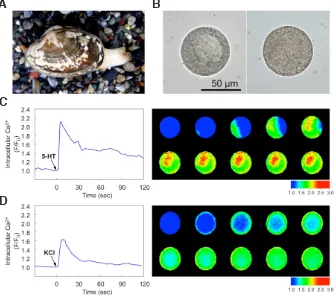
Fig. 3. Meiotic reinitiation by a 5HT-induced Ca2+ rise, but not by a K+-induced Ca2+ rise,
in oocytes of the Japanese littleneck bivalve mollusc Ruditapes philippinarum. (A) Adult clams (shell length: 3-6 cm) can be collected in the field or purchased from Japanese fish shops and supermarkets. (B, left)Ruditapes oocytes isolated from the ovaries remain arrested at prophase I. (B, right) Alternatively, oocytes exposed to 5HT at 1
mM undergo GVBD and subsequently arrest at metaphase I. (C, left) Addition of 5HT immediately causes an intraoocytic Ca2+ rise comprised of an
initial peak and a subsequent plateau, which are plotted in terms of the relative fluorescence inten-sity (F/F0) of the Ca2+ indicator Calcium Green-1. (C, right) Pseudocolored images taken every 0.5 sec reveal the initial rising phase as a point-source Ca2+ wave starting from the cortex at the opening
of a measurement chamber. (D) By contrast, in analyses using the same methods for monitoring and graphing calcium transients, a smaller Ca2+ rise
in the form of a cortical flash is triggered by excess K+ seawater (0.53 M KCl: normal seawater = 1: 4),
and such a flash fails to induce GVBD.
B
C
coupled with the presence or absence of GVBD, and 5HT cannot trigger GVBD without a significant intraoocytic Ca2+ rise (Guerrier et al., 1993; Gobet et al., 1994; Deguchi and Osanai, 1995; Fong et al., 1997; Leclerc et al., 2000; Deguchi and Morisawa, 2003).
Similarly, the stimulatory or inhibitory effects of various agonists or antagonists of GVBD correlate well with the amplitudes and dura-tions of the Ca2+ rises caused by these agents (Fong et al., 1997).
Based on studies using pharmacological modulators of mam-malian 5HT receptors (e.g. Gobet et al., 1994; Fong et al., 1997; Osada et al., 1998), 5HT receptors of bivalve oocytes appear to be distinct from any known mammalian 5HT receptor and instead tend to present a mixed pharmacological profile of 5HT1/5HT2/5HT3
receptors, with some subtypes of these three receptor classes having been shown to regulate calcium signals in somatic cells (Pytliak et al., 2011). Alternatively, a recently cloned 5HT receptor from the bivalve Patinopecten (5HTpy) exhibits specific sequence
homology to mammalian 5HT1 receptors, which can also modulate
intracellular calcium levels (Hill et al., 2000), and immunolocalization analyses reveal mRNA for such a receptor in intraovarian oocytes of Patinopecten (Tanabe et al., 2010).
Regardless of the type of 5HT receptor that may be expressed, intraovarian oocytes of bivalves are apparently protected from the premature action of 5HT by an “oocyte maturation arresting factor” (OMAF) that has been characterized in Patinopecten as a protein-containing substance with an estimated molecular mass of ~60 kDa (Tanabe et al., 2006; Yuan et al., 2012). Accordingly, OMAF inhibits a 5HT-induced Ca2+ rise and subsequent GVBD
in Ruditapes oocytes and prevents Patinopecten ovaries from releasing maturing oocytes (Tanabe et al., 2006).
The effects of various chemical agents other than 5HT also suggest essential roles for Ca2+ rises during GVBD in some bivalve
oocytes. For example, Ca2+ ionophores such as A23187 and
iono-mycin mobilize external and internal Ca2+ to produce an intraoocytic
Ca2+ rise that is followed by GVBD (Osanai and Kuraishi, 1988;
Guerrier et al., 1993; Deguchi and Osanai, 1994; 1995; Leclerc
et al., 2000; Zhang et al., 2009). Similarly, excess K+ seawater,
which evokes Ca2+ influx as a result of membrane depolarization,
is a reliable inducer of GVBD in bivalve oocytes that are normally fertilized at prophase I (Deguchi and Osanai, 1994 and references therein). In oocytes of Ruditapes, in contrast, excess K+ causes
a relatively small Ca2+ rise and fails to trigger GVBD (Guerrier et al., 1993; Leclerc et al., 2000; Fig. 3D). Moreover, although weak
bases such as ammonia are frequently used to cause intracellular alkalization, such bases can also accelerate a pre-GVBD Ca2+
rise in some bivalve oocytes mainly via the release of Ca2+ from
internal stores (e.g. Guerrier et al., 1993; Leclerc et al., 2000). Similarly, thapsigargin, which is known to induce an intracellular Ca2+ rise by inhibiting the Ca2+ pump of the ER, also triggers GVBD
in Ruditapes oocytes (Guerrier et al., 1993). In addition, injecting IP3 or photoreleasing caged IP3 induces Ca2+ release and GVBD
in Spisula, Mactra, and Ruditapes oocytes (Bloom et al., 1988; Guerrier et al., 1996; Deguchi and Morisawa, 2003). Accordingly, if the Ca2+ rises induced by the above mentioned chemicals are
inhibited, GVBD is also specifically blocked (Guerrier et al., 1993; Deguchi and Osanai, 1994; Leclerc et al., 2000; Deguchi and Mori-sawa, 2003). For example, in Mactra oocytes, pre-treatments with heparin and D-600 inhibit the Ca2+ rises and subsequent GVBD
that are normally induced by IP3 and excess K+, respectively, but
neither heparin nor D-600 blocks these processes in oocytes that
are stimulated by excess K+ and IP
3, respectively (Deguchi and
Osanai, 1994; Deguchi and Morisawa, 2003). Collectively, such findings indicate that an intraoocytic Ca2+ rise is a prerequisite for
meiotic reinitiation from prophase I in the bivalves described above. On the other hand, the role of calcium during GVBD either re-mains unclear or has been shown to be non-essential in bivalves such as Crassostrea, Limaria, and Mytilus, as well as in limpets such as Patella and Lottia, where naturally spawned oocytes arrest at metaphase I prior to fertilization. For example, in Crassostrea oocytes that undergo GVBD and arrest at metaphase I in response to 5HT (Osanai and Kuraishi, 1988), there are conflicting reports regarding the presence or absence of a Ca2+ rise during GVBD
(Kyozuka et al., 1997; Leclerc et al., 2000). Similarly, it is unclear whether or not GVBD is indeed triggered by artificially elevated Ca2+ levels in Crassostrea (Kyozuka et al., 1997; Leclerc et al.,
2000). In addition, the 5HT receptor profiles determined using spe-cific agonists and antagonists are quite different in two published papers (Kyozuka et al., 1997; Osada et al., 1998), although such discrepancies may well be due to the remarkable variability in maturity and responsiveness to 5HT that Crassostrea oocytes can exhibit during the breeding season (Leclerc et al., 2000).
In the bivalves Limaria and Mytilus, oocytes cannot be stimu-lated to mature by 5HT (Osanai and Kuraishi, 1988; Deguchi and Osanai, 1993). Moreover, A23187 either only weakly induces or does not stimulate GVBD in Limaria (Deguchi and Osanai, 1993; 1994) and Mytilus (Osanai and Kuraishi, 1988), respectively. Furthermore, an intraoocytic Ca2+ rise is neither necessary nor
sufficient for GVBD in the limpets Patella and Lottia (Guerrier et
al., 1986; Gould et al., 2001). Collectively, such findings indicate
a calcium-independent mechanism of GVBD, and indeed, meiosis reinitiation from prophase I arrest in these bivalves and limpets strictly depends on a pre-GVBD rise in intraoocytic pH (Guerrier et
al., 1986; Deguchi and Osanai, 1994; Gould et al., 2001). Similarly,
a pH-mediated mode of GVBD has also been reported for bivalve oocytes that require Ca2+ elevations for GVBD, as Ca2+ and pH
rises may act synergistically to reinitiate meiosis in such species (see Deguchi and Osanai, 1994; 1995; Dubé and Eckberg, 1997; Colas and Dubé, 1998 and references therein).
The pathways that potentially link pre-GVBD elevations in in-traoocytic Ca2+ and/or pH with the activation of cell cycle kinases
such as MPF and MAPKs remain largely unknown in molluscs (reviewed by Colas and Dubé, 1998). However, since phospholi-pase C presumably produces diacylglycerol (DAG) along with IP3
in oocytes stimulated by 5HT, it follows that DAG-sensitive isotypes of protein kinase C (PKC) would also be activated. Indeed, in
Spisula oocytes, PKC agonists such as TPA
(tetradecanoylphorbol-13-acetate) trigger GVBD, whereas antagonists of PKC can block GVBD that is normally induced by fertilization or TPA (Eckberg et
al., 1987). Alternatively, TPA inhibits rather than accelerates GVBD
in Ruditapes oocytes stimulated by 5HT, apparently by suppressing its action on a Ca2+ rise (Gobet et al., 1994).
Another potential calcium-sensitive mediator of GVBD is calcium/ calmodulin dependent kinase II (CaMKII), based on the inhibitory effects of calmodulin (CaM) antagonists on GVBD in some bivalve oocytes (Carroll and Eckberg, 1986; Zhang et al., 2009). However, as opposed to simply suppressing a pathway downstream to the maturation-associated Ca2+ elevation, such inhibitors can also
In any case, although the underlying regulatory mechanisms of GVBD have yet to be fully elucidated, many molluscan oocytes are known to enhance their abilities to generate proper fertilization-induced Ca2+ responses during maturation from prophase I to
metaphase I, with optimal responses generated during fertilization typically taking the form of repetitive Ca2+ oscillations in bivalves
versus a single but prolonged Ca2+ rise in limpets (Kashir et al.,
2013). Moreover, fertilization-induced Ca2+ signals are necessary
and sufficient for these oocytes to be released from a metaphase I arrest (Kashir et al., 2013), whereas an additional pH eleva-tion is not required for such cell cycle progression (Deguchi and Osanai, 1995).
Annelids (echiuran and polychaete worms)
Recent molecular-based phylogenies have indicated that the phylum Annelida with its ~14,000 marine species (Appeltans et
al., 2012) should also include echiuran worms that were formerly
viewed as constituting their own phylum (Struck et al., 2011). Thus, the echiuran Urechis is treated here as an annelid.
Oocytes of annelids are fertilized at prophase I (e.g. Urechis) or at metaphase I (e.g. Chaetopterus, Arenicola, and
Pseudo-potamilla) (Meijer, 1979; Stephano and Gould, 1997; Kashir et al., 2013), and in either case, fully-grown oocytes arrested at
prophase I accumulate in the coelom after having already been freed from follicle cells. As in molluscs, the connection between a Ca2+ rise and subsequent meiotic progression is well documented
for annelid oocytes that are normally fertilized at prophase I (Fig. 1). Accordingly, fertilized oocytes of Urechis exhibit a single Ca2+
rise initiated by a non-wave-like Ca2+ transient (Stephano and
Gould, 1997), which is probably homologous to the cortical flash seen in mollusc oocytes. Based on several lines of evidence, Ca2+
influx is responsible for the Ca2+ rise at fertilization, although the
supplemental contribution of IP3-dependent Ca2+ release remains
possible (Stephano and Gould, 1997). Moreover, a Ca2+ rise in Urechis oocytes is necessary but not sufficient for meiosis
reinitia-tion from prophase I, as GVBD proceeds only when an intraoocytic Ca2+ and pH rise occur simultaneously (Stephano and Gould, 1997
and references therein).
In oocytes of polychaete annelids that arrest at metaphase I before fertilization (Fig. 1), induction of GVBD in isolated oocytes is triggered in various ways, including: i) transfer into natural, but not artificial, seawater in Chaetopterus (Ikegami et al., 1976); ii) exposure to a protease purified from digestive tract fluids in
Sa-bellaria (Peaucellier, 1978), and iii) application of coelomic fluid
supplemented with prostomial homogenates in Arenicola (Watson
et al., 1998). Chaetopterus oocytes incubated in artificial seawater
can be stimulated to undergo GVBD by excess K+ in the presence
of external Ca2+ (Ikegami et al., 1976) and by A23187 even in Ca2+
-free seawater (Eckberg and Carroll, 1982). A23187 also triggers GVBD in Sabellaria oocytes (Peaucellier, 1978) but not in Arenicola oocytes (Meijer, 1979; Watson et al., 1998). Neither A23187 nor ionomycin induces GVBD in Pseudopotamilla oocytes (Nakano and Deguchi, unpublished observation), and instead an intraoocytic cAMP elevation seems to be essential for this process (Deguchi
et al., 2011). It should be noted that release from the secondary
arrest at metaphase I absolutely depends on an intraoocytic Ca2+
rise in all of the polychaetes listed above (Meijer, 1979; Kashir et
al., 2013), regardless of the importance of a Ca2+ rise at prophase I.
In Chaetopterus, it is thought that activations of PKC and CaM
are essential steps in the activation of MPF, and that PKC directly phosphorylates CDK1 (cdc2) to yield active MPF (Eckberg et al., 1996). However, the actual interactions between a Ca2+ rise and
PKC or CaMKII activities have not been fully documented.
Echinodermata (starfish, sea urchins)
Approximately 2,000 starfish species constitute the class Aster-oidea within the deuterostome phylum Echinodermata (Appeltans
et al., 2012). Most starfish have separate sexes, and ripe females
store numerous prophase-arrested oocytes in their ovaries. When triggered by environmental cues, a relaxin-like peptide is released from the nervous system of females to cause follicle cells around fully grown oocytes to produce a maturation-inducing substance (=1-methyladenine, 1-MA) that initiates the signaling cascade leading to GVBD (Mita, 2013). In prophase-arrested oocytes that are removed from the ovary and treated with 1-MA in vitro, meiosis is completed without a secondary arrest, whereas injecting 1-MA into females to replicate a more natural mode of maturation induc-tion causes oocytes to arrest at metaphase I, owing to a reduced intraoocytic pH that is established in the ovary, but not in seawater (Harada et al., 2003). Following the spawning of metaphase-arrested oocytes from 1-MA-injected females, seawater triggers an intraoo-cytic pH rise that eventually allows meiotic progression, and given that the lowest levels of polyspermy occur when inseminations are carried out between GVBD and first polar body formation, the metaphase-I arrest is viewed as an adaptation for ensuring that oocytes released during protracted spawnings in the field have not already undergone polar body formation before encountering sperm (Chiba, 2011).
Although the role of calcium during GVBD in starfish oocytes that undergo a metaphase I arrest has not been elucidated, numerous investigations have assessed calcium dynamics in isolated starfish oocytes that mature in vitro and complete their meiotic divisions without arresting at metaphase. For example, pioneering analyses of the starfish Marthasterias glacialis have shown that external ap-plications of 1-MA in CaFSW trigger a pre-GVBD Ca2+ transient and
that blocking the 1-MA-induced Ca2+ transient via EGTA injection
prevents GVBD (Moreau et al., 1978). These and various other findings support the view that a Ca2+ rise normally promotes GVBD
in starfish (Moreau et al., 1985). However, in additional studies of
M. glacialis and other starfish, GVBD can proceed in control 1-MA
treatments without a marked Ca2+ transient being generated, even
though subsequent inseminations trigger a recognizable Ca2+
re-sponse, and, contrary to previous reports, 1-MA-induced GVBD is not blocked by calcium chelators in such studies (Picard and Doree, 1983, Eisen and Reynolds, 1984; Witchel and Steinhardt, 1990; Kikuyama and Hiramoto, 1991; Stricker, 1995).
In attempting to reconcile these contradictory results, it should be noted that 1-MA causes oocytes of the starfish Asterina
pec-tinifera and Astropecten aranciacus to generate both cytoplasmic
and nuclear Ca2+ elevations before GVBD (Santella and Kyozuka,
1994; Santella et al., 1998; Lim et al., 2003), and that injections of BAPTA into the nucleus, but not into the cytoplasm, can inhibit 1-MA-induced GVBD (Santella and Kyozuka, 1994). Similarly, in 1-MA-treated A. pectinifera oocytes, all elevations in Ca2+ and
subsequent GVBD are eliminated by the membrane-permeable calcium release blocker TMB-8 (Tosuji et al., 2007), presumably because, unlike BAPTA, TMB-8 can cross the nuclear envelope to affect nuclear Ca2+ signals. Moreover, injecting IP
ribose (cADPR) into GVs of immature oocytes can trigger nuclear Ca2+ transients, and following such elevations, some of the injected
oocytes complete GVBD in the absence of 1-MA (Santella and Kyozuka, 1997a). Accordingly, some 1-MA-treated oocytes do not undergo GVBD, if IP3 and cADPR blockers are injected into the
GV (Santella and Kyozuka, 1997a; Stanella et al., 1998), whereas cytoplasmic delivery of the inhibitors routinely fails to prevent GVBD in 1-MA-stimulated oocytes (Santella and Kyozuka, 1997a; Iwasaki
et al., 2002). In addition, nuclear injections of antibodies to CaM,
a likely downstream target of Ca2+ signals, block GVBD in
1-MA-treated oocytes, whereas co-injecting the antibodies with excess CaM allows GVBD (Santella and Kyozuka, 1997b). Collectively, such findings suggest that in these two starfish nuclear Ca2+ transients
promote GVBD via a CaM-dependent mode of MPF activation. In terms of upstream processes that might regulate such Ca2+
signals in starfish oocytes, reorganizations of the cortical actin network may play a key role, based on: i) the rapid restructuring of actin filaments that routinely occurs before GVBD in 1-MA-treated controls; ii) the fact that the 1-MA-induced calcium wave propa-gates exclusively in the cortical region where actin filaments are concentrated, and iii) the findings that experimental manipulations to hypo- or hyperpolymerize cortical actin serve to disrupt normal Ca2+ dynamics and maturation (Kyozuka et al., 2008; 2009; Chun
and Santella, 2009; Chun et al., 2013). The precise mechanisms by which a remodeling of the actin network might affect Ca2+ release
capabilities in starfish oocytes are not known. However, based on results from analyses of somatic cells, changes in the organiza-tion of actin filaments could both directly and indirectly modulate IP3 receptor function (Chun and Santella, 2009). Alternatively, the
actin network itself might serve as an intracellular calcium pool that releases Ca2+ during 1-MA-induced restructuring (Chun and
Santella, 2009).
As opposed to the more variable results that have been reported regarding the role of calcium signals in triggering GVBD, starfish oocytes have consistently been demonstrated as developing an en-hanced capacity for IP3-mediated calcium release following GVBD,
owing to a heightened sensitivity to IP3 rather than to any marked
increase in IP3 receptor mass (Chiba et al., 1990; Iwasaki et al.,
2002; Lim et al., 2003). The enhanced capacity for Ca2+ release in
turn allows a more robust and effective fertilization-induced calcium response to be generated in post-GVBD vs. prophase-arrested specimens (Stricker et al., 1994), and although ER reorganizations have also been documented in maturing starfish oocytes (Jaffe and Terasaki, 1994), such changes occur well after actin remodeling is induced by 1-MA, suggesting that at least the early enhancements in calcium signaling capabilities rely more on actin reorganizations (Chun and Santella, 2009). Accordingly, the increased sensitivity to IP3 during starfish oocyte maturation depends on MPF activation,
which apparently modifies IP3-mediated signaling not by directly
phosphorylating IP3 receptors but instead by reorganizing the
cortical actin network (Lim et al., 2003).
Approximately 1,000 species of sea urchins and their allies constitute the class Echinoidea of the phylum Echinodermata (Appeltans et al., 2012). Sea urchins normally complete meiotic divisions inside the ovary without a metaphase arrest prior to spawn-ing, but fully-grown prophase-arrested oocytes of sea urchins can be removed from the ovary and allowed to undergo GVBD in vitro (Wessel et al., 2002). Although several types of inhibitors have been shown to block such in vitro GVBD (Wessel et al., 2002),
the roles played by calcium signals during meiotic reinitiation from prophase arrest have not been fully elucidated. However, calcium release capabilities mediated by both IP3- and non- IP3-dependent
pathways are enhanced during in vitro maturation of sea urchin oocytes, and sperm generate more robust calcium responses along with lower levels of polyspermy in post-GVBD vs. prophase-arrested specimens (Miyata et al., 2006).
Ascidians (sea squirts)
As deuterostome invertebrates closely related to vertebrates, ~3,000 species of ascidians belong to the subphylum Tunicata within the phylum Chordata (Appeltans et al., 2012). All ascidians are hermaphrodites that produce follicle-enclosed oocytes, and depending on the species examined, fully grown oocytes in the ovary can either remain arrested at prophase I or undergo GVBD (Lambert, 2005). After spawning, ascidian oocytes mature to a metaphase I arrest before being fertilized (Costache et al., 2014), although in vitro fertilization of certain GV-containing ascidian oocytes also yields some normal development (Gallo et al., 2013).
Immature oocytes of several ascidian species can be triggered to undergo GVBD by treatment with calcium-containing seawaters, whereas spontaneous maturation is reversibly inhibited if oocytes are incubated in CaFSW (Sakairi and Shirai, 1991; Cuomo et al., 2006; Lambert, 2011). Similarly, compared to the levels of forskolin-induced GVBD in low pH SW, oocytes of Boltenia villosa undergo less maturation following pre-treatment with either the calcium chelator BAPTA or with inhibitors of ryanodine receptor (RyR) functioning that normally allows internal calcium release via non-IP3-dependent mechanisms (Lambert, 2011). Conversely, GVBD
in Boltenia can be induced by an agonist of the exchange protein directly activated by cAMP (Epac) (Lambert, 2011), which has been shown to trigger calcium release from RyRs in other cells, and although actual evidence has yet to be published, preliminary observations of calcium elevations occurring during GVBD in
Bol-tenia oocytes have been reported (Lambert, 2011).
In addition, the activity of T-type calcium channels increases as immature oocytes of Styela plicata grow larger during prophase I arrest, and in this species, such increased activity is correlated with the ability of some fully-grown prophase-arrested oocytes to undergo normal post-fertilization GVBD and subsequent embryogenesis (Gallo et al., 2013). Furthermore, compared to metaphase-arrested oocytes of Ciona intestinalis, prophase-arrested specimens gener-ate lower amplitude calcium transients in response to either IP3
injections or ionophore treatments (Cuomo et al., 2006). Collectively, such findings support the views that in ascidians Ca2+ elevations
mediated by external calcium influx and/or internal calcium release help to promote GVBD and that calcium-mobilizing mechanisms are enhanced during oocyte growth and maturation. Exactly how Ca2+ elevations might elicit GVBD in ascidian oocytes remains
un-known, but based on tests using pharmacological reagents, it has been proposed that a calcium-dependent activation of CaMKII may facilitate activation of the Cdc25 phosphatase, which in turn could help convert inactive pre-MPF into active MPF (Lambert, 2011).
Conclusions
Moreover, in many cases, such enhanced capabilities depend on structural and functional reorganizations that the ER undergoes during oocyte maturation (Stricker, 2006). These findings in turn help to explain why prophase-arrested oocytes of most animals are incapable of generating an effective fertilization-induced calcium response that promotes normal meiotic completion and subsequent development (Stricker, 1999).
Alternatively, although the key roles of calcium transients dur-ing fertilization and the later phases of meiotic maturation are well established, there is little consensus regarding the contributions of calcium signals to the initial resumption of meiosis from prophase I arrest. The strongest data for an essential role being played by intraoocytic Ca2+ signals during GVBD come from analyses of
nuclear Ca2+ transients in starfish oocytes, as well as from
investi-gations of oocyte maturation in molluscs, annelids, and ascidians. However, other studies of cnidarian, nemertean, mollusc, annelid, and starfish oocytes provide evidence that tend to refute the view that calcium signals are routinely needed for GVBD.
In attempting to reconcile these apparently conflicting results, experimental protocols need to be carefully assessed. For example, along with exhibiting different patterns of meiotic arrest (Nishiyama
et al., 2010), marine invertebrate oocytes can also vary in the
tim-ing of GVBD onset relative to the initial stimulation of maturation. Thus, as proposed for mammals (Homa et al., 1993), variable results might be obtained, depending on exactly when an experi-mental manipulation such as calcium chelation is carried out with respect to the delivery of a maturation-inducing stimulus. Similarly, although the notion of nuclear envelopes being impermeable to calcium ions remains controversial (Bootman et al., 2009), variable experimental conditions have been postulated as causing starfish GVs to differ from being freely permeable to relatively impermeable to Ca2+ flow (Santella and Kyozuka, 1997a). Hence, depending on
the physiological state of the nucleus at the time that maturation is stimulated, differing conclusions regarding the roles of Ca2+ during
GVBD could be reached. Similarly, the precise mode of stimulus delivery could differ and thereby affect the presence or absence of calcium signals, as has been demonstrated for oocytes of the starfish Asterina miniata, where the relatively slow diffusion of subthreshold levels of maturation-inducing hormone fails to elicit a calcium response prior to GVBD, whereas a more rapid perfusion consistently generates a Ca2+ transient that nevertheless has been
deemed neither necessary nor sufficient for meiotic reinitiation (Witchel and Steinhardt, 1990).
In addition to such considerations, the potential contributions of false-positive and false-negative results should also be evalu-ated. For example, the ability of certain EGTA injections to block 1-MA-induced oocyte maturation has been attributed to possible toxic effects of the particular experimental protocol that was used (Picard and Doree, 1983). Similarly, the fact that BAPTA-loaded
Xenopus oocytes fail to mature may not be due to calcium chelation,
but rather to the sequestration of such transition metals as zinc (Machaca, 2011). Conversely, in cases where calcium chelators fail to block GVBD, the chelator treatment should also be demon-strated as actually preventing a pre-GVBD calcium rise. Moreover, the fact that GVBD might not be triggered by exogenous calcium modulators such as ionomycin does not rule out an essential role for endogenous calcium transients, as not all calcium-mobilizing agents may be capable of triggering signals that fully mimic the complex temporal and spatial features that are normally required
for maturation onset (Vasilev et al., 2012).
Aside from assessing experimental protocols, the possibility that variability in the reported results arises from inherent species-specific differences needs to be considered. In fact, given the wide diversity in body plans and modes of reproduction exhibited by extant marine invertebrates, variable patterns and mechanisms of meiotic progression could certainly have evolved from one taxon to another, or even among closely related species within the same group. For example, unfertilized oocytes of some starfish species are known to arrest at G1 after the second meiotic divi-sion, whereas others terminate at G2 (Nishiyama et al., 2010). Similarly, oocytes of one starfish (Asterina pectinifera), but not of another (Astropecten aranciacus), respond to cADPR (Chun and Santella, 2009). Collectively, such findings indicate that not all starfish oocytes regulate meiotic progression and calcium signaling in precisely the same way.
Perhaps by examining additional species and taxa, a clearer set of patterns will become evident across the animal kingdom, thereby helping to clarify the roles played by calcium signals during the onset of oocyte maturation. Similarly, although attempts have been made to identify specific downstream targets of Ca2+
eleva-tions during cases of calcium-mediated GVBD, further analyses are needed to link Ca2+ signals with MPF activation and GVBD. In any
case, various gaps in the existing literature as well as substantial variability in the reported results indicate that our understanding of calcium signals during oocyte maturation in marine invertebrates remains a work in progress.
Acknowledgements
The authors thank both the Asamushi Research Center for Marine Biology of Tohoku University and the Friday Harbor Laboratories of the University of Washington for use of their facilities. We also apologize to researchers whose articles were not cited owing to limitations on the number of allowable references. Funding support: JSPS KAKENHI Grant Number 26440177 to RD; JSPS KAKENHI Grant Number 26840073 to NT.
References
APPLETANS W, AHYONG S T, ANDERSON G, ANGEL M V, ARTOIS T, BAILLY N et al., (2012). The magnitude of global marine species diversity. Curr Biol 22: 2189-2202. BLOOM T L, SZUTS E Z and ECKBERG W R (1988). Inositol trisphosphate, inositol
phospholipid metabolism, and germinal vesicle breakdown in surf clam oocytes. Dev Biol 129: 532-540.
BOOTMAN M D, FEARNLEY C, SMYRNIAS I, MACDONALD F and RODERICK H L (2009). An update on nuclear calcium signaling. J Cell Sci 122: 2337-2350. CARROLL A G and ECKBERG W R (1986). Inhibition of germinal vesicle breakdown
and activation of cytoplasmic contractility in Spisula oocytes by calmodulin an-tagonists. Biol Bull 170: 43-50.
CHIBA K (2011). Evolution of the acquisition of fertilization competence and polyspermy blocks during meiotic maturation. Mol Reprod Dev 78: 808-813.
CHIBA K, KADO R T and JAFFE L A (1990). Development of calcium release mecha-nisms during starfish oocyte maturation. Dev Biol 140: 300-306.
CHUN J T and SANTELLA L (2009). Roles of actin-binding proteins in intracellular Ca2+ signalling. Acta Physiol 195: 61-70.
CHUN J T, VASILEV F and SANTELLA L (2013). Antibody against the actin-binding protein depactin attenuates Ca2+ signaling in starfish oocytes. Biochem Biophys Res Commun 441: 301-307.
COLAS P and DUBÉ F (1998). Meiotic maturation in mollusc oocytes. Semin Cell Dev Biol 9: 539-548.
COSTACHE V, MCDOUGALL A and DUMOLLARD R (2014). Cell cycle arrest and activation of development in marine invertebrate deuterostomes. Biochem Biophys Res Commun 450: 1175-1181.
CUOMO A, SILVESTRE F, DESANTIS R and TOSTI E (2006). Ca2+ and Na+ current patterns during oocyte maturation, fertilization, and early developmental stages of Ciona intestinalis. Mol Reprod Dev 73: 501-511.
DEGUCHI R and MORISAWA M (2003). External Ca2+ is predominantly used for cytoplasmic and nuclear Ca2+ increases in fertilized oocytes of the marine bivalve Mactra chinensis. J Cell Sci 116: 367-376.
DEGUCHI R and OSANAI K (1993). Artificial induction of meiosis reinitiation from the first prophase and from the first metaphase in oocytes of the marine bivalve Limaria hakodatensis. Bull Mar Biol Stn Asamushi Tohoku Univ 19: 29-39. DEGUCHI R and OSANAI K (1994). Meiosis reinitiation from the first prophase is
dependent on the levels of intracellular Ca2+ and pH in oocytes of the bivalves Mactra chinensis and Limaria hakodatensis. Dev Biol 166: 587-599.
DEGUCHI R and OSANAI K (1995). Serotonin-induced meiosis reinitiation from the first prophase and from the first metaphase in oocytes of the marine bivalve Hiatella flaccida: respective changes in intracellular Ca2+ and pH. Dev Biol 171: 483-496. DEGUCHI R, TAKEDA N and STRICKER S A (2011). Comparative biology of cAMP-induced germinal vesicle breakdown in marine invertebrate oocytes. Mol Reprod Dev 78: 708-725.
DUBÉ F and ECKBERG W R (1997). Intracellular pH increase driven by an Na+/H+ exchanger upon activation of surf clam oocytes. Dev Biol 190: 41-54.
ECKBERG W R and CARROLL A G (1982). Sequestered calcium triggers oocyte maturation in Chaetopterus. Cell Differ 11: 155-160.
ECKBERG W R, SZUTS E Z and CARROLL A G (1987). Protein kinase C activity, protein phosphorylation and germinal vesicle breakdown in Spisula oocytes. Dev Biol 124: 57-64.
ECKBERG W R, JOHNSON M R and PALAZZO R E (1996). Regulation of maturation-promoting factor by protein kinase C in Chaetopterus oocytes. Invert Reprod Dev 30: 71-79.
EISEN A and REYNOLDS G T (1984). Calcium transients during early development in single starfish (Asterias forbesi) oocytes. J Cell Biol 99: 1878-1882. FONG P P, DEGUCHI R and KYOZUKA K (1997). Characterization of serotonin
receptor mediating intracellular calcium increase in meiosis-reinitiated oocytes of the bivalve Ruditapes philippinarum from central Japan. J Exp Zool 279: 89-101. FREEMAN G and RIDGWAY E B (1993). The role of intracellular calcium and pH during fertilization and egg activation in the hydrozoan Phialidium. Dev Biol 156: 176-190. GALLO A, RUSSO G L and TOSTI E (2013). T-type Ca2+ current activity during oo-cyte growth and maturation in the ascidian Styela plicata. PLoS One 8: e54604. GOBET I, DUROCHER Y, LECLERC C, MOREAU M and GUERRIER P (1994). Re-ception and transduction of the serotonin signal responsible for meiosis reinitiation in oocytes of the Japanese clam Ruditapes philippinarum. Dev Biol 164: 540-549. GOREN S, ORON Y and DEKEL N (1990). Rat oocyte maturation: role of calcium in
hormone action. Mol Cell Endocrinol 72: 131-138.
GOULD M C, STEPHANO J L, ORTÍZ-BARRÓN B D and PÉREZ-QUEZADA I (2001). Maturation and fertilization in Lottia gigantea oocytes: intracellular pH, Ca2+, and electrophysiology. J Exp Zool 290: 411-420.
GUERRIER P, BRASSART M, DAVID C and MOREAU M (1986). Sequential control of meiosis reinitiation by pH and Ca2+ in oocytes of the prosobranch mollusk Patella vulgata. Dev Biol 114: 315-324.
GUERRIER P, LECLERC-DAVID C and MOREAU M (1993). Evidence for the involve-ment of internal calcium stores during serotonin-induced meiosis reinitiation in oocytes of the bivalve mollusc Ruditapes philippinarum. Dev Biol 159: 474-484. GUERRIER P, DUROCHER Y, GOBET I, LECLERC C and MOREAU M (1996).
Reception and transduction of the serotonin signal responsible for oocyte meiotic reinitiation in bivalves. Invert Reprod Dev 30: 39-45.
HARA M, ABE Y, TANAKA T, YAMAMOTO T, OKUMURA E and KISHIMOTO T (2012). Greatwall kinase and cyclin B-Cdk1 are both critical constituents of M-phase-promoting factor. Nature Commun 3: 1059, doi: 10.1038/ncomms2062. HARADA E, OITA E and CHIBA K (2003). Metaphase I arrest of starfish oocytes
induced via the MAP kinase pathway is released by an increase of intracellular pH. Development 130: 4581-4386.
HILL P B, DORA K A, HUGHES A D and GARLAND C J (2000). The involvement of intracellular Ca2+ in 5-HT
1B/1D receptor-mediated contraction of the rabbit isolated
renal artery. Br J Pharmacol 130: 835-842.
HOMA S T, CARROLL J and SWANN K (1993). The role of calcium in mammalian oocyte maturation and egg activation. Human Reprod 8: 1274-1281.
IKEGAMI S, OKADA T S and KOIDE S S (1976). On the role of calcium ions in oo-cyte maturation in the polychaete Chaetopterus pergamentaceus. Dev Growth Differ 18: 33-43.
IWASAKI H, CHIBA K, UCHIYAMA T, YOSHIKAWA F, SUZUKI F, IKEDA M, FURUICHI T and MIKOSHIBA K (2002). Molecular characterization of the starfish inositol 1,4,5-trisphosphate receptor and its role during oocyte maturation and fertilization. J Biol Chem 277: 2763-2772.
JAFFE L A and TERASAKI M (1994). Structural changes in the endoplasmic reticulum of starfish oocytes during meiotic maturation and fertilization. Dev Biol 164: 579-587. JAFFE L A and NORRIS R P (2010). Initiation of the meiotic prophase-to-metaphase
transition in mammalian oocytes. In Oogenesis: The Universal Process. (Eds. M-H. Verlhac and A Velleneuve). Wiley, Blackwell, Hoboken, NJ, pp. 181-198. KASHIR J, DEGUCHI R, JONES C, COWARD K and STRICKER S A (2013).
Com-parative biology of sperm factors and fertilization-induced calcium signals across the animal kingdom. Mol Reprod Dev 80: 787-815.
KIKUYAMA M and HIRAMOTO Y (1991). Change in intracellular calcium ions upon maturation in starfish oocytes. Dev Growth Differ 33: 633-638.
KISHIMOTO T (2003). Cell-cycle control during meiotic maturation. Curr Opin Cell Biol 15: 654-663.
KYOZUKA K, DEGUCHI R, YOSHIDA N and YAMASHITA M (1997). Change in intra-cellular Ca2+ is not involved in serotonin-induced meiosis reinitiation from the first prophase in oocytes of the marine bivalve Crassostrea gigas. Dev Biol 182: 33-41. KYOZUKA K, CHUN J T, PUPPO A, GRAGNANIELLO G, GARANTE E and SANTELLA
L (2008). Actin cytoskeleton modulates calcium signaling during maturation of starfish oocytes. Dev Biol 320: 426-435.
KYOZUKA K, CHUN J T, PUPPO A, GRAGNANIELLO G, GARANTE E and SAN-TELLA L (2009). Guanine nucleotides in the meiotic maturation of starfish oocytes: regulation of the actin cytoskeleton and of Ca2+ signaling. PLoS One 4: e6296. LAMBERT C C (2005). Signaling pathways in ascidian oocyte maturation: effects
of various inhibitors and activators on germinal vesicle breakdown. Dev Growth Differ 47: 265-272.
LAMBERT C C (2011). Signaling pathways in ascidian oocyte maturation: the roles of cAMP/Epac, intracellular calcium levels and calmodulin kinase in regulating GVBD. Mol Reprod Dev 78: 726-733.
LECLERC C, GUERRIER P and MOREAU M (2000). Role of dihydropyridine-sensitive calcium channels in meiosis and fertilization in the bivalve molluscs Ruditapes philippinarum and Crassostrea gigas. Biol Cell 92: 285-299.
LIM D, ERCOLANO E, KYOZUKA K, NUSCO G A, MOCCIA F, LANGE K and SANTELLA L (2003). The M-phase-promoting factor modulates sensitive of the Ca2+ stores to inositol 1,4,5-trisphosphate via the actin cytoskeleton. J Biol Chem 278: 42505-42514.
MACHACA K (2011). Ca2+ signaling, genes and the cell cycle. Cell Calcium 49: 323-330. MASSEAU I, BANNON P, ANCTIL M and DUBÉ F (2002). Localization and quan-tification of gonad serotonin during gametogenesis of the surf clam, Spisula solidissima. Biol Bull 202: 23-33.
MEHLMANN L M, KALINOWSKI R R, ROSS L K, PARLOW A F, HEWLETT E L and JAFFE L A (2006). Meiotic resumption in response to luteinizing hormone is independent of a Gi family G protein or calcium in the mouse oocyte. Dev Biol 299: 345-355.
MEIJER L (1979). Hormonal control of oocyte maturation in Arenicola marina L. (An-nelida, Polychaeta). II. Maturation and fertilization. Dev Growth Differ 21: 315-329. MITA M (2013). Relaxin-like gonad-stimulating substance in an echinoderm, the
starfish: a novel relaxin system in reproduction of invertebrates. Gen Comp Endocrinol 181: 241-245.
MIYATA K, NAKANO T, KURODA R and KURODA H (2006). Development of calcium releasing activity induced by inositol trisphosphate and cyclic ADP-ribose during in vitro maturation of sea urchin oocytes. Dev Growth Differ 48: 606-613. MIYAZAKI S (2006). Thirty years of calcium signals at fertilization. Semin Cell Dev
Biol 17: 233-243.
MOREAU M, VILAIN J P and GUERRIER P (1980). Free calcium changes associated with hormone action in amphibian oocytes. Dev Biol 78: 201-214.
MOREAU M, GUERRIER P and VILAIN J P (1985). Ionic regulation of oocyte matura-tion. In Biology of Fertilization, Vol. 1 Model Systems and Oogenesis (Eds. C B Metz and A Monroy). Academic Press, Orlando, FL, pp. 299-346.
NADER N, KULKARNI R P, DIB M and MACHACA K (2013) How to make a good egg! The need for remodeling of Ca 2+ signaling to mediate the egg-to-embryo transition. Cell Calcium 53: 41-54.
NISHIYAMA T, YOSHIZAKI N and KISHIMOTO T (2010). Cytostatic arrest: post-ovulation arrest until fertilization in metazoan oocytes. In Oogenesis: The Universal Process. (Eds. M-H. Verlhac and A Velleneuve). Wiley, Blackwell, Hoboken, NJ, pp. 357-384.
OSADA M, NAKATA A, MATSUMOTO T and MORI K (1998). Pharmacological char-acterization of serotonin receptor in the oocyte membrane of bivalve molluscs and its formation during oogenesis. J Exp Zool 281: 124-131.
OSANAI K and KURAISHI R (1988). Response of oocytes to meiosis-inducing agents in pelecypods. Bull Mar Biol Stn Asamushi Tohoku Univ 18: 45-56.
PEAUCELLIER G (1978). Acid release at meiotic maturation of oocytes in the poly-chaete annelid Sabellaria alveolata. Experientia 34: 789-790.
PICARD A and DOREE M (1983). Is calcium the second messenger of 1-methyl-adenine in meiosis reinitiation of starfish oocytes? Exp Cell Res 145: 325-337. PYTLIAK M, VARGOVA V, MECHIROVA V and FELSOCI M (2011). Serotonin
re-ceptors—from molecular biology to clinical applications. Physiol Rev 60: 15-25. SAKAIRI K and SHIRAI H (1991). Possible MIS production by follicle cells in spon-taneous oocyte maturation of the ascidian, Halocynthia roretzi. Dev Growth Differ 33: 155-162.
SANTELLA L and KYOZUKA K (1994). Reinitiation of meiosis in starfish oocytes requires an increase in nuclear Ca2+. Biochem Biophys Res Commun 203: 674-680. SANTELLA L and KYOZUKA K (1997a). Effects of 1-methyladenine on nuclear Ca2+ transients and meiosis resumption in starfish oocytes are mimicked by the nuclear injection of inositol 1,4,5-trisphosphate and cADP-ribose. Cell Calcium 22: 11-20. SANTELLA L and KYOZUKA K (1997b). Association of calmodulin with nuclear
structures in starfish oocytes and its role in the resumption of meiosis. Eur J Biochem 246: 602-610.
SANTELLA L, DE RISO L, GRANGANIELLO G and KYOZUKA K (1998). Separate action of cytoplasmic and nuclear calcium pools in maturing starfish oocytes. Biochem Biophys Res Commun 252: 1-4.
SILVESTRE F, BONI R, FISSORE R A and TOSTI E (2011). Ca2+ signaling during maturation of cumulus-oocyte complex in mammals. Mol Reprod Dev 78: 744-756. STEPHANO J L and GOULD M C (1997). The intracellular calcium increase at fertil-ization in Urechis caupo oocytes: activation without waves. Dev Biol 191: 53-68. STRICKER S A (1995). Time-lapse confocal imaging of calcium dynamics in starfish
embryos. Dev Biol 170: 496-518.
STRICKER S A (1996). Repetitive calcium waves induced by fertilization in the nemertean worm Cerebratulus lacteus. Dev Biol 176: 243-263.
STRICKER S A (1999). Comparative biology of calcium signaling during fertilization and egg activation. Dev Biol 211: 157-176.
STRICKER S A (2006). Structural reorganizations of the endoplasmic reticulum during egg maturation and fertilization. Semin Cell Dev Biol 17: 303-313.
STRICKER S A (2014). Calcium signaling and endoplasmic reticulum dynamics dur-ing fertilization in marine protostome worms belongdur-ing to the phylum Nemertea. Biochem Biophys Res Commun 450: 1182-1187.
STRICKER S A and SMYTHE T L (2000). Multiple triggers of oocyte maturation in nemertean worms: The roles of calcium and serotonin. J Expl Zool 287: 243-261. STRICKER S A and SMYTHE T L (2003). Endoplasmic reticulum reorganizations and
Ca2+ signaling in maturing and fertilized oocytes of marine protostome worms: The roles of MAPKs and MPF. Development 130: 2867–2879.
STRICKER S A, CENTONZE V E and MELENDEZ R F (1994). Calcium dynamics during starfish oocyte maturation and fertilization. Dev Biol 165: 33-58. STRICKER S A, SILVA R and SMYTHE T (1998). Calcium and endoplasmic reticulum
dynamics during oocyte maturation and fertilization in the marine worm Cerebratulus lacteus. Dev Biol 203: 305-322.
STRICKER S A, SMYTHE T L, MILLER L A and NORENBURG J L (2001). Compara-tive biology of oogenesis in nemertean worms. Acta Zool 82: 213-230. STRICKER S A, CLINE C and GOODRICH D (2013). Oocyte maturation and
fertiliza-tion in marine nemertean worms: using similar sorts of signaling pathways as in mammals, but often with differing results. Biol Bull 224: 137-155.
STRUCK T H, PAUL C, HILL N, HARTMANN S, HÖSEL C, KUBE M, LIEB B, MEYER A, TIEDEMANN R, PURSCHKE G and BLEIDORN C (2011). Phylogenomic analyses unravel annelid evolution. Nature 471: 95-98.
SUN L and MACHACA K (2004). Ca2+
cyt negatively regulates the initiation of oocyte maturation. J Cell Biol 165: 63-74.
TAKEDA N, KYOZUKA K and DEGUCHI R (2006). Increase in intracellular cAMPis a prerequisite signal for initiation of physiological oocyte meiotic maturation in the hydrozoan Cytaeis uchidae. Dev Biol 298: 248-258.
TANABE T, OSADA M, KYOZUKA K, INABA K and KIJIMA A (2006). A novel oocyte maturation arresting factor in the central nervous system of scallops inhibits serotonin-induced oocyte maturation and spawning of bivalve mollusks. Gen Comp Endocrinol 147: 352-361.
TANABE T, YUAN Y, NAKAMURA S, ITOH N, TAKAHASHI K G and OSADA M (2010). The role in spawning of a putative serotonin receptor isolated from the germ and ciliary cells of the gonoduct in the gonad of the Japanese scallop, Patinopecten yessoensis. Gen Comp Endocrinol 166: 620-627.
TOMBES R M, SIMERLY C, BORISY G G and SCHATTEN G (1992). Meiosis, egg activation and nuclear envelope breakdown are differentially reliant on Ca2+, whereas germinal vesicle breakdown is Ca2+ independent in the mouse. J Cell Biol 117: 799-811.
TOSUJI H, SEKI Y and KYOZUKA K (2007). Two phases of calcium requirement during starfish meiotic maturation. Comp Biochem Phyisol Part A 147: 432-437. VASILEV F, CHUN J T, GRAGNANIELLO G, GARANTE E and SANTELLA L (2012).
Effects of ionomycin on egg activation and early development in starfish. PLoS One 7: e39231.
VORONINA E and WESSEL G M (2003). The regulation of oocyte maturation. Curr Top Dev Biol 58: 53-110.
WAKAI T and FISSORE R A (2013). Ca2+ homeostasis and regulation of ER Ca2+ in mammalian oocytes/eggs. Cell Calcium 53: 63-67.
WANG C and MACHATY Z (2013). Calcium influx in mammalian eggs. Reproduction 145: R97-R105.
WATSON G J, CADMAN P S, PATERSON L A, BENTLEY M G and AUCKLAND M F (1998). Control of oocyte maturation, sperm activation and spawning in two lugworm species: Arenicola marina and A. defodiens. Mar Ecol Prog Ser 175: 167-176. WESSEL G M, CONNER S D and BERG L (2002). Cortical granule translocation is
microfilament mediated and linked to meiotic maturation in the sea urchin oocyte. Development 129: 4315-4325.
WITCHEL H J and STEINHARDT R A (1990). 1-methyladenine can consistently induce a fura-detectable transient calcium increase which is neither necessary nor sufficient for maturation in oocytes of the starfish Asternina minata. Dev Biol 141: 393-398.
Egg activation in physiologically polyspermic newt eggs: involvement of IP3 receptor, PLCγ, and microtubules in calcium wave induction
Tomoyo Ueno, Takehiro Ohgami, Yuichirou Harada, Shuichi Ueno and Yasuhiro Iwao Int. J. Dev. Biol. (2014) 58: 315-323
Could modifications of signalling pathways activated after ICSI induce a potential risk of epigenetic defects?
Brigitte Ciapa and Christophe Arnoult Int. J. Dev. Biol. (2011) 55: 143-152
Oscillatory Ca2+ dynamics and cell cycle resumption at fertilization in mammals: a modelling approach
Geneviève Dupont, Elke Heytens and Luc Leybaert Int. J. Dev. Biol. (2010) 54: 655-665
Sperm-activating peptides in the regulation of ion fluxes, signal transduction and motility
Alberto Darszon, Adán Guerrero, Blanca E. Galindo, Takuya Nishigaki and Christopher D. Wood
Int. J. Dev. Biol. (2008) 52: 595-606
The dynamics of calcium oscillations that activate mammalian eggs