Cite as: Can Urol Assoc J 2017;11(7):E261-5. http://dx.doi.org/10.5489/cuaj.4166 Published online July 11, 2017
Abstract
Introduction: Intraoperative warm ischemic time (WIT), associated with hilar clamping during partial nephrectomy (PN), is an estab-lished modifiable risk factor for renal dysfunction. We assessed early clamp release (ECR) as a strategy to reduce WIT and assess its impact on renal function and bleeding.
Methods: We retrospectively assessed patients who underwent minimally invasive PN by a single surgeon at our centre since December 2011. Comparing the standard technique to an ECR modification, WIT, complications, change in estimated glomerular filtration rate (eGFR), and change in differential function as dem-onstrated by MAG-3 nuclear renography were assessed. Followup blood work and renograms were performed at 6‒12 weeks post-operatively and compared to baseline in 70 patients (35 ECR: 35 control).
Results: The ECR and control groups were similar in age, sex, and tumour size, with only patient weight being higher in the ECR group (91.6 vs. 81.6 kg; p<0.05). WIT was significantly lower in ECR group compared to control (18.8 vs. 31.5 minutes; p<0.05). Although there was no significant difference in change from base-line eGFR in the early postoperative period (Day 3) or in followup (6‒12 weeks), the control group had a significantly greater loss of ipsilateral renal function from baseline compared to the ECR group (9 vs. 4% change; p<0.05). Blood loss and complication rate were similar between groups.
Conclusions: The ECR technique offers a safe, reproducible alterna-tive that reduces WIT during laparoscopic PN. ECR demonstrates a reduction in overall ipsilateral renal dysfunction, without increasing complication or intraoperative bleeding risk.
Introduction
Kidney cancer is a common malignancy, with almost 6000 new cases each year in Canada.1,2 The incidence of renal
cell carcinoma has been rising due to the frequent use of imaging modalities.3 Most recently, the standard of care for
small renal masses has been partial nephrectomy (PN). The oncological outcomes with PN are comparable to those of radical nephrectomy and demonstrate over 90% disease-specific survival for T1a disease.4 Minimally invasive
sur-gical approaches, including laparoscopic, hand-assisted, and robot-assisted, have become progressively more common over the past decade and are considered excellent alterna-tives to open PN.5-7
Vascular clamping is performed to prevent hemorrhage and allow visualization of the renal bed during the excision of renal tumours. The interruption and re-establishment of blood flow during clamping results in ischemia reperfusion injury, which has been shown to be associated with acute kidney injury and chronic renal damage. Accordingly, the degree of renal recovery postoperatively has been shown to be associated with warm ischemic time (WIT) during resection. Studies used to compare WIT have traditionally used markers like serum creatinine and estimated glomerular filtration rate (eGFR) as indicators of renal function;8 however, these measures are
believed to lack sensitivity due to the contralateral kidney compensation for renal functional loss. Although the quan-tification of acceptable WIT is controversial,9,10 one analysis
in patients with solitary kidneys undergoing PN revealed that WIT greater than 20 minutes is associated with increased risk of acute and chronic renal insufficiency.11
Many surgeons have investigated alternative options to reduce this risk of renal loss. Surgical techniques, such as “off-clamp,” “zero-ischemia,” and “segmental clamping,” have been used to reduce WIT.12-15 Although they have
typ-ically been demonstrated to be safe, these studies are limited by patient selection bias, small tumours, crude tests used to analyze renal function, and small patient populations.16-18
In effort to reduce our WIT, we used an early clamp release (ECR) technique during laparoscopic partial neph-rectomy (LPN). As a measure for quality assessment for this modification in surgical technique, we reviewed prospect-ively collected data to evaluate the safety of this technique
Jeffrey Campbell, MD; Garson Chan, MD; Patrick P. Luke, MD
Schulich School of Medicine and Dentistry and Department of Surgery, Division of Urology, Western University, London, ON, Canada
and assessed pre- and post-partial nephrectomy ipsilateral renal function with nuclear renography, serum creatinine, and eGFR.
Methods
The data collection for this study was approved by the ethics board at Western University in London, Ontario. A retro-spective chart review was carried out using proretro-spective data from patients who have undergone either robot-assist-ed, hand-assistrobot-assist-ed, or traditional LPN between December 2011and December 2015 at our centre by a single surgeon. Patients selected were consecutive patients undergoing ECR LPN (n=35) and the equivalent number of patients who had a LPN immediately preceding the initiation of the new technique. All patients were evaluated preoperatively with routine bloodwork and nuclear renography to assess split differential function and eGFR. Exclusion criteria included patients with bilateral renal masses, patients converted to complete nephrectomy, patients who did not receive pre-operative renograms, and one patient who was too obese to undergo nuclear renography testing.
We evaluated intraoperative WIT and surgical factors, such as estimated blood loss (EBL), operative time, and 90-day complication rate, in order to assess efficacy and safety of our ECR technique. Our primary outcomes were short and long-term renal dysfunction. We evaluated chan-ges in serum creatinine and eGFR immediately postopera-tively and at the three-month followup appointment. We also examined MAG-3 nuclear renogram split differential function at 6‒12 weeks postoperatively compared to pre-operative values.
Statistical analysis was carried out to compare patient factors between groups (student’s t-test and chi-square test) with p<0.05 considered statistically significant.
Technical description of ECR
Standard positioning and port placement were carried out for da Vinci robotic-assisted and standard LPN as previously described.19,20 The renal hilum and the vessels were
iden-tified and isolated. Intraoperative ultrasound was used to identify the borders of the mass, rule out other renal lesions, and assess renal vascular flow, as previously described by our group.21
Prior to resection, 0.5 mg/kg IV mannitol was admin-istered and both renal arteries and veins were clamped. Doppler ultrasound was used to confirm there was no flow throughout the kidney. The tumour was then resected sharply. Visible vessels were ligated or controlled with bipolar caut-ery. A running 2.0 PDS ‘whipstitch’ was used to reapproxi-mate the collecting system and the inner aspect of the renal parenchyma. Our closure was tested using an injection of
methylene blue through the 5 French, open-ended catheter placed preoperatively. If a collecting system leak was identi-fied, it was closed with another layer of 2.0 PDS. Once the first layer closure was complete, Floseal® (Baxter, IL, U.S.)
was applied to the surgical bed and clamps were released. A second external layer of closure was then carried out using interrupted 0 PDS with the sliding locking clip technique described by Benway et al.22 Hemostatic agents were applied
as necessary after the reconstruction. The perinephric fat was closed over our reconstruction and a drain was placed.
By comparison, with the standard clamp release technique, the vascular clamps were released after the completion of the second layer closure using interrupted 0 PDS suture.
Results
Patient demographics
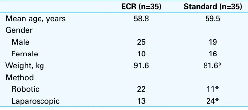
The patient demographics were similar between ECR and standard clamp release groups (Table 1); however, the ECR group had a mean weight of 91.6 kg, which was significantly heavier compared to standard clamp release group (81.6 kg). As well, the ECR group was associated with more robotic-assisted operations vs. control.
Tumour characteristics
Tumour size was smaller in the ECR vs. control group (36 cm3, range 1‒133cm3 vs. 55.4 cm3, range 1‒170cm3,
respectively), but was not statistically significant (Table 2). Nephrometry scores were blindly calculated by a third party in a retrospective fashion and confirmed by one author (GC). Mean nephrometry scores were 6.8 and 7.7 in the ECR and control groups, respectively. There was not a significant difference between scores.In both groups, most tumours were pathologically T1, with only two ECR and three control group patients having T2 tumours. Both groups had seven patients with positive or indeterminate surgical margins. None of the specimen had multifocal positive margins. Most
Table 1. Patient characteristics of the ECR and control groups.
ECR (n=35) Standard (n=35)
Mean age, years 58.8 59.5
Gender
tumours were clear-cell renal cell carcinomas (ccRCC), with an equally small distribution of other variants.
Operative outcomes
To determine the safety and efficacy of the ECR procedure, surgical factors were compared between the two groups (Table 3). WIT is represented by the “on-clamp time.” Clamp time in the ECR group was significantly lower vs. standard technique (18.8 vs. 31.5 minutes; p<0.05). This included one patient in the ECR group who needed to go back on clamp for bleeding and had a total clamp time of 31 minutes. Although there was a higher percentage of robotic vs. laparoscopic PNs in the ECR vs. standard group, there was no difference in clamp times between laparoscopic vs. robotic operations (mean 17.6 vs. 19.5 minutes, respectively). Importantly, there were no significant differences in EBL, transfusion rates, oper-ating time, length of stay in hospital, or the 90-day complica-tion rate between ECR and standard LPN groups. One Clavien IV and three Clavien III complications were seen in each group. Postoperative complications in the ECR group
includ-ed: delayed bleeding requiring transfusion and embolization, urine leak, aspiration pneumonitis requiring reintubation and intensive care unit admission, perinephric abscess requiring insertion of a drain, and a postoperative splenectomy. Similar complications arose in the standard approach and included urine leak, pseudoaneurysm bleeding requiring transfusion and embolization, and respiratory distress requiring intensive care admission and ventilation.
Functional outcomes
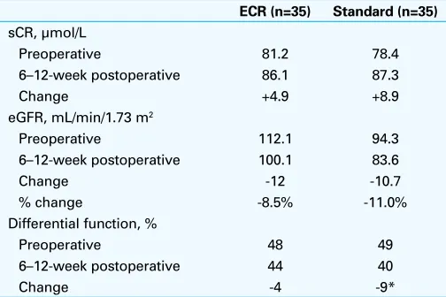
The primary intent of this study was to evaluate the impact of ECR on renal function. Renal function was compared using standard serum creatinine and eGFR measures (Table 4). Preoperative serum creatinine was similar between groups (mean of 81.2 and 78.4 mmol/L in the ECR and control groups, respectively). Three months postoperatively, the decline in renal function was similar and both groups showed an increase in mean creatinine (ECR 4.9 mmol/L vs. control 8.9 mmol/L). Accordingly, the calculated reduction of eGFR in both groups was similar (12.0 vs. 10.7 mL/min/1.73 m2).
In addition to biochemical analysis of renal function, com-parison of renal function/excretion using MAG3 nuclear reno-grams preoperatively and 6‒12 weeks postoperatively was per-formed. Preoperative split function of the affected kidney was similar between groups (48 and 49% in the ECR and standard groups, respectively). Importantly, the decline in split function was significantly greater in the standard technique group (9%) compared to the ECR group (4%) (Table 4).
Discussion
WIT is a well-documented risk factor for postoperative renal impairment and a number of studies have been published Table 2: Tumour pathological characteristics comparing
ECR and control group
ECR (n=35) Standard (n=35)
Size, cm3 (range) 36.1 (1–133) 55.4 (1–170)
Nephrometry score, mean 6.8 7.7
Stage
Positive margin status 7 7
*Oncocytoma, metanephric adenoma, or unclassified. ccRCC: clear-cell renal cell carcinoma; ECR: early clamp release.
Table 3. Surgical factors comparing ECR and control group
ECR ( n=35) Standard (n=35)
Clamp time, min (range) 18.8 (10–31) 31.5* (22–68)
EBL, ml 301 284
OR time, min 234 243
LOS (days) 4.6 5.3
90-day complication rate, n 7 5
Clavien score
*Statistically significant value, p<0.05. EBL: estimated blood loss; ECR: early clamp release; LOS: length of stay; OR: operating room (time from start of procedure to completion).
Table 4. Changes in renal function comparing ECR and control groups
recently evaluating efficacy of different techniques.11,23-25
Since it was adopted in 2013, we have performed ECR exclusively for both laparoscopic and robotic PN, which reduced WIT by nearly 14 minutes. Although there have been previous studies describing the ECR technique, this is the first study to assess its impact on WIT and renal dam-age compared with the standard technique.26 As a matter of
quality assurance, we also wanted to ensure that the ECR technique did not lead to a higher rate of bleeding, compli-cations, or length of stay.
Other groups have assessed LPN techniques using crude tests, such as serum creatinine and eGFR, which are often inaccurate and subject to day-to-day variability.15 In
addi-tion, in patients with a normal contralateral kidney, serum creatinine (and derived eGFR) does not accurately assess renal damage due to compensation by the contralateral kid-ney. Uniquely, our study used MAG-3 nuclear renography to assess the change in renal function (via assessment of tubular secretion and glomerular filtration) in the ipsilateral unit. Using renography, loss of renal function in the affected kidney was assessed by comparing renographic changes with the unaffected unit. Accordingly, when preoperative to postoperative renal function were assessed by creatinine and eGFR in our patient population, no difference was noted between ECR and standard LPN. It is only when assessment of ipsilateral renal damage was assessed by renography that the impact of ECR and shorter WIT was noted.
The safety and efficacy of ECR was assessed by comparing operative and postoperative factors. There was no difference in blood loss, length of operative time, length of stay in hos-pital, or complication rate between ECR and standard LPN groups. Our outcomes are reassuring, as complication rates are similar to other LPN series.16 By comparison, off-clamp
or selective clamping PNs have been reported to be associ-ated with higher EBL and complication rates.11,17,27 In the past,
techniques permitting reduced clamp time were performed in selected patients with smaller tumours and lower nephrometry scores, whereas in our study, no selection bias occurred, since the technique was applied to all LPN cases in succession.
Limitations of our study include a relatively small patient population, owing from a single surgeon experience. Although this was a retrospective study, data was collected within a long-standing LPN database that prospectively assessed preoperative and postoperative renal function at precise time points. Aside from a higher body mass index in the ECR group, the two groups were similar demograph-ically. As well, there were more robotic PNs performed in the ECR group, but WIT between robotic and standard LPN groups were similar, indicating that a higher percentage of robotic PNs did not affect the difference in WIT between ECR and standard LPN groups.
Before universal use of the described ECR technique, careful multicentre assessment of the technique should be
performed. We believe that this technique can be performed by experienced surgeons who are currently performing robotic or standard LPN. Unlike a technically demanding off-clamp or selective clamping technique that may be asso-ciated with higher bleeding and positive margin rates (owing to a wet renal resection bed), the ECR technique is technic-ally simple, requiring only that the clamps be released prior to the second closure layer. If excessive bleeding occurs as a result of the ECR technique, the clamp need only be reapplied, as it did in one of our procedures.
Conclusion
ECR is a safe method of LPN that significantly reduced WIT and renal damage vs. standard LPN according to assessment by nuclear renography. Intraoperative and postoperative complication rate and blood loss was not affected by ECR.
Competing interests: The authors report no competing personal or financial interests.
This paper has been peer-reviewed.
References
1. Canadian Cancer Society’s Advisory Committee on Cancer Statistics (2014) Canadian cancer statistics 2014. Canadian Cancer Society, Toronto
2. Siegel R, Ma J, Zou Z, et al. Cancer statistics 2014. CA Cancer J Clin 2014;64:9-29. https://doi.org/10.3322/caac.21208
3. Hollingsworth JM, Miller DC, Daignault S, et al. Rising incidence of small renal masses: A need to reassess treatment effect. J Nat Cancer Inst 2006;98:1331-4. https://doi.org/10.1093/jnci/djj362 4. Sun M, Bianchi M, Trinh QD, et al. Comparison of partial vs. radical nephrectomy with regard to other-cause
mortality in T1 renal cell carcinoma among patients aged ≥75 years with multiple comorbidities. BJU Int 2013;111: 67-73. https://doi.org/10.1111/j.1464-410X.2012.11254.x
5. Leslie S, Goh AC, Gill IS. Partial nephrectomy—contemporary indications, techniques, and outcomes. Nat Rev Urol 2013;10:275-83. https://doi.org/10.1038/nrurol.2013.69
6. Choi JD, Park JW, Lee HW, et al. A comparison of surgical and functional outcomes of robot-assisted vs. pure laparoscopic partial nephrectomy. JSLS 2013;17:292-9. https://doi.org/10.4293/10868081 3X13693422521359
7. Van Poppel H. Efficacy and safety of nephron-sparing surgery. Int J Urol 2010;17:314-26. https://doi.org/10.1111/j.1442-2042.2010.02482.x
8. Thompson RH, Lane BR, Lohse CM, et al. Every minute counts when the renal hilum is clamped during partial nephrectomy. Eur Urol 2010;24:1283-7. https://doi.org/10.1016/j.eururo.2010.05.047 9. Becker F, Van Poppel H, Hakenburg OW, et al. Assessing the impact of ischemia time during partial
nephrectomy. Eur Urol 2009;56:625-34. https://doi.org/10.1016/j.eururo.2009.07.016 10. Funahashi Y, Hattori R, Yamamoto, T et al. Effect of warm ischemia on renal function during partial
nephrectomy: Assessment with new 99 mTc-mercaptoacetyltriglycine scintigraphy parameter. J Urol 2012;78:160-4. https://doi.org/10.1016/j.urology.2011.08.071
11. Thompson RH, Frank I, Lohse CM, et al. The impact of ischemia time during open nephron-sparing surgery on solitary kidneys: A multi-institutional study. J Urol 2007;177:471-6. https://doi.org/10.1016/j. juro.2006.09.036
12. Kowalczyk KJ, Alemozaffar M, Hevelone ND, et al. Partial clamping of the renal artery during robot-assisted laparoscopic partial nephrectomy: Technique and initial outcomes. J Endourol 2012;26:469-73. https://doi.org/10.1089/end.2011.0511
13. Lamoshi AY, Salkini MW. Off-clamp robotic partial nephrectomy: Technique and outcome. Urol Ann 2015;7:226-30. https://doi.org/10.4103/0974-7796.150529
15. Simone G, Gill IS, Mottrie A, et al. Indications, techniques, outcomes, and limitations for minimally ischemic and off-clamp partial nephrectomy: A systematic review of the literature. Eur Urol 2015;68:632-40. https://doi.org/10.1016/j.eururo.2015.04.020
16. Baumert H, Ballaro A, Shah N, et al. Reducing warm ischemia time during laparoscopic partial nephrectomy: A prospective comparison of two renal closure techniques. Eur Urol 2007;52:1164-9. https://doi.org/10.1016/j.eururo.2007.03.060
17. Kaczmarek BF, Tanagho YS, Hillyer SP, et al. Off-clamp robot-assisted partial nephrectomy preserves renal function: A multi-institutional propensity score analysis. Eur Urol 2013;64:988-93. https://doi. org/10.1016/j.eururo.2012.10.009
18. Martin GL, Warner JN, Nateras RN, et al. Comparison of total, selective, and non-arterial clamping techniques during laparoscopic and robot-assisted partial nephrectomy. J Endourol 2012; 26:152-6. https://doi.org/10.1089/end.2011.0304
19. Beasley KA, Al Omar M, Shaikh A, et al. Laparoscopic vs. open partial nephrectomy. Urology 2004;64:458-61. https://doi.org/10.1016/j.urology.2004.04.028
20. Warren J, da Silva V, Caumartin Y, et al. Robotic renal surgery: The future or a passing curiosity? Can Urol Assoc J 2009;3:231-40. https://doi.org/10.5489/cuaj.1080
21. Fazio LM, Downey D, Nguan CY, et al. Intraoperative laparoscopic renal ultrasonography: Use in advanced laparoscopic renal surgery. Urology 2006;68:723-7. https://doi.org/10.1016/j.urology.2006.04.022
22. Benway BM, Wang AJ, Cabello J, et al. Robotic partial nephrectomy with sliding-clip renorrhaphy: Technique and outcomes. Eur Urol 2009;55:592-9. https://doi.org/10.1016/j.eururo.2008.12.028 23. Mir MC, Ercole C, Takagi T, et al. Decline in renal function after partial nephrectomy: Etiology and
preven-tion. J Urol 2015;15:185-8. https://doi.org/10.1016/j.juro.2015.01.093
24. Schmid M, Abd-El-Barr AE, Gandaglia G, et al. Predictors of 30-day acute kidney injury following radical and partial nephrectomy for renal cell carcinoma. Urol Oncol 2014;32:1259-66. https://doi.org/10.1016/j. urolonc.2014.05.002
25. Lane BR, Babineau DC, Poggio ED, et al. Factors predicting renal functional outcome after partial neph-rectomy. J Urol 2008;180:2363-8. https://doi.org/10.1016/j.juro.2008.08.036
26. Nguyen MM, Gill IS. Halving ischemia time during laparoscopic partial nephrectomy. J Urol 2008;179:627-32. https://doi.org/10.1016/j.juro.2007.09.086
27. Rais-Bahrami S, George AK, Herati AS et al. Off-clamp vs. complete hilar control laparoscopic partial neph-rectomy: Comparison by clinical stage. BJU Int 2012;109:1376-81.https://doi.org/10.1111/j.1464-410X.2011.10592.x