Establishment of a proneural field in the inner ear
GINA ABELLÓ and BERTA ALSINA*
Developmental Biology, Dept. of Experimental Sciences and Health, Pompeu Fabra University, Barcelona Biomedical Research Park, Barcelona, Spain
ABSTRACT Hair-cells, supporting cells and sensory neurons are the main specialized cell-types responsible for mechanotransduction in the inner ear. They derive from precursors expressing proneural genes and recent data has underlined the importance of SoxB1 genes as upstream activators of proneural genes during cranial placode development. Here we review the steps of establishing a proneural field and propose several models for how early otic regionalization into a proneural territory is achieved.
KEY WORDS:
bHLH, proneural gene, notch signaling, FGF signaling, SoxB1, neural induction
Introduction
Sensory organs provide key and species-specific information that allows animals to effectively forage, find mates and avoid predators. The inner ear is one of the main sensory organs of the head and responsible for the perception of sound and balance. In adults, the organ is arranged in a highly complex three-dimen-sional structure, named the membranous labyrinth, composed of ducts, recesses and six to eight sensory epithelia. The latter are composed of hair-cells - the mechanotransducers - and support-ing cells arranged in a cellular mosaic. Mechanosensory informa-tion is then transmitted to the brainstem by afferent bipolar sensory neurons. Albeit the structural complexity of the adult inner ear, it derives from a simple anlage, the otic placode (Torres and Giraldez, 1998, Baker and Bronner-Fraser, 2001, Fritzsch and Beisel, 2001, Riley and Phillips, 2003). Cranial placodes are specialized areas of ectoderm outside the neural plate that contribute to all the cranial paired sensory organs and most of the sensory neurons from the peripheral nervous system of the head. The current view is that placodes share a common developmental origin, the preplacodal region (PPR), a horseshoe band encircling the neural plate from which individual placodes emerge (Jacobson, 1963, Torres and Giraldez, 1998, Streit, 2004, Schlosser, 2006). Except the adenohypophyseal and the lens placode, the rest of placodes (olfactory, profundal and trigeminal, otic, lateral line, epibranchial) are neurogenic, this means that they give rise to
sensory neurons among other specialized cell-types (Adam et al.,
1998, Ma et al., 2000, Schlosser and Northcutt, 2000, Andermann et al., 2002, Begbie et al., 2002).
During inner ear development, sensory neurons and hair-cells derive from otic precursors in which proneural genes are
acti-*Address correspondence to: Berta Alsina. DCEXS-Universitat Pompeu Fabra, Parc de Recerca Biomèdica de Barcelona, c/ Dr. Aiguader 88, 08003 Barcelona, Spain. Tel: +34-93-316-0837. Fax: +34-93-316-0901. e-mail: [email protected]. Web: http://www.upf.edu/cexs/recerca/biodes/index.htm
0214-6282/2007/$30.00 © UBC Press
Printed in Spain
www.intjdevbiol.com
vated. Despite the knowledge of proneural function and a general view of placodes as lateral neurogenic domains, little is known about how and when proneural competence is acquired in the otic placode. The aim of this review is to summarize the data concern-ing the mechanisms of proneural activation in the inner ear. Which proneural genes are functional during otic development in the different species? When and where are they activated? Which is the origin of proneural otic competence? The regulation of pro-neural function is tightly linked to Notch signaling activity. The second part of the review intends to highlight the function of Notch pathway in proneurosensory development.
Activation of proneural genes in the inner ear
After otic placode formation, the otic tissue invaginates and initiates its autonomous program of development. The most obvious change, apart from its continuous modulation of shape, is its progressive generation of specialized cells in precise loca-tions. Once the placode invaginates, some epithelial cells identi-fied as neuroblasts loose connectivity and delaminate into the mesenchymal space where they remain as a group forming the cochleo-vestibular ganglion (D’Amico-Martel, 1982, Hemond and Morest, 1991, Adam et al., 1998). There neuroblasts suffer a
series of transit-amplifying rounds of division (Alsina et al., 2003,
Camarero et al., 2003), to then enter into differentiation, extend
their axons and innervate the epithelial regions near where they have been generated (Fritzsch et al., 2002). In the past decade,
many genes implicated in the process of neurosensory develop-ment have been identified both in invertebrates and vertebrates (Ghysen and Richelle, 1979, Torres and Giraldez, 1998, Baker and Bronner-Fraser, 2001). Now it is clear that the early steps of cell fate specification in the different sense organs are controlled by proneural genes, which were first identified in Drosophila
mutants lacking the ability to develop external sense organs and bristles (Ghysen and Richelle, 1979). Proneural genes encode for transcription factors of the basic Helix-Loop-Helix (bHLH) class
that bind to a common DNA sequence called the E-box sequence (Bertrand et al., 2002). In Drosophila, two major proneural gene
families control neuronal development of the PNS, the achaete-scute (asc) and the atonal (ato) gene family (Simpson, 1990). Asc
genes in Drosophila specify external sense organs, while ato
gene specifies the photoreceptors and chordotonal organs, among them the specialized Johnston´s organ (Ghysen and Dambly-Chaudiere, 1989, Treisman, 2004). The latter, located in the fly antenna, appears to be the homologous hearing organ in Droso-phila given its capacity of sound evoked potentials (reviewed in
Caldwell and Eberl, 2002, Falk, 2005;Eberl and Boekhoff-Falk in this issue). In vertebrates, in addition to the achaete-scute homologues (ash) and the atonal homologues (atoh), other new
related proneural families are encoded in the genome: the E proteins, Olig, NeuroD, Neurogenin and Nscl protein families (Bertrand et al., 2002), with some of them having been recruited
for the peripheral sensory developmental program in craniates. It has been postulated that the appearance of new proneural genes in vertebrates allowed the ancestral functions of mechanotransduction and electrical propagation performed by a single cell to be segregated and performed by two different cells: the hair-cell specified by atoh and, the neuron, specified by Neurogenin (Neurog, also–named Ngn) (see Fritzsch and Beisel,
2001).
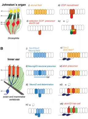
In Drosophila chordotonal organs, as a result of the activation
of the Notch pathway, the sense organ precursor (SOP) is singled out from a group of equivalent cells expressing the ato proneural
gene. After the selection of the SOP, proliferation initiates giving rise to the main cell types. Contrary to external bristles, in some chordotonal organs secondary precursors are recruited by exter-nal sigexter-nals, such as EGFR sigexter-naling (zur Lage et al., 2004)
(Figure 1). In amniote vertebrates, a proneural field expressing
Neurog1 is distinguished in an anterior subdomain of the otic
placode. In this domain, scattered cells with increased levels of
Neurog1 and Delta1 appear in the otic cup in mouse and chick
(Adam et al., 1998, Ma et al., 2000, Alsina et al., 2004). Similar to
the chordotonal organs, several signaling molecules (in this case from the FGF family: FGF3, FGF8, FGF10) are expressed in the proneural field in zebrafish, chick and mice, their function prob-ably also related to the recruitment of cells into the neurosensory lineage (Leger and Brand, 2002, Pirvola et al., 2002, Alsina et al.,
2004). Following Neurog1 expression, other genes of the
proneu-ral bHLH family (i.e. NeuroD and NeuroM) switch on, the
expres-sion detected in epithelial and delaminated neuroblasts (Figure 1). Functional analysis of Neurog1 and NeuroD suggests that they
are necessary and sufficient for neurogenesis. Overexpression of
Neurog1 drives formation of ectopic neurons (Perron et al., 1999,
Kim et al., 2001), while targeted inactivation of Neurog1 results in
loss of proximal cranial sensory ganglia (Ma et al., 2000).
Disrup-tion of NeuroD function also results in a severe loss of sensory
neurons associated with the inner ear (Kim et al., 2001). The
sequence of activation of Neurog1, followed by NeuroD/NeuroM
expression plus the phenotypes observed supports the notion that Neurog genes have conserved the neuronal determination
functions of the atonal Drosophila counterparts, whereas the NeuroD/NeuroM bHLH transcription factors exhibit neuronal
dif-ferentiation and survival functions.
Hair-cell specification lags neuronal cell specification by ap-proximately two days in amniotes. Few scattered cells expressing Atoh1 are detected in the prosensory patches in chick E3.5 otic
vesicles (Pujades et al., 2006) and the sensory epithelia of
E11-E12 mice embryos (Shailam et al., 1999; Matei et al., 2005). By
contrast, in zebrafish, sensory and neuronal precursors are specified almost about the same time (Andermann et al., 2002).
Low expression of atoh1b in zebrafish precedes the appearance
of individual atoh1a-positive hair-cells in a broad sensory field of
equivalent cells. The initial development of this domain is regu-lated by Notch signaling like in Drosophila, suggesting a classical
proneural role of atoh1b in zebrafish (Millimaki et al., 2007). In
mammals, this role is less clear due to the appearance of Atoh1
mainly in post-mitotic cells and restricted to the cells that will differentiate into hair-cells (Chen et al., 2002), although in mice
expression has been detected before the appearance of defined sensory patches by RT-PCR (Matei et al., 2005). In amniotes,
neuronal and hair-cell determination events are segregated in time, with gross morphogenetic changes during this period. Thus, it is yet not resolved if Neurog1 and Atoh1 proneural activation
occurs in a common proneurosensory domain or from segregated or partially overlapping neural (neuronal) and sensory domains. Lineage studies have determined that the macula utriculae
con-tains progenitors with shared neuro-sensory lineage in chick and
Neurog1 mice mutants display also loss of hair-cells, indicating
that at least in some areas exist common progenitors for both cell lineages (Satoh and Fekete, 2005). However, as Neurog1 and Atoh1 genes are expressed broadly at nearly undetectable levels
and only enhanced at determined cells, analysis of the distribution of Sox proteins may constitute better readouts of proneurosensory committed territory/ies. In neural development, Sox genes are expressed before proneural genes (see below) and Sox2 mutants
present severe defects in sensory organ development, lacking also Atoh1 expression (Kiernan et al., 2005). Sox3 and Sox2
proteins are both present in the anterior ventral otic cup but later on, exclusively Sox2 map in the sensory patches (Neves et al.,
2007).
Inducing neural competence in the otic placode
Smad-interacting protein 1, is regulated by Churchill, a
gene isolated after neural induction mediated by FGF (Sheng et al., 2003). The relationship of FGF signaling
and proneural activation comes from studies in the
emerging caudal CNS, where Cash4 is activated by
FGF (Henrique et al., 1997). However, in most cases
switch on of proneural genes is delayed to neural induction and their activation is mediated by early in-duced pan-neural genes (Sox, ENRI, Zic) that then regulate proneural activity.
Sox proteins form a family of HMG-box transcription factors related to SRY, the mammalian testis determin-ing factor. On the basis of sequence similarity, both in the DNA-binding domain and in other, group-specific conserved motifs, Sox proteins have been divided into at least seven subgroups (A-G). Throughout evolution,
the expression of the SoxB1 genes (Sox1, Sox2 and
Sox3), directly correlates first, with ectodermal cells that are competent to acquire a neural fate and second, with the commitment of cells to a neural fate (Rex et al.,
1997, Pevny and Placzek, 2005). The Drosophila
SoxNeuro, a putative ortholog of the vertebrate Sox1-3 proteins, is one of the earliest transcription factors to be
expressed pan-neuroectodermally (Cremazy et al.,
2000) and it acts upstream and in parallel with the
achaete-scute genes. Interestingly in Drosophila,
SoxNeuro is only involved in CNS but not in PNS development, being suggested that recruitment of Sox proteins into placode development is a novelty of crani-ates to expand the ectodermal anlage rapidly (Fritzsch
et al., 2006b). Direct evidence for the involvement of SoxB1 genes in neural commitment comes from in vitro
stem cell studies, where it was shown that the Sox1
gene can induce neural fate in competent ectodermal cells (Pevny et al., 1998). Consistent with SOXB1
fac-tors marking ectodermal cells with neural potential their expression is modified by neural inducing signals (Streit
et al., 2000). Moreover, strong evidence of the link
between neural inducing signals and Sox activation is
derived from the identification of FGF and Wnt response binding domains in the same enhancer region of the
Sox2 locus (Takemoto et al., 2006).
The induction of the preplacodal region from which sensory cranial placodes emerge is mediated by the same signaling pathways. In this case, the activation of the FGF pathway, together with WNT and BMP antago-nists specifies the preplacodal field (Ahrens and Schlosser, 2005, Litsiou et al., 2005). After the
forma-tion of the preplacodal region, a second wave of induc-tion in particular locainduc-tions, promotes a series of subdomains. Although, at the end in amniotes six
pla-are initially intermingled and then sort out to different regions (Bhattacharyya et al., 2004). The Pax3 and the Pax2 domain
contain precursors for the trigeminal placode and the epibranchial/otic precursors respectively (Baker and Bronner-Fraser, 2000, Schlosser and Ahrens, 2004). At the end, indi-vidual placodes will be identified by a combination of Pax, Six/ Eya, Foxi and Sox transcription factors. Six proteins are usually
expressed from the initial preplacodal field to the late individual
Fig. 1. Steps of proneural gene activation in vertebrate and invertebrate sensory organs. (A) Schematic drawing of three chordotonal sensilla of Droso-phila (modified from Yager, 1999) and sequential steps of insect sensory devel-opment. i) a neurosensory field is specified by atonal proneural gene, ii) a SOP is singled out by the N-Dl pathway, iii) EGF recruits secondary precursors and iv) sensory neuron and accessory cells are generated. at, attachment cell; sc, scolopale cell; gl, glial cell; sn, sensory neuron. (B) Sensory patch of the vertebrate inner ear (Fettiplace and Hackney, 2006) and outline of major steps of sensory development. i) A proneural field is specified by Neurogenin, ii) neuronal precur-sors are singled out by the N-Dl pathway, iii) NeuroD is switched on, v) neuroblasts delaminate and differentiate, vi) atoh starts to be expressed in a prosensory field, vii) sensory precursors are singled out, viii) hair-cells and supporting cells are determined by the activity of Notch pathway. sn, sensory neuron; hc, hair cell; su, supporting cell.
codes develop (adenohypophyseal, lens, olfactory, trigeminal, epibranchial and otic), an intermediate step of placodal devel-opment is identified by the appearance of three broad domains of Pax gene family expression: an anterior domain Pax6
posi-tive, a second domain Pax3-postive next to the
midbrain-hindbrain boundary, plus a caudal region expressing Pax2
(Bhattacharyya and Bronner-Fraser, 2004). In the Pax6
do-main, precursors of the adenohypophyseal, lens and olfactory
placodes. In some vertebrate species, the expression of Sox2
and Sox3 in the ectoderm arises in two waves; initially they are
expressed at low levels in the preplacodal region, then expres-sion is transiently shut off from the presumptive placode do-mains and finally it resumes when individual placodes are specified and express Pax genes (Abu-Elmagd et al., 2001,
Schlosser and Ahrens, 2004, Sun et al., 2007). For example in
the lens field, Sox2 expression is enhanced once the lens
contacts the optic vesicle (Kamachi et al., 1998). All cranial
placodes, neurogenic and non-neurogenic, except the trigemi-nal placode, express SoxB1 transcription factors. In Xenopus Neurog1 expression is initiated in the entire preplacodal
ecto-derm but subsequently is shut off in lens, suggesting that all placodes in fact have an initial neural bias (Schlosser and Ahrens, 2004).
In chick, the otic proneural territory expressing Neurog1
emerges as a triangle in the anterior half of the flat otic placode. Initially the proneural domain is the anterior-medial aspect of the otic placode to end up, after invagination, in an anterior-medial and ventral position of the otic vesicle (Alsina et al.,
2004). Before the otic placode is morphologically visible, Sox3
is expressed in a broad band that encompasses the otic and epibranchial territory to get restricted to the proneural region of the otic placode (Sun et al., 2007; G.A and B.A, unpublished
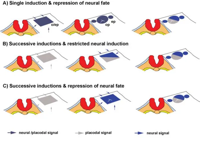
observations). Two main questions arise when analyzing Sox function during otic development. First, does the process of placodal induction, by default, involve proneural induction or, alternatively, do successive induction events lead first to placodal and subsequently to neural fate? Secondly, is neural compe-tence only acquired in a subdomain of the otic fated cells or is repression of neural fate required in the non-neural competent region? We propose several models on how the proneural
domain may be established (Figure 2) by using the Sox3
expression as the earliest marker of neural induction and Pax2
as markers of placode induction.
Model A: Single induction and repression of neural fate
This model suggests that the competence of neural fate is conditioned by the acquisition of a placodal identity (Figure 2A). The fact that FGF signaling is required both for otic/epibranchial induction, as well as for Sox1-3 gene expression in the otic/
epibranchial field in zebrafish (Sun et al., 2007) suggest that
both events could, in fact, be part of the same process. In addition, Sox2 is able to regulate the expression of a typical
placodal marker such as Pax6 in lens and nasal placodes
(Donner et al., 2007) and in Medaka ectopic expression of Sox3
induces ectopic otic and lens placodes expressing Pax2 and Pax6 respectively (Koster et al., 2000). In the posterior
preplacodal region Six1 and Sox3 expression correlate in a
higher degree but not in the anterior preplacodal field of Xeno-pus (Schlosser and Ahrens, 2004). Again at the otic-epibranchial
field, Pax2 and Sox3 are also co-expressed in zebrafish and
chick, although in the latter the appearance of Sox3 expression
is delayed to Pax2 (Groves and Bronner-Fraser, 2000,
Abu-Elmagd et al., 2001, Sun et al., 2007). Altogether, Sox2-3
expression and placodal marker expression such as Pax and Six are strongly paralleled. However, albeit some data may
suggest that placode and neural fate could be linked, there is little functional evidence supporting this model. In this model,
as in model c, proneural fate is inhibited in the posterior otic territory after broad placodal induction (see below).
Model B: Successive inductions and restricted neural in-duction
This model postulates that placodal and neural induction are successive events (Figure 2B). Graft experiments at different axial levels performed by Noden and Van De Water, (1986) revealed that presumptive otic placode ectoderm could ectopi-cally generate otic vesicles without the ability to form neurons. Again, Groves and Bronner-Fraser (2000) observed that quail anterior epiblast grafted in the presumptive otic region of host of 3-10 somites could start to express Pax2 and Sox3, while grafts
performed at 11-21 somites, only expressed Sox3 but not Pax2,
suggesting that Pax2-inducing signals are lost before
Sox3-inducing signals. Thus, depending on the lenght of exposure of signals the appearance of some molecular markers, such as Pax2
and Sox3, can be dissociated. Inhibition of FGF signaling in
zebrafish indicates that induction of Sox3 and Pax2 requires FGF
signaling at the otic-epibranchial region but not in an interdepen-dent manner (Groves and Bronner-Fraser, 2000, Nikaido et al.,
2007, Sun et al., 2007). Consistent with a view of a multi-step
inductive process for full otic development, it is well established that induction of the otic placode demands first the induction of the preplacodal ectoderm, biasing the ectoderm for receiving a sec-ond wave of induction (Litsiou et al., 2005, Martin and Groves,
2006).
This model also hypothesizes for a restricted source of neural inducers in the anterior otic field. So far, requirement of FGF signaling has been mainly associated with otic induction, but as reported recently this process probably also involves epibranchial induction (Hans et al., 2007, Nikaido et al., 2007, Sun et al., 2007,
Nechiporuk et al., 2007). Several FGFs are expressed in
sur-rounding tissues at the time of otic and Sox3 induction. Several
laboratories have addressed the issue concerning the identifica-tion of the signals involved in otic inducidentifica-tion. The current view is that FGF8 from the endoderm, FGF19/ FGF10 from the meso-derm and FGF3/FGF8/FGF10 from the hindbrain are involved sequentially and sometimes synergistically to induce the already biased ectoderm to develop the inner ear (for review see Groves, 2005; Groves, Schimmang in this issue). FGF molecules (22 in mammals) regulate a wide variety of functions such as cell fate, patterning, proliferation, differentiation, survival and morphogen-esis. During inner ear development, FGF also regulate some of these later events, such as patterning and growth (Wright and Mansour, 2003). Single mutants for FGF3 or hindbrain patterning genes like kreisler, in which FGF3 levels in the posterior hindbrain
are reduced/lost show otic patterning defects such as expansion of the cochlea and absence of endolymphatic duct (Mansour et al., 1993, McKay et al., 1996, Choo et al., 2006). In zebrafish, the valentino mutant (zebrafish orthologue of kreisler) displays an
expansion of the anterior otic domain and overproduction of hair-cells, probably due to the posterior expansion of FGF3 expression within the hindbrain (Kwak et al., 2002). Likewise, the vHNF1
mutant embryos, also displaying an anteriorization of the hind-brain, again shows a caudal expansion of neurogenic and sen-sory domains at the expense of posterior and dorsal markers (Lecaudey et al., 2007). Some of these effects can be also
promote neural character. However, one should bear in mind that some of the inner ear phenotypes affecting expansion and/or reduction of neuronal or sensory territories have been analyzed at otic vesicle stage when major morphogenesis and cell growth also occurs, with some of these pathways participating also in the mentioned processes. In summary, many FGFs and WNT mol-ecules are locally expressed around the presumptive otic field in different time periods, therefore it is reasonable to believe that i) type, ii) time and, iii) space activation of these signals may differentially regulate placode and otic neural induction. In chick, FGF2 was able to induce a vast array but not all early otic markers (i.e. Pax2, EphA4, Nkx5.1, SOHo1, but not BMP7, Dlx5), while
FGF8 had some but little inducing properties (Adamska et al.,
2001, Martin and Groves, 2006). Altogether, in this moment, a detail study on the roles of different FGFs on activating specific early otic genes is needed for drawing the full picture.
Model C: Successive inductions and repression of neural fate
This model predicts on successive inductions, as in model b, but unrestricted neural inducing signals activate Sox genes in a broad otic/epibranchial field. Broad proneural competence is subsequently restricted to the anterior otic region by an inhibitory signal from the posterior region (Figure 2C). To date, Tbx1, a
transcription factor belonging to the T-box gene family, is the only factor that clearly has been demonstrated to inhibit neurogenesis function in the posterior inner ear (Vitelli et al., 2003, Raft et al.,
2004, Xu et al., 2007). In mice, Tbx1 is expressed in pharyngeal
arches, head mesenchyme, the secondary heart field, the poste-rior otocyst and sclerotome (Chapman et al., 1996). Homozygous
mutation of Tbx1 gene in mouse causes birth defects derived from
developmental abnormalities of the embryonic pharyngeal
sys-It has been shown that hairy controls the pattern of sensory organ
formation by repressing achaete-scute (Orenic et al., 1993). In the
zebrafish midbrain-hindbrain boundary, mouse olfactory placode and the inter-proneural stripes of Xenopus, Hairy and Enhancer of Split homologues (Hes/Her) repress neural fate (Cau et al.,
2000, Geling et al., 2003, Bae et al., 2005). In chick otic cups, Hairy1 (Hes1 in mammals) is expressed restricted in the nonneural
domain, disclosing a putative role in repressing proneural fate in the posterior region (Abelló et al., 2007). This links us to the
following section in which the role of Notch pathway in defining sensory and neural domains are discussed.
In all three models, proneural function is probably enhanced by local otic FGF signals. For example, FGF3, FGF10 and FGF8 expressions are distinguished in the proneural territory in various species with reported neurogenic functions for some of them.
Notch signaling during early otic development
Principles of the Notch signaling pathway
Notch signaling pathway is involved in almost every develop-mental decision and process and together with the FGF, WNT, TGFβ and HH family proteins constitute one of the five main pathways required for metazoan development. The first identifi-cation of the Notch receptor arose in laboratories working on the fruit fly, Drosophila melanogaster, when a series of mutants that
generated a Notched (indented) wing phenotype plus an overpro-duction of neural cell-types at the expense of nonneural cells were isolated. The core signaling pathway consists of the Notch recep-tor, with a large extracellular domain with 36 tandem epidermal growth factor (EGF)–like repeats and three cysteine-rich Notch/ LIN-12 repeats. Six tandem ankyrin repeats, a glutamine-rich domain (opa) and a PEST sequence are found within the
intrac-Fig. 2. Proposed models for neural induction in the otic/epibranchial field. Dorsal views of the cranial ectoderm at the level of the hindbrain. o/ep, otic-epibranchial placode; ep, epibranchial placode; op, otic placode.
tem. The otocyst also presents major defects, being small and sensory organs do not form (Vitelli et al., 2003). Interestingly, Tbx1 is
expressed in the nonneural otic vesicle re-gion, complementary to NeuroD positive
do-main. Loss of Tbx1 gene causes the
expan-sion and duplication of the neural region while neural determination is suppressed in Tbx1
gain-of-function otocysts, suggesting that its function is required to inhibit neuronal fate (Raft et al., 2004). This has been further
demonstrated by fate mapping; Tbx1 positive
cells identify a population of cells that give rise to most of the otocyst, excluding the neurogenic and endolymphatic duct regions (Xu et al., 2007). In chick, two complementary
domains are detected at early otic cup stage: a proneural region expressing LFng, FGF10/ FGF3, Delta1 and Hes5 and a non-neural
region expressing Lmx1, Irx1, Tbx1, Hairy1
and Serrate1 (Alsina et al., 2004, Abelló et al.,
2007).
Hairy another bHLH gene is in Drosophila
expressed in broad areas in which proneural function is repressed (Orenic et al., 1993).
The legs of the Drosophila melanogaster adult
ellular domain (reviewed in Artavanis-Tsakonas et al., 1999). The
ligands that bind to the Notch receptor are two single-pass proteins, Delta and Serrate (Jagged in mammals). As a result of receptor activation, Notch receptor suffers a series of proteolytic cleavages that frees the intracellular domain of the Notch receptor (NICD) into the cytoplasm. The NICD then translocates to the nucleus, recruits the transcription factor Suppressor of Hairless
[Su(H)] (CBF1/RJBk in mammals, LAG-1 in C. elegans) and
mastermind co-factor protein, to form a complex that activates downstream target genes; the Enhancer of split (E(spl) genes in
Drosophila and the HES or HESR in vertebrates as the main
primary targets. This general picture of Notch signaling is a mere cartoon of the pathway further complicated with the isolation of many modulators of the Notch pathway(Artavanis-Tsakonas et al., 1999, Schweisguth, 2004, Louvi and Artavanis-Tsakonas,
2006).
Notch pathway regulates a long list of different biological processes: cell fate decisions, proliferation, apoptosis, cell differ-entiation, patterning and asymmetric divisions. Notch functions might differ by the modulation on the amount of the receptor or the ligand on the cell surface, by feedback loops that potentiate or shut off the signal, or by tissue specific co-factors (Schweisguth, 2004). The most obvious role of Notch pathway is its ability to influence its neighbouring cells, by one cell promoting one state and the adjacent cell adopting the antagonist state. Notch plays an indisputable role in neurogenesis, in which the Delta-positive
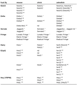
cell promotes neuronal fate and the Notch-activated cell re-presses neuronal fate (reviewed in Lewis, 1998). This phenom-enon, in which Notch activity generates a negative feed-back loop by repressing its ligands, is referred as lateral inhibition. Gener-ally, in those cases, the Notch activated cell remains as an undifferentiated progenitor cell and represses proneural gene activity, while the Delta-positive cell enters into differentiation. However, recent data also suggests that Notch pathway can promote differentiation, i.e. in glial differentiation. In Table 1 is listed the mouse, chick and zebrafish orthologues of Drosophila
members of the Notch pathway.
Notch pathway also has the ability to segregate specific cell lineages from fields of developmentally equivalent cells; com-monly, in this mechanism Notch operates by inducing its ligands, thus referred to as lateral induction. During neural vertebrate development, it has been reported to be essential for the segmen-tation of the hindbrain and the formation of the zona limitans intrathalamica (Lewis, 1998, Pasini and Wilkinson, 2002, Kiecker and Lumsden, 2005). Although the exact mechanism by which adjacent regional domains acquire distinct properties in verte-brates is less clear, the underlying model extracted from Droso-phila predicts that Notch pathway is differentially activated by the
different expression of Delta and Serrate ligands and the modu-lator Fringe in adjacent territories. Drosophila Fringe (Fng) and
the vertebrate homologues Lunatic fng, Manic fng and Radical fng
are glycosyl-transferases that glycosylate Notch EGF repeats, resulting in a bias of Notch receptor activation by Delta versus Serrate ligand (Rampal et al., 2005).
Expression patterns and roles of Notch in the inner ear
Mostly, Notch role in inner ear development has focused on sensory development due to the stunning phenotype on hair-cell production when Notch signaling is disrupted. A picture of the role of Notch pathway in inner ear development is emerging with the aid of a long list of mouse mutants and reported lines, comple-mented by genetic analysis in zebrafish (Lewis, 1998, Eddison et al., 2000, Landford and Kelley, 2005). Here we will briefly
summa-rize the expression patterns of Notch elements and roles during sensory development to focus then on the earliest role of Notch on the regulation of the proneural domain based on our studies in chick.
Notch function in sensory epithelia
It has been reported that in mice, by E12, Notch1, Jag1 and LFng expression start to be concentrated in the entire epithelium
of the six sensory epithelia (Morrison et al., 1999).
Simulta-neously, Dll1 (Dl1 in non mammalian vertebrates) is expressed in
scattered cells, presumably the nascent hair-cells of the vestibu-lar epithelia (differentiates before the auditory epithelium). Later on, in E14-E15, Dll1 and Jagged2 (Jag2) appear to be expressed
also in the cochlea epithelium, again restricted to the cells that will become hair-cells (Morsli et al., 1998, Morrison et al., 1999),
starting in the basal end of the cochlea to progressively extend to the apex. Hes1 and Hes5 transcripts have been detected only by
RT-PCR at these stages (Shailam et al., 1999, Lanford et al.,
2000). At E15, Hes5 expression is detected by in situ hybridization
at the base of the cochlea and by E17, when most cells have stopped dividing, LFng, Jagged1 (Jag1) and Hes5 expression
fruit fly mouse chick zebrafish
Notch Notch1 + Notch1 + Notch1a, Notch1b +
Notch2 nd Notch2 - Notch2 (Notch6) nd
Notch3 nd Notch3 (Notch5) nd
Notch4 nd
Fringe Lunatic Fringe + Lunatic Fringe + Lunatic Fringe + Manic Fringe + Manic Fringe - Manic Fringe + Radical Fringe - Radical Fringe - Radical Fringe
-Hairy Hes1 + Hairy2 + her6 (Hes14) nd
ORTHOLOGUES OF NOTCH PATHWAY COMPONENTS TABLE 1
restrict to supporting cells in the cochlea, while Jag2 and Dll1
expressions are restricted to hair-cells (see revision in Landford and Kelley, 2005). Similar expression patterns are found in other species such as chick and zebrafish, where it has mostly been studied (Adam et al., 1998, Haddon et al., 1998, Eddison et al.,
2000).
Defective Notch signaling in zebrafish or mammals (mutants for Jag2, Hes5, Dll1, mind bomb) results in overproduction of
hair-cells at the expense of supporting hair-cells, indicating that Notch signaling regulates sensory cell fate specification by a mecha-nism of lateral inhibition (Haddon et al., 1998, Lanford et al., 1999,
Zine et al., 2000, Kiernan et al., 2001). Thus, Jag2/Dll1 ligands
lead to the activation of Notch pathway and Hes5 in adjacent cells
that blocks hair-cell determination. Similar phenotypes of extra hair-cells are present in Foxg1 null mice, suggesting possible
interactions of Hes proteins with forkhead box G1 proteins
bal-ancing cell proliferation and cell fate determination in the inner ear (Pauley et al., 2006, Fritzsch et al., 2006a). On the other hand, Jag1 is broadly expressed in the entire prosensory patches and
different Jag1 mice mutants result in total or partial loss of sensory
epithelium, indicating that Jag1 is required for the specification of
the prosensory patch (Tsai et al., 2001, Daudet and Lewis, 2005,
Brooker et al., 2006, Kiernan et al., 2006). Moreover, Jag1 is also
required for maintenance of cells into a sensory progenitor state (Kiernan et al., 2006). The prevailing view is that in addition to
regulate hair-cell specification, Notch-Jag1 signaling initially speci-fies sensory versus nonsensory epithelium within the ear, placing the limits by a lateral inductive mechanism (Adam et al., 1998,
Lewis, 1998, Eddison et al., 2000, Landford and Kelley, 2005).
One of the possible models suggests that Notch activity in the nonsensory epithelium driven by Jag1 binding from the sensory
epithelium represses sensory fate. Consistent with this hypoth-esis, Notch inactivation in zebrafish leads to an expansion of sensory fate and similar results where obtained in the cochlea of conditional mutants for Notch1 (Kiernan et al., 2001). However, as
mentioned, conditional Jag1 deletion in the otocyst does not
phenocopy the results of Notch1 inactivation but rather suggests that Notch pathway is activated in the sensory epithelium. More-over, in chick overexpression of NICD causes the induction of
extra sensory patches (Daudet and Lewis, 2005). Altogether, it has been proposed that Notch activity is required for sensory development to first, make cells competent to form a prosensory patch conferring them a prosensory character and, subsequently, inhibit hair-cell differentiation and establish a mosaic cellular pattern (Daudet and Lewis, 2005).
Notch function in the proneural domain
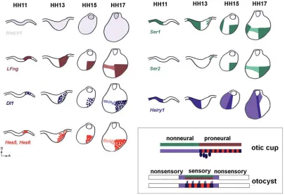
Before the appearance of the sensory patches, when an early proneural domain establishes, most members of Notch pathway are already expressed in the otic placode/cup. In chick, Notch1 is
expressed in the entire otic epithelium from 11 somites to late otocyst stage (Groves and Bronner-Fraser, 2000). LFng is
ex-pressed throughout the proneural domain, while Dl1 is detected
in a salt and pepper pattern (Adam et al., 1998, Cole et al., 2000,
Alsina et al., 2004). In the same domain, Hes5 is expressed in
cells adjacent to Dl1-positive cells as a consequence of the N-Dl
lateral inhibition process (Fig. 3) (Abelló et al., 2007). Hes6 in the
neural tube is expressed both in precursor and differentiated
neurons (Fior and Henrique, 2005) and in the chick inner ear shows a mosaic pattern in the proneural domain (G.A and B.A unpublished observations). In the sensory patches, Hes6
expres-sion is very similar to Atoh1 (Qian et al., 2006). Therefore, LFng, Hes5, Dl1 and Hes6 domains of expression extend from otic cup
to late otocyst, as a common toolkit mechanism for the selection of first, neuronal and, second, sensory cells. As expected from the lateral inhibition model, disruption of Notch signaling in the mind bomb mutant zebrafish or in chick by inhibiting Notch intracellular
cleavage leads to the production of excess neuronal precursors in the inner ear concomitant to the suppression of Hes5 activation
(Haddon et al., 1998, Abelló et al., 2007). Altogether, Notch
signaling is required in otic neurogenesis to regulate the number of neural committed cells that enter into neuronal differentiation. Interestingly, Ser1 (or Jag1) is also expressed already at early
otic cup stage, well before the appearance of prosensory patches. In this case, Ser1 appears broadly in a domain complementary to
the proneural domain. Later on, Ser1 also is distinguished in the
proneural domain together with Ser2 in a scattered manner.
About the same initial stage, Hes1 is also restricted in the
nonneural region, initially with a broad posterior expression to progressively increase the levels in a narrow lateral band adja-cent to the proneural domain. In summary, the nonneural region is initially characterized by a domain with Notch, Jag1 and Hes1
and devoid of LFng and Dl1 (Fig. 3). The early complementary
expressions of Ser1/Hes1 and Dl1/LFng/Hes5 may reflect a role
of Notch in distinguishing a proneural domain from a nonneural domain. During the stabilization of the proneural domain, one can propose that Notch activity through Ser1 binding in the nonneural
domain, may result in Hes1 activation to inhibit there proneural
activity. However, blockade of Notch activity in chick before the appearance of Hes1 expression does not expand the proneural
domain, albeit Hes1 is downregulated (Abelló et al., 2007). This
indicates that either other factors, such as Tbx1, may repress
proneural fate in the nonneural region or that Notch activity is not required to suppress neural fate. This is in accordance with the results in chick previously mentioned in which Notch activity promotes sensory fate rather that inhibiting it. In mice, NICD is only detected in the proneural domain at otic vesicle stage (Xu et al., 2007). Detail mapping of NICD and Hes1 expressions from
otic placode/cup stage to otic vesicle stage is required in mam-mals and chick. Early blockade of Notch at 6-9 somites, does not hamper the development of proneural domain but affects the restricted expression of nonneural genes such as Lmx1 (Abelló et al., 2007). In agreement with this idea, in embryonic stem cells
Notch activity promotes neural competence, synchronizes cells with Sox1 and promotes neural character by inhibiting nonneural fates (Lowell et al., 2006).
Summary
The early development of the inner ear is tightly linked to the emergence of a proneural domain with neurogenic capacity expressing the proneural gene Neurogenin1. Preceding the
es-tablishment of the proneural domain, SoxB1 genes distinguish a
broad otic and epibranchial field foreshadowing the neural com-petent domain. Several models have been proposed for how neural competence is acquired. Yet, lacking many functional studies we envisage a multi-inductive process in which first, a
placodal induction is followed by a neural induction. Restricted anterior proneural function would result of a combination of i) inhibitory factors acting in the posterior region plus ii) anterior enhancement of proneural activity by local otic FGFs. Notch activity in the proneural field is not required for the establishment of the proneural field but to maintain a pool of progenitors with proneural character.
Acknowledgments
We are grateful to Domingos Henrique for helpful discussions. We are indebted to the colleagues who participated in work reviewed in this paper: Joana Neves, Cristina Pujades and Fernando Giraldez. The Ministerio de Ciencia y Tecnología supported us.
References
ABELLO, G., KHATRI, S., GIRALDEZ, F. and ALSINA, B. (2007). Early regionaliza-tion of the otic placode and its regularegionaliza-tion by the Notch pathway. Mech. Dev. 124:
631-645. DOI10.1016/j.mod.2007.04.002.
ABU-ELMAGD, M., ISHII, Y., CHEUNG, M., REX, M., LE ROUEDEC, D. and SCOTTING, P. J. (2001). cSox3 expression and neurogenesis in the epibranchial placodes. Dev. Biol., 237:258-269.
ADAM, J., MYAT, A., LE, R., I, EDDISON, M., HENRIQUE, D., ISH-HOROWICZ, D. and LEWIS, J. (1998). Cell fate choices and the expression of Notch, Delta and Serrate homologues in the chick inner ear: parallels with Drosophila sense-organ development. Development, 125:4645-4654.
ADAMSKA, M., HERBRAND, H., ADAMSKI, M., KRUGER, M., BRAUN, T. and BOBER, E. (2001). FGFs control the patterning of the inner ear but are not able to induce the full ear program. Mech. Dev. 109:303-313.
AHRENS, K. and SCHLOSSER, G. (2005). Tissues and signals involved in the induction of placodal Six1 expression in Xenopus laevis. Dev. Biol. 288: 40-59.
ALSINA, B., ABELLO, G., ULLOA, E., HENRIQUE, D., PUJADES, C. and GIRALDEZ, F. (2004). FGF signaling is required for determination of otic neuroblasts in the chick embryo. Dev. Biol., 267:119-134.
ALSINA, B., GIRALDEZ, F. and VARELA-NIETO, I. (2003). Growth factors and early development of otic neurons: interactions between intrinsic and extrinsic signals. Curr. Top. Dev. Biol., 57:177-206.
ANDERMANN, P., UNGOS, J. and RAIBLE, D. W. (2002). Neurogenin1 defines zebrafish cranial sensory ganglia precursors. Dev. Biol., 251:45-58.
ARTAVANIS-TSAKONAS, S., RAND, M. D. and LAKE, R. J. (1999). Notch signal-ing: cell fate control and signal integration in development. Science,
284:770-776.
BAE, Y. K., SHIMIZU, T. and HIBI, M. (2005). Patterning of proneuronal and inter-proneuronal domains by hairy- and enhancer of split-related genes in zebrafish neuroectoderm. Development, 132:1375-1385.
BAKER, C. V. and BRONNER-FRASER, M. (2000). Establishing neuronal identity in vertebrate neurogenic placodes. Development, 127:3045-3056.
BAKER, C. V. and BRONNER-FRASER, M. (2001). Vertebrate cranial placodes I. Embryonic induction. Dev. Biol., 232:1-61.
BEGBIE, J., BALLIVET, M. and GRAHAM, A. (2002). Early steps in the production of sensory neurons by the neurogenic placodes. Mol. Cell Neurosci.,
21:502-511.
BERTRAND, N., CASTRO, D. S. and GUILLEMOT, F. (2002). Proneural genes and the specification of neural cell types. Nat. Rev Neurosci., 3:517-530.
BHATTACHARYYA, S., BAILEY, A. P., BRONNER-FRASER, M. and STREIT, A. (2004). Segregation of lens and olfactory precursors from a common territory: cell sorting and reciprocity of Dlx5 and Pax6 expression. Dev. Biol.,
271:403-414.
BHATTACHARYYA, S. and BRONNER-FRASER, M. (2004). Hierarchy of regula-tory events in sensory placode development. Curr. Opin. Genet. Dev.,
14:520-526.
BOEKHOFF-FALK, G. (2005). Hearing in Drosophila: development of Johnston’s organ and emerging parallels to vertebrate ear development. Dev. Dyn.,
BROOKER, R., HOZUMI, K. and LEWIS, J. (2006). Notch ligands with contrasting functions: Jagged1 and Delta1 in the mouse inner ear. Development,
133:1277-1286.
CALDWELL, J. C. and EBERL, D. F. (2002). Towards a molecular understanding of Drosophila hearing. J. Neurobiol., 53:172-189.
CAMARERO, G., LEON, Y., GOROSPE, I., DE PABLO, F., ALSINA, B., GIRALDEZ, F. and VARELA-NIETO, I. (2003). Insulin-like growth factor 1 is required for survival of transit-amplifying neuroblasts and differentiation of otic neurons.
Dev. Biol., 262:242-253.
CAU, E., GRADWOHL, G., CASAROSA, S., KAGEYAMA, R. and GUILLEMOT, F. (2000). Hes genes regulate sequential stages of neurogenesis in the olfactory epithelium. Development, 127:2323-2332.
CHAPMAN, D. L., GARVEY, N., HANCOCK, S., ALEXIOU, M., AGULNIK, S. I., GIBSON-BROWN, J. J., CEBRA-THOMAS, J., BOLLAG, R. J., SILVER, L. M. and PAPAIOANNOU, V. E. (1996). Expression of the T-box family genes, Tbx1-Tbx5, during early mouse development. Dev. Dyn., 206:379-390.
CHEN, P., JOHNSON, J. E., ZOGHBI, H. Y. and SEGIL, N. (2002). The role of Math1 in inner ear development: Uncoupling the establishment of the sensory primordium from hair cell fate determination. Development. 129:2495-2505.
CHOO, D., WARD, J., REECE, A., DOU, H., LIN, Z. and GREINWALD, J. (2006). Molecular mechanisms underlying inner ear patterning defects in kreisler mutants. Dev. Biol. 289:308-317.
COLE, L. K., LE, R., I, NUNES, F., LAUFER, E., LEWIS, J. and WU, D. K. (2000). Sensory organ generation in the chicken inner ear: contributions of bone morphogenetic protein 4, serrate1 and lunatic fringe. J. Comp Neurol.,
424:509-520.
CREMAZY, F., BERTA, P. and GIRARD, F. (2000). Sox neuro, a new Drosophila Sox gene expressed in the developing central nervous system. Mech. Dev.
93:215-219.
D’AMICO-MARTEL, A. (1982). Temporal patterns of neurogenesis in avian cranial sensory and autonomic ganglia. Am. J Anat., 163:351-372.
DAUDET, N. and LEWIS, J. (2005). Two contrasting roles for Notch activity in chick inner ear development: specification of prosensory patches and lateral inhibi-tion of hair-cell differentiainhibi-tion. Development, 132:541-551.
DONNER, A. L., EPISKOPOU, V. and MAAS, R. L. (2007). Sox2 and Pou2f1 interact to control lens and olfactory placode development. Dev. Biol.
303:784-799.
EDDISON, M., LE, R., I and LEWIS, J. (2000). Notch signaling in the development of the inner ear: lessons from Drosophila. Proc. Natl. Acad. Sci. USA,
97:11692-11699.
FETTIPLACE, R. and HACKNEY, C. M. (2006). The sensory and motor roles of auditory hair cells. Nat. Rev. Neurosci., 7:19-29.
FIOR, R. and HENRIQUE, D. (2005). A novel hes5/hes6 circuitry of negative regulation controls Notch activity during neurogenesis. Dev. Biol., 281:318-333.
FRITZSCH, B. and BEISEL, K. W. (2001). Evolution and development of the vertebrate ear. Brain Res. Bull., 55:711-721.
FRITZSCH, B., BEISEL, K. W. and HANSEN, L. A. (2006a). The molecular basis of neurosensory cell formation in ear development: a blueprint for hair cell and sensory neuron regeneration? Bioessays, 28:1181-1193.
FRITZSCH, B., BEISEL, K. W., JONES, K., FARINAS, I., MAKLAD, A., LEE, J. and REICHARDT, L. F. (2002). Development and evolution of inner ear sensory epithelia and their innervation. J. Neurobiol., 53:143-156.
FRITZSCH, B., PAULEY, S. and BEISEL, K. W. (2006b). Cells, molecules and morphogenesis: the making of the vertebrate ear. Brain Res., 1091:151-171.
GELING, A., ITOH, M., TALLAFUSS, A., CHAPOUTON, P., TANNHAUSER, B., KUWADA, J. Y., CHITNIS, A. B. and BALLY-CUIF, L. (2003). bHLH transcrip-tion factor Her5 links patterning to regional inhibitranscrip-tion of neurogenesis at the midbrain-hindbrain boundary. Development, 130:1591-1604.
GHYSEN, A. and DAMBLY-CHAUDIERE, C. (1989). Genesis of the Drosophila peripheral nervous system. Trends Genet., 5:251-255.
GHYSEN, A. and RICHELLE, J. (1979). Determination of sensory bristles and pattern formation in Drosophila. II. The achaete-scute locus. Dev. Biol.,
70:438-452.
GROVES, A. K. (2005). The Induction of the Otic Placode. In Development of the
Inner Ear. Handbook of Auditory Reserarch. Kelley M., Wu D.K, Popper A.N.,
Fay R.R., ed. Springer, 26, 2:10-42.
GROVES, A. K. and BRONNER-FRASER, M. (2000). Competence, specification and commitment in otic placode induction. Development, 127:3489-3499.
HADDON, C., JIANG, Y. J., SMITHERS, L. and LEWIS, J. (1998). Delta-Notch signalling and the patterning of sensory cell differentiation in the zebrafish ear: evidence from the mind bomb mutant. Development, 125:4637-4644.
HANS, S., CHRISTISON, J., LIU, D. and WESTERFIELD, M. (2007). Fgf-depen-dent otic induction requires competence provided by Foxi1 and Dlx3b. BMC. Dev. Biol. 7:5.
HEMOND, S. G. and MOREST, D. K. (1991). Ganglion formation from the otic placode and the otic crest in the chick embryo: mitosis, migration and the basal lamina. Anat. Embryol. (Berl), 184:1-13.
HENRIQUE, D., TYLER, D., KINTNER, C., HEATH, J. K., LEWIS, J. H., ISH-HOROWICZ, D. and STOREY, K. G. (1997). cash4, a novel achaete-scute homolog induced by Hensen’s node during generation of the posterior nervous system. Genes Dev., 11:603-615.
JACOBSON, A. G. (1963). THE DETERMINATION AND POSITIONING OF THE NOSE, LENS AND EAR. I. INTERACTIONS WITHIN THE ECTODERM and BETWEEN THE ECTODERM AND UNDERLYING TISSUES. J. Exp. Zool.,
154:273-283.
KAMACHI, Y., UCHIKAWA, M., COLLIGNON, J., LOVELL-BADGE, R. and KONDOH, H. (1998). Involvement of Sox1, 2 and 3 in the early and subsequent molecular events of lens induction. Development, 125:2521-2532.
KIECKER, C. and LUMSDEN, A. (2005). Compartments and their boundaries in vertebrate brain development. Nat. Rev. Neurosci., 6:553-564.
KIERNAN, A. E., AHITUV, N., FUCHS, H., BALLING, R., AVRAHAM, K. B., STEEL, K. P. and HRABE, d. A. (2001). The Notch ligand Jagged1 is required for inner ear sensory development. Proc. Natl. Acad. Sci. USA, 98:3873-3878.
KIERNAN, A. E., PELLING, A. L., LEUNG, K. K., TANG, A. S., BELL, D. M., TEASE, C., LOVELL-BADGE, R., STEEL, K. P. and CHEAH, K. S. (2005). Sox2 is required for sensory organ development in the mammalian inner ear. Nature,
434:1031-1035.
KIERNAN, A. E., XU, J. and GRIDLEY, T. (2006). The Notch ligand JAG1 is required for sensory progenitor development in the mammalian inner ear. PLoS. Genet.,
2:e4.
KIM, W. Y., FRITZSCH, B., SERLS, A., BAKEL, L. A., HUANG, E. J., REICHARDT, L. F., BARTH, D. S. and LEE, J. E. (2001). NeuroD-null mice are deaf due to a severe loss of the inner ear sensory neurons during development. Develop-ment, 128:417-426.
KOSTER, R. W., KUHNLEIN, R. P. and WITTBRODT, J. (2000). Ectopic Sox3 activity elicits sensory placode formation. Mech. Dev. 95:175-187.
KWAK, S. J., PHILLIPS, B. T., HECK, R. and RILEY, B. B. (2002). An expanded domain of fgf3 expression in the hindbrain of zebrafish valentino mutants results in mis-patterning of the otic vesicle. Development. 129:5279-5287.
LANDFORD, P. J. and KELLEY, M. W. (2005). Notch Signaling and Cell Fate Specification. In Development of the Inner Ear. Handbook of Auditory Reserarch.
Kelley M., Wu D.K, Popper A.N., Fay R.R., ed. Springer, 26, 5:122-157. LANFORD, P. J., LAN, Y., JIANG, R., LINDSELL, C., WEINMASTER, G., GRIDLEY,
T. and KELLEY, M. W. (1999). Notch signalling pathway mediates hair cell development in mammalian cochlea. Nat. Genet., 21:289-292.
LANFORD, P. J., SHAILAM, R., NORTON, C. R., GRIDLEY, T. and KELLEY, M. W. (2000). Expression of Math1 and HES5 in the cochleae of wildtype and Jag2 mutant mice. J. Assoc. Res. Otolaryngol. 1:161-171.
LECAUDEY, V., ULLOA, E., ANSELME, I., STEDMAN, A., SCHNEIDER-MAUNOURY, S. and PUJADES, C. (2007). Role of the hindbrain in patterning the otic vesicle: a study of the zebrafish vhnf1 mutant. Dev. Biol. 303:134-143.
LEGER, S. and BRAND, M. (2002). Fgf8 and Fgf3 are required for zebrafish ear placode induction, maintenance and inner ear patterning. Mech. Dev.,
119:91-108.
LEWIS, J. (1998). Notch signalling and the control of cell fate choices in vertebrates.
Semin. Cell Dev. Biol., 9:583-589.
132:4051-4062.
LOUVI, A. and ARTAVANIS-TSAKONAS, S. (2006). Notch signalling in vertebrate neural development. Nat. Rev. Neurosci., 7:93-102.
LOWELL, S., BENCHOUA, A., HEAVEY, B. and SMITH, A. G. (2006). Notch promotes neural lineage entry by pluripotent embryonic stem cells. PLoS. Biol.
4:e121.
MA, Q., ANDERSON, D. J. and FRITZSCH, B. (2000). Neurogenin 1 null mutant ears develop fewer, morphologically normal hair cells in smaller sensory epithelia devoid of innervation. J Assoc. Res. Otolaryngol., 1:129-143.
MANSOUR, S. L., GODDARD, J. M. and CAPECCHI, M. R. (1993). Mice homozy-gous for a targeted disruption of the proto-oncogene int-2 have developmental defects in the tail and inner ear. Development., 117:13-28.
MARTIN, K. and GROVES, A. K. (2006). Competence of cranial ectoderm to respond to Fgf signaling suggests a two-step model of otic placode induction.
Development. 133: 877-887.
MATEI, V., PAULEY, S., KAING, S., ROWITCH, D., BEISEL, K. W., MORRIS, K., FENG, F., JONES, K., LEE, J. and FRITZSCH, B. (2005). Smaller inner ear sensory epithelia in Neurog 1 null mice are related to earlier hair cell cycle exit.
Dev. Dyn., 234:633-650.
MCKAY, I. J., LEWIS, J. and LUMSDEN, A. (1996). The role of FGF-3 in early inner ear development: an analysis in normal and kreisler mutant mice. Dev. Biol.,174:
370-378.
MILLIMAKI, B. B., SWEET, E. M., DHASON, M. S. and RILEY, B. B. (2007). Zebrafish atoh1 genes: classic proneural activity in the inner ear and regulation by Fgf and Notch. Development. 2007. Jan.;134. (2):295. -305. Epub. 2006. Dec. 13., 134:295-305.
MORRISON, A., HODGETTS, C., GOSSLER, A., HRABE, d. A. and LEWIS, J. (1999). Expression of Delta1 and Serrate1 (Jagged1) in the mouse inner ear.
Mech. Dev., 84:169-172.
MORSLI, H., CHOO, D., RYAN, A., JOHNSON, R. and WU, D. K. (1998). Develop-ment of the mouse inner ear and origin of its sensory organs. J. Neurosci.,
18:3327-3335.
NECHIPORUK, A., LINBO, T., POSS, K. D. and RAIBLE, D. W. (2007). Specifica-tion of epibranchial placodes in zebrafish. Development, 134:611-623.
NEVES, J., KAMAID A., ALSINA B. and GIRALDEZ F. (2007). Differential expres-sion of Sox2 and Sox3 in neuronal and sensory progenitors of the developing inner ear. J. Comp. Neurol., 503: 487-500. DOI 10.1002/cne.21299.
NIKAIDO, M., DOI, K., SHIMIZU, T., HIBI, M., KIKUCHI, Y. and YAMASU, K. (2007). Initial specification of the epibranchial placode in zebrafish embryos depends on the fibroblast growth factor signal. Dev. Dyn. 236:564-571.
NODEN, D. M. and VAN DE WATER, T. R. (1986). The developing ear: Tissue origins and interactions. The Biology of Change in Otolaryngology,15-46.
ORENIC, T. V., HELD, L. I., Jr., PADDOCK, S. W. and CARROLL, S. B. (1993). The spatial organization of epidermal structures: hairy establishes the geometrical pattern of Drosophila leg bristles by delimiting the domains of achaete expres-sion. Development.,118: 9-20.
PASINI, A. and WILKINSON, D. G. (2002). Stabilizing the regionalisation of the developing vertebrate central nervous system. Bioessays, 24:427-438.
PAULEY, S., LAI, E. and FRITZSCH, B. (2006). Foxg1 is required for morphogen-esis and histogenmorphogen-esis of the mammalian inner ear. Dev. Dyn., 235:2470-2482.
PERA, E. M., IKEDA, A., EIVERS, E. and DE ROBERTIS, E. M. (2003). Integration of IGF, FGF and anti-BMP signals via Smad1 phosphorylation in neural induction. Genes Dev. 2003. 17:3023-3028.
PERRON, M., OPDECAMP, K., BUTLER, K., HARRIS, W. A. and BELLEFROID, E. J. (1999). X-ngnr-1 and Xath3 promote ectopic expression of sensory neuron markers in the neurula ectoderm and have distinct inducing properties in the retina. Proc. Natl. Acad. Sci. USA, 96:14996-15001.
PEVNY, L. and PLACZEK, M. (2005). SOX genes and neural progenitor identity.
Curr. Opin. Neurobiol., 15:7-13.
PEVNY, L. H., SOCKANATHAN, S., PLACZEK, M. and LOVELL-BADGE, R. (1998). A role for SOX1 in neural determination. Development., 125:1967-1978.
PIRVOLA, U., YLIKOSKI, J., TROKOVIC, R., HEBERT, J. M., MCCONNELL, S. K. and PARTANEN, J. (2002). FGFR1 is required for the development of the auditory sensory epithelium. Neuron, 35:671-680.
PUJADES, C., KAMAID, A., ALSINA, B. and GIRALDEZ, F. (2006). BMP-signaling regulates the generation of hair-cells. Dev. Biol., 292:55-67.
QIAN, D., RADDE-GALLWITZ, K., KELLY, M., TYRBERG, B., KIM, J., GAO, W. Q. and CHEN, P. (2006). Basic helix-loop-helix gene Hes6 delineates the sensory hair cell lineage in the inner ear. Dev. Dyn. 235: 1689-1700.
RAFT, S., NOWOTSCHIN, S., LIAO, J. and MORROW, B. E. (2004). Suppression of neural fate and control of inner ear morphogenesis by Tbx1. Development,
131:1801-1812.
RAMPAL, R., LI, A. S., MOLONEY, D. J., GEORGIOU, S. A., LUTHER, K. B., NITA-LAZAR, A. and HALTIWANGER, R. S. (2005). Lunatic fringe, manic fringe and radical fringe recognize similar specificity determinants in O-fucosylated epider-mal growth factor-like repeats. J. Biol. Chem. 280: 42454-42463.
REX, M., ORME, A., UWANOGHO, D., TOINTON, K., WIGMORE, P. M., SHARPE, P. T. and SCOTTING, P. J. (1997). Dynamic expression of chicken Sox2 and Sox3 genes in ectoderm induced to form neural tissue. Dev. Dyn.,209: 323-332.
RILEY, B. B. and PHILLIPS, B. T. (2003). Ringing in the new ear: resolution of cell interactions in otic development. Dev. Biol., 261:289-312.
SATOH, T. and FEKETE, D. M. (2005). Clonal analysis of the relationships between mechanosensory cells and the neurons that innervate them in the chicken ear.
Development, 132:1687-1697.
SCHLOSSER, G. (2006). Induction and specification of cranial placodes. Dev. Biol., 294:303-351.
SCHLOSSER, G. and AHRENS, K. (2004). Molecular anatomy of placode devel-opment in Xenopus laevis. Dev. Biol. 271:439-466.
SCHLOSSER, G. and NORTHCUTT, R. G. (2000). Development of neurogenic placodes in Xenopus laevis. J. Comp Neurol., 418:121-146.
SCHWEISGUTH, F. (2004). Notch signaling activity. Curr. Biol., 14:R129-R138.
SHAILAM, R., LANFORD, P. J., DOLINSKY, C. M., NORTON, C. R., GRIDLEY, T. and KELLEY, M. W. (1999). Expression of proneural and neurogenic genes in the embryonic mammalian vestibular system. J. Neurocytol., 28:809-819.
SHENG, G., DOS, R. M. and STERN, C. D. (2003). Churchill, a zinc finger transcriptional activator, regulates the transition between gastrulation and neurulation. Cell. 115:603-613.
SIMPSON, P. (1990). Lateral inhibition and the development of the sensory bristles of the adult peripheral nervous system of Drosophila. Development,
109:509-519.
STERN, C. D. 2005. Neural induction: old problem, new findings, yet more questions. Development. 132:2007-2021.
STREIT, A. (2004). Early development of the cranial sensory nervous system: from a common field to individual placodes. Dev. Biol., 276:1-15.
STREIT, A., BERLINER, A. J., PAPANAYOTOU, C., SIRULNIK, A. and STERN, C. D. (2000). Initiation of neural induction by FGF signalling before gastrulation.
Nature, 406:74-78.
SUN, S. K., DEE, C. T., TRIPATHI, V. B., RENGIFO, A., HIRST, C. S. and SCOTTING, P. J. (2007). Epibranchial and otic placodes are induced by a common Fgf signal, but their subsequent development is independent. Dev. Biol. 303:675-686.
TAKEMOTO, T., UCHIKAWA, M., KAMACHI, Y. and KONDOH, H. (2006). Conver-gence of Wnt and FGF signals in the genesis of posterior neural plate through activation of the Sox2 enhancer N-1. Development. 133:297-306.
TORRES, M. and GIRALDEZ, F. (1998). The development of the vertebrate inner ear. Mech. Dev., 71:5-21.
TREISMAN, J. E. (2004). Coming to our senses. Bioessays, 26:825-828.
TSAI, H., HARDISTY, R. E., RHODES, C., KIERNAN, A. E., ROBY, P., TYMOWSKA-LALANNE, Z., MBURU, P., RASTAN, S., HUNTER, A. J., BROWN, S. D. and STEEL, K. P. 2001. The mouse slalom mutant demonstrates a role for Jagged1 in neuroepithelial patterning in the organ of Corti. Hum. Mol. Genet. 10:507-512.
VITELLI, F., VIOLA, A., MORISHIMA, M., PRAMPARO, T., BALDINI, A. and LINDSAY, E. (2003). TBX1 is required for inner ear morphogenesis. Hum. Mol. Genet. 12:2041-2048.
WRIGHT, T. J. and MANSOUR, S. L. (2003). FGF signaling in ear development and innervation. Curr. Top. Dev. Biol. 57:225-259.
and BALDINI, A. (2007). Tbx1 regulates population, proliferation and cell fate determination of otic epithelial cells. Dev. Biol. 302:670-682.
YAGER, D. D. (1999). Structure, development and evolution of insect auditory systems. Microsc. Res. Tech., 47:380-400.
ZINE, A., VAN DE WATER, T. R. and DE RIBAUPIERRE, F. (2000). Notch signaling regulates the pattern of auditory hair cell differentiation in mammals.
Develop-Related, previously published Int. J. Dev. Biol. articles
See our Special Issue Ear Development edited by Fernando Giraldez and Bernd Fritzsch at: http://www.ijdb.ehu.es/web/contents.php?vol=51&issue=6-7
A change in response to Bmp signalling precedes ectodermal fate choice
Chris T. Dee, Abigail Gibson, Andrea Rengifo, Shun-Kuo Sun, Roger K. Patient and Paul J. Scotting Int. J. Dev. Biol. (2007) 51: 79-84
Analysis of Netrin 1 receptors during inner ear development
Tanja Matilainen, Maarja Haugas, Jordan A. Kreidberg and Marjo Salminen Int. J. Dev. Biol. (2007) 51: 409-414
Cell proliferation during the early compartmentalization of the Xenopus laevis inner ear Quincy A. Quick and Elba E. Serrano
Int. J. Dev. Biol. (2007) 51: 201-210
Retinoic acid is required for specification of the ventral eye field and for Rathke's pouch in the avian embryo Malcolm Maden, Aida Blentic, Susan Reijntjes, Sophie Seguin, Emily Gale and Anthony Graham
Int. J. Dev. Biol. (2007) 51: 191-200
Expression of Bmp ligands and receptors in the developing Xenopus retina Jennifer C. Hocking and Sarah McFarlane
Int. J. Dev. Biol. (2007) 51: 161-165
Interplay between FGF10 and Notch signalling is required for the self-renewal of pancreatic progenitors Francisco Miralles, Luciane Lamotte, Dominique Couton and Rajiv L. Joshi
Int. J. Dev. Biol. (2006) 50: 17-26
Notch in vertebrates - molecular aspects of the signal Ken-Ichi Katsube and Kei Sakamoto
Int. J. Dev. Biol. (2005) 49: 369-374
Activin-like signaling activates Notch signaling during mesodermal induction
Takanori Abe, Miho Furue, Yasufumi Myoishi, Tetsuji Okamoto, Akiko Kondow and Makoto Asashima Int. J. Dev. Biol. (2004) 48: 327-332
Evolution of cis-regulation of the proneural genes. Jean-Michel Gibert and Pat Simpson
Int. J. Dev. Biol. (2003) 47: 643-651
The bHLH genes in neural development. C Dambly-Chaudière and M Vervoort Int. J. Dev. Biol. (1998) 42: 269-273
ment, 127:3373-3383.
ZUR LAGE, P. I., POWELL, L. M., PRENTICE, D. R., MCLAUGHLIN, P. and JARMAN, A. P. (2004). EGF receptor signaling triggers recruitment of Droso-phila sense organ precursors by stimulating proneural gene autoregulation.
Dev. Cell, 7:687-696.