Expression of protocadherin-19 in the nervous system
of the embryonic zebrafish
QIN LIU*
,1, YUN CHEN
1, FUMITAKA KUBOTA
2, JEAN J. PAN
1and TOHRU MURAKAMI
31Department of Biology, University of Akron, Akron, Ohio, USA, 2Gunma University Hospital, Maebashi,
Gunma, Japan and 3Department of Anatomy, Gunma University Graduate School of Medicine, Maebashi,
Gunma, Japan
ABSTRACT We have analyzed the expression pattern of protocadherin-19, a member of the δ 2-protocadherins, in the nervous system of developing zebrafish using in situ hybridization methods. mRNA encoding protocadherin-19 (Pcdh19) began to be expressed at about 12 hours post fertilization (hpf) showing a segmental expression pattern in the anterior 1/3 of the neural keel, with strong expression in the presumptive forebrain, cerebellum/rhombomere 1 and rhombomere 4. Pcdh19 expression in the posterior neural keel was continuous and confined to the midline region. By 24 hpf, Pcdh19 was expressed widely in the brain and spinal cord, with higher expression levels in the ventral telencephalon, dorsal and central thalamus, optic tectum, central tegmentum, cerebellum and dorsolateral regions of the hindbrain. As development proceeded, Pcdh19 expression domains became restricted to the dorsal and/or lateral regions of the central nervous system, and Pcdh19 expression was not detected in the spinal cord of two- and three-day old embryos. Pcdh19 was also expressed by the eye primordium, developing retina, lens and otic vesicle. Similar to its expression in the brain, Pcdh19 expression in the eye and ear was also spatially and temporally regulated.
KEY WORDS:
development, cell adhesion molecule, central nervous system, retina, otic vesicle
Introduction
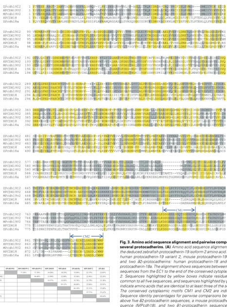
Cell adhesion molecules cadherins play important roles in tissue and organ development, function and maintenance of adult structures (Yagi and Takeichi 2000; Gumbiner 2005; van Roy and Berx 2008). So far more than 100 cadherins have been identified, and they are grouped into several subfamilies, including classical cadherins, protocadherins, desmosomal cadherins and flamingo cadherins (Nollet et al. 2000). The protocadherins (Pcdhs) sub-family contain more members than any other cadherin subfami-lies, and Pcdhs are divided into several groups, such as clustered Pcdhs: α-, β-, and γ-Pcdhs, and non-clustered δ-Pcdhs (Suzuki 1996; Frank and Kemler 2002; Noonan et al. 2004; Wu 2005). The δ-Pcdhs are further divided into δ1-Pcdhs and δ2-Pcdhs based mainly on presence of several conserved motifs in the cytoplas-mic domains, with the δ1-Pcdhs (e.g. Pcdh1, 7 and 9) having three conserved motifs CM1-CM3, whereas the δ2-Pcdhs (e.g. Pcdh10, 18 and 19) containing only two (CM1 and CM2) of the motifs (Redies et al. 2005; Vanhalst et al. 2005).
BIOLOGY
www.intjdevbiol.com*Address correspondence to: Dr. Qin Liu. Department of Biology, University of Akron, 185 East Mill Street, Akron, Ohio 44325, U.S.A. Fax: +1-330-972-8445. e-mail: [email protected]
Accepted: 24 July 2009. Final author-corrected PDF published online: 14 August 2009. Associate Editor: Makoto Asashima.
ISSN: Online 1696-3547, Print 0214-6282
© 2009 UBC Press Printed in Spain
Abbreviations used in this paper: hpf, hours post fertilization; Pcdh19, protocadherin-19 gene or mRNA; Pcdh19, protocadherin-19 protein; Pcdhs, protocadherins;.
Results and Discussion
Zebrafish Pcdh19
Pcdh domains, the zebrafish CM1 and CM2 are more similar to those of Pcdh18 (δ2) than Pcdh1 (δ1) (data not shown). Zebrafish Pcdh19 has two isoforms, which are identical except that isoform 1 is slightly shorter (missing amino acids 755-826 in the cytoplas-mic domain, Fig. 1) than isoform 2. The human PCDH19 also has two variants but they differ by only one amino acid, with variant 1 containing an extra serine at amino acid 847 in the protein.
Pcdh19 expression
Compared to our extensive knowledge of classical cadherins (e.g. cadherin-1 and cadherin-2, also known as E- and N-cadherins, respectively) expression and function, little is known about Pcdh19 expression and function in developing vertebrates, and to the best of our knowledge, there is no published report on Pcdh19 expres-sion in nonmammalian vertebrates.
Using reverse transcriptase-polymerase chain reaction (RT-PCR) and whole mount in situ hybridization methods, we analyzed expression of Pcdh19 in embryonic zebrafish from 6 hours post fertilization (hpf) to 72 hpf. RT-PCR experiments showed that both Pcdh19 isoforms were expressed by embryos of 10 hpf to 72 hpf, with isoform 2 expression slightly stronger than isoform 1 in 18-22 hpf and 72 hpf embryos (Fig. 4A). cRNA probes designed to detect both pcdh19 isoforms were used to perform whole mount in situ hybridization. There was no Pcdh19 expression found in young embryos of 6 hpf (Fig. 4B) and 9 hpf (data not shown). At 12-13 hpf, Pcdh19 expression was observed in the neural keel. In the anterior neural keel, Pcdh19 expressing domains in the presumptive fore-brain and hindfore-brain were separated by regions with little or no Pcdh19 expression (Fig. 4 C,D). The eye primordia also contained Pcdh19 (Fig. 4D). To determine the relative positions of the Pcdh19 expression domains in the presumptive hindbrain, we performed
double-labeling experiments using digoxigenin-la-beled Pcdh19 cRNA probes coupled with fluorescein-labeled pax2a (labeling the boundary of the mid- and hindbrains, Krauss et al. 1991) or krox20 (labeling the rhombomeres 3 and 5, Oxtoby and Jowett 1993). The first Pcdh19 expression domain (indicated by an arrow) in the presumptive hindbrain was located immediately posterior to the boundary between the mid- and hindbrains (Fig. 4F), while the second Pcdh19 expression domain (indicated by an arrowhead) in the presumptive hindbrain was located between the rhombomeres 3 and 5 labeled by the krox20 probe (Fig. 4 G,H). Therefore, the first and second Pcdh19 expression domains in the presumptive hindbrain were likely situated in the presumptive cerebellum/ rhombomere 1 and rhombomere 4, respectively. Pcdh19 expression in the posterior neural keel was continuous, but appeared to be restricted to regions along the midline (indicated by two arrows in Fig. 4E). At 18 hpf, Pcdh19 was expressed in both the brain and spinal cord (Fig. 5 A-D), with obvious regional differences in expression levels, judged by staining intensities, in the fore- and midbrains (Fig. 5B). The ventral telencephalon, ventral diencephalon and teg-mentum showed stronger Pcdh19 expression than the remaining regions of the fore- and midbrains. Similar to Pcdh19 expression in the younger embryos (Fig. 4), reduced Pcdh19 expression was found be-Sig
MHSKDMDFVQMFVCFLLCWTGVDAVFNLK 29
EC1
YTVEEELRAGTKIANVTADAKVAGFALGNRQPYLRVISNSEPRWVNLSPAGLLITKQKID
RDAVCRQTPKCFISLEVMSNSMEICVIKIEIIDVNDNAPRF 130 EC2
PTNHIDIEISENAAPGTRFPLEGASDPDSGSNGIQTYTITPNDIFGLEIKTRGDGSKIAE
LVVEKTLDRETQSRYTFELTAEDGGDPPKSGTVQLNIKVIDSNDNNPVF 239 EC3
DEPVYTVNVLENSPINTLVIDLNATDPDEGTNGEVVYSFINFVSNLTKQMFKIDPKTGVI
TVNGVLDHEELHIHEIDVQAKDLGPNSIPAHCKVIVNVIDINDNAPEI 347 EC4
KLLSENSEMVEVSENAPLGYVIALVRVSDNDSGANGKVQCRLQGNVPFRLNEFESFSTLL
VDGRLDREQRDMYNLTILAEDSGYPPLRSSKSFAVKVTDENDNPPYF 454 EC5
TKPHYQAMVLENNVPGAFLLAVSARDPDLGMNGTVSYEIIKSEVRGMSVESYVTVNSNGE
IYGVRAFNHEDTRTFEFKVSAKDGGDPPLTSNATVRIVVLDVNDNTPVM 563 EC6
TTPPLVNGTAEVSIPKNAGVGYLVTQIKADDYDEGENGRLTYSISEGDMAYFEIDQINGE
VRTTKTFGENAKPSYQITVVAHDHGQTSLSASAYIVIYLSPDLNAQEQIGPVN 676 TM
LSLIFIIALGSIAVILFVTMIFVAV 701
Cyto
KCKRDNKEIRTYNCRVAEYSYGNQKKSSKKKKLSKNDIRLVPRDVEETDKMNVVSCSSLT SSLNYFDYHQQTLPLGCRRSESTFLNVENQNSRNAAPNHGYHHTFTGQGPQQPDLIINGM
PLPETENYSIDSSYVNSRAHLIKSTSTFKDMEGNSLKDSGHEESDQTDSEHDVQRGHYAD CM1 TAVNDVLNMTVPSNNSQIPDQDQSEGFHCQDECRILGHSDRCWMPRVPIPARAKSPEHGR CM2 NVIALSIEATTVDVPHYEDCGTTKRTFATFGKDGPDEDRAEQRGRRQTAEPAVCSPKTNG
AVREAGNGREAVSPITSPVHLKSPQSKAPSTYNTLKCRDAERIANHSLLRQPEGKDSEPA
MREINTLLQDGRDKESPGSKRLKDIVL 1088
Fig. 1.Deduced amino acid sequence of zebrafish Pcdh19. The putative hydropho-bic signal sequence (Sig), and the conserved cytoplamic motifs 1 and 2 (CM1 and CM2, respectively) are underlined. Other abbreviations: cyto, cytoplasmic domain; EC1-EC6, extracellular domains 1-6; TM, transmembrane domain.
tween the cerebellum and optic tectum (arrowhead in Fig. 5B). No regional difference in expression levels was detected in the hindbrain (Fig. 5C), except there was a narrow region, between the cerebellum and the remaining hindbrain, with reduced Pcdh19 expression (arrow in Fig. 5 B,C). This region was likely derived from the region between the first Pcdh19 expression domain and rhombomere 3 in the presumptive hindbrain of younger embryos (Fig. 4 G,H). In the spinal cord, Pcdh19 expression appeared to be stronger in the floor plate region (Fig. 5D). In addition to the
CM1
CM2
ZPcdh19I2 1 YTVEEELRAGTKIANVTADAKVAGFALGNRQP-YLRVISNSEPRWVNLSP-AGLLITKQKIDRDAVCRQTPKCFISLEVMSN---SMEICVIKIEIID HPCDH19V2 1 YSVEEEQRAGTVIANVAKDAREAGFALDPRQASAFRVVSNSAPHLVDINPSSGLLVTKQKIDRDLLCRQSPKCIISLEVMSS---SMEICVIKVEIKD MPcdh19V2 1 YSVEEEQRAGTVIANVAKDAREAGFALDPRQASAFRVVSNSAPHLVDINPSSGLLVTKQKIDRDLLCRQSPKCIISLEVMSS---SMEICVIKVEIKD HPCDH18 1 YRIYEEQRVGSVIARLSEDVADVLLKLPNPSTVRFRAMQRGNSPLLVVNEDNGEISIGATIDREQLCQKNLNCSIEFDVITLPTEHLQLFHIEVEVLD ZPcdh18a 1 YQVYEEQKVGTVIARLREDVADVLSKLPSSIPLRFRAMQRGSASLLSVRDQDGEISIRTKIDREKLCEKNLNCTIEFDVLTLPTEHLQLFHIEVEILD
ZPcdh19I2 93 VNDNAPRFPTNHIDIEISENAAPGTRFPLEGASDPDSGSNGIQTYTITPNDIFGLEIKTRGDGSKIAELVVEKTLDRETQSRYTFELTAEDGGDPPKS HPCDH19V2 95 LNDNAPSFPAAQIELEISEAASPGTRIPLDSAYDPDSGSFGVQTYELTPNELFGLEIKTRGDGSRFAELVVEKSLDRETQSHYSFRITALDGGDPPRL MPcdh19V2 95 LNDNAPSFPAAQIELEISEAASPGTRIPLDSAYDPDSGSFGVQTYELTPNELFGLEIKTRGDGSRFAELVVEKSLDRETQSHYSFRITALDGGDPPHM HPCDH18 98 INDNSPQFSRSLIPIEISESAAVGTRIPLDSAFDPDVGENSLHTYSLSANDFFNIEVRTRTDGAKYAELIVVRELDRELKSSYELQLTASDMGVPQRS ZPcdh18a 98 INDNAPQFARPVIPIEISETAAVGTRIPLDSATDPDVGENSLNTYSLTPSGFFKIDILTRTDGAKYAELVVLKELDREVRASYELQLTASDRGVPPKF
ZPcdh19I2 191 GTVQLNIKVIDSNDNNPVFDEPVYTVNVLENSPINTLVIDLNATDPDEGTNGEVVYSFINFVSNLTKQMFKIDPKTGVITVNGVLDHEELHIHEIDVQ HPCDH19V2 193 GTVGLSIKVTDSNDNNPVFSESTYAVSVPENSPPNTPVIRLNASDPDEGTNGQVVYSFYGYVNDRTRELFQIDPHSGLVTVTGALDYEEGHVYELDVQ MPcdh19V2 193 GTVGLSIKVTDSNDNNPVFGESTYSVSVPENSPPNTPVIRLNASDPDEGTNGQVVYSFYGYVNDRTRELFQIDPHSGLVTVTGALDYEEGHVYELDVQ HPCDH18 196 GSSILKISISDSNDNSPAFEQQSYIIQLLENSPVGTLLLDLNATDPDEGANGKIVYSFSSHVSPKIMETFKIDSERGHLTLFKQVDYEITKSYEIDVQ ZPcdh18a 196 GTTLLKISIADSNDNNPVFEKPSYVINLLENSPLGSLLIDLNATDPDEGTNGKIIYSFSSHVSPKILETFKINSDNGHLTLMRKVDFESTNSYDIDVQ
ZPcdh19I2 289 AKDLGPNSIPAHCKVIVNVIDINDNAP--EIKLLSE-NSEMVEVSENAPLGYVIALVRVSDNDSGANGKVQCRLQGNVPFRLN-EFESFSTLLVDGRL HPCDH19V2 291 AKDLGPNSIPAHCKVTVSVLDTNDNPP--VINLLSV-NSELVEVSESAPPGYVIALVRVSDRDSGLNGRVQCRLLGNVPFRLQ-EYESFSTILVDGRL MPcdh19V2 291 AKDLGPNSIPAHCKVTVSVLDTNDNPP--IINLLSV-NSELVEVSESAPPGYVIALVRVSDRDSGLNGRVQCRLLGNVPFRLQ-EYESFSTILVDGRL HPCDH18 294 AQDLGPNSIPAHCKIIIKVVDVNDNKPEININLMSPGKEEISYIFEGDPIDTFVALVRVQDKDSGLNGEIVCKLHGHGHFKLQKTYENNYLILTNATL ZPcdh18a 294 AQDMGPNSMPAHCKVIIKVVDVNDNKPDISVNLMSTGNEEIAYISETAPVDTFVALVSVNDLDSGLNGEVECRLYGQGHFRLQKSYEKNYMILTNVTL
ZPcdh19I2 383 DREQRDMYNLTILAEDSGYPPLRSSKSFAVKVTDENDNPPYFTKPHYQAMVLENNVPGAFLLAVSARDPDLGMNGTVSYEIIKSEVRGMSVESYVTVN HPCDH19V2 385 DREQHDQYNLTIQARDGGVPMLQSAKSFTVLITDENDNHPHFSKPYYQVIVQENNTPGAYLLSVSARDPDLGLNGSVSYQIVPSQVRDMPVFTYVSIN MPcdh19V2 385 DREQHDQYNLTIQARDSGVPMLQSAKSFTVRITDENDNHPHFSKPYYQVIVQENNTPGAYLLSVSARDPDMGLNGSVSYQIVPSQVRDMPVFTYVSIN HPCDH18 392 DREKRSEYSLTVIAEDRGTPSLSTVKHFTVQINDINDNPPHFQRSRYEFVISENNSPGAYITTVTATDPDLGENGQVTYTILESFILGSSITTYVTID ZPcdh18a 392 DREKRSEFSLTVIAEDKGSPSLSTIKNFIVEVQDENDNAPSFAKSRYEISKAENNSPGAYLSSVKASDPDLGPNGQVSYSILESMVHGSSISTYVTID
ZPcdh19I2 481 SN-GEIYGVRAFNHEDTRTFEFKVSAKDGGDP-PLTSNATVRIVVLDVNDNTPVMTTPPLVNGTAEVSIPKNAGVGYLVTQIKADDYDEGENGRLTYS HPCDH19V2 483 PNSGDIYALRSFNHEQTKAFEFKVLAKDGGLP-SLQSNATVRVIILDVNDNTPVITAPPLINGTAEVYIPRNSGIGYLVTVVKAEDYDEGENGRVTYD MPcdh19V2 483 PNSGDIYALRSFNHEQTKAFEFKVLAKDGGLP-SLQSNATVRVIILDVNDNTPVITAPPLINGTAEVYIPRNSGIGYLVTVVKADDYDEGENGRVTYD HPCDH18 490 PSNGAIYALRIFDHEEVSQITFVVEARDGGSPKQLVSNTTVVLTIIDENDNVPVVIGPALRNNTAEITIPKGAESGFHVTRIRAIDRDSGVNAELSCA ZPcdh18a 490 PSNGDIYALRTFDREDVSQISFLVQARDSGNP-PLRSNVTVVLTVLDENDNRPVIMMPQLWNHTADVPVSKYAEIGDVVTVVRAMDHDAGANGDLSCS
ZPcdh19I2 577 ISEG-DMAYFEIDQINGEVRTTKTFGENAKPSYQITVVAHDHGQTSLSASAYIVIYLSPDLNAQEQ---IGPVNLSLIFIIALGSIAVILFV HPCDH19V2 580 MTEG-DRGFFEIDQVNGEVRTTRTFGESSKSSYELIVVAHDHGKTSLSASALVLIYLSPALDAQES---MGSVNLSLIFIIALGSIAGILFV MPcdh19V2 580 MTEG-DRGFFEIDQVNGEVRTTRTFNENSKPSYELIVVAHDHGKTSLSASALVLIYLSPALDAQES---MGSVNLSLIFIIALGSIAGILFV HPCDH18 588 IVAGNEENIFIIDPRSCDIHTNVSMDSVPYTEWELSVIIQDKGNPQLHTKVLLKCMIFEYAESVTSTAMTS--VSQASLDVSMIIIISLGAICAVLLV ZPcdh18a 587 VVGGNEAGYFNMDPKTCEIRTNVSIQEVPQDHVELTILVQDHGTPTLSARALLRLSLYENIENLMNPHLTGGRNGDGPLDVSMIIIISLGAICAVLLL
ZPcdh19I2 665 TMIFVAVKCKRDNKEIRTYNCRVAEYSYGNQKKSSKKKKLSKNDIRLVPRDVEETDKMNVVSCSSLTSSLNYFDYHQQTLPLGCRRSESTFLNVENQN HPCDH19V2 668 TMIFVAIKCKRDNKEIRTYNCRIAEYSYGHQKKSSKKKKISKNDIRLVPRDVEETDKMNVVSCSSLTSSLNYFDYHQQTLPLGCRRSESTFLNVENQN MPcdh19v2 668 TMIFVAIKCKRDNKEIRTYNCRIAEYSYGHQKKSSKKKKISKNDIRLVPRDVEETDKMNVVSCSSLTSSLNYFDYHQQTLPLGCRRSESTFLNVENQN HPCDH18 684 IMVLFATRCNREKKDTRSYNCRVAESTYQHHPKRPSRQ-IHKGDITLVP-TINGTLPIRSHHRSSPSSSP---TLERGQMGSRQSHNSHQSLNSLV ZPcdh18a 685 IMVAFALRCSREKKDTRSYNCRVAESTYQQHPKKPSRQ-IHKGDITLMP-TVNGTLPIRAHHR-SPTSSP---GAERAHMGSRQSQHSRQSLNSLV
ZPcdh19I2 763 SRNAAPNHGYHHTFTGQGPQQPDLIINGMPLPETENYSIDSSYVNSRAHLIKSTSTFKDMEGNSLKDSGHEESDQTDSEHDVQRGHYADTAVNDVLNM HPCDH19V2 766 TRNTSANHIYHHSFNSQGPQQPDLIINGVPLPETENYSFDSNYVNSRAHLIKS-STFKDLEGNSLKDSGHEESDQTDSEHDVQRSLYCDTAVNDVLNT MPcdh19V2 766 TRNTTASHIYHHSFNSQGPQQPDLIINGVPLPETENYSFDSNYVNSRAHLIKS-STFKDLEGNSLKDSGHEESDQTDSEHDVQRSLYCDTAVNDVLNT HPCDH18 775 TISSNHVPENFSLELTHATPAVEQVSQLLSMLHQGQYQPRPSFRGNKYSRSYR-YALQDMDKFSLKDSGRGDSEAGDSDYDLGRDSPIDRLLGEGFSD ZPcdh18a 775 TISSNHIPENFALELTHATPPVE---GQYQPRPSFRGNKYSRSYR-YALQDMDKFSLKDSGRGDSDAGDSDCEMGRDSPIDRLLGDGFGD
ZPcdh19I2 861 TVPSNNSQIPDQDQSEGFHCQDECRILGHSDRCWMP HPCDH19V2 863 SVTSMGSQMPDHDQNEGFHCREECRILGHSDRCWMP MPcdh19V2 863 SVTSMGSQMPDHDQNEGFHCREECRILGHSDRCWMP HPCDH18 872 LFLTDG--RIPAAMR---LCTEECRVLGHSDQCWMP ZPcdh18a 861 LFHSDGHHRLHPVMR---LCTEECRVLGHSDQCWMP
ZPcdh19I2 HPCDH19V2 MPcdh19V2 HPCDH18 MPcdh18 ZPcdh18a HPCDHV2 ZPcdh1 ZPcdh19I2 71.28% 71.76% 34.20% 34.29% 35.84% 22.75% 22.05%
HPCDH19V2 96.46% 33.99% 32.80% 35.56% 22.46% 22.29%
MPcdh19V2 33.57% 32.95% 35.78% 22.79% 23.30%
HPCDH18 93.12% 66.84% 22.06% 22.95%
MPcdh18 66.10% 22.31% 23.60%
ZPcdh18a 23.71% 23.41%
HPCDH1V2 68.64%
ZPcdh1
B
A
G
B
C
D
E
F
H
A
G
O
B
C
D
E
F
H
I
J
K
L
P
A
M
N
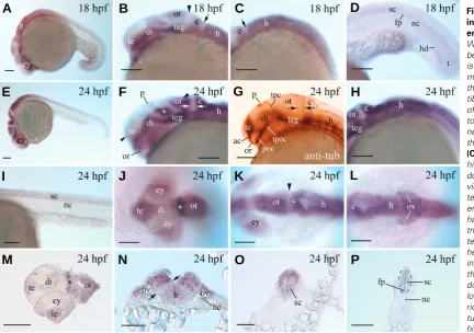
Fig. 4.Pcdh19 expression in 6-13 hpf zebrafish embryos. (A) RT-PCR analysis of Pcdh19 isoforms 1 and 2 expression in embryonic zebrafish using total RNAs. RT-PCR for cdh1 was performed as loading control. The remaining panels show whole mount embryos labeled with Pcdh19 cRNA probes (B-E),Pcdh19 and pax2a cRNA probes (F), or Pcdh19 and krox20 cRNA probes (G,H). (B,C) Lateral views of the entire embryos (head towards the lower left corner for C). (D,E) Dorsal views (anterior to the left) of the entire embryos. (F,G) Lateral views of the anterior half of the embryos (anterior to the left and dorsal up), while (H) is a dorsal view of the presumptive hindbrain region of an embryo (anterior to the left). The arrow and arrowhead in (C, D, F, G and H) point to the first and second Pcdh19 expression domains, respectively, in the presumptive hindbrain. The two arrows in panel E indicate Pcdh19 expression in the middle neural keel. Abbreviations: bmh, boundary of the mid- and hindbrains; ep, eye premordium; f, presumptive forebrain; h, presumptive hindbrain; r3 and r5, rhombomeres 3 and 5, respectively. Scale bars, 100 μm.
nervous tissue, Pcdh19 was also seen in the hypochord below the notochord near the tail (Fig. 5D). Embryos of 24 hpf showed somewhat similar Pcdh19 expression patterns in the fore- and midbrains as 18 hpf embryos, except that the staining was stronger in the dorsal thalamus (ventroanteral to the optic tectum, and adjacent to the tract of the posterior commissure, indicated by an
Fig. 6.Pcdh19 expression in 36 hpf embryos. (A,B,C) Lateral views of whole mount embryos showing the anterior 2/3 of the fish, the anterior head, and posterior head regions, respectively (anterior to the left and dorsal up). (D-I) Cross sections (dorsal up) from whole mount embryos processed for Pcdh19in situ hybridization. Levels of the sections are shown in (A). The arrowhead in (B) points to the region with reduced Pcdh19 expression (also see Fig. 5F). Arrows in (E,F) indicate pigmented epithelium. The arrowhead in (F) points to Pcdh19 expression in the epithelial layer of the lens. Asterisks in (B,E) indicate the same thalamic regions with stronger
Pcdh19 expression. The opposing arrows in (G,H) indicate the band of tissue with reduced Pcdh19 expression (also see Fig. 5N). Abbreviations are the same as in Fig. 5. Scale bars, 100 μm.
asterisk in Fig. 5F) and optic tectum (Fig. 5F) than 18 hpf embryos (Fig. 5B). In the telencephalon, the Pcdh19 expressing domain was located anterodorsal to the optic recess (Fig. 5 F,G). The stronger labeled regions in the telencephalon, central thalamus and tegmentum appeared to form a continuous thick bank viewing laterally (Fig. 5F). The stripe of tissue between the posterior border of the optic tectum and the boundary of the mid- and hindbrains continued to show much re-duced staining (Fig. 5 F,G, indicated by two opposing arrows). In the hindbrain, Pcdh19 expression was con-tinuous from the cerebellum to the spinal cord, with stronger expression levels detected in the cerebellum and dorsolateral hindbrain (Fig. 5 H,N). A band of tissue between the dorsolateral and the ventromedial hind-brain showed reduced staining (indicated by two oppos-ing arrows in Fig. 5N). In the anterior spinal cord, its dorsal 1/3 area was also more strongly labeled than the ventral spinal cord (Fig. 5O), while in the mid-trunk and tail regions of the spinal cord, regional differences in the staining was not detected and the floor plate region continued to express Pcdh19 (Fig. 5P). Pcdh19 was also expressed by the retina, lens (Fig. 5 J,M), and otic vesicle (5L and N). The lens and peripheral retina (future retinal marginal zones) were more strongly la-beled than the central retina (Fig. 5M). Epithelial cells in the lateral otic vesicle and the hair cells were strongly labeled (Fig. 5N).
Pcdh19 expression was reduced and regional differ-ences in expression levels became more pronounced in some regions of the CNS in 36 hpf embryos (Fig. 6). Stronger Pcdh19 staining continued to be observed in the dorsal and central thalamus (Fig. 6 B,E,F), the cerebellum (Fig. 6 B,C), and the dorsolateral regions of the hindbrain (Fig. 6 B,C,G,H). In the telecephalon, stronger Pcdh19 expression was found in the lateral portion (Fig. 6D). In the diencephalon, the strongly labeled Pcdh19 expression domains in the dorsal and central thalamus
G
B
C
D
E
F
H
I
A
G
B
C
D
E
F
H
I
J
K
L
A
M
had the appearance of two horizontal bands in cross sections (Fig. 6E, indicated by two asterisks). In the midbrain, Pcdh19 expression was confined mainly to the optic tectum and dorsal tegmentum (Fig. 6F). Again, the region between the posterior border of the optic tectum and the boundaries of mid- and hindbrains (arrowhead in Fig. 6B), and the region between the dorsolateral and the ventromedial hindbrain (opposing arrows in Fig. 6 G,H) had less Pcdh19 expression than their neighboring areas. The dorsal spinal cord contained higher Pcdh19 expressing cells than the ventral spinal cord even in the mid-trunk region (Fig. 6I). Pcdh19 expres-sion in the retina and lens was reduced compared to 24 hpf embryos, except in the retinal marginal zones and the lens epithe-lial layer (Fig. 6 E,F). Strong Pcdh19 expression continued in the lateral epithelial cells of the otic vesicle, but its expression was reduced in the hair cells (Fig. 6G).
Generally speaking, Pcdh19 expression in 50 hpf embryos was similar to those of 72 hpf (Fig. 7). Similar to the younger embryos, stronger Pcdh19 staining was seen in the dorsal thalamus and dorsal tegmentum (Fig. 7B), optic tectum (Fig. 7 B,F,J), cerebellum (Fig. 7 B,K), and dorsolateral hindbrain (Fig. 7 D,H,K), but Pcdh19 expression in the central thalamus, tegmentum and hindbrain was reduced compared to 24 and 36 hpf embryos. At 50 hpf, Pcdh19 expression in the telencephalon (Fig. 7C) was mainly in the lateral regions (similar to 36 hpf embryos, Fig. 6D), but stronger expres-sion became restricted to the dorsal telencephalon by 72 hpf (Fig. 7I). The region between the posterior border of the optic tectum and the boundary of the mid-hindbrains continued to show reduced staining (Fig. 7F), while Pcdh19 expression in the ventromedial hindbrain was greatly reduced (Fig. 7K) compared to younger embryos (Figs. 5N and 6G). Pcdh19 expression in the spinal cord was no longer detectable at both 50 hpf (Fig. 7D, data not shown for spinal cord sections) and 72 hpf (Fig. 7 H,M). In the eye, Pcdh19 expression became mainly confined to the retinal ganglion cell layer, while there was no Pcdh19 expression in the lens (Fig. 7 C,G). Pcdh19 expression in the otic vesicle became further re-duced (Fig. 7K).
Pcdh19 expression in the embryonic zebrafish is somewhat similar to that in embryonic mice (Gaitan and Bouchard, 2006). In both species, early Pcdh19 expression is mainly confined to the nervous system. Pcdh19 is found in the telencephalon and dien-cephalon, in the spinal cord, in the developing retinal ganglion cell layer and lens. In adult rat brain, Pcdh19 was expressed in specific regions of the telencephalon, diencephalon, midbrain and hind-brain (Kim et al. 2007). In addition to the neural tissue, Pcdh19 is also expressed by several other tissues in human (RT-PCR analysis, Wolverton and Lalande 2001) and mouse (Gaitan and Bouchard 2006), including the heart and kidney. It remains to be determined if Pcdh19 is expressed by these tissues in larval and adult zebrafish.
Materials and Methods
Zebrafish embryos were obtained from in house breeding, and main-tained as described in the Zebrafish Book (Westerfield 2005). Embryos for whole mount in situ hybridization were raised in PTU (1-phenyl-2-thiourea,
0.003%) at 28.5oC, staged in hours post fertilization, and fixed in phosphate
buffered 4% paraformaldehyde.
Cloning of zebrafish Pcdh19
A TBlastn search using human PCDH19 protein sequence as a query
resulted in a zebrafish genomic DNA sequence CR318607. A zebrafish
Pcdh19 cDNA fragment was amplified by PCR from a zebrafish embryonic
cDNA Uni-ZAP XR library (discontinued; Stratagene, La Jolla, CA) using primers (forward primer 0, 5’-CGTTAGTCATAGACCTGAACGCCACTGA CC -3, reverse primer 0, 5’-TTACTACCAAGCCACGATGACAGTCTGAGC -3’) flanking a predicted zebrafish Pcdh19 coding region. The PCR product
was cloned in the pCR-Blunt II-TOPO vector (Invitrogen, Carlsbad, CA). Sequences of the clones indicated 2 isoforms, Pcdh19 isoforms 1 and 2
(GenBank, accession number: AB 362378 and AB 362379, respectively, F. Kubota and T. Murakami). Analysis of the deduced amino acid sequences of these isoforms revealed that they lack the N-terminal 1/3 (from signal sequence to about half of EC3) of the proteins compared to other protocadherins. A BLAST search using the Pcdh19 isoform 2 sequence
resulted in a zebrafish cDNA clone sequence (wu:fc83e05, GenBank accession number: BC129243, Strausberg et al.) that contains all the
incomplete Pcdh19 isoform 2 sequence, plus most of the missing
N-terminal sequence. Compared to other protocadherins including the mam-malian Pcdh19 proteins, this zebrafish sequence (zebrafish protocadherin-19 isoform 2) appeared to be missing some of the signal sequence in the 5’ region. Using primers designed according to genomic sequence of wu:fc83e05 (GenBank accession number: NW 001884478) and mRNA sequence of the wu:fc83e05 (forward primer 1, CGCGTGAAGACAGACATCAA-3’, forward primer 2, 5’-TTCCAAGGACATGGATTTCG-3’, 58 and 25 nucleotides, respectively, 5’ to the starting codon of the published wu:fc83e05 sequence; reverse primer 1, 5’-AGTAGACCACCTCGCCATTG-3’, corresponding to the nucleotides 748 to 767 of the wu:fc8305), and total RNAs from 24-70 hpf whole zebrafish embryos, we performed RT-PCR and obtained the missing signal sequence (Fig. 1).
RT-PCR analysis of Pcdh19 expression in developing zebrafish
RT-PCR analysis of Pcdh19 isoforms temporal expression profiles
was performed using Pcdh19 specific primers (forward primer 3,
GCCTTGGGCTCTATTGCAGTCA-3’; reverse primer 2, 5’-AGCATAGTGCCCTCTCTGGA-3’, corresponding to nucleotides 1284-1305 and 1855-1874, respectively, of zebrafish Pcdh19 isoform 2, GenBank
accession number AB 362379). These primers amplified bands of 377 bp and 590 bp for Pcdh19 isoforms 1 and 2, respectively (Fig. 4A). Zebrafish cdh1 transcripts (Liu et al. 2007) were used as the control for the RT-PCR
experiments because cdh1 was shown to be strongly expressed by young
zebrafish embryos (Babb et al. 2005).
In situ hybridization and immunocytochemistry
For obtaining a Pcdh19 DNA fragment (corresponding to the
nucle-otides 43-985 of the incomplete zebrafish Pcdh19 isoforms 1 and 2) as a
template for synthesizing cRNA probes, zebrafish Pcdh19 specific
prim-ers (forward primer 4, 5’-CAATGGCGAGGTGGTCTACT-3’; revprim-erse primer 3, 5’-CAACTCCAGCGTTTTTAGGG-3’), and total RNA isolated from 20-50 hpf whole zebrafish embryos were used for RT-PCR. This cDNA fragment was cloned into the pCRII-TOPO vector (Invitrogen), and was verified by restriction mapping and a PCR experiment using a pair of
Pcdh19 specific primers that were internal to the previous set of primers
(forward primer 5, 5’-GCCCGAAATCAAACTGTTGT-3’; reverse primer 4, 5’-GCACCTCCGATTTGATGATT-3’). This experiment produced a cDNA fragment corresponding to the nucleotides 266-729 of the incom-plete zebrafish Pcdh19 isoforms 1 and 2). The larger Pcdh19 cDNA
fragment in the pCRII-TOPO vector was used as a template for the synthesis of digoxigenin-labeled zebrafish Pcdh19 RNA sense or
anti-sense probes for in situ hybridization. Both Pcdh19 isoforms contained
this cDNA fragment. cDNAs used to generate the fluorescein-labeled antisense pax2a and krox20 were provided by Drs. Pamela Raymond
(University of Michigan) and Lisa Maves (Fred Hutchinson Cancer Re-search Center), respectively. Detailed procedures for the cRNA probe synthesis and whole mount in situ hybridization were described
staining in zebrafish embryos from 24-50 hpf using the sense Pcdh19
probes (data not shown).
Anti-acetylated tubulin antibody (Sigma, St Louis, MO) was used at 1:3000. The secondary antibody (used at 1:250) was biotinylated anti-mouse IgG (Vector laboratories, Burlingame, CA). Whole-mount immu-nocytochemistry was carried out according to protocols described in the Zebrafish Book (Westerfield 2005).
Acknowledgements
This work was supported by grants from NIH EY13879 to Q. L., Grants-in-Aid awarded to T.M. from the Ministry of Education, Culture, Sports, Science, and Technology of Japan (MEXT), and the Initiative for Attrac-tive Education in Graduate Schools awarded to Gunma University from MEXT. We thank Drs. Pamela Raymond (University of Michigan, Ann Arbor, MI) and Lisa Maves (Fred Hutchinson Cancer Research Center, Seattle, WA) for the pax2a and krox20 containing plasmids, respectively.
References
BABB S.G., BARNETT J., DOEDENS A.L., COBB N., LIU Q., SORKIN B.C., YELICK P.C., RAYMOND P.A. and MARRS J.A. (2001). Zebrafish E-cadherin: expression during early embryogenesis and regulation during brain develop-ment. Dev Dyn 221: 231-237.
FRANK M. and KEMLER R. (2002). Protocadherins. Curr Opin Cell Biol 14:
557-562.
GAITAN Y. and BOUCHARD M. (2006). Expression of the δ–protocadherin gene
Pcdh19 in the developing mouse embryo. Gene Expr Patterns 6: 893-899.
GUMBINER B.M. (2005). Regulation of cadherin-mediated adhesion in morpho-genesis. Nat Rev Mol Cell Biol 6: 622-634.
KIM S.-Y, SUN CHUNG H, SUN W. and KIM H. (2007). Spatiotemporal expression pattern of non-clustered protocadherin family members in the developing rat brain. Neurosci 147: 996-1021.
KRAUSS S, JOHANSEN T, KORZH V, and FJOSE A. (1991). Expression of the zebrfish paired box gene pax[zf-b] during early neurogenesis. Development
113: 1193-1206.
KUBOTA F, MURAKAMI T, TAJIKA Y, and YORIFUJI H. (2008). Expression of protocadherin-18 in the CNS and pharyngeal arches of zebrafish embryos. Int J Dev Biol 52: 397-405.
LIU Q, SANBORN K.L, COBB N, RAYMOND P.A, and MARRS J.A. (1999). R-cadherin expression in the developing and adult zebrafish visual system. J Comp Neurol 410: 303-319.
LIU B, DUFF R.J, LONDRAVILLE R.L, MARRS J.A, and LIU Q. (2007). Cloning and expression analysis of cadherin7 in the central nervous system of the embryonic zebrafish. Gene Expr Patterns 7: 15-22.
NOONAN J.P, GRIMWOOD J, SCHMUTZ J, DICKSON M, and MYERS R.M. (2004). Gene conversion and the evolution of protocadherin gene cluster diversity. Genome Res 14: 354-366.
NOLLET F, KOOLS P, and VAN ROY F. (2000). Phylogenetic analysis of the cadherin superfamily allows identification of six major subfamilies besides several solitary members. J Mol Biol 299: 551-572.
OXTOBY L, and JOWETT T. (1993). Cloning of the zebrafish krox-20 (krx-20) and
its expression during hindbrain development. Nucleic Acids Res 21: 1087-1095.
REDIES C, VANHALST K, and VAN ROY F. (2005). δ–Protocadherins: unique structures and functions. Cell Mol Life Sci 62: 2840-2852.
SUZUKI S.T. (1996). Protocadherins and diversity of the cadherin superfamily. J Cell Sci 109: 2609-2611.
VAN ROY F, and BERX G (2008). The cell-cell adhesion molecule E-cadherin. Cell Mol Life Sci 65: 3756-3788.
VANHALST K, KOOLS P, STATES K, VAN ROY F. and REDIES C. (2005). δ -Protocadherins: a gene family expressed differentially in the mouse brain. Cell Mol Life Sci 62: 1247-1259.
WESTERFIELD M. (2005). The zebrafish Book. Eugene, OR: University of Oregon
Press.
WOLVERTON T, and LALANDE M. (2001). Identification and characterization of three members of a novel subclass of protocadherins. Genomics 76: 66-72.
WU Q. (2005). Comparative genomics and diversifying selection of the clustered vertebrate genes. Genetics 169: 2179-2188.
YAGI T. and TAKEICHI M. (2000). Cadherin superfamily genes: functions, genomic organization, and neurologic diversity. Genes Dev 14: 1169-1180.
Further Related Reading, published previously in the Int. J. Dev. Biol.
See Special Issue Pattern Formation edited by Michael K. Richardson and Cheng-Ming Chuong at: http://www.ijdb.ehu.es/web/contents.php?vol=53&issue=5-6
Expression of protocadherin 18 in the CNS and pharyngeal arches of zebrafish embryos
Fumitaka Kubota, Tohru Murakami, Yuki Tajika and Hiroshi Yorifuji Int. J. Dev. Biol. (2008) 52: 397-405
Cadherin-6 is required for zebrafish nephrogenesis during early development
Fumitaka Kubota, Tohru Murakami, Kenji Mogi and Hiroshi Yorifuji Int. J. Dev. Biol. (2007) 51: 123-129
Cadherin-mediated cell-cell adhesion and tissue segregation in relation to malignancy
Ramsey A. Foty and Malcolm S. Steinberg Int. J. Dev. Biol. (2004) 48: 397-409
Discovery and characterization of the cadherin family of cell adhesion molecules. An interview with Masatoshi Takeichi
Douglas Sipp
Int. J. Dev. Biol. (2004) 48: 387-396
Transcriptional regulation of cadherins during development and carcinogenesis